Autores
Daniel da Almeida Thiengo 1,2
Jocemir Ronaldo Lugon 2
Miguel Luis Graciano 2
1 Hospital Icaraí. 2 Universidade Federal
Fluminense.
Data de submissão: 23/02/2015. Data de aprovação: 01/07/2015.
Correspondência para:
Daniel da Almeida Thiengo. Universidade Federal Fluminense, Centro de Diálise, Hospital Universitário Antônio Pedro. Rua Marques do Parana, nº 303, 2º andar; Niterói, RJ, Brasil. CEP: 24033-900
E-mail: [email protected]
Troponin I serum levels predict the need of dialysis in incident
sepsis patients with acute kidney injury in the intensive care unit
Níveis séricos de troponina predizem a necessidade de diálise em
pacientes sépticos com injúria renal aguda renal na unidade de
terapia intensiva
Introdução: Sepse é uma condição
extremamente prevalente na unidade de terapia intensiva, geralmente associada com disfunção orgânica que pode afetar o coração e os rins. Objetivo:
Determinar se a disfunção cardíaca e a troponina I preveem a ocorrência de lesão renal aguda na sepse. Métodos:
A disfunção cardíaca foi avaliada por ecocardiografia e pelos níveis de troponina I sérica; e a lesão renal aguda pelos critérios AKIN e necessidade de diálise. Vinte e nove pacientes com sepse foram recrutados. Resultados
e Discussão: Os pacientes tinham em
média 75,3 ± 17,3 anos e 55% eram do sexo masculino. O escore de gravidade APACHE II médio de internação na UTI foi de 16 (9,7-24,2) e taxa de mortalidade em 30 dias foi de 45%. No quinto dia, 59% tinham disfunção ventricular. O nível sérico de troponina no dia 1 nos pacientes afetados foi de 1,02 ± 0,6 ng/mL em comparação com 0,23 ± 0,18 ng/mL em pacientes sem disfunção cardíaca (p = 0,01). Dezoito dos 29 pacientes (62%) foram submetidos à terapia renal substitutiva e a porcentagem de pacientes com disfunção ventricular que necessitou de diálise foi maior (94% vs. 16%, p
= 0,0001) nesse grupo. A área sob uma curva ROC desenhada para prever a necessidade de diálise de acordo com o nível sérico de troponina no dia 1 foi de 0,89 e o valor de corte foi de 0,4 ng/mL. Conclusão: Verificou-se que uma elevação nos níveis séricos de troponina pode prever a necessidade de diálise em pacientes sépticos com lesão renal aguda.
RESUMO
Palavras-chave: lesão renal aguda; sepse; troponina.
Introduction: Sepsis, an extremely prevalent condition in the intensive care unit, is usually associated with organ dysfunction, which can affect heart and kidney. Objective: To determine whether the cardiac dysfunction and the Troponin I forecast the occurrence of acute renal failure in sepsis. Methods:
Cardiac dysfunction was assessed by echocardiography and by the serum troponin I levels, and renal impairment by AKIN criteria and the need of dialysis. Twenty-nine patients with incident sepsis without previous cardiac or renal dysfunction were enrolled. Results and Discussion: Patients averaged 75.3 ± 17.3 years old and 55% were male. Median APACHE II severity score at ICU admission was 16 (9.7 - 24.2) and mortality rate in 30 days was 45%. On the fifth day, 59% had ventricular dysfunction. Troponin serum levels on day 1 in the affected patients were 1.02 ± 0.6 ng/mL compared with 0.23 ± 0.18 ng/ mL in patients without heart dysfunction (p = 0.01). Eighteen out of 29 patients (62%) underwent renal replacement therapy (RRT) and the percent of patients with ventricular dysfunction who required dialysis was higher (94% vs. 16%, p = 0.0001). Values of troponin at day 1 were used to develop a ROC curve to determine their ability to predict the need of dialysis. The area under the curve was 0.89 and the cutoff value was 0.4 ng/mL. Conclusion: We found that an elevation in serum troponin levels, while guarding a relationship with ventricular dysfunction, can be a precious tool to predict the need for dialysis in sepsis patients.
A
BSTRACTKeywords: acute kidney injury; sepsis; troponin.
INTRODUCTION
Despite technological advances, sepsis still remains a major cause of mortality in intensive care units around the world.1 Sepsis is diagnosed by the presence of
clinical signs of infections or systemic inflammation regardless of the site of the infection or the causative microbe involved. It can be classified as severe when occur in association with signs of organ dysfunction such as kidney failure or myocardial dysfunction. The kidney is frequently affected in sepsis. Indeed, renal injury is commonly find in severe sepsis and is associated with increased mortality.2 Assessment
of renal function in critical care patients is now frequently performed scoring the AKIN criteria which take into account serum creatinine and urinary output.3 Myocardial dysfunction can also occur in
sepsis. Accordingly, recent studies have showed that the myocardial dysfunction in sepsis, evaluated by echocardiogram, EKG or troponin measurement, is present in 40% of these patients and is associated with an increase of 20% to 30% in the mortality rate.4,5 The pathophysiology of renal injury in sepsis
is multifactorial and may involve crosstalk between the heart and the kidney.
OBJECTIVES
To determine whether the cardiac dysfunction and the Troponin I forecast the occurrence of acute kidney injury or dialysis on patients with sepsis.
METHODS
In this study, we conducted a prospective, longitudinal study in 29 patients with sepsis, admitted in the intensive care unit (ICU) of a private hospital located in the state of Rio de Janeiro - Brazil, in the period of July 2012 to February 2013. The study was approved by the ethical committee of Medical School/University Hospital of Universidade Federal Fluminense. An informed consent was signed by every patient or a relative.
STUDYSUBJECTS
Patients were recruited by systematic sampling as they were being admitted to the ICU, as long as they fulfill the following inclusion criteria: age between 18-90 years, 24 hours or less from the onset of symptoms of sepsis, normal left ventricular function in a transthoracic echocardiogram (TTE) performed in
the first 24 hours in the ICU (left ventricular ejection fraction, LVEF > 45%). Admitted patients were excluded from the study if the following exclusion criteria were found: previous either myocardial dysfunction or coronary disease, kidney disease, hepatic failure, use of iodinated contrast media, AIDS, neoplastic disease under treatment with radiotherapy or chemotherapy.
DEFINITIONS AND PROCEDURES
Sepsis was defined by the evidence of bacterial growth in blood, urine or tracheal secretion in addition to at least two of the following criteria: temperature > 38°C or < 36°C, heart rate > 90 beats per minute, respiratory rate > 20 breaths per minute or pCO2 < 32 mmHg, white blood cell count > 12.000 cells/ mm3, or < 4.000 cells/mm3 or > 10% of young cells
accordingly to the definition of the Surviving Sepsis Campaign.6 On admission, age, sex, and the severity
score APACHE II7 were documented. On the fifth
day of hospitalization, a second echocardiogram was performed to discriminate patients without or with ventricular dysfunction, defined by a LVEF value lower than 45%. All patients also had the troponin I serum levels measured in the first, third, fifth and seventh days of hospitalization to assess the role of this biomarker as to the prediction of renal dysfunction. Kidney dysfunction was characterized accordingly to the Acute Kidney Injure Network - AKIN criteria.2 During the period of observation the
degree of renal dysfunction as well as the number of patients undergoing dialysis was recorded on a daily basis. All enrolled patients were followed by 30 days or until discharge or death.
STATISTICAL ANALYSIS
troponin measurement to predict the need of dialysis was also evaluated by calculation of the sensitivity, specificity, positive and negative predictive value, and positive and negative likelihood ratio. Statistical analysis was performed with the statistic software MedCalc 11.4.2.0 (Mariakerke, Oost- Vlaanderen, Belgium). P values <.05 were considered significant.
RESULTS
Twenty-nine patients were included in this analysis after exclusion of three patients, two of them due to withdrawal of the informed consent and one due to a diagnosis of AIDS. The mean age was 75.3 ± 17.3 years and 16 subjects (55%) were male. In view of the high grade of miscegenation of Brazilian population we did not stratified the sample according to race. At the end of the study, 13 deaths (45%) and 9 (31%) discharges were observed with seven patients remaining in the ICU after 30 days of observation. The Charlson comorbidity index varied from 1 to 5, with a mean of 3.2 ± 1.1 and the median APACHE II severity score at ICU admission was 16 (9.7 - 24.2). Regarding myocardial function, the mean LVEF on the first day was 48.2 ± 3.1% and no segmental alterations in ventricular function were found. On the fifth day, 17 patients (59%) had myocardial dysfunction; the mean LVEF of the entire sample at this time was 37.8 ± 5.4%. As expected, troponin values reflected myocardial dysfunction whenever measured during the study. Interestingly, on day 1, the troponin serum levels in patients with ventricular dysfunction on the fifth day were 1.02 ± 0.6 ng/mL, compared with 0.23 ± 0.18 in the remaining ones (p = 0.01). Similarly, the mean serum levels of troponin on the third day was 1.53 ± 0.72 ng/mL in patients with myocardial dysfunction and 0.55 ± 0.25 ng/ mL in the group without dysfunction (p = 0.001). Correspondent data on fifth and seventh days were 1.8 ± 0.35 vs. 0.35 ± 0.77 (p = 0.001) and 1.70 ± 0.62 vs. 0.34 ± 0.35 (p = 0.0001) (Table 1). All patients showed an increase in AKIN. Eighteen out of 29 patients (62%) underwent renal replacement therapy (RRT). The mean age among dialysis patients was 80.5 ± 11.7 years and the majority (67%) was male. Thirteen patients who required RRT had a fatal outcome (72%) whereas only 1 out of 11 non-dialysis patients (9%) did so. On average RRT was initiated on the eighth day of ICU admission varying from as early as day 2 to as late as day 15. Accordingly, the
higher values of AKIN peaked on the eighth day of observation. As expected, AKIN values were higher in patients who had undergone RRT (1.58 ± 0.55 vs. 0.21 ± 0.11, p = 0.0001). The LVEF in the fifth day was 34.7 ± 4.1% in the group requiring RRT compared with 42.8 ± 3.0% in the group that remained free of dialysis (p < 0.001). From a different perspective, RRT requirement was higher in the group with myocardial dysfunction in TTE performed on the fifth day compared to the group that had no dysfunction. Accordingly, patients with myocardial dysfunction were dialyzed more frequently than patients without dysfunction (94% vs. 16%, p = 0.0001). The group with myocardial dysfunction also exhibited a higher mean AKIN scoring along the ICU stay compared with patients with normal heart function (2.8 ± 0.25 vs. 1.8 ± 0.4, p = 0.03). All these data collectively suggest an association between the presence of myocardial dysfunction and acute renal injury. The mean troponin serum levels were significantly higher in patients who received RRT in each day measured. Values for the first, third, fifth and seventh days (RRT vs. non-RRT) were, respectively: 0.98 ± 0.65 vs. 0.18 ± 0.21, p = 0.008; 1.47 ± 0.75 vs. 0.56 ± 0.26, p = 0.007; 1.78 ± 0.72 vs. 0.28 ± 0.70, p = 0.001; and 1.61 ± 0.26 vs. 0.58 ± 0.09, p < 0.0001 (Table 2). Values of troponin from day 1 were used to develop a ROC curve to determine their ability to predict dialysis. The area under the curve obtained was 0.89. Cutoff value was 0.4 ng/mL (Figure 1). For this value, sensitivity, specificity, positive and negative predictive value, and negative likelihood ratio were 78%, 100%, 100%, 73%, and 0.22. Less certain was the association of troponin and the highest AKIN score reached observed during hospital stay. Accordingly, when we compared the troponin values in three groups divided according to the highest AKIN score reached during hospitalization (≤ 1, 2, and 3) mean serum troponin levels measured on day 1 were 0.24 ± 0.21 ng/mL, 0.27 ± 0.55 ng/mL, and 1.00 ± 0.64 ng/ mL, respectively (p < 0.001 vs. AKIN ≤ 1 or AKIN = 2). Troponin serum levels were also statistically higher in patients with AKIN scoring of 3 on days 3, 5 and 7 (Table 3).
DISCUSSION
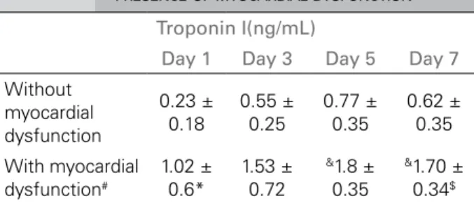
TABLE 1 MEANVALUESOFTROPONINIACCORDINGTO PRESENCEOFMYOCARDIALDYSFUNCTION
Troponin I(ng/mL)
Day 1 Day 3 Day 5 Day 7
Without myocardial dysfunction
0.23 ± 0.18
0.55 ± 0.25
0.77 ± 0.35
0.62 ± 0.35
With myocardial
dysfunction#
1.02 ± 0.6*
1.53 ± 0.72
&1.8 ±
0.35
&1.70 ±
0.34$
# Defined as left ventricular ejection fraction < 45% on the 5th day of
ICU. *p < 0.01, &p < 0.001, $p < 0.0001.
TABLE 2 MEANVALUESOFTROPONINIACCORDINGTOTHE NEEDOFRENALREPLACEMENTTHERAPY (RRT)
Troponin I (ng/mL) Need of
RRT Day 1 Day 3 Day 5 Day 7
Yes 0.21 ±
0.18
0.56 ± 0.26
0.70 ± 0.28
0.58 ± 0.09
No 0.98 ±
0.65*
1.47 ± 0.75*
1.78 ± 0.72*
1.61 ± 0.26#
RRT, renal replacement therapy. *p < 0.001, #p < 0.0001.
TABLE 3 MEANVALUESOFTROPONINIACCORDINGTOTHE AKINSCORE
Troponin I (ng/mL)
Day 1 Day 3 Day 5 Day 7
AKIN 1 0.24 ±
0.18
0.59 ± 0.26
0.72 ± 0.30
0.59 ± 0,34
AKIN 2 0.27 ±
0.23
0.50 ± 0.26
0.67 ± 0.12
0.43 ± 0.06
AKIN 3 1.00 ±
0.63*
1.58 ± 0.72*
1.93 ± 0.64*
1.79 ± 1.00* *p < 0.05 vs. AKIN 1 and AKIN 2. AKIN, Acute Kidney Injury Network score.
Figure 1. Receiver-operating characteristic curve displaying the potential troponin I values on day 1 to of intensive care unit stay to predict renal replacement therapy need. AUC, area under the receiver-operating characteristic curve. Sens, sensitivity; Spec, specificity.
sepsis in the intensive care unit. In this prospective observational study, conducted from July 2012 to February 2013, 29 patients were included. Patients were derived from the emergency or surgical room, were unaware of cardiac disease and had sepsis diagnosed within the last 24 hours, representing the typical incident patient with sepsis in the ICU. Myocardial failure is a common organic dysfunction in sepsis.9,10 Accordingly, it has already been reported
that impaired cardiac function is frequent in septic patients and considerably increases mortality rate in ICU patients.4 A broad spectrum of ventricular
impairment can be found in sepsis, ranging from diastolic dysfunction to severe systolic dysfunction.11
Myocardial involvement in sepsis, regardless of the pathophysiological mechanisms can be detected by abnormalities in EKG or TTE, particularly decrement of ejection fraction,11 and also by elevation of serum
biomarkers like troponin I and BNP.12 The initial
myocardial injury characterized by elevated serum troponin eventually leads to organ dysfunction11
in most of these patients expressed as a fall in the LVEF.11 The results of the present study confirm
the relationship between elevated troponin levels and the development of myocardial dysfunction in septic patients.12,13 The sepsis associated myocardial
dysfunction appears to be in some way related to the development of renal injury in such patients.14 This
However, Troponin I is less predictably increased in renal insufficiency and its level usually is not affected by the renal functional status. Moreover, in the current study, it is implausible that troponin I levels reflects renal function as it increase preceded the reduction in glomerular filtration rate or the decrease in urinary output. Perhaps inspired in the concept of hepatorenal syndrome the term cardiorenal syndrome has been coined.18 Contrariwise to the first entity
however, the so called cardiorenal syndromes lack a common pathophysiological background and are better understood as a collection of different entities affecting both the kidneys and the heart. Sepsis would be categorized as CRS type 5 in which both the kidney and the heart are contemporaneously affected by the same pathological process. It should be remembered, nevertheless, that intravascular volume contraction brought by systemic vasodilation as well as diminished myocardial contractility induced by the inflammatory milieu are sufficient reasons to trigger renal malperfusion and AKI, at least in the absence of fluid resuscitation and early management of the septic syndrome. The fact that the current series involves patients in the early phase of sepsis renders the hypothesis of renal injury caused by hypoperfusion highly unlikely. Moreover, the finding of Langenberg et al.19 that hyperdynamic sepsis in large mammals
involves renal hyperemia instead of ischemia heightens the probability that AKI in the scenario of sepsis is not determined by renal hypoperfusion. It is more conceivable that the systemic inflammatory environment triggers concomitantly both renal and cardiac dysfunction in the context of septic multiple organ dysfunction. However, it is intriguing how experimental ischemic AKI can determine distant cardiac dysfunction denoting significant crosstalk between the kidneys and heart. In that specific study, renal ischemia, incapable of producing oliguria or uremia, induced long distance heart damage characterized by altered echocardiography, as well as leukocyte infiltration and apoptosis induction in the cardiac tissue. Additionally, it was shown that these abnormalities were partially mediated by TNF-alfa a known mediator of the inflammatory cascade.20 In
the present study echocardiographic abnormalities were observed earlier than the AKI diagnosis was made either through the AKIN score or by the commencement of RRT. As undisputed clinical heart failure was never diagnosed in our patients it is very
unlikely that low cardiac output triggered renal injury. Naturally, both organs might be involved at the same time by the inflammatory/cytopathic phenomena associated with sepsis; however, it is tempting to speculate that initial renal damage could eventually lead to cardiac damage. Of course, in the absence of an earlier marker of renal injury, such reasoning is pure speculation, at best. Not surprisingly, there has been a widespread search for such early markers of renal injury by the nephrology and renal scientists’ community. Suspicious they are that clinical diagnosis of AKI is a late phenomenon, they longed for a biomarker able to predict renal outcome in AKI. Accordingly, NGAL, KIM-1, IL-18 and several other molecules are now considered putative markers of renal dysfunction.21-24 All those markers perform
fairly well in predicting AKI in timed events such as cardiac surgery and contrast media administration. However, in the setting of a more blurred insult (in the timeline) to the kidneys, such as the case of sepsis, the same biomarkers perform poorly. Interestingly, in the search of such gold standard biomarker scientists used to say they were looking for the “troponin” of acute kidney injury.25,26 It would be extremely
ironic that in the end troponin itself resulted being such Holy Grail of nephrology. Regardless of the pathophysiological mechanisms involved, the results shown here may have an important and practical clinical implication as troponin I might be used as a biomarker of AKI in sepsis. Most importantly, and which cannot be underemphasized, troponin I not only predict “soft” outcomes such as minor elevations of serum creatinine levels, but actually predict “hard” ones like necessity of dialysis and even mortality. The fact that this result was observed in such a complex disease as sepsis renders it even more compelling.
CONCLUSION
In conclusion, the measurement of troponin I seem to be able to measure accurately the presence of cardiac dysfunction in septic patients and predict renal dysfunction in these same patients. Of course, this important clinical observation has to be confirmed by larger studies.
REFERENCES
2. Gopaluni S, Lines S, Lewington AJ. Acute kidney injury in cri-tically ill patient. Curr Anaesthesia Crit Care 2010;21:60-4. DOI:http://dx.doi.org/10.1016/j.cacc.2009.09.006
3. Levin A, Kellum JA, Mehta RL; Acute Kidney Injury Network (AKIN). Acute kidney injury: toward an integrated understan-ding through development of a research agenda. Clin J Am Soc Nephrol 2008;3:862-3. DOI: http://dx.doi.org/10.2215/ CJN.04841107
4. Dhainaut JF, Cariou A, Laurent I. Myocardial dysfunc-tion in sepsis. Sepsis 2000;4:89-97. DOI: http://dx.doi. org/10.1023/A:1011446602717
5. Ammann P, Maggiorini M, Bertel O, Haenseler E, Joller-Jeme-lka HI, Oechslin E, et al. Troponin as a risk factor for morta-lity in critically ill patients without acute coronary syndromes. J Am Coll Cardiol 2003;41:2004-9. PMID: 12798573 DOI: http://dx.doi.org/10.1016/S0735-1097(03)00421-2
6. Dellinger RP, Levy MM, Rhodes A, Annane D, Gerlach H, Opal SM, et al.; Surviving Sepsis Campaign Guidelines Com-mittee including the Pediatric Subgroup. Surviving sepsis cam-paign: international guidelines for management of severe sepsis and septic shock: 2012. Crit Care Med 2013;41:580-637. DOI: http://dx.doi.org/10.1097/CCM.0b013e31827e83af
7. Larvin M, Mcmohon MJ. APACHE-II score for assessment and monitoring of acute pancreatitis. Lancet 1989;2:201-5. DOI:http://dx.doi.org/10.1016/S0140-6736(89)90381-4 8. Swets JA. Measuring the accuracy of diagnostic systems.
Scien-ce 1988;240:1285-93. PMID: 3287615 DOI: http://dx.doi. org/10.1126/science.3287615
9. Ognibene FP, Parker MM, Natanson C, Shelhamer JH, Par-rillo JE. Depressed left ventricular performance. Response to volume infusion in patients with sepsis and septic sho-ck. Chest 1988;93:903-10. DOI: http://dx.doi.org/10.1378/ chest.93.5.903
10. Cunnion RE, Parrillo JE. Myocardial dysfunction in sepsis. Crit Care Clin 1989;5:99-118.
11. Poelaert J, Declerck C, Vogelaers D, Colardyn F, Visser CA. Left ventricular systolic and diastolic function in septic sho-ck. Intensive Care Med 1997;23:553-60. DOI: http://dx.doi. org/10.1007/s001340050372
12. John J, Woodward DB, Wang Y, Yan SB, Fisher D, Kinasewitz GT, et al. Troponin-I as a prognosticator of mortality in severe sepsis patients. J Crit Care 2010;25:270-5. DOI: http://dx.doi. org/10.1016/j.jcrc.2009.12.001
13. Røsjø H, Varpula M, Hagve TA, Karlsson S, Ruokonen E, Pet-tilä V, et al.; FINNSEPSIS Study Group. Circulating high sensi-tivity troponin T in severe sepsis and septic shock: distribution, associated factors, and relation to outcome. Intensive Care Med 2011;37:77-85. DOI:http://dx.doi.org/10.1007/s00134-010-2051-x
14. Chelazzi C, Villa G, De Gaudio AR. Cardiorenal syndromes and sepsis. Int J Nephrol 2011;2011:652967. PMID: 21603105 DOI:http://dx.doi.org/10.4061/2011/652967
15. Song D, de Zoysa JR, Ng A, Chiu W. Troponins in acute kid-ney injury. Ren Fail 2012;34:35-9. DOI: http://dx.doi.org/10. 3109/0886022X.2011.623440
16. Beciani M, Tedesco A, Violante A, Cipriani S, Azzarito M, Sturniolo A, et al. Cardiac troponin I (2nd generation assay) in chronic haemodialysis patients: prevalence and prognostic value. Nephrol Dial Transplant 2003;18:942-6. DOI: http:// dx.doi.org/10.1093/ndt/gfg057
17. Abbas NA, John RI, Webb MC, Kempson ME, Potter AN, Price CP, et al. Cardiac troponins and renal function in non-dialysis patients with chronic kidney disease. Clin Chem 2005;51:2059-66. DOI: http://dx.doi.org/10.1373/clin-chem.2005.055665
18. Ronco C, Haapio M, House AA, Anavekar N, Bellomo R. Cardiorenal syndrome. J Am Coll Cardiol 2008;52:1527-39. PMID: 19007588 DOI:http://dx.doi.org/10.1016/j. jacc.2008.07.051
19. Langenberg C, Wan L, Egi M, May CN, Bellomo R. Renal blood flow in experimental septic acute renal failure. Kidney Int 2006;69:1996-2002. PMID:16641923 DOI: http://dx.doi. org/10.1038/sj.ki.5000440
20. Kelly KJ. Distant effects of experimental renal ischemia/reper-fusion injury. J Am Soc Nephrol 2003;14:1549-58. DOI:http:// dx.doi.org/10.1097/01.ASN.0000064946.94590.46
21. McCullough PA, Bouchard J, Waikar SS, Siew ED, Endre ZH, Goldstein SL, et al. Implementation of novel biomarkers in the diagnosis, prognosis, and management of acute kidney injury: executive summary from the tenth consensus conference of the Acute Dialysis Quality Initiative (ADQI). Contrib Nephrol 2013;182:5-12. PMID: 23689652
22. Bonventre JV. Kidney Injury Molecule-1 (KIM-1): a specific and sensitive biomarker of kidney injury. Scand J Clin Lab Invest Suppl 2008;241:78-83. PMID: 18569971 DOI: http:// dx.doi.org/10.1080/00365510802145059
23. Soto K, Papoila AL, Coelho S, Bennett M, Ma Q, Rodrigues B, et al. Plasma NGAL for the diagnosis of AKI in patients ad-mitted from the emergency department setting. Clin J Am Soc Nephrol 2013;8:2053-63. DOI: http://dx.doi.org/10.2215/ CJN.12181212
24. Hall IE, Coca SG, Perazella MA, Eko UU, Luciano RL, Pe-ter PR, et al. Risk of poor outcomes with novel and tradi-tional biomarkers at clinical AKI diagnosis. Clin J Am Soc Nephrol 2011;6:2740-9. DOI: http://dx.doi.org/10.2215/ CJN.04960511
25. Abdallah E, Waked E, Al-Helal B, Asad R, Nabil M, Harba T. Novel troponin-like biomarkers of acute kidney injury. Saudi J Kidney Dis Transpl 2013;24:1111-24. DOI: http://dx.doi. org/10.4103/1319-2442.121267