Fisioter. Mov., Curitiba, v. 30, n. 3, p. 473-484, Jul./Sep. 2017 Licenciado sob uma Licença Creative Commons DOI: http://dx.doi.org/10.1590/1980-5918.030.003.AO05
Association between kinesiologic dysfunctions, lumbar
disability and lumbopelvic pain in pregnancy
Associação entre disfunções cinético-funcionais,
incapacidade lombar e dor lombopélvica na gestação
Letícia Fujimaki de Paula[a], Raíssa Gabriela Cabral Silva[a], Letícia Fernandes Andres[b], Raciele Ivandra Guarda Korelo[c]*
[a] Universidade Federal do Paraná (UFPR), Matinhos, PR, Brazil [b] Secretaria de Saúde de Pontal do Paraná, Pontal do Paraná, PR, Brazil [c] Universidade Federal do Paraná (UFPR), Curitiba, PR, Brazil
[R]
Abstract
Introduction: Low back pain in pregnancy is highly prevalent and multifactorial. However, it is still unclear if the back pain is associated with functional kinetic changes that occur during pregnancy. Objective: To evaluate the occurrence of low back pain in pregnancy and to investigate the association of low back pain disability with intensity, pain origin and kinesilogic dysfunction (range of motion of the lumbar spine, lum-bar flexibility and trunk mobility). Methods: Women (n = 32) with gestational age equal or less than 20 weeks, assisted in one health centers on the coast of Paraná. Obstetric and historical lumbopelvic of pain, musculoskeletal discomfort, intensity lumbopelvic pain, low back disability, the source of pain through spe-cific clinical trials, joint range of motion of the lumbar spine, the lumbar flexibility and general mobility of the trunk were evaluated. Results: The lumbar region was the most reported and higher frequency (p = 0.000) for the occurrence of musculoskeletal discomforts. The prevalence of lumbopelvic pain was 93.8%. Most reported the first episode after the 14th week of pregnancy (90%), on a daily frequency (63.3%), high
2
intensity (50%), limiting the activities of daily living (50%) and generating low back disability (moderate to severe in 56.9%). Lumbar disability levels were significantly correlated to gestational age (r = 0.353), pain intensity (r = 0.402), positive results in clinical trials (except for the Lasègue test), range of motion for flexion (r = -0.280) and lumbar extension (r = -0.301), lumbar flexibility (r = -0.371) and general mobility trunk (r = 0.503). Conclusion: The greater gestational age, the greater intensity of pain, positivity in clinical trials, decreased range of motion, flexibility and lumbar trunk mobility constitute major lumbar disability.
Keywords: Low Back Pain. Pelvic Pain. Pregnancy. Pregnancy Complications.
Resumo
Introdução: Lombalgia gestacional é de alta prevalência e multifatorial. No entanto, ainda não está total-mente esclarecido se a dor lombar está associada com as alterações cinético funcionais que ocorrem na gesta-ção. Objetivo: Estimar a ocorrência de lombalgia gestacional e verificar a associação da incapacidade de dor lombar com intensidade, origem da dor e disfunções cinético funcionais (amplitude de movimento da coluna
lombar, flexibilidade lombar e mobilidade do tronco). Métodos: Mulheres (n = 32) com idade gestacional igual ou superior a 20 semanas, assistidas em um centro de saúde do litoral do Paraná. Foram avaliados dados obs-tétricos e históricos da dor lombopélvica, desconfortos musculoesqueléticos, intensidade da dor lombopélvica,
a incapacidade lombar, a origem da dor por meio de testes clínicos específicos, amplitude de movimento arti
-cular da coluna lombar, a flexibilidade lombar e a mobilidade geral do tronco. Resultados: A região lombar foi
a mais relatada e de maior periodicidade (p = 0.000) para a ocorrência de desconfortos musculoesqueléticos.
A prevalência da dor lombopélvica foi de 93,8%. A maioria relatou o primeiro episódio após a 14a semana
gestacional (90%), com frequência diária (63,3%), de alta intensidade (50%), limitando as atividades de vida diária (50%) e gerando incapacidade lombar (moderada a severa para 56,9%). Os níveis de incapacidade lom
-bar apresentaram correlação significativa para idade gestacional (r = 0,353), intensidade da dor (r = 0,402), positividade nos testes clínicos (exceto para o teste de Lásegue), amplitude articular para flexão (r = -0,280) e extensão lombar (r = -0,301), flexibilidade lombar (r = -0,371) e mobilidade geral de tronco (r = 0,503). Conclusão: Maior idade gestacional, maior intensidade da dor, positividade nos testes clínicos, menor
amplitu-de articular, menor flexibilidaamplitu-de lombar e menor mobilidaamplitu-de amplitu-de tronco acarretam maior incapacidaamplitu-de lombar.
Palavras-chave: Dor Lombar. Dor Pélvica. Gestação. Complicações na Gravidez.
Introduction
Musculoskeletal discomforts are experienced dur -ing pregnancy, caus-ing impact-ing everyday
limita-tions on life quality (1, 2). Among these discomforts,
lumbopelvic pain has received much attention in the
last years by the scientific community (3, 4, 5, 6, 7). It
is estimated that lumbopelvic pain affects one in two pregnant women, causing great concern (8). Basing on that concern, its high prevalence, intensity and discomfort, the pain limits daily life activities, reason why most of the sick days occur on this period of
time (7, 8, 9).
For example, Vermani et al. (10) reported that the pain lumbopelvic starts from 18 gestational weeks, and the highest intensities occur between the 24th
to 36th week. Malmqvist et al. (4) reported that most
women who develop moderate to severe pain, had this symptom from the 20th gestational week.
According to research conducted in Brazil, the prevalence of gestational lumbopelvic pain ranges
from 73% to 95.2% (11, 12, 13, 14). This variation
may be due to differences in the population of the study, the evaluation period (before or after gesta-tional period), the study design (prospective, cross-sectional or retrospective) or due to their different forms of presentation (low back pain, pelvic pain or combination of both), known popularly, regardless of the origin of pain and characterization, as low back pain in pregnancy (5).
3
The formula (1) used for that is below, with a 95% confidence interval and 5% sampling error:
n = z 2
α/2 * N * P * (1 – P) ε2 * (N – 1) + z2
α/2 * (1 – P)
(1)
Where: n - sample size to be calculated; z2
α/2 -
crit-ical value to the desired degree of confidence; N -
population size; P - population proportion of preg-nant women in the municipality of Pontal do Paraná;
ε2: - sampling error. Assuming these parameters, the
result was 68 pregnant women.
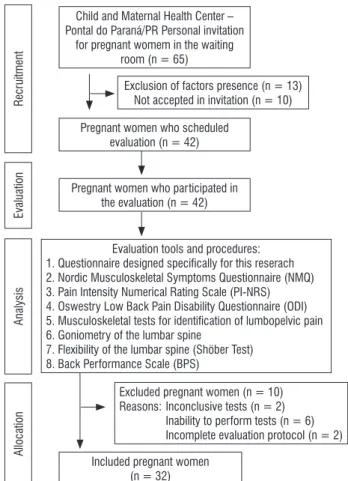
For this study, pregnant women were enrolled in the program for the period (from March to August
/ 2104) who met the inclusion criteria (n = 65), but 32 were included (Figure 1). While they were wait -ing their consultation with an obstetrician and/or physiotherapist in the Health Center, they were in-vited to participate in the study individually and after
explanation about that, they signed written consent.
Child and Maternal Health Center – Pontal do Paraná/PR Personal invitation
for pregnant womem in the waiting room (n = 65)
R
ecr
uitment
Evaluation
Analysis
Allocation
Exclusion of factors presence (n = 13) Not accepted in invitation (n = 10)
Pregnant women who scheduled evaluation (n = 42)
Pregnant women who participated in the evaluation (n = 42)
Evaluation tools and procedures:
1. Questionnaire designed speciically for this reserach
2. Nordic Musculoskeletal Symptoms Questionnaire (NMQ) 3. Pain Intensity Numerical Rating Scale (PI-NRS) 4. Oswestry Low Back Pain Disability Questionnaire (ODI)
5. Musculoskeletal tests for identiication of lumbopelvic pain
6. Goniometry of the lumbar spine
7. Flexibility of the lumbar spine (Shöber Test) 8. Back Performance Scale (BPS)
Excluded pregnant women (n = 10) Reasons: Inconclusive tests (n = 2)
Inability to perform tests (n = 6) Incomplete evaluation protocol (n = 2)
Included pregnant women (n = 32)
Figure 1 - Experimental design with flow diagram regarding the stages of study
pain in pregnancy has a multifactorial etiology and is related to physiological factors, biomechanics,
vascu-lar, psychological and hormonal (6). By involving the
combination of several factors, this painful condition is not yet fully understood and little is known about the relationship between the disability caused by this condition with the limitations and functional kinetic disorders of the lumbar spine.
Thus, we investigate the musculoskeletal
dis-comfort expressed by pregnant women and their
frequency. Also to estimate the occurrence of lum-bopelvic pain and to describe its main characteristics and triggering/aggravating factors. Then, the purpose of this paper is to determine the association of low back pain disability with history, intensity and origin of the pain, and the association with functional ki-netic disorders, such as range of motion of the lumbar
spine, lumbar flexibility and trunk mobility.
Methods
Study design
This research is an exploratory and descriptive
cross-sectional study with non probabilistic conve-nience sample. The study was approved by the Ethics in Research of the Health Sciences Sector of the Federal
University of Paraná Committee (504.452), under Resolution 466/12 of the National Health Council,
conducted according to the Helsinki Declaration re-vised in 2008, and it was registered in the Brazilian
Registry of Clinical Trials (RBR-7pbcdw).
This study included pregnant women enrolled for the program Happy Mother of the Health Secretary
of Pontal do Paraná - Brazil, aged between 18-35
years and gestational age greater than or equal to 20 weeks, representing more than 50% of pregnant
women in those health centers. They excluded those
with a clinical diagnosis of high-risk pregnancy; twins pregnancy; presence of cognitive/mental disabilities; urinary tract infection; neurological dysfunction; her-niated lumbar disc; and previous surgery in the spine, pelvis, hip or knee.
For sample size calculation, we assumed the
proportion of pregnant women in 8.51% (236) of all women of the city in the age group 15-39 years
4
reliable tests for the diagnosis of posterior pelvic
pain. So for statistical analysis and confirmation of
these claims, in our study the following criteria was
established for defining the location of pain: a posi
-tive result in, at least, two of the first three tests, the
pain was rated as lumbar origin. And in positivity cases, at least, two of the last three tests, the pain was rated as sacroiliac or posterior pelvic origin. If the participants presented positive result in two of three tests lumbar origin associated with positivity in two of three tests of sacroiliac origin, pain was rated as the origin in both regions.
6) Goniometry of lumbar spine: to measure the maximum range active joint of the lumbar spine in
degrees was used goniometry protocol (22) of the
lumbar spine (flexion, extension, lateral flexion and
rotation on both sides), with the 20 cm goniometer (Carci®), by the same examiner.
7) Schober Test (19): in order to check the lum
-bosacral flexibility of the study participants.
8) Back Performance Scale (23): used to check overall trunk mobility and physical performance. It consists of 5 standardized tests (Sock Test, Pick-up Test, Roll-up Test, Fingertip-to-Floor Test and Lift Test). The score for each test was graded as 0 (can
easily perform), 1 (can perform with less effort), 2
(can perform with effort) and 3 (does not perform completely or needs help), all of which are added to the general score.
Statistical analysis
The Statistical Package for the Social Sciences
(SPSS) software, version 21.0 for Windows was
used. In their totality, the data were submitted to the Shapiro Wilk test to test the normality of distri-bution. The categorical variables were described in absolute frequency (n) and relative frequency (%), while numerical variables were described in mean ± standard error of the mean.
For the analysis of the origin of the low back pain
and the data referring to the Nordic Musculoskeletal
Questionnaire the Pearson chi-square test was used, corrected by the Likelihood Ratio for small samples.
The level of significance was set at p < 0.05.
Aiming to identify aggravating factors for the func-tional limitation of the lumbar spine (ODI), correla-tion tests were performed by individually associating each variable collected with the score obtained in Experimental procedure
The participants were evaluated with:
1) Questionnaire designed specifically for the
research: the questionnaire contained socio demo-graphic, obstetric and historical low back pain data;
2) Nordic Musculoskeletal Symptoms Questionnaire validated in Brazil (15): to evaluate
the occurrence of musculoskeletal symptoms of pain, numbness, tingling or discomfort in the nine regions of the body (neck, shoulder, arms, elbows, forearms,
wrists/hands/fingers, dorsal, lower back and hip/ lower limbs), from the 4-point Likert scale (no, rarely,
often and always). Study participants reported the occurrence of symptoms considering the gestational months and seven days prior to the interview;
3) Pain Intensity Numerical Rating Scale (PI-NRS) (16): to evaluate the intensity of low back pain in
the seven days preceding the interview was used
11-point pain intensity numerical rating scale that consists of 11 numbers from 0 (no pain) to 10 (maxi -mum pain) in a horizontal line associated with
fa-cial expressions;
4) Oswestry Lowe Back Pain Disability Questionnaire or ODI (Oswestry Disability Index) validation into Portuguese language (17): to evalu
-ate the lumbar disability index. It consists of 10 ques
-tions in 6-point Likert scale where the highest score
indicates greater functional limitation. The total score was calculated by summing the points transformed into percentage and interpreted as follows: 0 to 20%
= minimal disability; 20 to 40% = moderate disability; 40 to 60% = severe disability; 60 to 80% = very severe disability; 80 and 100% = complete failure.
5) Musculoskeletal clinical tests to identify the
lumbopelvic pain: six clinical trials were selected, conducted by the same examiner to identify the origin
and site of low back pain: provocation test lumbar
pain (18), Laségue test (19), SLR - Straight Leg Raise test (19), PPPP - Posterior Pelvic Pain Provocation test (5, 18, 20), Gaenslen test (19, 20) and ASLR -
Active Straight Leg Raise (5, 20). The PPPP, due to stretching of structures of this region, allows the detection of the presence of pain in pregnant
wom-en (11, 18). The other two tests, Gawom-enslwom-en test and
ASLR, were chosen because they are established in the literature on differential diagnosis of the lower
back pain source with neural involvement (18). The
last three tests are recommended by European
5
the ODI. For the parametric data the Pearson
cor-relation test was used and the level of significance was set at p < 0.05. The magnitude scale (24) used to interpret the correlation coefficients was considered: r < 0.1 = trivial; between 0.1 - 0.29 = small; 0.30 - 0.49 = moderate; 0.50 - 0.69 = high; 0.70 - 0.90 = very high
and r > 0.90 = almost perfect.
Results
Table 1 shows the characteristics of the sample.
Table 1 - Sociodemographic and obstetric characteristics.
Variable
Age in years (mean ± SEM) 25.72 ± 1.02 Gestational age in weeks (mean ± SEM) 30.09 ± 1.11 Schooling (n, %)
Incomplete Middle School 2 (6.3)
Complete Middle School 2 (6.3)
Incomplete High School 5 (15.6)
Complete High School 13 (40.6)
Incomplete Higher Education 6 (18.8)
Graduated 4 (12.5)
Occupation (n,%)
Housewife 16 (50)
Student 3 (9.4)
Teacher 3 (9.4)
Others 10 (31.3)
Physical Activity Practice (n, %)
Walking 3 (9.4)
Gym 14 (43.8)
Yoga 1 (3.1)
Does not practice 14 (43.8)
Number of pregnancies (n, %)
Only one 12 (37.5)
Two 12 (37.5)
Three 4 (12.5)
Four or more 4 (12.5)
Number of Abortions (n, %)
None 26 (81.3)
Only one 5 (15.6)
Two 1 (3.1)
Number of vaginal deliveries (n, %)
None 18 (56.3)
Only one 8 (25)
Two 3 (9.4)
Three 3 (9.4)
Number of cesarean deliveries (n, %)
None 29 (90.6)
Only one 3 (9.4)
Note: SEM = Standard Error of the Mean.
Regarding to the history of low back pain, 13 (40.6%) women reported their occurrence before
gestation, with frequency of two to more times per
month for most of them (46.2%), followed by only once a month (23.1%), and two more times each 6 months (30.8%). Interestingly, of the 13 women only 4 (30.7%) were under treatment (medicated) and 69.3% did not undergo any treatment for symptom
relief. Differently, during pregnancy, 30 (93.8%) preg-nant women reported the presence of low back pain,
and the majority (90%) had the first episode of pain after 14 gestational weeks, against only 10% of them experiencing this condition in the first trimester of
Gestation. The daily frequency of this condition was
reported by 63.3% of the pregnant women in the last four weeks; 23.3% with a frequency of 3 to 6 times a week and 10% of 1 to 2 times a week, and only 10%
sought treatment for this condition. Still, the long-term walk without professional guidance/follow-up
was cited as the main triggering factor in 36.7% of
pregnant women, and the fact of lying down, sitting
or relaxing, decreased pain by 76.7%.
A survey of the occurrence of
musculoskel-etal discomfort using the Nordic Musculoskelmusculoskel-etal
Questionnaire revealed that the presence of pain, tingling or numbness in the lumbar region was the
most reported (90.6%) in the different gestational months, followed by the dorsal region (62.5%) and the hip region (56.2%), neck (40.7%), ankle/feet (34.3%), wrists/hands (31.3%), shoulders (28.1%), knees (15.6%) and elbows (3.1%). Thus, the discom -forts felt in the lumbar region were responsible for preventing the activities of daily living in 50% of pregnant women. Still, from the 32 pregnant women
evaluated, 25 (78.1%) reported the presence of pain in this region in the last 7 days. Pearson’s chi-square
test (corrected by the Likelihood Ratio) applied to verify the periodicity of the referred symptom in the different regions showed that the lumbar region was the only one, as always, present in the gestational period, χ² (3, n = 30) = 17.75, p = 0.000, differing from the others when compared statistically to other fre-quency possibilities, most often reported to other regions as never before.
From the 30 pregnant women who reported pain on the initial questionnaire, their intensity
was graded by PI-NRS, which presented a median of 7.5 (0 - 10), being classified as low to 6.7%, mod
6
Table 2 - Association of symptoms and positivity in specific tests of gestational lumbopelvic pain with the origin/site of pain
Lumbopelvic pain
test Description Lumbar Pelvic Both Total LR p–value Cramer’s V
Lumbar pain provocation Positive Negative 4 7 0 7 12 0 16
14 28.16 0.000* 0.600
Laségue Positive
Negative 3 8 1 6 8 4 12
18 6.472 0.039* 0.456
SLR Positive Negative 6 5 1 6 11 1 18
12 12.59 0.002* 0.612
PPPP Positive Negative 1 10 3 4 9 3 13
17 11.295 0.004* 0.582
Gaenslen Positive Negative 0 11 3 4 11 1 14
16 25.011 0.000* 0.805
ASLR Positive Negative 4 7 2 5 10 2 16
14 7.846 0.020* 0.495
Total 11 7 12 30
Note: * p < 0.05, Pearson’s chi-square test (corrected by Likelihood Ratio). Abbreviations: LR, Likelihood Ratio; SLR, Straight Leg Raise; PPPP, Posterior Pelvic Pain Provocation; ASLR, Active Straight Leg Raise.
disability index assessed from the ODI revealed a mean of 23.9 ± 3.2 points, the majority (40.6%) being classi
-fied as low disability, 37.5% moderate, 15.6% severe and 6.3% very severe.
Through the specific tests of lumbopelvic pain
used in this study, it was possible to identify that
23.4% presented positivity in the tests indicating
the sacroiliac/posterior pelvic region as its origin,
followed by 36.6% for low back pain and 40% pre -sented a combination of the pain site. Positively, all
of the specific lumbopelvic pain tests used in this
study were associated with the origin of the analyzed pain (lumbar or posterior pelvic region), using the Pearson chi-square test, corrected by the Likelihood
Ratio (Table 2). For example, in the tests for verifica -tion of sacroiliac pain origin, as in the Posterior Pelvic Pain Provocation test, statistical analyses (χ² (2, n =
30) = 11.29, p = 0.004, Cramer’s V = 0.582) suggest that based on the relative risk rule, all those tested
positive were 4.6 times more likely to originate from
sacroiliac pain than lumbar origin.
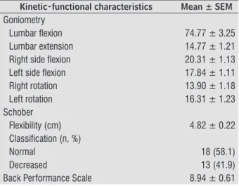
The assessment of joint amplitude of the lumbar
spine using goniometry revealed that most women presented limitation in all movements, reducing the
mean of the maximum active joint amplitude in this sample (Table 3). At the same time, the flexibility of
the lumbar spine measured by the Schober test was
decreased in 41.9% of the pregnant women (Table 3).
Likewise, the results obtained by the Back Performance Scale presented, on average, low scores
(8.94 ± 0.61), with the majority classified with mod -erate limitation in general trunk mobility and physi-cal performance. The tests performed revealed that
most of the evaluated ones (53.1%) did not reach their
malleolus to put their Sock Test in need of help to do
so; 34.4% had difficulty reaching objects on the floor,
needing support on one or both legs to do it (Pick-up
test); 59.4% presented great limitation to sit from the
position of dorsal position (Roll-up test). However,
54.8% could reach the ground from the orthostatic
position with little effort, while 83.8% presented a low
(46.9%) to moderate (46.9%) limitation to support a
weight of 5 kg for one minute (Lift test).
Table 3 - Lumbar spine joint amplitude, lumbar flexibility and general trunk mobility of the evaluated preg-nant women
Kinetic-functional characteristics Mean ± SEM
Goniometry
Lumbar lexion 74.77 ± 3.25
Lumbar extension 14.77 ± 1.21
Right side lexion 20.31 ± 1.13
Left side lexion 17.84 ± 1.11
Right rotation 13.90 ± 1.18
Left rotation 16.31 ± 1.23
Schober
Flexibility (cm) 4.82 ± 0.22
Classiication (n, %)
Normal 18 (58.1)
Decreased 13 (41.9)
Back Performance Scale 8.94 ± 0.61
7
disability also showed moderate correlations to seek professional help to reduce pain (r = -0.346), lower
level of lumbar extension measured by goniometry
(r = -0.301), and limitation of lumbar flexibility mea -sured by the Schober test (r = -0.371). Furthermore,
a significant negative correlation of weak magnitude was observed for a lower degree of lumbar flexion
measured by goniometry (r = -0.280). The analysis of the correlation of the various
vari-ables of the study with lumbar incapacity, quantified by the ODI score are presented in Table 4. Significant
positive correlation of moderate magnitude was ob-served in most of the presented variables, the highest magnitude being the provocation test (r = 0.540),
presence of pain in the last seven days (r = 0.510),
Pick-up test (r = 0.565) and Back Performance Scale
(r = 0.503) that presented high magnitude. Lumbar
Table 4 - Correlation of lumbar incapacity (Oswestry Disability Index) with obstetric characteristics, history of low back pain and functional kinetic conditions (specific tests for lumbopelvic pain, lumbar spine articular range of motion, lumbar flexibility and general trunk mobility)
Variables r p-valor R2
Obstetric
Gestational age 0.353 0.006* 0.12
History of low back pain during pregnancy
Presence of pain during pregnancy 0.341 0.024* 0.11
Frequency of pain in the last 4 weeks 0.398 0.008* 0.15
Pain worsens when walking 0.428 0.006* 0.18
PI-NRS Score 0.402 0.003* 0.16
PI-NRS Classiication 0.460 0.002* 0.21
Seek professional help to decrease pain -0.345 0.027* 0.11
Musculoskeletal tests to identify lumbopelvic pain
Lumbar pain provocation 0.540 0.000* 0.29
Straight Leg Raise 0.373 0.013* 0.13
Posterior Pelvic Pain Provocation 0.356 0.018* 0.12
Gaenslen Test 0.473 0.002* 0.22
Active Straight Leg Raise 0.455 0.003* 0.20
Lumbar discomfort by Nordic Questionnaire
Impediment of performing physical activities 0.470 0.002* 0.22
Presence of pain in the last 7 days 0.510 0.001* 0.26
Goniometry
Lumbar lexion -0.280 0.031* 0.07
Lumbar extension -0.301 0.027* 0.09
Flexibility (Shober in cm) -0.371 0.006* 0.13
Back Performance Scale
Sock test 0.338 0.017* 0.11
Pick-up test 0.565 0.000* 0.31
Roll-up test 0.431 0.003* 0.18
Fingertip-to-loor test 0.447 0.002* 0.20
Lift test 0.444 0.002* 0.19
Total do Back Performance Scale 0.503 0.000* 0.25
Note: * Pearson correlation test with signiicance level of p < 0.05. Abbreviation: PI-NRS, Pain Intensity Numerical Rating Scale.
Discussion
This study was conducted with the main objective
of relating the lumbar incapacity provided by lum-bopelvic pain from the twentieth gestational week
with the limitations and kinetic-functional dysfunc-tions of the lumbar spine. Relevance to this study is due to the fact that up to now, adaptations of the musculoskeletal system during morphophysiological
8
73% to 95.2%. In our study, the majority (63.3%) of the pregnant women reported experiencing this pain
daily, being graded to 50% of them as high intensity
and moderate to severe low back disability to 56.9%
of the sample. Characteristics of gestational low back pain may vary throughout pregnancy, and even in the same pregnancy from one pregnancy to anoth-er, because it is assumed to depend on the balance between perceived pain, limitation, and individual
ability to seek coping strategies (6). Another study (8) stated that 73% of the pregnant women reported
low back pain, most of them felt as a spike, with daily frequency, duration of at least one hour and average intensity around seven. This same study observed an association of moderate magnitude between pain scores and lumbar incapacity. In contrast to these
findings, a study (5) of 182 Dutch women found a prevalence of 60.4% of pelvic low pain, with a mean score of 3.6 (greater than 5 only 20% of the sample)
and minimal disability measured by the Quebec Back Pain Disability Scale (severe incapacity was found for only 20% of pregnant women).
We also observed in our study that lumbopelvic pain was responsible for preventing the performance of daily life activities in 50% of pregnant women, with a positive correlation of moderate magnitude (r =
0.470) with lumbar incapacity measured by ODI.
Previous study (8) have already suggested that low back pain interferes in some way with the functional-ity of the pregnant women, with work impairment, generating absenteeism and frequent leaves from work. But it did not take into account the relationship between the degree of disability and the intensity of pain, which is related to the higher number of sick
leaves (7). This corroborates our findings, since the
greater the intensity of pain (r = 0.402) or more se
-vere its classification (r = 0.460) by the PI-NRS, the
greater the lumbar incapacity, causing difficulties for
their productivity. However, contrary to these results,
a study (26) of 100 Turkish pregnant women showed
no correlation between pain intensity and lumbar incapacity. The different results may be due to the fact that pregnant women included in their study had
low values of pain intensities measured by the Visual
Analog Scale.
As expected, our study revealed that gestational
age is associated with the appearance of this con-dition, that is, the greater the gestational age, the greater the possibility of occurrence of lumbopelvic pain (r = 0.353). The majority (90%) of the pregnant
(25), especially those related to kinetic-functional dysfunctions. The strength of the current study is the multidimensional assessment approach used,
including specific clinical trials for identification of
the origin of low back pain, assessment of range of
motion and lumbar flexibility, as well as overall trunk
mobility and/or physical performance. The evalua-tion was performed by a single evaluator, but was limited to only one repetition of each test, making it impossible to evaluate its reliability, due to the
long evaluation protocol (an average of 40 minutes).
This factor restricted the participation of pregnant women who were waiting for obstetrical consulta-tion, arguing that there was no time to participate in the evaluation. Differently, the pregnant women who were waiting for physical therapy consultation for prenatal orientation did not report this inconve-nience. However, the reasons related to this difference were not investigated.
In the present study, musculoskeletal discomforts
ascertained by the Nordic Questionnaire during ges
-tation revealed that the lumbar region (90.6%) was
the most cited for the occurrence of symptoms such
as pain, numbness, tingling or discomfort. Still, 78.1%
reported the presence of pain in this region in the
last 7 days, being their periodicity the only region to
present as always present in the gestational period.
This differs from a study (1) conducted in India to
validate a questionnaire to identify musculoskeletal discomforts during gestation. In the third gestational trimester, the most frequent discomforts were calf
muscle weakness (64.6%), followed by foot pain (37.1%), and low back pain (33.7%). Already, in the
second trimester, this study revealed that calf pain
was the most frequent complaint (47.8%), followed by low back pain (42%) and pelvic pain (37%). The researchers found that pregnant women experience
more of these symptoms during their hygiene activi-ties, when trying to put on their shoes, when getting up from chairs or sitting with the lower limbs in
extension. Contradictions in the results may be due
to the evaluation instrument used and the cultural
and environmental differences experienced by the
pregnant women.
Differently, a narrative study (2) on musculoskel-etal discomforts in gestation reports the occurrence of low back pain in 50 to 80% of pregnant women,
as the most prevalent. Studies (11, 12, 13, 14) on the
9
women had the first episode of pain after 14 ges -tational weeks, being compatible with most of the
previous studies (3, 4, 8, 27), possibly related to
direct fetal pressure against the neural elements of the lumbar spine and greater overload in the lumbar
muscles (13).
However, there are differences in the literature regarding the different risk factors. In our study, we observed no association of lumbar disability with age, level of schooling, number of previous births or
type of delivery, as previously reported (4) in 569
women. Although these results were contradictory, a
study (3) with 1158 women presented an association
of gestational low back pain with educational level (corresponding to secondary education) and younger. Possibly, this association may be related to greater sensitivity to the changes in this period, provided by
the action of hormones or collagen laxity that is more
pronounced in younger women (8).
Unexpectedly, there was no association in our
study with reports of pain prior to gestation or the practice of physical activity before or during gesta-tion. Previous reports (5, 8) have shown that the presence of pain before pregnancy is a potential risk factor and, conversely, the practice of physical activity
is a preventive factor (4). However, corroborating our findings, previous studies (3, 6) found no association
with previous physical activity or during pregnancy, but found a strong association with a previous his-tory of pain.
Observational (28) and clinical (29) studies demon-strated that the practice of physical activity before or during pregnancy reduced the risk of lumbopelvic pain. However, a study (30) was carried out with 855 pregnant women aimed to evaluate a 12-week exercise program during the second half of pregnancy on the prevalence of lumbopelvic pain and the number of medical licenses in the period; revealed that the program did not appear to inluence the prevalence, but signiicantly reduced the proportion of women applying for sick leaves for this condition. Those results suggested that they can cope better with the disorder compared to women who received standard prenatal care.
Inactivity may be a predisposing factor to
pain-ful experiences (30) and a cross-sectional study (3)
showed that there is an association of gestational low back pain with the longer time spent in the lying position, independent of the mattress characteristic. However, for pregnant women with low back pain, rest may be a strategy to relieve symptoms, since our
study revealed that the majority (76.7%) found pain relief at bedtime, sit or relax, not related to lumbar incapacity. Previous studies (8, 14) also reported im -provement of the symptom with rest, possibly related
to muscle relaxation (12), and worsened with the
orthostatic position. Also, according to the partici-pants, our study revealed that the walking, and this
is significantly related to lumbar incapacity, possi -bly due to overload in the musculoskeletal system
(14), but also, to the increase of the width of the step
that consumes more energy and the longer time of maintenance of the feet on the ground, decreasing the simple support time, used as strategy for greater stability and support of body weight (25).
Different clinical tests can be used to identify the origin of this painful condition. The European
com-munity (20) has defined that pelvic pain is a specific
type of low back pain, which can occur alone or in
conjunction with low back pain. Its diagnosis should
be based on the use of different clinical tests, includ-ing: Posterior Pelvic Pain Provocation test, Gaenslens
test and ASLR (13, 20, 21). As well as the PPPP test (6, 31) has high sensitivity and specificity for pos -terior pelvic pain, the lumbar pain provocation test can be used in the diagnosis of gestational low back
pain (11, 18). Our study revealed that posterior pel
-vic pain was less frequent (23.4%) followed by low back pain (36.6%), most of which were combined (40%). Previous study (14) of prevalence that have used specific clinical trials for low back pain are rare
because different criteria can be used to characterize
the origin of pain. A study (6) of 64 women reported gestational lumbopelvic pain, found that 17% identi
-fied the lumbar region as a site of pain, 33% of the
pelvic region and 50% of both. However, this study differentiated the origin of the pain only by the use
of a body diagram. Another study (4) found a similar
prevalence for the origin of pain in the body diagram, and revealed that the combination of the two pain sites results in a higher rate of lumbar incapacity in the ODI compared to women who present these pain conditions in only one region. In agreement with such researchers, the fact that different regions have been
identified as a site of pain between the different stud
-ies may be explained by the inability of most women
to anatomically locate pain in a drawing/diagram,
making the specific clinical test extremely important to their clarification.
10
of gestational low back pain with yoga affirmed that before the intervention, 17% of pregnant women pre -sented lumbar pain (proven by means of the lumbar pain provocation test), 20% presented posterior
pel-vic pain (proven by the PPPP test), but the majority (63.3%) presented a combination of both. The dif -ferent results found with the proposed intervention indicate that the success of the kinesio therapeutic intervention has some relation with the correct
iden-tification of the origin of the pain.
Also, in our study, it was possible to notice that the positivity in the different tests presented a cor-relation with the severity of the lumbar incapacity,
except for the Laségue test, suggesting that this popu -lation rarely presents neural involvement during this
period (11, 19). These data reveal the importance of using specific clinical tests to identify the origin of the pain, but the combination of tests should be explored
in future research to assist in the determination of lumbopelvic pain (5).
It is recognized in the literature that the relaxin
hormone promotes greater ligament elasticity in the
joints, leading to increased joint mobility (14), which
leads to greater demands on muscle and ligament
stabilization (31). However, our study revealed that women with lumbopelvic pain had a decrease in joint amplitude (especially for flexion and extension) of
the lumbar spine in all movements, as well as lumbar
flexibility, these being related to the increase of the
lumbar incapacity. This may reveal that the presence
of pain can lead to joint restraints, possibly due to
the needs imposed on the ligaments and stabilizing muscles, causing muscle spasm.
It is known that physical capacity decreases in the last gestational trimester, being even more limited
to pregnant women with low back pain (23, 26). In
our study, physical performance and overall trunk mobility was assessed by the Back Performance Scale
in terms of five domains. That is, the ability to wear a sock test, to reach objects on the floor from the sit-up
(Pick-up test), to sit (Roll-up test), to reach the ground from the orthostatic position (Fingertip-Floor test) and withstand a weight of 5 kg for one minute (Lift
test). It was verified that the majority of the women
presented limitation to the accomplishment of these activities and there was a positive correlation with the lumbar incapacity (measured by the ODI), that is, the higher the limitation in the physical performance and general mobility of the trunk, the more severe the incapacity low back.
In contrast to these findings, a study with preg
-nant in the third gestational trimester (26) found
that all of them were able to perform the functional
activities evaluated by Katz’s Activity Daily Index
(bathing, dressing, going to the toilet, performing transfers, maintaining continence and eating) inde-pendently, with no correlation with lumbar disability. The contradictory results may be due to the different instruments evaluated for physical capacity measure-ment. However, the functional scores obtained in the study were worse in comparison to those who did not report low back pain. In this way, pregnant with low back pain presented a strong correlation between pain intensity and physical capacity, corroborating
our findings.
It is emphasized that the results of this study should be interpreted with caution, as it has a cross-sectional design, and the correlations found do not
necessarily imply causality (3). For example, the asso -ciation of lumbar disability with higher levels of pain intensity, presence of lumbar pain musculoskeletal
specific tests, greater limitation of lumbar amplitude (flexion and extension), lumbar flexibility, and lower
trunk mobility may be associated with higher lev-els of pain intensity. Interpreted as indicating that these factors increase disability or, conversely, as an indication that they are consequences of disability. However, it is unlikely that a causal factor shows no association with the corresponding condition (3). Therefore, cross-sectional studies are useful in identifying the variables that should be included in prospective studies. However, the sample size of
our study has no statistical power to draw defini -tive conclusions.
Conclusion
According to the results of this study, it was possi-ble to identify that 93.8% of the women reported the presence of pain in the lumbar region during the ges-tational period. This was the most cited region for the occurrence of musculoskeletal discomforts, reported
by the majority of women (56.3%) with periodicity
always present in the gestational period. Pain is
char-acterized in the majority of pregnant women, having a daily frequency (63.3%), with the first episode after 14 weeks (90%), being the main triggering factor
11
(36.7%) classified as high intensity (50%), generating mild incapacity (40.6%).
We have also shown that increased lumbar inca-pacity during pregnancy is associated with positive clinical trials (in particular, the lumbar pain
provoca-tion test), decreased joint amplitude (especially lum
-bar spine flexion and extension), decreased lum-bar flexibility and of general trunk mobility. This repre -sents an important aspect to be observed in the ob-stetric evaluation and they become important goals in the program planning of preventive or corrective
exercises for this condition.
We suggest, for further researches, randomized
clinical trials to evaluate the efficacy of treatments
or preventive measures for lumbopelvic pain
gesta-tional, and should include specific tests for low back pain, assessment of range of motion and flexibility, as
well as evaluation of general trunk mobility, to help in clarifying these issues.
References
1. Ramachandra P, Maiya AG, Kumar P, Kamath A. Preva-lence of musculoskeletal dysfunctions among Indian Pregnant Women. J Pregnancy. 2015;2015:437105.
2. Thabah M, Ravindran V. Musculoeskeletal problems in pregnancy. Rheumatol Int. 2015;35(4):581-7.
3. Kovacs FM, Garcia E, Royuela A, Gonzalez L, Abraira V. Prevalence and factors associated with low back pain and pelvic girdle pain during pregnancy: a multicenter study conducted in the Spanish National Health Ser -vice. Spine. 2012;37(17):1516-33.
4. Malmqvist S, Kjaermann I, Andersen K, Økland I, Brønnick K, Larsen JP. Prevalence of low back and pelvic pain during pregnancy in a Norwegian Popula -tion. J Manipulative Physiol Ther. 2012;35(4):272-8.
5. Mens JM, Huis in’t Veld YH, Pool-Goudzwaard A. Sever -ity of signs and symptoms in lumbopelvic pain during pregnancy. Man Ther. 2012;17(2):175-9.
6. Pierce H, Homer CSE, Dahlen HG, King J. Pregnancy-Related Lumbopelvic Pain: listening to Australian women. Nurs Res Pract. 2012;2012:387428.
7. Gutke A, Olsson CB, Völlestad N, Öberg B, Wikmar LN, Robinson HS. Association between lumbopelvic pain, disability and sick leave during pregnancy – a comparison of three Scandinavian cohorts. J Rehabil Med. 2014;46(5):468-74.
8. Madeira HGR, Garcia JBS, Lima MVV, Serra HO. Inca -pacidade e fatores associados à lombalgia durante a gravidez. Rev Bras Ginecol Obstet. 2013;35(12):541-8.
9. Bastiaenen CH, de Bie RA, Vlaeyen JW, Goossens ME, Leffers P, Wolters PM, et al. Long-term effectiveness and costs of a brief self-management intervention in women with pregnancy-related low back pain after delivery. BMC Pregnancy Childbirth. 2008;8:19. 10. Vermani E, Mittal R, Weeks A. Pelvic girdle pain and
low back pain in pregnancy: a review. Pain Pract. 2010;10(1):60-71.
11. 11. Martins RF, Silva JLP. Prevalência de dores nas costas na gestação. Rev Assoc Med Bras. 2005;51(3):144-7.
12. Santos MM, Gallo AP. Lombalgia gestacional: prevalên -cia e características de um programa pré-natal. Arq Bras Ciên Saúde. 2010;35(3):174-9.
13. Lima AS, Gomes MRA, Araújo RC, Pitangui ACR. Análise da postura e frequência de lombalgia em gestantes: estudo piloto. J Health Sci Inst. 2011;29(4):290-3. 14. Gomes MRA, Araújo RC, Lima AS, Pitangui ACR.
Lombalgia gestacional: prevalência e característi -cas clíni-cas em um grupo de gestantes. Rev Dor. 2013;14(2):114-7.
15. 15. Pinheiro FA, Troccoli BT, Carvalho CV. Validação do Questionário Nórdico de Sintomas Osteomuscula -res como medida de morbidade. Rev Saúde Pública. 2002;36(3):307-12.
16. Farrar JT, Young JP Jr, LaMoreaux L, Werth JL, Poole RM. Clinical importance of changes in chronic pain intensity measured on an 11-point numerical pain rating scale. Pain. 2001;94(2):149-58.
12
18. Martins RF, Pinto e Silva JL. Treatment of pregnancy-related lumbar and pelvic girdle pain by the yoga method: a randomized controlled study. J Altern Complement Med. 2014;20(1):24-31.
19. Magee DJ. Avaliação Musculoesquelética. 5th ed. São Paulo: Manole; 2010.
20. Vleeming A, Albert HB, Östgaard HC, Sturesson B, Stuge B. European guidelines for the diagno-sis and treatment of pelvic girdle pain. Eur Spine J. 2008;17(6):794-819.
21. Kanakaris NK, Roberts CS, Giannoudis PV. Pregnan -cy-related pelvic girdle pain: an update. BMC Med. 2011;9:15.
22. Marques AP. Manual de goniometria. 3rd ed. São Paulo: Manole; 2014.
23. Strand LI, Moe-Nilssen R, Ljunggren AE. Back Per -formance Scale for the assessment of mobility-re-lated activities in people with back pain. Phys Ther. 2002;82(12):1213-23.
24. Hopkins WG. Measures of reliability in sports medi-cine and science. Sports Med. 2000;30(1):1-15.
25. Branco M, Santos-Rocha R, Vieira F. Biomechanics of gait during pregnancy. Sci World J. 2014;2014:527940. 26. Coban A, Arslan GG, Colakfakioglu A, Sirlan A. Impact on quality of life and physical ability of pregnancy-related back pain in the third trimester of pregnan-cy. J Pak Med Assoc. 2011;61(11):1122-4.
27. Yoo H, Shin D, Song C. Changes in the spinal curvature, degree of pain, balance ability, and gait ability accord-ing to pregnancy period in pregnant and nonpregnant woman. J Phys Ther Sci. 2015;27(1):279-84.
28. Mogren IM. Previous physical activity decreases the risk of low back pain and pelvic pain during preg-nancy. Scand J Public Health. 2005;33(4):300-6.
29. Mørkved S, Salvesen KA, Schei B, Lydersen S, Bø K. Does group training during pregnancy prevent lum-bopelvic pain? A randomized clinical trial. Acta Obstet Gynecol Scand. 2007;86(3):276-82.
30. Stafne SN, Salvesen KÅ, Romundstad PR, Stuge B, Mørkved S. Does regular exercise during pregnancy influence lumbopelvic pain? A randomized controlled trial. Acta Obstet Gynecol Scand. 2012;91(5):552-9. 31. Olsén MF, Gutke A, Elden H, Nordenman C, Fabri
-cius L, Gravesen M, et al. Self-administered tests as a screening procedure for pregnancy-related pelvic girdle pain. Eur Spine J. 2009;18(8):1121-9.
Received in 09/11/2015 Recebido em 11/09/2015