C A SE R EPORT
186 J Vasc Bras. 2015 Apr.-June; 14(2):186-188 http://dx.doi.org/10.1590/1677-5449.0077
Leiomyosarcoma of the great saphenous vein
Leiomiossarcoma de veia safena magna
Alexandre Campos Moraes Amato1
*
, Ana Júlia de Deus Silva1, Ricardo Virgínio dos Santos1, Salvador José de Toledo Arruda Amato2
Abstract
A 56-year-old male patient presented with a complaint of two painful, hard, palpable nodules in the right lower limb. A Doppler ultrasound scan revealed the presence of nodules, likely to be neoplastic. Computed angiography showed two solid hypervascular nodules in the right great saphenous vein, fed by branches of the posterior tibial artery. Embolization of the nodules using surgical cyanoacrylate was performed, followed by an excisional biopsy. Anatomical pathology and immunohistochemical analysis identiied the nodule as a high-grade leiomyosarcoma, characterized by ten mitotic igures per ten high-power ields, necrosis and cell pleomorphism. Immunohistochemical analysis results were positive for caldesmon and desmin labeling. A second surgical procedure was performed to enlarge the free margins.
Keywords: neoplasms; vascular tissue neoplasms; blood vessels.
Resumo
Paciente do sexo masculino, 56 anos, com queixa de dois nódulos palpáveis, dolorosos e rígidos, em membro inferior direito. O eco-Doppler colorido evidenciou nódulos de provável natureza neoplásica na veia safena magna direita distal, com luxo de baixa velocidade no seu interior. A angiotomograia evidenciou dois nódulos sólidos hipervascularizados no trajeto da veia safena magna direita, possuindo ramos nutridores provenientes da artéria tibial posterior. Para realizar a biópsia excisional dos nódulos, optou-se inicialmente pela embolização do tumor com cola cirúrgica de cianoacrilato, devido à sua alta vascularização. Os exames anatomopatológico e imuno-histoquímico evidenciaram leiomiossarcoma de alto grau, com dez mitoses por dez campos de grande aumento, necrose tumoral e pleomorismo celular. A imuno-histoquímica demonstrou positividade para os marcadores Caldesmon (anticorpo hCD) e Desmina (anticorpo D33). O paciente realizou uma nova cirurgia para ampliação de margens comprometidas e está em acompanhamento clínico com a Oncologia.
Palavras-chave: neoplasias; neoplasias de tecido vascular; vasos sanguíneos.
1Universidade de Santo Amaro – UNISA, São Paulo, SP, Brazil. 2Amato - Instituto de Medicina Avançada, São Paulo, SP, Brazil. Financial support: None.
Conlicts of interest: No conlicts of interest declared concerning the publication of this article. Submitted: October 15, 2014. Accepted: November 20, 2014.
187
J Vasc Bras. 2015 Apr.-June; 14(2):186-188 Alexandre Campos Moraes Amato, Ana Júlia de Deus Silva et al.
INTRODUCTION
Leiomyosarcomas of the vascular system are
extremely rare tumors, accounting for approximately
2% of this type of neoplasm,
1and most often
affecting the gastrointestinal tract or the uterus.
2Leiomyosarcomas of major blood vessels account
for 0.95% of all sarcomas and the inferior vena
cava is the most common site for these cases. This
type of sarcoma originates in smooth muscle cells
and, despite its rarity, leiomyosarcoma is the most
frequent type of primary malignant vascular tumor.
Leiomyosarcoma is ive times more frequent in the
veins than in the arteries,
1-4and this type of sarcoma
affects men and women equally, with peak incidence
from 55 to 61 years of age.
4CASE REPORT
A 56-year-old male patient presented with a
complaint of two painful, hard, palpable nodules in the
right lower limb. A Doppler ultrasound scan revealed
the presence of nodules (most likely neoplastic) with
both intraluminal and extraluminal components and
reduced blood low in the distal portion of the right
great saphenous vein (Figure 1). Computed angiography
showed two solid hypervascular nodules in the right
great saphenous vein, measuring 1.7 × 0.8 cm and 1.9 ×
0.8 cm and fed by branches of the posterior tibial artery
(Figure 2
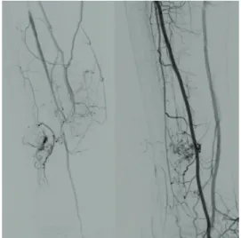
). In view of the signiicant vascularization
of the tumors (Figure 3, left), it was decided that
the nodules should be embolized
(Figure 3, right)
prior to the excisional biopsy using cyanoacrylate
surgical glue (Glubran
) combined with iodinated
poppy seed oil (Lipiodol
). The tumors were excised
4 days after embolization and the specimen was sent
for anatomical pathology and immunohistochemical
analysis, which identiied the nodule as a high-grade
leiomyosarcoma (Figure 4
), characterized by ten mitotic
igures per ten high-power ields, necrosis and cell
pleomorphism. Immunohistochemical analysis results
were positive for caldesmon (anti-hCD antibody)
and desmin (antibody clone D33) labeling. A second
surgical procedure was performed to enlarge the free
margins. The patient is currently being followed up
by the oncology department. The patient authorized
publication of his case and signed a consent form.
DISCUSSION
Because the disease is malignant, fast-evolving
and demands wide surgical margins,
5halting the
progression of leiomyosarcoma depends on early
Figure 1. Neoplastic and vascularized nodules shown on Doppler ultrasound.
Figure 2. hree-dimensional angiotomography reconstruction revealed the presence of two solid hypervascular nodules in the right great saphenous vein, fed by branches of the posterior tibial artery.
188 J Vasc Bras. 2015 Apr.-June; 14(2):186-188 Leiomyosarcoma
diagnosis, high degree of clinical suspicion and
effective intervention.
3Amputation should be considered where lesions
are located below the knee, especially in cases with
high-grade sarcoma,
6as well as in patients with bone or
nerve involvement.
7When the great saphenous vein is
affected, as in the case described here, surgical treatment
increases survival rates by 80-90%.
8Metastases are
present in 44% of cases at the time of diagnosis
9and
in 60% by the time of surgery.
8Diagnosis is quite
dificult, as the case reported here illustrates, since
this patient saw several specialists before a diagnosis
of leiomyosarcoma of the great saphenous vein was
established. The diagnostic dificulty is due to the
great similarity of the clinical examination indings
for this type of tumor and for its differential diagnoses
of lipoma, epithelioid hemangioendothelioma and
angiosarcoma.
4REFERENCES
1. Song KY, Jang YW, Kim MK, Lee GY, Sung RH. Leiomyosarcoma arising in the great saphenous vein: a case report. J Korean Med Sci. 1991;6(4):372-5. http://dx.doi.org/10.3346/jkms.1991.6.4.372. PMid:1844647
2. Fujimoto S, Mizuno R, Saito Y, Nakamura S. Clinical application of wave intensity for the treatment of essential hypertension. Heart Vessels. 2004;19(1):19-22. http://dx.doi.org/10.1007/s00380-003-0725-9. PMid:14685750
3. Marle AG, Bronkhorst MW, Brouwers MA. Leiomyosarcoma of the great saphenous vein: a case report and review of the Figure 4. Microscopy of vein wall, showing high-grade leiomyosarcoma 2 (FNCLCCC) and high grade TNM, tumor necrosis with ischemic pattern, marked cellular pleomorphism and 10 mitoses per 10 high power ields.
literature. Sarcoma. 2004;8(4):135-9. http://dx.doi.org/10.1155/ S1357714X04000234. PMid:18521408
4. Bibbo C, Schroeder M. Review of vascular leiomyosarcoma and report of a case localized to the greater saphenous vein of the ankle. J Foot Ankle Surg. 2011;50(3):329-35. http://dx.doi.org/10.1053/j. jfas.2011.01.002. PMid:21435912
5. Cormier JN, Pollock RE. Soft tissue sarcomas. CA Cancer J Clin. 2004;54(2):94-109. http://dx.doi.org/10.3322/canjclin.54.2.94. PMid:15061599
6. Banks AS, Downey MS, Martin DE, Miller SJ. Tumors: part 2, soft tissue masses. In: Doeney MS, Gredlein CM, editors. McGkamry`s Comprehensive Textbook of Foot and Ankle Surgery. 3rd ed. Philadelphia: Lippincott Williams & Wilkins; 2001. p. 1339-1368.
7. Reix T, Sevestre H, Sevestri-Pietri M-A, Szychta P, Pietri J. Primary malignant tumors of the venous system in the lower extremities. Ann Vasc Surg. 1998;12(6):589-96. http://dx.doi.org/10.1007/ s100169900205. PMid:9841691
8. Humphrey M, Neff J, Lin F, Krishnan L. Leiomyosarcoma of the saphenous vein: a case report and review of the literature. J Bone Joint Surg Am. 1987;69(2):282-6. PMid:3805093.
9. Abed R, Abudu A, Grimer RJ, Tillman RM, Carter SR, Jeys L. Leiomyosarcomas of vascular origin in the extremity. Sarcoma. 2009;2009:385164. http://dx.doi.org/10.1155/2009/385164. PMid:19587823
*
Correspondence
Alexandre Campos Moraes Amato Av. Brasil, n. 2283 - Jardim America CEP 01431-001 - São Paulo (SP), Brazil E-mail: [email protected]
Author information
ACMA – professor of Vascular Surgery at Universidade de Santo Amaro (UNISA), São Paulo, SP, Brazil; a member with Sociedade Brasileira de Angiologia e Cirurgia Vascular; Board certiied in Vascular and Endovascular Surgery from Sociedade Brasileira de Angiologia e Cirurgia Vascular; and Board-certiied in Vascular Doppler Ultrasound from pelo Colégio Brasileiro de Radiologia. AJDS – medical student at Universidade de Santo Amaro (UNISA). RVS – professor of Vascular Surgery at Universidade de Santo Amaro (UNISA). SJTAA – chief of the Vascular Surgery team at Amato, Instituto de Medicina Avançada.
Author contributions
Conception and design: ACMA, SJTAA Analysis and interpretation: ACMA, RVS, SJTAA Data collection: ACMA, SJTAA Writing the article: ACMA, AJDS Critical revision of the article: ACMA, SJTAA Final approval of the article*: ACMA, AJDS, RVS, SJTAA Statistical analysis: N/A Overall responsibility: ACMA