A CLINICO-INVESTIGATIVE STUDY OF ALOPECIA AREATA WITH SPECIAL
REFERENCE TO ITS ASSOCIATION WITH VARIOUS SYSTEMIC AND
DERMATOLOGICAL DISORDERS.
Gopal M.G1, Praveen Kumar S2, Sharath Kumar B.C3, Ramesh M4
HOW TO CITE THIS ARTICLE:
Gopal MG, Praveen Kumar S, Sharath Kumar BC, Ramesh M. A clinico-investigative study of alopecia areata with special reference to its association with various systemic and dermatological disorders . Journal of Evolution of Medical and Dental Sciences 2013; Vol2, Issue 48, December 02; Page: 9239-9249.
ABSTRACT: BACKGROUND: Alopecia areata is believed to be an autoimmune condition with a worldwide occurrence. It usually presents as patchy, non-scarring hair loss. There is a paucity of clinical data in Asians. OBJECTIVES: To describe the demographic profile, clinical profile and histopathology of alopecia areata d out the association with various systemic and dermatological disorders. METHODS: 120 cases were included for the study, over a period of 18 months from January 2007 to June 2008. A descriptive study with purposive sampling was done. Demographic data and a detailed history of the patient to rule out associated systemic and dermatological disorders and various autoimmune disorders were documented. RESULTS:45 (37.5%) out of 120 cases were in the age group of 21-30 years. Male:Female ratio was 2:1. 103 (85.8%) patients were Hindus from urban background who were skilled workers. Parietal area was the commonest area involved over scalp, in 38 (31.67%) patients. All these data are statistically significant. 7 (5.8%) patients had seborrhoeic dermatitis of scalp, 2 (1.6%) patients had lichen planus and Vitiligo. 1 (0.8%) patient had psoriasis. Family history of alopecia areata was noted in 8 (6.7%) patients. Pitting was noted in 14 (11.7%) patients. 41 (93.2%) patients in urban background had emotional stress.
KEY WORDS: Alopecia areata, Autoimmunity, systemic disorders
INTRODUCTION: Alopecia areata is characterized by the loss of hair in circumscribed patches. The patches may be later being round or oval or irregular in shape, of varying size and accompanied by little or no evidence of inflammation. Alopecia areata is a multifactorial disease with autoimmune components, which although seen in genetically predisposed individuals, the real causes have yet to be determined and various factors should be considered. A variety of environmental factors including infections, drugs, trauma, and stress have been suggested as triggering factors for alopecia areata, although most patients of alopecia areata are unaware of any obvious precipitating factors, it is possible though hard to prove, that a diversity of insults, physical or psychological, may trigger episodes of alopecia areatabut there is no influence that they affect prognosis. There are reported associations between alopecia areata and classic auto-immune disorders. The main ones being thyroid disorders and vitiligo. The presence of peribulbar lymphocytic inflammatory infiltrate is a histopathologic characteristic found in most of the terminal hair in one evolutionary stage: catagen or telogen. The follicles become smaller during the course, forming miniaturized hair and are substituted by fibrous tracts.
120 consecutive cases were included for the study. After obtaining the written consent, the study subjects were subjected to detailed history taking including demographic data, drug history, personal history, family history, present and past medical history and history of emotional stress exposure to STD and drug intake. A detailed history of the patient to rule out associated systemic and dermatological disorders and various autoimmune disorders were made. The available records were scrutinized to collect any valid data. A thorough clinical evaluation was done: to assess the clinical pattern, extent severity and duration of alopecia areata, to detect any predisposing or any underlying disease/pathological factors, to assess any other organ system involvement and assess the presence of other dermatological disorders as suggested by detailed history were done. Nail changes were documented in all patients. Histopathology of alopecia areata were studied in selected.
Inclusion criteria: All diagnosed and suspected cases of alopecia areata visiting or referred to Department of Dermatology, Kempegowda Institute of Medical Sciences and Research Centre, Bangalore.
Exclusion Criterion: Patients not willing to give written or informed consent and not willing to participate in the study and various types of alopecias, other than alopecia areata.
Statistical methods: The data was analyzed statistically using descriptive statistics namely Mean, Proportion and Standard Deviation and Chi Square test. Wherever necessary the results are depicted in the form of graphs of percentages.
D
DIISSCCUUSSSSIIOONN::Alopecia areata is a common, unpredictable, non-scarring form of hair loss1. Although
alopecia areata can occur at any ages from infancy to old age2, the first attack is most commonly seen
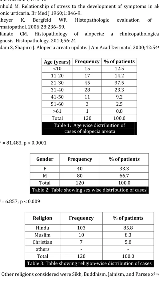
between the age of 5 and 40 years and accounts for 70 to 80% of cases.The onset of alopecia areata may be at any age, peaking between second and fourth decades. In our study, the commonest age group of occurrence was 21-30 years accounting to 37.5%. The next common age group in our study was 31 to 40 years accounting to 23.3%. This clearly indicates that, in our study the commonest age group was between 21 to 40 years, which is in line with the studies done before.
Sex incidence is reported to be equal in UK and USA, but figures from France, Italy and Spain show a considerably higher incidence in males about 2:1 of male:female ratio. Our study shows 66.7% of patients were males and 33.3% were females (approximately the ratio being 2:1). This in line with the studies done before, and is in accordance with the studies done in south-west European countries.
The percentage of Hindus among the patients was 85.8%, next common being Muslims at 8.3% and Christians being 5.8%. The majority of the patients were Hindus. 72.5% of patients were from urban background, and 27.5% were from rural background. 35% of patients were in income range >Rs.3375-6749. 32.5% of patients were in income range Rs.6750-13499. 19.2% of patients were in income range <Rs.3374. 13.3% of patients were in income range >Rs.13,500.This clearly indicates that most of the patients were in the income range Rs. 3375-6749.
The total number of patients in the group of educational status degree and above were 33.3%. The total number of literate population was 20%. The total number of patients in P.U.C group were 17.5%. The total number of illiterate population was 11.7%. The total number of patients in primary school group were 9.2%. The total number of patients in high school group were 8.3%. This clearly indicates that the majority of the patients are in group degree and above and next common ones are in literate group. This can be attributed to the fact educated people have more knowledge about seeking professional medical advice for their health related problems.
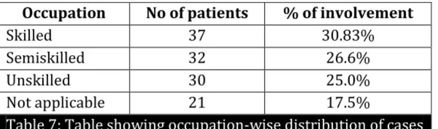
Most of the patients in our study were skilled workers amounting to 30.83%, patients with semi-skilled occupation being 26.6%, unskilled ones being 25%, and not applicable ones being 17.5%. This clearly indicates that alopecia areata in our study was commonly seen in skilled workers.
Alopecia areata is usually confined to one portion of the body, but not necessarily the scalp. In
Muller and Winkelman’s3 study 95% of the patients had scalp affection.19 The study done by Awachat
AK et al4 shows that involvement of various regions of scalp were as follows [Table 16]. This indicates
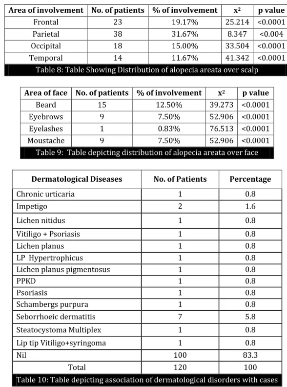
that the commonest area of involvement over scalp was the Parietal area. This is statistically significant and correlates with the previous study.
The percentage of beard involvement according to Awachat AK et al4 was 12%[Table 17]. This
shows that beard was involved in 12.5% of cases and is statistically significant and is in line with the studies done before. In our study beard was the commonest area involved over the face. Eyebrows were involved in 7.5% of cases, eyelashes in 0.83% and mustache in 7.5% of cases, extremities in 5.83% and pubic hair in 1 case 0.83%. Alopecia totalis was seen in 2 cases and 1 case progressed from alopecia totalis to universalis.
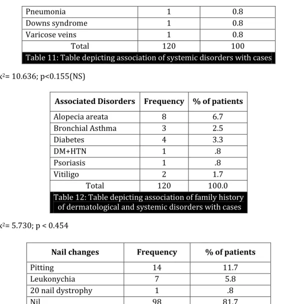
ASSOCIATED DERMATOLOGICAL DISORDERS: Seborrhoeic dermatitis was seen in 5.8% of cases. Psoriasis and impetigo were seen in 1.6% cases each. The other dermatological disorders5 associated
Palmoplantar keratoderma, Schambergs purpura, Steatocystoma multiplex in (0.8%) of cases each. Totally 2 cases of Vitiligo was seen amounting to (1.6%), of which 1 case each were associated with Psoriasis and Syringomas respectively. Muller and Winkelman studies have shown that association of alopecia areata with Vitiligo to be 4%. In our study association with Vitiligo is 1.6%. Lichen planus, Psoriasis and Vitiligo have a proposed autoimmune etiology. In our study, with these associations trends are available that could indicate that alopecia areata could be an autoimmune disorder. But further studies are needed in this regard.
ASSOCIATION WITH SYSTEMIC DISORDERS: Bronchial asthma was seen in 4.2% of cases. Diabetes mellitus was seen in 5.8% cases. Hypothyroidism was seen in 2.5% cases. Down’s syndrome, chronic pyelonephritis, pneumonia and varicose veins were seen in 0.8% cases each. Hypothyroidism, bronchial asthma and diabetes mellitus have a proposed autoimmune6 etiology. Muller and
Winkelman3 studies showed association of alopecia areata with atopy in 18% of children and 9% of
adults. In study by Ikeda1 percentage of association with atopy was 10%. In our study, with these
associations trends are available that could indicate that alopecia areata could be an autoimmune disorder. But further studies are needed in this regard.
ASSOCIATED FAMILY HISTORY: In our study, family history of alopecia areata was seen in 6.7% cases, Bronchial asthma in 2.5% cases. This attributes to 9.2% of patients, associated with atopy. Family history of diabetes mellitus was seen in 3.3% cases. One case had both diabetes mellitus (Type II) and hypertension (0.8%). One case had history of psoriasis (0.8%) cases. 2 cases had family history of vitiligo (1.7%). In our study 14.1% cases had association with other autoimmune disorders mentioned above. In our study, with these associations trends are available that could indicate that alopecia areata could be an autoimmune disorder. But further studies are needed in this regard. There are several reports of occurrence of alopecia areata in families and occurrence of the same in twins at the same site and also the onset of time being the same. Percentage of family history ranges from 10-27% as per Dan A. Nelson et al in their study.
NAIL INVOLVEMENT IN ALOPECIA AREATA: In our study, Pitting was seen in 11.7% cases and was the commonest finding. King Muller3, and Read noted pitting in 66%. Leukonychia was seen in 5.8%
cases. 20 nail dystrophy was seen in 0.8% of cases. In our study pitting was the commonest nail finding.
STRESS AND ALOPECIA AREATA: In Muller and Winkelman3 studies 12% patients had emotional
living in the urban areas. There are previous studies7 to show the correlation between stress and
alopecia areata.
HISTOPATHOLOGY8,9 OF ALOPECIA AREATA: Biopsy was done in selected cases (in a total of 25
patients) and most of the cases showed features of peribulbar lymphocytic inflammatory infiltrate(Figure 1,2). In our study more number of telogen follicles were found in comparison to anagen hair follicles. Around Telogen hair inflammatory infiltrate was less than anagen hair follicles. Telogen hairs show little or no perifollicular inflammation. The number of Catagen and telogen follicles found may be marked, approaching 100%. Follicles may enter a persistent phase of telogen in which the hair shaft has already been shed, manifested by the telogen germinal unit. Whiting and others have emphasized the use of follicular counts to aid in the diagnosis of alopecia areata when the characteristic peribulbar inflammation is missing, with a high percentage of catagen or telogen hairs and miniaturized hairs as a strong sign of alopecia areata.
TRICHOGRAM ANALYSIS: In our study, Trichogram analysis was done in 5 cases and in most of the patients more of Telogen hairs compared to anagen hairs were noted. In a 2 patients, dystrophic hairs and catagen hairs were more compared to anagen hairs. Exclamatory mark hairs10 were seen in all
cases.
TREATMENT OPTIONS IN ALOPECIA AREATA: The treatment options for alopecia areata are diverse, and the treatment options are based on the age of the patient and the extent of disease. For children less than 10 years topical minoxidil, corticosteroids and in resistant cases anthralin therapy is used. In children more than 10 years, if more than 50% area of involvement, immunotherapy is used as a first line therapy. If the response in 6 months is poor, than systemic corticosteroids and PUVA therapy is tried as a second line therapy. If the response to this is poor then azathioprine, cyclosporine area used as a third line therapy. Excimer laser therapy is tried in lesions less than 50% of scalp surface area.
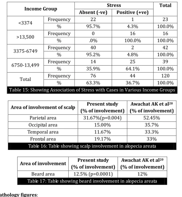
CONCLUSION: In our study trends are available that indicate that alopecia areata could be autoimmune but further studies are needed in this regard. Patients had significant association with emotional stress and stress was common in the income group Rs.6750 to Rs.13, 499/-. A holistic approach is important in the management of alopecia areata as the disease can have a severe psychologic impact on an individual's well-being.
REFERENCES:
1. 1 .Ikeda T. A new classification of alopecia areata. Dermatologica 1965; 131:421-45.
2. Mitchell AJ, Krull EA. Alopecia areata: Pathogenesis and treatment. J Am Acad Dermatol.1984;11:763-75.
3. Muller HK, Winkelmam RK. Alopecia areata. Arch Dermatol 1963;88:290-97. 4. Awachat AK, Sharma ML, et al . Alopecia areata. Arch Dermatol 1960; 26: 59 - 70.
6. Hordinsky M, Ericson M. Autoimmunity: Alpecia areata. J Investig Dermatol SympProc. 2004;9:73–8.
7. Reinhold M. Relationship of stress to the development of symptoms in alopecia areata and chronic urticaria. Br Med J 1960;1:846-9.
8. Sellheyer K, Bergfeld WF. Histopathologic evaluation of alopecias. Am J Dermatopathol. 2006;28:236–59.
9. Stefanato CM. Histopathology of alopecia: a clinicopathological approach to diagnosis. Histopathology. 2010;56:24
10.Madani S, Shapiro J. Alopecia areata update. J Am Acad Dermatol 2000;42:549- 66.
Age (years) Frequency % of patients
<10 15 12.5
11-20 17 14.2
21-30 45 37.5
31-40 28 23.3
41-50 11 9.2
51-60 3 2.5
>61 1 0.8
Total 120 100.0
Table 1: Age wise distribution of cases of alopecia areata
x2 = 81.483, p < 0.0001
Gender Frequency % of patients
F 40 33.3
M 80 66.7
Total 120 100.0
Table 2: Table showing sex wise distribution of cases
x2= 6.857; p < 0.009
Religion Frequency % of patients
Hindu 103 85.8
Muslim 10 8.3
Christian 7 5.8
others - -
Total 120 100.0
Table 3: Table showing religion-wise distribution of cases
Location Frequency % of patients
Rural 33 27.5
Urban 87 72.5
Total 120 100.0
Table 4: Table Showing Location Wise Distribution of Cases
x2=12.79; p<0.0001
Income in Rs./- Frequency % of patients
<3374 23 19.2
3375-6749 42 35.0
6750-13499 39 32.5
>13,500 16 13.3
Total 120 100.0
Table 5: Table showing income wise distribution of cases
₪ Based on Kuppuswamy socioeconomic status scale (Modified for 1998) x2= 8.359; p < 0.039
Educational status Frequency % of patients
Illiterate 14 11.7
literate 24 20.0
Primary school 11 9.2
High school 10 8.3
P.U.C 21 17.5
Degree/above 40 33.3
Total 120 100.0
Table 6: Table showing educational status wise distribution of cases
x2= 14.060; p<.015
The total number of illiterate population was 11.7%.
Occupation No of patients % of involvement
Skilled 37 30.83%
Semiskilled 32 26.6%
Unskilled 30 25.0%
Not applicable 21 17.5%
Table 7: Table showing occupation-wise distribution of cases
x2 =2.384; p < 0.497
Area of involvement No. of patients % of involvement x2 p value
Frontal 23 19.17% 25.214 <0.0001
Parietal 38 31.67% 8.347 <0.004
Occipital 18 15.00% 33.504 <0.0001
Temporal 14 11.67% 41.342 <0.0001
Table 8: Table Showing Distribution of alopecia areata over scalp
Area of face No. of patients % of involvement x2 p value
Beard 15 12.50% 39.273 <0.0001
Eyebrows 9 7.50% 52.906 <0.0001
Eyelashes 1 0.83% 76.513 <0.0001
Moustache 9 7.50% 52.906 <0.0001
Table 9: Table depicting distribution of alopecia areata over face
Systemic Disorders Frequency % of patients
Diabetes mellitus 7 5.8
Bronchial Asthma 5 4.2
Anemia 5 4.2
Hypothyroidism 3 2.5
Chronic Pyelonephritis 1 0.8
Dermatological Diseases No. of Patients Percentage
Chronic urticaria 1 0.8
Impetigo 2 1.6
Lichen nitidus 1 0.8
Vitiligo + Psoriasis 1 0.8
Lichen planus 1 0.8
LP Hypertrophicus 1 0.8
Lichen planus pigmentosus 1 0.8
PPKD 1 0.8
Psoriasis 1 0.8
Schambergs purpura 1 0.8
Seborrhoeic dermatitis 7 5.8
Steatocystoma Multiplex 1 0.8
Lip tip Vitiligo+syringoma 1 0.8
Nil 100 83.3
Total 120 100
Pneumonia 1 0.8
Downs syndrome 1 0.8
Varicose veins 1 0.8
Total 120 100
Table 11: Table depicting association of systemic disorders with cases
x2= 10.636; p<0.155(NS)
Associated Disorders Frequency % of patients
Alopecia areata 8 6.7
Bronchial Asthma 3 2.5
Diabetes 4 3.3
DM+HTN 1 .8
Psoriasis 1 .8
Vitiligo 2 1.7
Total 120 100.0
Table 12: Table depicting association of family history of dermatological and systemic disorders with cases
x2= 5.730; p < 0.454
Nail changes Frequency % of patients
Pitting 14 11.7
Leukonychia 7 5.8
20 nail dystrophy 1 .8
Nil 98 81.7
Table 13: Table showing various nail changes associated with cases
x2=9.839; p < 0.009
Stress LOCATE Total
Rural Urban
Absent (-ve) Frequency 30 46 76
% 39.5% 60.5% 100.0%
Positive (+ve) Frequency 3 41 44
% 6.8% 93.2% 100.0%
Total Frequency 33 87 120
% 27.5% 72.5% 100.0%
Table 14: Showing association of stress with cases in rural and urban areas
Income Group Stress Total Absent (-ve) Positive (+ve)
<3374 Frequency 22 1 23
% 95.7% 4.3% 100.0%
>13,500 Frequency 0 16 16
% .0% 100.0% 100.0%
3375-6749 Frequency 40 2 42
% 95.2% 4.8% 100.0%
6750-13,499 Frequency 14 25 39
% 35.9% 64.1% 100.0%
Total Frequency 76 44 120
% 63.3% 36.7% 100.0%
Table 15: Showing Association of Stress with Cases in Various Income Groups
Area of involvement of scalp Present study (% of involvement)
Awachat AK et al20
(% of involvement) Parietal area 31.67%(p=0.004) 52.45%
Occipital area 15.00% 35.7%
Temporal area 11.67% 33.3%
Frontal area 19.17% 33%
Table 16: Table showing scalp involvement in alopecia areata
Area of involvement Present study (% of involvement)
Awachat AK et al20
(% of involvement) Beard area 12.5% (p=0.0001) 12%
Table 17: Table showing beard involvement in alopecia areata
Histopathology figures:
Fig. 1: Dermis shows hair follicles surrounded by inflammatory infiltrate.
AUTHORS: 1. Gopal M.G. 2. Praveen Kumar S. 3. Sharath Kumar B.C. 4. Ramesh M.
PARTICULARS OF CONTRIBUTORS:
1. Professor & HOD, Department of Dermatology, Kempegowda Institute of Medical Sciences, Bangalore.
2. Assistant Professor, Department of Dermatology, M.S. Ramaiah Medical College and Teaching Hospitals, Bangalore.
3. Professor, Department of Dermatology, Kempegowda Institute of Medical Sciences, Bangalore.
4. Associate Professor, Department of Dermatology, Kempegowda Institute of Medical Sciences, Bangalore.
NAME ADRRESS EMAIL ID OF THE CORRESPONDING AUTHOR: Dr. Praveen Kumar S.
Sai Krupa, 1547, 26th Main, 26th Cross, 2nd Sector, HSR Layout, Bangalore – 560034. Email – [email protected]