DNA Barcoding of Neotropical Sand Flies
(Diptera, Psychodidae, Phlebotominae):
Species Identification and Discovery within
Brazil
Israel de Souza Pinto1,2
*, Bruna Dias das Chagas1, Andressa Alencastre Fuzari Rodrigues3, Adelson Luiz Ferreira2, Helder Ricas Rezende2, Rafaela
Vieira Bruno1,4, Aloisio Falqueto2, José Dilermando Andrade-Filho5, Eunice Aparecida Bianchi Galati6, Paloma Helena Fernandes Shimabukuro5, Reginaldo Peçanha Brazil3, Alexandre Afranio Peixoto1†
1Lab. de Biologia Molecular de Insetos, Instituto Oswaldo Cruz, FIOCRUZ, Brasil Ave. 4365, 21040360, Rio de Janeiro, RJ, Brazil,2Unidade de Medicina Tropical, UFES, Marechal Campos Ave. 1468, 29043900, Vitória, Espírito Santo, Brazil,3Lab. de Doenças Parasitárias, Instituto Oswaldo Cruz, FIOCRUZ, Brasil Ave. 4365, 21040360, Rio de Janeiro, RJ, Brazil,4Instituto Nacional de Ciência e Tecnologia em Entomologia Molecular–INCT-EM/CNPq, Rio de Janeiro, Brazil,5Grupo de Estudos em Leishmanioses, Centro de Pesquisas René Rachou, FIOCRUZ, Augusto de Lima Ave. 1715, 30190002, Belo Horizonte, MG, Brazil,6Departamento de Epidemiologia, Faculdade de Saúde Pública, USP, Dr. Arnaldo Cerqueira Cesar Ave. 715, 01246904, São Paulo, SP, Brazil
†Deceased.
Abstract
DNA barcoding has been an effective tool for species identification in several animal groups. Here, we used DNA barcoding to discriminate between 47 morphologically distinct species of Brazilian sand flies. DNA barcodes correctly identified approximately 90% of the sampled taxa (42 morphologically distinct species) using clustering based on neighbor-joining distance, of which four species showed comparatively higher maximum values of divergence (range 4.23–19.04%), indicating cryptic diversity. The DNA barcodes also
cor-roborated the resurrection of two species within the shannoni complex and provided an effi-cient tool to differentiate between morphologically indistinguishable females of closely related species. Taken together, our results validate the effectiveness of DNA barcoding for species identification and the discovery of cryptic diversity in sand flies from Brazil.
Introduction
Sand flies (Diptera, Psychodidae, Phlebotominae) are small insects that are the main vectors of
LeishmaniaRoss parasites, the etiologic agents of the leishmaniases. These insects are also vec-tors of other pathogens, includingBartonellaStronget al. and Phleboviruses [1]. Currently, there are over 520 species of sand flies recorded for the Neotropical region, with higher species
a11111
OPEN ACCESS
Citation:Pinto IdS, Chagas BDd, Rodrigues AAF, Ferreira AL, Rezende HR, Bruno RV, et al. (2015) DNA Barcoding of Neotropical Sand Flies (Diptera, Psychodidae, Phlebotominae): Species Identification and Discovery within Brazil. PLoS ONE 10(10): e0140636. doi:10.1371/journal.pone.0140636
Editor:Helge Thorsten Lumbsch, Field Museum of Natural History, UNITED STATES
Received:July 7, 2015
Accepted:August 19, 2015
Published:October 27, 2015
Copyright:© 2015 Pinto et al. This is an open access article distributed under the terms of the
Creative Commons Attribution License, which permits unrestricted use, distribution, and reproduction in any medium, provided the original author and source are credited.
Data Availability Statement:All relevant data are within the paper and its Supporting Information files.
Funding:This work was supported by grants from the CNPq and FIOCRUZ.
richness in Brazil [2,3]. This high species richness is associated with the diversity of biomes in Brazil, which range from rainforests such as the Amazon and the Atlantic forests, to dry areas ofcerradoandcaatinga. Each biome has a different biogeographic history, created by events that shaped the landscape and molded the geographical distribution of animals, including sand flies and their amphibian, reptilian, avian, and mammalian hosts [4,5,6,7,8]. Vicariance, a his-torical event characterized by the geographical separation and isolation of a subpopulation, for example, can lead to speciation due reduced gene flow between subpopulations of the same species [9,10]. As a result of these geographical barriers, subpopulations begin to evolve inde-pendently and to accumulate genetic differences that can lead to speciation. In the event of spe-ciation, a subpopulation loses the ability to mate successfully with other subpopulations, even if the geographical barrier is later removed. Over time, distinct morphological characters can also evolve; however, morphological differentiation tends to take longer because changes in morphological traits require changes in multiple genes [10,11]. Morphological differences among species have been the basis of taxonomy and classification for several groups of animals, including phlebotomine sand flies [2,12,13,14,15].
Currently, there are two classification systems for American sand flies. The first classifica-tion divided the New World sand fly species into three genera:WarileyaHertig,Brumptomyia
França & Parrot, andLutzomyiaFrança.Lutzomyiawas subdivided further into several subge-nera and species groups [12,13]. The second classification also recognized the gesubge-neraWarileya,
BrumptomyiaandLutzomyia, but elevated several subgenera and species groups within the
Materials and Methods
Sand fly Collection and Morphological Identification
All the sand flies collections were performed using a permanent license to collect zoological material (process number: 32102) provide by the Instituto Brasileiro do Meio Ambiente e dos Recursos Naturais Renováveis (IBAMA). The collections performed in private areas also received a verbal permission to conduct the study on the site by the land owner. Sand flies were collected between 2011 and 2013 from 19 municipalities distributed across five Brazilian states: State of Mato Grosso: 1) Cáceres (16°24’08”S; 57°29’55”W; 286 meters at the sea level—a.s.l); State of Minas Gerais: 2) Lagoa Santa (10°45’48”S; 48°06’32”W; 740 m a.s.l.); State of Bahia: 3) Wenceslau Guimarães (13°35’04”S; 39°42’32”W; 455 m a.s.l); State of Rio de Janeiro: 4) Bom Jesus do Itabapoana (21°03’25”S; 41°47’31”W; 500 m a.s.l.); State of Espírito Santo: 5) Afonso Cláudio (20°12’53”S; 41°02’31”W; 1030 m a.s.l.), 6) Alfredo Chaves (20°29’25”S; 40°57’28”
W; 1069 m a.s.l.), 7) Alto Rio Novo (18°58’35”S; 41°00’43”W; 762 m a.s.l.), 8) Baixo Guandu (19°21’04”S; 40°49’48”W; 719 m a.s.l.), 9) Domingos Martins (20°24’01”S; 40°45’11”W; 673 m a.s.l.), 10) Ibitirama (20°28’41”S; 40°42’19”W; 842 m a.s.l.), 11) Itaguaçu (19°44’13”S; 40° 58’09”W; 871 m a.s.l.), 12) Iúna (20°21’02”S; 41°43’27 W; 851 m a.s.l.), 13) João Neiva (19° 48’07”S; 40°30’23”W; 632 m a.s.l.), 14) Mantenópolis (18°51’09”S; 41°03’59”W; 661 m a.s.l.), 15) Marilândia (19°19’04”S; 40°31’01”W; 581 m a.s.l.), 16) Pancas (19°13’44”S; 40°45’31”W; 133 m a.s.l.), 17) Santa Leopoldina (20°08’16”S; 40°30’57 W; 51 m a.s.l.), 18) Santa Maria de Jetibá (19°58’54”S; 40°48’46”W; 844 m a.s.l.), 19) Santa Teresa (19°54’30”S; 40°39’25”W; 754 m a.s.l.). A map with these localities is shown inFig 1.
The sand flies were captured using light traps (HP model) [41] placed within the peridomi-ciliary environment (represented by the domiciles and their annexes, and domestic animal shelters) and/or forest environment from 18:00 to 06:00. The insects were stored in 80% etha-nol and transported to the laboratory where they were screened and separated. Sand fly legs were separated for subsequent DNA extraction, and the sand fly body was mounted on glass slides as reported by [42] for identification of the species based on morphological characters [2,43], using the classification of [14,16]. The generic names abbreviations followed [44]. Sand fly vouchers were deposited in the Coleção de Flebotomíneos (COLFLEB) of the Centro de Pes-quisas René Rachou, FIOCRUZ, Belo Horizonte, state of Minas Gerais.
Species were selected based on their epidemiological importance, includingLutzomyia long-ipalpis(Lutz & Neiva) andLutzomyia cruzi(Mangabeira) vectors ofLeishmania infantum
Nicolle,Bichromomyia flaviscutellata(Mangabeira) vector ofLeishmania(Leishmania) amazo-nensisLainson & Shaw andNyssomyia whitmani(Antunes & Coutinho) andNyssomyia inter-media(Lutz & Neiva) vectors ofLeishmania(Viannia)braziliensisVianna Other species, includingMigonemyia migonei(França),Pintomyia fischeri(Pinto),Psychodopygus ayrozai
(Barretto & Coutinho),Psychodopygus davisi(Root) andPsychodopygus hirsutus(Mangabeira), were selected because they were previously found to be infected byLeishmaniaparasites in some regions of Brazil [45,46,47,48,49,50,51,52,53,54,55].
Genomic DNA Extraction and Analysis
centrifuged at 15,000gfor 5 min at 4°C and remove the supernatant to a fresh tube. We used 20μl of extraction buffer to macerate the legs which incubated overnight at 37°C. Polymerase chain reaction (PCR) was performed to amplify a 658 bp fragment of theCOIgene, to a final volume of 50μL reaction mixture containing 2μL of genomic DNA template, 25μL 2X Pro-mega GoTaqGreen™Master Mix (Promega, Madison, WI, USA), and the primers LCO1490 (50-GGTCAACAAATCATAAAGATATTGG-30) and HCO2198 (50-TAAACTTCAGGGTGAC CAAAAAATCA-30) to a final concentration of 1.0μM [57]. The reaction cycle consisted of an initial denaturation step of 95°C for 3 min, followed by 37 cycles of 95°C for 1 min, 50°C for 1 min, 72°C for 1.5 min, and a final extension cycle 72°C for 7 min. The amplified fragments were separated by agarose (2%) gel electrophoresis and purified using Illustra GFX PCR DNA™
and Gel Band Purification Kit (GE Healthcare, Pittsburgh, PA, USA). The purified fragments were sequenced bidirectionally at Fundação Oswaldo Cruz (PDTIS/FIOCRUZ), Rio de Janeiro, Brazil with an ABI 3730 sequencer, using the same primers that were used in PCR.
Fig 1. Map showing the sampling sites across the Brazilian territory.(1) Cáceres. (2) Lagoa Santa. (3) Wenceslau Guimarães. (4) Bom Jesus do Itabapoana. (5) Afonso Cláudio. (6) Alfredo Chaves. (7) Alto Rio Novo. (8) Baixo Guandu. (9) Domingos Martins. (10) Ibitirama. (11) Itaguaçu. (12) Iúna. (13) João Neiva. (14) Manenópolis. (15) Marilândia. (16) Pancas. (17) Santa Leopoldina. (18) Santa Maria de Jetibá. (19) Santa Teresa.
Sequence Analysis
The forward and reverse sequences for each specimen were aligned using Clustal W [58] and edited to generate a consensus sequence using BioEdit 7.0 [59]. The“Sequence Composition”
tool in BOLD (Barcode of Life Database–www.boldsystems.org) was used to evaluate nucleo-tide content of the sequences.
Pairwise nucleotide sequence divergence was estimated between all sequences using the Kimura 2-parameter (K2P) model [60] implemented in MEGA 6.0 [61]. We used the K2P model for two reasons: (1) it makes fewer assumptions about the nature of sequence changes than more heavily parameterized models [35,62] and (2) it provides a conservative estimate of long branches than more complex models, by underestimating the number of multiple hits [63]. Levels of genetic divergence within genera and species were calculated in BOLD using the
“Distance Summary”tool. Calculations of intraspecific divergence were limited to those species that were represented by at least three specimens and whose DNA sequences showed nucleo-tide substitutions. Female specimens that were identified only at genus level due to morpholog-ical similarities among congeners were not used in the analyses at the species level.
The K2P distances were used in MEGA 6.0 to conduct a neighbor-joining (NJ) analysis and to build a dendrogram showing the similarity among taxa, including bootstrap analysis (1000 replications). One black fly species (Diptera, Simuliidae),Simulium metallicum s.l. (GenBank number: KC015102.1), and two mosquitoes species (Diptera, Culicidae),Ochlerotatus cana-densis(GenBank number: JX259544.1), andCulex pipiens pallens(GenBank number:
JQ350727.1) were used as outgroups. MEGA 6.0 was also used to inspect the parsimony infor-mative sites among closest species with little to no morphological differentiation and to high-light the fixed differences (defined as sites at which all of the sequences in one sample are different from all of the sequences in a second sample) among its sequences.
Molecular Identification of Species
The ABGD software (Automatic Barcode Gap Discovery) [64] was used to find potential bar-code gaps and for primary species delimitation. ABGD sorts sequences into hypothetical spe-cies based on the barcode gap, which can be observed whenever the intraspecific divergence is smaller than the interspecific divergence. ABGD uses a range of prior intraspecific divergences to infer a confidence limit for intraspecific divergence from within the data and then identifies and uses the barcode gap to partition the data. The inferred confidence limit and barcode gap are then recursively applied until the data is maximally partitioned. We limited the default range of intraspecific divergence between 0.001 and 0.1. Species were assigned the status of pro-visional species, indicated by the addition of the suffix PS to the species name, if they split into two or more distinct groups with sequence divergences between them that greater than value of the barcode gap found by ABGD. We used NJ bootstrap support values (80%) for the groups to corroborate the data and morphological identification to solve problems regarding species groups with low congener divergence.
Statistical Analysis
Results
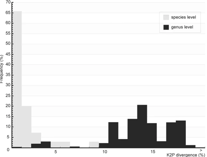
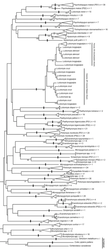
A total of 576 specimens of Brazilian sand flies, belonging to 47 morphologically classified spe-cies and 14 genera, were collected and subjected to barcode analysis (S1 Table). A 658 bp length of theCOIamplicon was recovered from all 576 specimens. Sequences and original trace files are available in the“AFBR-DNA barcoding for identification of sand flies (Diptera, Psychodi-dae, Phlebotominae) from Brazil”project in BOLD and in GenBank (accessions numbers: KP112487—KP113062) (S2 Table). TheCOIsequences from the sampled species showed an A + T bias (mean = 0.671) relative to the C + G content (mean = 0.329), as has been found previ-ously for arthropods [33,67]. Individual mean nucleotide content was as follows: A = 0.290, G = 0.159, C = 0.170 and T = 0.381. The mean K2P sequence distance within nominal species was 1.41%, while the mean divergence between congeners was approximately 10-fold higher (13.34%). Frequency histograms of meanCOIsequence divergences (K2P) at both the species and genus levels of the taxonomic hierarchy are shown inFig 2. The NJ tree, built using the K2P distances between the specimens, is shown inFig 3andS1 Fig.
Using ABGD, we identified nine values for potential barcode gaps (S2 Fig). We only consid-ered barcode gaps with values of prior intraspecific divergence between 1% and 2.5% because values below 1% or above 2.5% can overestimate or underestimate, respectively, the number of species. We found two values for barcode gaps within this range: 1.29% and 2.15%. Using the value of 2.15%, the species were partitioned into 48 groups, as indicated by one black dash (Fig 3). Using the value of 1.29%, the species were partitioned into 49 groups because one of the original 48 groups [Psychodopygus matosi(Barretto & Zago)] was split in two groups, as indi-cated using one white dash (Fig 3). Seven species were not recognized by the ABGD partition-ing:Nyssomyia intermediawas grouped withNy.whitmani;Ev.carmelinoi(Ryanet al.) was grouped withEvandromyia lenti(Mangabeira) as indicated with two thin black dashes in the NJ tree (Fig 3); andLu.longipalpis, which if analyzed separately, showed deep intraspecific var-iation that suggested it should form two groups. One of the two groups, comprisingLu. longi-palpisspecimens from the central-western region of Brazil, clustered withLu.cruzispecimens, while the other group, comprisingLu.longipalpisfrom southeastern of Brazil, clustered with
Lutzomyia alencariMartins, Souza & Falcão (Fig 3). Four nominal species,Psathyromyia bigeniculata(Floch & Abonnenc),Evandromyia edwardsi(Mangabeira),Pintomyia monticola
(Costa Lima), andSciopemyia microps(Mangabeira), showed deep intraspecific variation, forming two or more intraspecific barcode groups with mean divergence greater than 2.15% (Fig 3).Psychodopygus matosishowed moderate intraspecific variation and only formed two intraspecific barcode groups with mean divergence greater than 1.29% (Fig 3).
We found fixed differences withinPa.bigeniculata,Ev.edwardsi,Pi.monticola, and Brump-tomyiagenus and betweenEvandromyia tupynambai(Mangabeira) andEvandromyiaspp. (S3 Fig). Because of the morphological similarities between the species, we also comparedPa.
bigeniculata(PS1 and PS2) sequences to the sequences fromPa.limai(Fonseca).Psathyromyia limaishowed 41 fixed differences in relation to both provisional species ofPa.bigeniculata
(PS1 and PS2), whilePa.bigeniculataPS1 showed nine fixed differences in relation to bothPa.
bigeniculataPS2 andPa.limai. Likewise,Pa.bigeniculataPS2 showed 13 fixed differences in relation to bothPa.bigeniculataPS1 andPa.limai. These three species still showed one fixed difference among each other that can be used as diagnostic site (421stposition of theCOI
sequence) (S3A Fig). The three species within theEv.edwardsinominal species lacked a diag-nostic site among them. However,Ev.edwardsiPS1 showed eight fixed differences in relation toEv.edwardsiPS2 and PS3, whileEv.edwardsiPS2 showed ten fixed differences in relation to
Ev.edwardsiPS1 and PS3, andEv.edwardsiPS3 showed 16 fixed differences in relation toEv.
fixed (S3C Fig). The three species of theBrumptomyiagenus showed seven fixed differences that can be used as diagnostic sites (S3D Fig).Evandromyia tupynambaishowed 27 fixed dif-ferences in relation toEvandromyiaspp. (S3E Fig).
The mean intraspecific sequence divergence values (adjusted R2= -0.0265, p = 0.587) (S4A Fig) and maximum intraspecific sequence divergence values (adjusted R2= -0.0372, p = 0.861) (S4B Fig) were not significantly correlated to sample size. After a Bonferroni correction sequences divergences were significantly correlated to geographical distance within ten species (S4C, S4D, S4E, S4F, S4G, S4H, S4I, S4J, S4L, and S4M Fig).
Discussion
The molecular identification of species basedCOIDNA barcoding has been useful for species recognition and the discovery of different taxa of insect vectors of parasites [33,34,39]. Here, Fig 2. Frequency histograms of meanCOIsequence divergences (K2P) among sand flies from Brazil.Gray bars represent meanCOIsequence
divergences (K2P) for species levels and black bars for genus levels of the taxonomic hierarchy.
Fig 3. Neighbor-joining tree ofCOIsequence divergences (K2P) obtained from 567 specimens of sand flies analyzed.Numbers next to the branches indicate Bootstrap percentages80%. One black dash
we show thatCOIDNA barcoding is an effective tool for the discovery and recognition of sand fly species from Brazil.
First, there existed a possibility that we would not accurately detect amplification of theCOI
gene when using the sand fly genomic DNA. This concerned was based on the amplification of
COInuclear pseudogenes of mitochondrial origin (NUMTs) in a small number of previous DNA barcode studies using animal DNAs [68,69]. The presence of NUMT contamination is often indicated by the presence of PCR ghost bands, extra bands in restriction profiles, sequence ambiguities in polymorphic sites, or when both template strands are sequenced, by the presence of frameshift mutations, stop codons, and unexpected phylogenetic placements [68]. Additionally, in the DNA barcode studies, the amplicons generated from NUMT sequences often were shorter than the full-length barcode sequences ofCOI[69]. As shown above, all of the sequence reads from this study recovered full-length barcodes sequence, with no evidence of insertions, deletions, or stop codons, indicating the absence of NUMTs in the sequences analyzed. This finding is similar to a previous barcoding study that did not find NUMTs in Colombian sand fly species [34]. From our results, we concluded thatCOI
sequences could reliably be used for barcode analysis of sand flies collected from Brazil. The DNA barcode analyses using the automatic partitioning performed by ABGD allowed us to correctly discriminate approximately 85% (40 species) of all previously morphologically identified species (Fig 3). This percentage becomes closer to 90% when we consider that the
COIgene can be used to discriminate betweenNy.intermediaandNy.whitmani. Although the genetic divergence between these species was too low to allow partitioning by ABGD,Ny. inter-mediaandNy.whitmaniboth are composed of monophyletic groups with high bootstrap sup-port values (Fig 3). Even at 90%, this identification percentage was somewhat lower than the percentages found in DNA barcode studies of other sand fly fauna. The identification percent-ages from studies using sand flies from Colombia and India, for example, were close to 100% [34,39]. Nevertheless, a number of DNA barcode studies in which specimen for each species were collected from several localities showed discrimination percentages similar to those reported here [33,70]. Differences between the species discrimination percentages of sand flies from Brazil and others regions may be related to sampling effects. In the study of sand flies from Colombia, 36 species were collected from different geographic localities and analyzed, 31 of which included the analysis of more than one specimen; however, only ten of these 31 spe-cies were sampled from more than one locality. Two of these ten spespe-cies,Lutzomyia gomezi
(Nitzulescu) andPsychodopygus panamensisShannon, showed the maximum intraspecificCOI
sequence divergence within one species [34]. This result suggests that the increase in sampling localities for each species could influence their discrimination percentages. Our data supports this possibility, since ABGD accurately delimited between species collected from the same locality. In the study of sand flies from India, although the species were all collected from mul-tiple localities, the high percentage of discrimination between the species may be explained by the small number of species analyzed [39]. The genetic distances between species also can be larger if some species are missing from the assemblage and, consequently, the delimitation becomes easier [71]. This raised concerns regarding the effect of sample size and geographical scale of sampling in theCOIsequence divergence (intraspecific genetic variation and interspe-cific genetic divergence to congeners) for sand flies from Brazil.
that intraspecific sequence divergence (K2P) inCOIwas significantly correlated to geographi-cal distance (S4 Fig) within several, but not all, of the species analyzed. OnlyPa.bigeniculata
(PS1 from Santa Leopoldina, state of Espírito Santo; and PS2 from Cáceres, state of Mato Grosso) andSc.microps(PS1 from Iúna, state of Espírito Santo; and PS2 from Pancas, state of Espírito Santo) were clustered according to geographical distance. From these samples, we were able to infer that the barcoding gene reliably discriminates between the nominal species, even with correlation between the intraspecific sequence divergence (K2P) atCOIand the geo-graphical distance within some species.
The effects of geographical scale on sampling in theCOIsequence divergence has been pro-posed to occur because the distance of one species to the closest morphologically different spe-cies significantly decreases with increasing geographical scale of sampling [71]; however, our data did not corroborate this hypothesis. ThoughLu.longipalpis,Lu.cruziandLu.alencariare closely related species [2,19,74], for example, specimens from these three species of the same geographical region showed less sequence divergence than the closest morphologically differ-ent species from others regions (Fig 3).Lutzomyia longipalpisspecimens from Pancas, state of Espírito Santo, were closely related toLu.alencarispecimens from the same region, andLu.
longipalpisspecimens from Cáceres, state of Mato Grosso clustered withLu.cruzispecimens from the same region (Fig 3). Despite the morphological differences between these species, this low level of sequence divergence, suggests thatLu.alencarialso belongs to theLu.longipalpis
complex. This possibility is further supported by the presence of a spot in the abdominal tergite of both species, which appears to be a synapomorphy of theLu.longipalpiscomplex [19]. The low sequence divergence found between these sympatric species can be the result of introgres-sion [75,76], a common phenomenon among closely species that tends to occur much more readily in the mitochondrial DNA than in the nuclear DNA [77,78].
Introgression can also explain the low divergence found amongNy.intermediaandNy.
whitmani(Fig 3). The evidence of introgression among these species is corroborated by studies using mitochondrial DNA (mtDNA) and using nuclear DNA [79,80,81]. Introgression of mtDNA was detected in studies analyzingcytochrome bgene from females that morphologi-cally were identified asNy.intermediaspecimens from Viana, state of Espírito Santo, but showed mtDNA haplotypes fromNy.whitmani[79]. Likewise, introgression of nuclear DNA was detected by studies analyzing theperiodgene in order to differentiate species of theNy.
intermediacomplex. This latter analysis indicated a low level of divergence, high similarities among sympatric sequences, and the presence of shared haplotypes betweenNyssomyiaspecies specimens from the locality of Posse, state of the Rio de Janeiro [80,81]. It is hard to distinguish introgression from the persistence of ancestral polymorphisms, but the introgression hypothe-sis has been the preferred model based on data analyhypothe-sis which suggested thatNy.intermedia
andNy.whitmanimight be exchanging alleles [80,81].
Unlike the results forNy.intermediaandNy.whitmani, the bootstrap values did not sup-port the clades ofEv.carmelinoiandEv.lenti, and ABGD was not able to discriminate between them (Fig 3), indicating very low sequence divergence between these species. These two species are very similar morphologically, but can be differentiated by the male genital filamental tips (genital filament tips arrow-like and elongate 1/8ththe length of the genital filaments forEv.
carmelinoifrom Cáceres, state of Mato Grosso andEv.lentifrom Pancas, state of Espírito Santo), would favor reduction of the gene flow, thereby increasing sequence divergence. Fur-thermore, this similarity of sequences cannot be explained by the phenomenon of decreasing interspecific divergence with increasing geographical scale of sampling, because the geographi-cal distance among the areas (S3 Table) is not enough to achieve such similarity (See Figure 4 from [71]). If these two nominal species really represent two different taxa, the only likely explanation is that strong introgressive hybridization occurs betweenEv carmelinoiandEv.
lentiin Cáceres, where these two species are sympatric [83]. It should be noted, however, that onlyEv.lentihas been reported in Pancas. Molecular and morphological revision of the species group using a larger number of specimens from several localities throughout its distribution range is required to resolve this discrepancy.
While our study was in progress, the shannoni group of the genusPsathyromyiawas revised [43], including species previously reported for the state of Espírito Santo asPsathyromyia shan-noni(Dyar) andPsathyromyia pestanai(Barretto & Coutinho) [84,85,86]. From the revision, two species of the shannoni complex,Pa.bigeniculataandPa.limai, were resurrected from the synonymy ofPa.shannoni, while a third species,Pa.pestanai, was proposed to be a junior syn-onym ofPa.limaion the basis of morphological characters. Therefore, we reexamined all slides of the specimens morphologically identified asPa.shannonifrom states of Espírito Santo state and Mato Grosso and found them to bePa.bigeniculatafrom Cáceres, Mato Grosso and from Santa Leopoldina, state of Espírito Santo. We also foundPa.limaifrom the remaining areas of the state of Espírito Santo. Corroborating the results of the morphological revision, the DNA barcode discriminated betweenPa.limaiandPa.bigeniculata, showing a high mean sequence divergence (10%) between them (Fig 3). It is noteworthy that the DNA barcode analysis from our study split the specimens ofPa.bigeniculatainto two groups (PS1 from state of Espírito Santo and PS2 from Cáceres from state of Mato Grosso) that represent putative species within thePa.bigeniculatacomplex (Fig 3). These groups had a high number of fixed differences among them, including a diagnostic site (421stposition of the 658 bp fragment ofCOIgene) that enabled identification ofPa.bigeniculata(PS1 and PS2) andPa.limai(S3A Fig). Based on the distribution of the analyzed specimens,Pa.bigeniculataPS1 represented a coastal species (Atlantic Forest) andPa.bigeniculataPS2 represented an interior species (cerrado). Besides supporting the resurrection of two species within the shannoni group that are morphologically similar, the DNA barcode also provided a reliable method to discriminate among them, cor-roborating the utility of the DNA barcode for sand fly identification.
Our analyses also suggested that three other nominal species belongs to cryptic species com-plex:Ev.edwardsi,Pi.monticola, andSc.microps.Evandromyia edwardsiwas split into three species (PS1, PS2, and PS3) (Fig 3), each with fixed differences in relation to the other species (S3B Fig), but without a diagnostic site. The fixed differences set can be used to differentiate between these three species within theEv.edwardsicomplex.Evandromyia edwardsidoes not morphologically resemble other sand fly species, and therefore, can be easily identified based on the male paramere and shape of the spermathecae of the female. This morphological dis-tinction and the low number of specimens collected throughout the distribution range ofEv.
specimens, identified asSciopemyiasp. The females grouped asSc.micropsPS2 were morpho-logically identified asSciopemyiasp. because, due to slide preparation, it was not possible to exam the spermathecae, and because the cibarium (horizontal teeth less-development) was very different from the morphological description of the females ofSc.microps(horizontal teeth well-developed). Unlike theSc.micropsPS2 females, the female ofSc.micropsPS1 was morphologically similar to the description ofSc.microps[87], with well-developed horizontal teeth in the cibarium. Two other female specimens (Sc_sp255f and Sc_sp299f,S1 Fig) were also identified asSciopemyiasp. and were partitioned into a third species based on our DNA bar-code analyses (Fig 3). This result supports a previous sand fly survey of the area, which identi-fied these specimens as belonging toSc. aff.micropsrather thanSc.microps[24] because of differences in the diameter of the female spermathecae ducts (AL Ferreira,personal communi-cation). These slight morphological differences are difficult to detect and require considerable technical skills. Therefore, DNA barcode analysis is a useful alternative for discriminating among specimens with small morphological differences.
Finally, the DNA barcode analysis was able to associate morphologically indistinguishable females with males of the generaBrumptomyia,Evandromyia, andPressatia. Based on the male morphology, three species ofBrumptomyia[Brumptomyia cunhaiMangabeira, Brumpto-myia ortiziMartins, Silva & Falcão, andBrumptomyia nitzulescui(Costa Lima)] were identified and all the females were identified asBrumptomyiasp. The specimens ofBr.cunhaiwere col-lected from a sand fly survey from Pancas, Espírito Santo and females from this locality were not analyzed. Unfortunately, this species was not included in the sand fly survey from that area because, at that time,Br.cunhaispecimens were misidentified asBr.nitzulescui[24]. Since the initial survey, we performed a morphological revision of the specimens based on a preliminary barcode analyses which indicated two groups ofBr.nitzulescui(one from Pancas and others from others areas). Indeed, we find that the group from Pancas was composed of specimens of
Br.cunhai. This result highlights the importance of integrative approaches based on morphol-ogy and molecular biolmorphol-ogy to reach accurate results with sand fly identification. In our study,
Br.cunhaishowed 46 fixed differences in relation toBr.ortiziandBr.nitzulescui, including seven fixed differences that can be used as diagnostic sites among these three species. Besides the seven diagnostic sites,Br.ortizishowed 26 fixed differences in relation toBr.cunhaiand
Br.nitzulescui(S3 Fig). Five of the ten females identified asBrumptomyiasp. were grouped with theBr.ortizimales, sharing its fixed differences in relation to othersBrumptomyiaspecies. The automatic partitioning by ABGD also indicated that these females belong toBr.ortizi
taxon. In addition to the similarities between theCOIgene sequences, the specimens were col-lected at the same locality, which further supports that they belong to the same taxon. This finding is very interesting and underscores the suitability of DNA barcoding for describingBr.
ortizifemales, since females ofBr.ortizihave not been previously described [2] because it is morphologically similar to other females of theBrumptomyiagenus. Females ofBr.nitzulescui
can be morphologically differentiated from five othersBrumptomyiaspecies, but when sympat-ric with others species [88], the differentiation is difficult to achieve. Our DNA barcode analysis clustered fiveBrumptomyiafemales specimens within theBr.nitzulescuiclade, which showed a high number of fixed differences and, consequently, high sequence divergence in relation to othersBrumptomyiaspecies. As noted forBr.ortizi, this result highlights the utility of the DNA barcode tool for sand fly species identification and its potential for female and male spe-cies association, even for spespe-cies without formal morphological description of both sexes. Our DNA barcode analysis also indicated an association between the males and the females of the
Evandromyiagenus.Evandromyia tupynambaiwas described for both sexes, but when found to be sympatric withEvandromyia callipyga(Martins & Silva) orEvandromyia costalimai
the males ofEv.callipygaorEv.costalimai; however our data suggest that the barcode gene can discriminate among these species since the females identified asEvandromyiasp. were clus-tered in two different groups with high sequence divergence and fixed differences among them (Fig 3). The first group belonged toEv.tupynambaiand was composed of females. The second group of three females belonged to either toEv.callipygaorEv.costalimai, but could not be dis-tinguished further because of morphological similarities with the females ofEv.tupynambaiof the first group and because we did not analyze DNA barcode from their males. The high genetic difference among the females further underscores the usefulness of the DNA barcode method for associating males with females of a species. Finally, we provided a DNA barcode profile for the females of thePressatiagenus, which indicated that these females belong to
Pressatia choti(Floch & Abonnenc) (Fig 3). This result is supported by the absence of other
Pressatiaspecies in the region where our specimens were collected [24].
In conclusion, except in cases of species that have undergone introgressive hybridization, DNA barcoding allowed discrimination of sand fly species from Brazil and the discovery of species within putative sand fly species complex. This method also reliably detected sand fly species misidentification and allowed the association between males and females among species that are morphologically similar. Finally, this study highlights the importance of utilizing inte-grative approaches for sand fly species identification in order to achieve accurate and reliable results.
Supporting Information
S1 Fig. A neighbor-joining tree ofCOIsequence divergences (K2P) in 47 sand fly species from Brazil. Only bootstrap values higher than 80 are shown.Each tip label in the tree con-tains the sand fly species name abbreviation (five words), the sample ID (number) and sex of the specimen (f = female or m = male). The sand fly species names were abbreviated as follows: Bi_fla =Bichromomyia flaviscutellata;Br_nit =Brumptomyia nitzulescui; Br_cun = Brumpto-myia cunhai; Br_ort =Brumptomyia ortizi; Br_sp =Brumptomyiaspp.; Ev_car =Evandromyia carmelinoi; Ev_edw =Evandromyia edwardsi; Ev_len =Evandromyia lenti; Ev_sp = Evandro-myiaspp.; Ev_ter =Evandromyia termitophila; Ev_tup =Evandromyia tupynambai; Ex_fir =
Expapillata firmatoi; Lu_ale =Lutzomyia alencari; Lu_cru =Lutzomyia cruzi; Lu_dis = Lutzo-myia dispar; Lu_ren =Lutzomyia renei; Lu_sp =Lutzomyiasp.; Mi_cap =Micropygomyia capixaba; Mi_ech =Micropygomyia echinatopharynx; Mi_fer =Micropygomyia ferreirana; Mi_per =Micropygomyia peresi; Mi_qui =Micropygomyia quinquefer; Mi_sch = Micropygo-myia schreiberi; Mg_mig =Migonemyia migonei; Ny_int =Nyssomyia intermedia; Ny_whi =
Nyssomyia whitmani; Ny_yui =Nyssomyia yuilli yuilli; Pi_bia =Pintomyia bianchigalatiae; Pi_fis =Pintomyia fischeri; Pi_mis =Pintomyia misionensis; Pi_mon =Pintomyia monticola; Pr_cho =Pressatia choti; Pr_sp =Pressatiaspp.; Pa_big =Psathyromyia bigeniculata; Pa_lim =
Psathyromyia limai; Pa_lut =Psathyromyia lutziana; Pa_pas =Psathyromyia pascalei; Pa_pel =Psathyromyia pelloni; Ps_ayr =Psychodopygus ayrozai; Ps_dav =Psychodopygus davisi; Ps_hir =Psychodopygus hirsutus; Ps_mat =Psychodopygus matosi; Sc_mic =Sciopemyia microps; Sc_sor =Sciopemyia sordellii; Sc_sp =Sciopemyiaspp.; Th_via =Trichophoromyia viannamartinsi.
(PDF)
S2 Fig. Number of groups among the 576 specimens of sand flies from Brazil based on the values of prior intraspecific divergence found by the Automatic Barcode Gap Discovery (ABGD) software as potential barcode gaps using a range of 0.001 to 0.1 for prior intraspe-cific divergence.
S3 Fig. Parsimony informative sites from a fragment of the 658 bp of thecytochrome c oxi-dase subunit I(COI) gene among closest related species of sand flies from Brazil.A)
Psathyromyia bigeniculata(PS1 and PS2); B)Evandromyia edwardsi(PS1, PS2, and PS3); C)
Pintomyia monticola(PS1 and PS2); D)Brumtomyiagenus (Brumptomyia cunhai, Brumpto-myia ortizi, andBrumptomyia nitzulescui); and E)Evandromyia tupynambaiandEvandromyia
spp. (yellow = fixed differences; red = diagnostic sites). (PDF)
S4 Fig. Correlation analyses showing the relationship between mean intraspecific sequence divergence (K2P) atCOIand sample size (A), the relationship between maximum interspe-cific sequence divergence and sample size (B), and the relationship between sequence diver-gence and geographical distance among each species collected from more than one site (C-M).
(PDF)
S1 Table. Table showing the collections sites and number and sex of sand fly specimens for each analyzed species used to create the DNA barcoding tree in theFig 2.Maximum and mean intraspecific values of genetic divergence (Kimura 2-parameter pairwise distances) are shown. As implemented by the Barcode of Life Database (BOLD), values are given only to spe-cies represented by three or more individuals and showing at least one nucleotide substitution. Nominal species marked with one asterisk showed distinct intraspecific lineages suggesting cryptic species complexes. Classification follows [14,16] and the generic abbreviations follow [44].
(PDF)
S2 Table. Phlebotominae taxa sampled, museum specimen number, BOLD and GenBank accession numbers.
(PDF)
S3 Table. Matrix of geographical distance between the sampling localities estimated using the Geographic Distance Matrix Generator version 1.2.3.All the distances were estimated in kilometers. (BA = state of Bahia; ES = state of Espírito Santo; MG = state of Minas Gerais; MT = Mato Grosso; RJ = state of Rio de Janeiro).
(PDF)
Acknowledgments
We thank Agenor Barbosa, Antonio Teva, Daniel Kiefer, Felipe Vigoder, Gabriela Thomazim, João F. R. Tonini, Luiz G. S. R. Bauzer and Mateus Borges for the help in the fieldwork. We also thank Gustavo R. Leite for the preparation of the map, Elisa Cupolillo for comments on an earlier version of the manuscript, and Instituto Brasileiro do Meio Ambiente e dos Recursos Naturais Renováveis (IBAMA) for provide the collecting permissions. This work was sup-ported by the Conselho Nacional de Desenvolvimento Científico e Tecnológico (CNPq, Brazil) and FIOCRUZ. We are grateful to the Plataforma Genomica-Sequenciamento de DNA-RPT01A- PDTIS/FIOCRUZ.
Author Contributions
References
1. Ready P. Biology of Phlebotomine sand flies as vectors of disease agents. Annu Rev Entomol. 2013; 58: 227–250. doi:10.1146/annurev-ento-120811-153557PMID:23317043
2. Galati EAB. Morfologia, terminologia de adultos e identificação dos táxons da América. In: Rangel EF, Lainson R, editors. Flebotomíneos do Brasil. Rio de Janeiro: Fiocruz; 2003. pp. 53–175.
3. Galati EAB. Phlebotominae (Diptera, Psychodidae). Classificação, morfologia, terminologia e identifi-cação de Adultos. Apostila da Disciplina HEP 5752—Bioecologia e Identificação de Phlebotominae 2014, Programa de Pós-Graduação em Saúde Pública, Faculdade de Saúde Pública, Universidade de São Paulo. 22 august 2014. Available:<www.fsp.usp.br/~egalati>.
4. Costa LP, Leite YLR, Fonseca GAB, Fonseca MT. Biogeography of South American forest mammals: endemism and diversity in the Atlantic Forest.Biotropica. 2000; 32: 872–881.
5. Costa LP. The historical bridge between the Amazon and the Atlantic Forest of Brazil: a study of molec-ular phylogeography with small mammals. J Biogeogr. 2003; 30: 71–86.
6. Watts PC, Hamilton JG, Ward RD, Noyes HA, Souza NA, Kemp SJ, et al. Male sex pheromones and the phylogeographic structure of the Lutzomyia longipalpis species complex (Diptera: Psychodidae) from Brazil and Venezuela. Am J Trop Med Hyg. 2005; 73: 734–743. PMID:16222018
7. Santos AMM, Cavalcanti DR, Silva JMC, Tabarelli M. Biogeographical relationships among tropical for-ests in north-eastern Brazil. J Biogeogr. 2007; 34: 437–446.
8. Carnaval AC, Moritz C. Historical climate modelling predicts patterns of current biodiversity in the Bra-zilian Atlantic Forest. J Biogeogr. 2008; 35: 1187–1201.
9. Avise JC. Phylogeography: the history and formation of species. Harvard University Press Cambridge, MA: Harvard University Press; 2000.
10. Ridley M. Evolution. 3rd edition. Malden: Blackwell Publishing; 2004.
11. Coyne JA, Orr HA. Speciation. Sunderland, MA: Sinauer Associates, Inc; 2004.
12. Lewis DJ, Young DG, Minter DM, Fairchild GB. Proposals for a stable classification of the Phlebotomine sandflies (Diptera: Psychodidae). Syst Entomol. 1977; 2: 235–262.
13. Young DG, Duncan MA. Guide to the identification and geographic distribution ofLutzomyiasand flies
in Mexico, the West Indies, Central and South America (Diptera: Psychodidae). Gainesville, FL: Asso-ciated Publishers; (1994.
14. Galati EAB. Classificação de Phlebotominae. In: Rangel EF, Lainson R, editors. Flebotomíneos do Bra-sil. Rio de Janeiro: Fiocruz; 2003. pp. 23–51.
15. Sinclair BJ, Borkent A, Wood DM. The male genital tract and aedeagal components of the Diptera with a discussion of their phylogenetic significance. Zool J Linn Soc-Lond. 2007; 150: 711–742.
16. Galati EAB. Phylogenetic systematics of the Phlebotominae (Diptera, Psychodidae) with emphasis on American groups. Bol Dir Malariol San Amb. 1995; 35: 133–142.
17. Beati L, Cáceres AG, Lee JA, Munstermann LE. Systematic relationships among Lutzomyia sand flies (Diptera: Psychodidae) of Peru and Colombia based on the analysis of 12S and 28S ribosomal DNA sequences. Int J Parasitol. 2004; 34: 225–234. PMID:15037108
18. Andrade-Filho JD, Galati EAB, Brazil RP. Review of American fossil Phlebotominae (Diptera: Psycho-didae) with a description of two new species. J Med Entomol. 2009; 46: 969–979. PMID:19769025
19. Pinto IS, Filho JD, Santos CB, Falqueto A, Leite YL. Phylogenetic relationships among species of Lut-zomyia, subgenus Lutzomyia (Diptera: Psychodidae). J Med Entomol. 2010; 47: 16–21. PMID:
20180303
20. Rodrigues AAF, Barbosa VA, Andrade-Filho JD, Brazil RP. The sandfly fauna (Diptera: Psychodidae: Phlebotominae) of the Parque Estadual da Serra da Tiririca, Rio de Janeiro, Brazil. Mem Inst Oswaldo Cruz. 2013; 108: 943–946. doi:10.1590/0074-0276130688PMID:24141956
21. Virgens TM, Santos CB, Pinto IS, Silva KS, Leal FC, Falqueto A. Phlebotomine sand flies (Diptera, Psy-chodidae) in an American tegumentary leishmaniasis transmission area in northern Espírito Santo state, Brazil. Cad Saúde Pública. 2008; 24: 2969–2978. PMID:19082291
22. Virgens TM, Rezende HR, Pinto IS, Falqueto A. Sand fly fauna (Diptera: Psychodidae) from the Goyta-cazes National Forest and surrounding areas of southeastern Brazil. J Vector Ecol. 2015; 40: 28–35.
doi:10.1111/jvec.12129PMID:26047181
24. Ferreira AL, Falqueto A, Grimaldi G, Peixoto AA, Pinto IS. Ecological and epidemiological aspects of the sand fly (Diptera, Psychodidae) fauna of the National Monument of Pontões Capixabas, state of Espírito Santo, southeastern Brazil. J Med Entomol. 2013; 50: 1215–1223. PMID:24843925
25. Figueira EAG, Silva G, Chagas ECS, Shimabukuro PHF. Phlebotomine sandflies (Diptera: Psychodi-dae) from Lábrea, state of Amazonas, Brazil, with a description of Evandromyia (Aldamyia) apurinan Shimabukuro, Figueira & Silva, sp. nov. Mem Inst Oswaldo Cruz. 2013; 108: 280–287.
26. Ladeia-Andrade S, Fé NF, Sanguinette CC, Andrade-Filho JD. Description of Trichophoromyia uninien-sis, a new phlebotomine species (Diptera: Psychodidae: Phlebotominae) of Amazonas State, Brazil. Parasit Vectors. 2014; 7: 400. doi:10.1186/1756-3305-7-400PMID:25168121
27. Andrade-Filho JD, Carvalho GML, Saraiva L, Falcão AL. Bilateral anomaly in the style of Micropygo-myia schreiberi (Martins, Falcão & Silva) (Diptera, Psychodidae). Rev Bras Entomol. 2004; 48: 583–585.
28. Galati EAB, Marassá AM, Fonseca MB, Gonçalves-Andrade RM, Consales CA, Bueno EFM. Phleboto-mines (Diptera, Psychodidae) in the Speleological Province of the Ribeira Valley: 3. Serra district—
area of hostels for tourists who visit the Parque Estadual do Alto Ribeira (PETAR), state of São Paulo, Brazil. Rev Bras Entomol. 2010; 54: 665–676.
29. Pinto IS, Santos CB, Ferreira AL, Falqueto A. Variations in the gonostyle of Nyssomyia intermedia (Lutz & Neiva) (Diptera: Psychodidae). Neotrop Entomol. 2010; 39: 732–735. PMID:21120381
30. Sanguinette CC, Faustino JX, Meira PCS, Botelho HA, Carvalho GM, Gontijo CM, et al. Anomalies in the sand fly Lutzomyia longipalpis (Diptera: Psychodidae) in Brazil. J Am Mosq Control Assoc. 2013; 29: 54–58. PMID:23687856
31. Shannon RC, Del Ponte E. Cuatro notas sobre especies nuevas de dipteros nematóceros, hematófa-gos o no, de la República Argentina. Rev Inst Bacteriol Dep Nac Hig. 1927; 4: 724–736.
32. Coutinho JO, Barretto MP. Contribuição para o conhecimento dos flebótomos do estado de São Paulo. III. Descrição do macho de Phlebotomus alphabeticus Fonseca, 1936 e de Phlebotomus pascalei n. sp. (Diptera: Psychodidae). An Fac Med Univ São Paulo. 1940; 16: 193–206.
33. Rivera J, Currie DC. Identification of Nearctic black flies using DNA barcodes (Diptera: Simuliidae). Mol Ecol Resour. 2009; 9: 224–236. doi:10.1111/j.1755-0998.2009.02648.xPMID:21564982
34. Gutiérrez MAC, Vivero RJ, Vélez ID, Porter CH, Uribe S. DNA Barcoding for the identification of sand fly species (Diptera, Psychodidae, Phlebotominae) in Colombia. Plos One. 2014; 9: e85496. doi:10. 1371/journal.pone.0085496PMID:24454877
35. Hebert PDN, Cywinska A, Ball SL, deWaard JR. Biological identifications through DNA barcodes. Proc R Soc Lond B Biol Sci. 2003; 270: 313–321.
36. Hebert PDN, Ratnasingham S, deWaard JR. Barcoding animal life: cytochrome c oxidase subunit 1 divergences among closely related species. Proc R Soc Lond B Biol Sci. 2003; 270: S96–S99.
37. Hebert PDN, Penton EH, Burns JM, Janzen DH, Hallwachs W. Ten species in one: DNA barcoding reveals cryptic species in the Neotropical skipper butterflyAstraptes fulgerator. Proc Natl Acad Sci USA. 2004; 101: 14812–14817. PMID:15465915
38. Ruiz-Lopez F, Wilkerson RC, Conn JE, McKeon SN, Levin DM, Quiñones ML, et al. DNA barcoding reveals both known and novel taxa in the Albitarsis Group (Anopheles: Nyssorhynchus) of Neotropical malaria vectors. Parasit Vectors. (2012; 5: 44. doi:10.1186/1756-3305-5-44PMID:22353437
39. Kumar NP, Srinivasan R, Jambulingam P. DNA barcoding for identification of sand flies (Diptera: Psy-chodidae) in Índia. Mol Ecol Resour. 2012; 12: 414–420. doi:10.1111/j.1755-0998.2012.03117.x
PMID:22277023
40. Azpurua J, De La Cruz D, Valderama A, Windsor D. Lutzomyia sand fly diversity and rates of infection by Wolbachia and an exotic Leishmania species on Barro Colorado Island, Panama. PLoS Negl Trop Dis. 2010; 4: e627. doi:10.1371/journal.pntd.0000627PMID:20231892
41. Pugedo H, Barata RA, França-Silva JC, Silva JC, Dias ES. HP: um modelo aprimorado de armadilha luminosa de sucção para a captura de pequenos insetos. Rev Soc Bras Med Trop. 2005; 38: 70–72. PMID:15717102
42. Barretto MP, Coutinho JO. Contribuicão ao conhecimento dos Flebotomus de São Paulo. II—
Descrição do macho de Phlebotomus limai Fonseca, 1935 e de duas novas espécies: Phlebotomus ayrozai e P. amarali (Diptera, Psychodidae).ArqFacHig Saude Publica Univ São Paulo. 1940; 6:
34–35.
43. Sábio PB, Andrade AJ, Galati EAB Assessment of the taxonomic status of some species included in the Shannoni complex, with the description of a new Species of Psathyromyia (Diptera: Psychodidae: Phlebotominae). J Med Entomol. 2014; 51: 331–341. PMID:24724281
45. Lainson R, Shaw JJ, Ward RD, Fraiha H. Leishmaniasis in Brazil, IX. Considerations on the Leishmania braziliensis complex, importance of sandflies of the genus Psychodopygus (Mangabeira) in the trans-mission of L. braziliensis braziliensis in north Brazil. Trans R Soc Trop Med Hyg. 1973; 67: 184–196.
PMID:4784055
46. Rangel EF, Ryan L, Lainson R, Shaw JJ. Observations on the sandfly (Diptera: Psychodidae) fauna of Além Paraíba, state of Minas Gerais, Brazil, and the isolation of a parasite of the Leishmania brazilien-sis complex from Psychodopygus hirsuta hirsuta. Mem Inst Oswaldo Cruz. 1985; 80: 373–374. PMID:
3837173
47. Arias JR, Miles MA, Naiff RD, Povoa MM, Freitas RA, Biancardi CB, et al. Flagellate infections of Brazil-ian sand flies (Diptera: Psychodidae): isolation in vitro and biochemical identification of Endotrypanum and Leishmania. Am J Trop Med Hyg. 1985; 34; 1098–1108. PMID:3938924
48. Gil LHS, Basano SA, Souza AA, Silva MGS, Barata I, Ishikawa EA, et al. Recent observations on the sand fly (Diptera: Psychodidae) fauna of the State of Rondônia, Western Amazônia, Brazil: the impor-tance of Psychdopygus davisi as a vector of zoonotic cutaneous leishmaniasis. Mem Inst Oswaldo Cruz. 2003; 98: 751–755. PMID:14595450
49. Rangel EF, Lainson R. Flebotomíneos do Brasil. Rio de Janeiro: Editora Fiocruz; 2003.
50. Pita-Pereira D, Alves CR, Souza MB, Brazil RP, Bertho AL, Barbosa AF, et al. Identification of naturally infected Lutzomyia intermedia and Lutzomyia migonei with Leishmania (Viannia) braziliensis in Rio de Janeiro (Brazil) revealed by a PCR multiplex non-isotopic hybridization assay. Trans R Soc Trop Med Hyg. 2005; 99: 905–913. PMID:16143358
51. Pita-Pereira D, Cardoso MAB, Alves CR, Brazil RP, Britto C. Detection of natural infection in Lutzomyia cruzi and Lutzomyia forattinii (Diptera: Psychodidae: Phlebotominae) by Leishmania infantum in an endemic area of visceral leishmaniasis in Brazil using PCR multiplex assay. Acta Trop. 2008; 107: 66–69. doi:10.1016/j.actatropica.2008.04.015PMID:18502392
52. Carvalho MR, Valença HF, Silva FJ, Pita-Pereira D, Pereira TA, Britto C, et al. Natural Leishmania infantum infection in Migonemyia migonei (França, 1920) (Diptera: Psychodidae: Phlebotominae) the putative vector of visceral leishmaniasis in Pernambuco State, Brazil. Acta Trop. 2010; 116: 108–110.
doi:10.1016/j.actatropica.2010.03.009PMID:20457120
53. Rocha LS, Falqueto A, Santos CB, Ferreira AL, Graça GC, Grimaldi G Jr, et al. Survey of natural infec-tion byLeishmaniain sand fly species collected in southeastern Brazil. Trans R Soc Trop Med Hyg. 2010; 104: 461–466. doi:10.1016/j.trstmh.2010.02.005PMID:20346478
54. Missawa NA, Veloso MA, Maciel GB, Michalsky EM, Dias ES. Evidence of transmission of visceral leishmaniasis by Lutzomyia cruzi in the municipality of Jaciara, State of Mato Grosso, Brazil. Rev Soc Bras Med Trop. 2011; 44: 76–78. PMID:21340413
55. Falqueto A, Ferreira AL. Reservatórios extra-humanos do complexo Leishmânia e dinâmica de trans-missão da infecção ao homem. In Coura JR, editor. Dinâmica das doenças infecciosas e parasitárias. 2ed. Rio de Janeiro: Guanabara Koogan; 2013. pp. 786–797.
56. Jowett T. Preparation of nucleic acids. In: Roberts DB, ed. Drosophila: A practical approach. Oxford: IRL Press; 1998. pp. 347–371.
57. Folmer O, Black M, Hoeh W, Lutz R, Vrijenhoek R. DNA primers for amplification of mitochondrial cyto-chrome c oxidase subunit I from diverse metazoan invertebrates. Mol Mar Biol Biotechnol. 1994; 3: 294–299. PMID:7881515
58. Thompson JD, Higgins DG, Gibson TJ. CLUSTAL W: improving the sensitivity of progressive multiple sequence alignment through sequence weighting, position-specific gap penalties and weight matrix choice. Nucleic Acids Res. 1994; 22: 4673–4680. PMID:7984417
59. Hall TA. BioEdit: a user-friendly biological sequence alignment editor and analysis program for Win-dows 95/98/NT. Nucleic Acids Symp Ser. 1999; 41: 95–98.
60. Kimura M. A simple method for estimating evolutionary rate of base substitutions through comparative studies of nucleotide sequences. J Mol Evol. 1980; 16: 111–120. PMID:7463489
61. Tamura K, Stecher G, Peterson D, Filipski A, and Kumar S. MEGA6: Molecular Evolutionary Genetics Analysis version 6.0. Mol Biol Evol. 2013; 30; 2725–2729. doi:10.1093/molbev/mst197PMID: 24132122
62. Barrett RDH, Hebert PDN. Identifying spiders through DNA barcodes. Can J Zool. 2005; 83: 481–491.
63. Nei M, Kumar S. Molecular Evolution and Phylogenetics. New York: Oxford University Press; 2000. 64. Puillandre N, Lambert A, Brouillet S, Achaz G. ABGD, Automatic Barcode Gap Discovery for primary species delimitation. Mol Ecol. 2012; 21: 1864–1877. doi:10.1111/j.1365-294X.2011.05239.xPMID: 21883587
66. Ersts PJ. Geographic Distance Matrix Generator (version 1.2.3). American Museum of Natural History, Center for Biodiversity and Conservation. 22 august 2014. Available:http://biodiversityinformatics. amnh.org/open_source/gdmg.
67. Crease TJ. The complete sequence for the mitochondrial genome of Daphnia pulex (Cladocera: Crus-tacea). Gene. 1999; 233: 89–99. PMID:10375625
68. Bensasson D, Zhang D, Hartl DL, Hewitt GM. Mitochondrial pseudogenes: evolution’s misplaced
wit-nesses. Trends Ecol Evol. 2001; 16; 314–321. PMID:11369110
69. Kerr KCR, Stoeckle MY, Dove CJ, Weigt LA, Francis CM, Hebert PDN. Comprehensive DNA barcode coverage of North American birds. Mol Ecol Notes. 2007; 7: 535–543. PMID:18784793
70. Clare EL, Lim BK, Engstrom MD, Eger JL, Hebert PDN. DNA barcoding of Neotropical bats: species identification and discovery within Guyana. Mol Ecol Notes. 2007; 7: 184–190.
71. Bergsten J, Bilton DT, Fujisawa T, Elliott M, Monaghan M, Balke M, et al. The effect of geographical scale of sampling on DNA barcoding. Syst Biol. 2012; 61: 851–869. PMID:22398121
72. Wright S. (1943) Isolation by distance. Genetics. 1943; 31: 114–138.
73. Nekola JC, White PS. The distance decay of similarity in biogeography and ecology. J Biogeogr. 1999; 26: 867–878.
74. Vigoder FM, Araki AS, Bauzer LG, Souza NA, Brazil RP, Peixoto AA. Lovesongs and period gene poly-morphisms indicate Lutzomyia cruzi (Mangabeira, 1938) as a sibling species of the Lutzomyia longipal-pis (Lutz and Neiva, 1912) complex. Infect Genet Evol. 2010; 10: 734–739. doi:10.1016/j.meegid. 2010.05.004PMID:20478408
75. Lins RMMA, Souza NABrazil RPMaingon RDCPeixoto AA. Fixed differences in theparalyticgene define two lineages within theLutzomyia longipalpiscomplex producing different types of courtship
songs. PLoS ONE. 2012; 7: e44323. doi:10.1371/journal.pone.0044323PMID:22970200
76. Araki AS, Ferreira GEM, Mazzoni CJ, Souza NA, Machado RC, Bruno RV, et al. Multilocus analysis of divergence and introgression in sympatric and allopatric sibling species of the Lutzomyia longipalpis complex in Brazil. PLoS Negl Trop Dis. 2013; 7: e2495. doi:10.1371/journal.pntd.0002495PMID:
24147172
77. Powell J. Interspecific cytoplasmic gene flow in the absence of nuclear gene flow: evidence from Dro-sophila. Proc Natl Acad Sci USA. 1983; 80: 492–495. PMID:6300849
78. Bachtrog D, Thornton K, Clark A, Andolfatto P. Extensive introgression of mitochondrial DNA relative to nuclear genes in the Drosophila yakuba species group. Evolution. 2006; 60: 292–302. PMID: 16610321
79. Marcondes CB, Day JC, Ready PD. Introgression betweenLutzomyia intermediaand bothLu.neivai
andLu.whitmani, and their roles as vectors ofLeishmania braziliensis. Trans R Soc Trop Med Hyg.
1997; 91: 725–726. PMID:9509190
80. Mazzoni CJ, Souza NA, Andrade-Coelho C, Kyriacou CP, Peixoto AA. Molecular polymorphism, differ-entiation and introgression in theperiodgene betweenLutzomyia intermediaandLutzomyia whitmani.
BMC Evol Biol. 2006; 6: 85. PMID:17069656
81. Mazzoni CJ, Araki AS, Ferreira GEM, Azevedo RVDM, Barbujani G, Peixoto AA. Multilocus analysis of introgression between two sand fly vectors of leishmaniasis.BMC Evol Biol. 2008; 8: 141
82. Ryan L, Fraiha H, Lainson R, Shaw JJ. New phlebotomine sandflies of the Walkeri group (Diptera: Psy-chodidae) from Pará state, Brazil, with a pictorial key 1. Mem Inst Oswaldo Cruz. 1986; 81: 323–331.
83. Alves GB, Oshiro ET, Leite MC, Melão AV, Ribeiro LM, Mateus NLF, et al. Phlebotomine sandflies fauna (Diptera: Brazil) (Diptera: Psychodidae) at rural settlements in the municipality of Cáceres, State of Mato Grosso, Brazil. Rev Soc Bras Med Trop. 2012; 45: 437–443. PMID:22836661
84. Pinto IS, Santos CB, Ferreira AL, Falqueto A. American visceral leishmaniasis dissociated from Lutzo-myia longipalpis (Diptera, Psychodidae) in the State of Espírito Santo, Brazil. Cad Saúde Pública. 2010; 26: 365–372. PMID:20396851
85. Pinto IS, Santos CB, Ferreira AL, Falqueto A. Richness and diversity of sand flies (Diptera, Psychodi-dae) in an Atlantic rainforest reserve in southeastern Brazil. J Vect Ecol. 2010; 35: 325–332. 86. Pinto IS, Tonini JFR, Ferreira AL, Falqueto A. A brief inventory of sand flies (Diptera, Psychodidae)
from the National Forest of the Rio Preto, state of the Espírito Santo, southeastern Brazil. Biota Neo-trop. 2012; 12: 323–326.
87. Martins AV, Falcão AL, Silva JE. Estudos sobre os flebótomos do estado de Minas Gerais—X—
Descrição das fêmeas de Lutzomyia microps (Mangabeira, 1942) e Lutzomyia firmatoi (Barretto, Mar-tins & Pellegrino, 1956) (Diptera, Psychodidae, Phlebotominae). Rev Bras Biol. 1975; 35: 259–263. 88. Shimabukuro PHF, Tolezano JE, Galati EAB. Chave de identificação ilustrada dos Phlebotominae