Epigenetic Regulatory Effect of Exercise on
Glutathione Peroxidase 1 Expression in the
Skeletal Muscle of Severely Dyslipidemic Mice
Albert Nguyen1,2, Natacha Duquette2, Maya Mamarbachi2, Eric Thorin1,2,3*
1Department of Pharmacology, Faculty of Medicine, Université de Montréal, Montreal, Quebec, Canada,
2Montreal Heart Institute, Research Center, Montreal, Quebec, Canada,3Department of Surgery, Faculty of Medicine, Université de Montréal, Montreal, Quebec, Canada
*eric.thorin@umontreal.ca
Abstract
Exercise is an effective approach for primary and secondary prevention of cardiovascular diseases (CVD) and loss of muscular mass and function. Its benefits are widely docu-mented but incompletely characterized. It has been reported that exercise can induce changes in the expression of antioxidant enzymes including Sod2, Trx1, Prdx3 and Gpx1 and limits the rise in oxidative stress commonly associated with CVD. These enzymes can be subjected to epigenetic regulation, such as DNA methylation, in response to environ-mental cues. The aim of our study was to determine whether in the early stages of athero-genesis, in young severely dyslipidemic mice lacking LDL receptors and overexpressing human ApoB100 (LDLR-/-; hApoB+/+), exercise regulates differentially the expression of antioxidant enzymes by DNA methylation in the skeletal muscles that consume high levels of oxygen and thus generate high levels of reactive oxygen species. Expression ofSod2, Txr1,Prdx3andGpx1was altered by 3 months of exercise and/or severe dyslipidemia in 6-mo dyslipidemic mice. Of these genes, onlyGpx1exhibited changes in DNA methylation associated with dyslipidemia and exercise: we observed both increased DNA methylation with dyslipidemia and a transient decrease in DNA methylation with exercise. These epige-netic alterations are found in the second exon of theGpx1gene and occur alongside with inverse changes in mRNA expression. Inhibition of expression by methylation of this spe-cific locus was confirmedin vitro. In conclusion,Gpx1expression in the mouse skeletal muscle can be altered by both exercise and dyslipidemia through changes in DNA methyla-tion, leading to a fine regulation of free radical metabolism.
Introduction
Chronic inflammatory diseases such as atherosclerosis are characterized in part by high levels of free radicals, inducing oxidative stress [1]. This oxidative stress targets DNA, lipids and pro-teins, altering cellular functions and leading to organ failure [2]. To maintain a healthyredox equilibrium, cells normally express endogenous antioxidant enzymes including peroxiredoxins (Prx), thioredoxins (Trx), superoxide dismutases (SOD) and gluthatione peroxidases (Gpx). OPEN ACCESS
Citation:Nguyen A, Duquette N, Mamarbachi M, Thorin E (2016) Epigenetic Regulatory Effect of Exercise on Glutathione Peroxidase 1 Expression in the Skeletal Muscle of Severely Dyslipidemic Mice. PLoS ONE 11(3): e0151526. doi:10.1371/journal. pone.0151526
Editor:Laszlo Csernoch, University of Debrecen, HUNGARY
Received:September 22, 2015
Accepted:February 28, 2016
Published:March 24, 2016
Copyright:© 2016 Nguyen et al. This is an open access article distributed under the terms of the
Creative Commons Attribution License, which permits unrestricted use, distribution, and reproduction in any medium, provided the original author and source are credited.
Data Availability Statement:All relevant data are within the paper and its Supporting Information files.
Funding:This work was supported by Canadian Institutes of Health Research operating grant numbers 14496 and 133649 to ET ( http://www.cihr-irsc.gc.ca/e/193.html). The funders had no role in study design, data collection and analysis, decision to publish, or preparation of the manuscript.
These enzymes inactivate reactive oxygen species (ROS) and maintain them to physiological levels. ROS are indeed signalling molecules and natural by-products of the metabolic machin-ery and are therefore necessary for cellular function [3].
ROS are poorly regulated in the presence of a chronic inflammatory state such as diabetes, cancer and dyslipidemia [4]. With inflammation, theredoxequilibrium tips towards the gener-ation of free radicals by simultaneous occurrences of pro-oxidative events, including dysregula-tion of ROS-generating enzymes such as NADPH oxidases (NOX) normally involved in cell survival, growth and death [5], as well as regulators of the mitochondrial respiratory chain such as uncoupling proteins (UCP) [6]. The primary consequence of a rise in oxidative stress is a vascular endothelial dysfunction, a marker of future cardiovascular events [7]. However, clin-ical trials using antioxidants in patients with cardiovascular diseases (CVD) have provided con-troversial and still inconclusive results [8,9]. In contrast, while physical exercise paradoxically increases ROS production both in animal studies [10] and humans [11], physical training is an excellent primary and secondary prevention strategy in CVD [12] and delay the loss muscle mass and function (sarcopenia) [13], including in patients suffering from atherosclerosis [14, 15]. While an acute bout of exercise increases ROS production, chronic regular exercise upre-gulates antioxidant defenses [16–18].
Little is known, however, about the molecular mechanisms increasing stress resistance. Dur-ing physical exercise, the main source of ROS is the skeletal muscle [13]. Muscles of the lower limbs are high producers of mitochondria-derived metabolites, and it is therefore likely that ROS arising from the metabolic activity of the skeletal muscle could potentially damage the vascular endothelium if the appropriate defense mechanisms are not maintained properly [13]. This supposes that genes coding for antioxidant proteins are partly regulated by the level of physical activity.
Epigenetic regulation of gene expression in response to environmental stimuli is well docu-mented [19]. In mammals, the addition of a methyl group to a cytosine preceding a guanine (CpG), usually leads to gene silencing if located in CpG-dense promoter regions. However, the regulatory effects of methylation in the gene body are more ambiguous [20–22]. DNA methyla-tion is a dynamic process that can respond to most extracellular cues including diet [23] and physical activity [24]. This sensitivity of DNA methylation to environmental signals is sug-gested to contribute to the development of various pathological conditions since gene-specific and genome-wide DNA methylation have been linked to a wide variety of disease states rang-ing from inflammatory [25] and CVD [26] includrang-ing atherosclerosis [27,28] and skeletal mus-cle [29]. This highlights the importance of epigenetic plasticity to the environment.
Studies have shown that antioxidant enzymes could be induced or repressed by methylation of regions in the corresponding gene, while aberrant methylation patterns have been associated with pro-oxidative and pro-inflammatory diseases [30–32]. We therefore hypothesised that in young mice, severe dyslipidemia would alter antioxidant gene expression while chronic physi-cal exercise would maintain it, in part due to epigenetic regulation. In this study, we demon-strate the dynamic nature of antioxidant genes expression in the mouse skeletal muscle in sedentary and active dyslipidemic mice. Our results show for the first time thatGpx1 expres-sion is associated with changes in DNA methylation in a specific region in the gene body. Hence, physical exercise influencesGpx1gene expression through epigenetic regulation and may ultimately contribute to the cellular defense against metabolic stress.
Materials and Methods
Approval by the Montreal Heart Institute Animal Ethical Committee (#R2014-62-02) was given for all animal experiments, which were performed in accordance with theGuide to Care
and Use of Experimental Animals(vol.1, 2nded., 1993) of the Canadian Council on Animal Care. No animals became ill or died prior to the experimental endpoint. Experiments were con-ducted on the femoral artery and the skeletal muscle (soleus and gastrocnemius) isolated from 6-months old (6-mo) male C57/bl6 control wild type (WT) compared to severely dyslipidemic and spontaneously atherosclerotic (ATX) mice. Transgenic LDLr-/-:hApoB+/+ATX mice dis-play high levels of cholesterol, they spontaneously develop atherosclerotic lesions (under a nor-mal diet) and endothelial dysfunction in the aorta, carotids and renal arteries [33,34]. Both 3-mo WT and ATX mice were randomly assigned to two groups; one remained in control sed-entary (SED) conditions (n = 10) and one was exposed to 3 months of voluntary exercise (EX) (n = 10). To this end, mice were kept individually in cages instrumented with a running wheel (Lafayette Instrument Company, Lafayette, IN) [34]. Heart rate, systolic and diastolic blood pressure were monitored weekly by tail-cuff (Kent Scientific Corporation, Torrington, CT). Mice were studied at 6-mo and were sacrificed after anesthesia with a 1:1 mixture of Xylazine (Bayer Inc, Toronto, ON, Canada) and Ketamine hydrochloride (Bioniche, Belleville, ON, Can-ada) at two different times of the day, either at 10:00 AM while being inactive or at 2:00 AM, i.e. during their running time (Fig 1).
DNA and RNA extraction
Upon sacrifice, skeletal muscle tissues were harvested and snap-frozen. Total DNA and RNA were extracted using Qiagen RNeasy mini kit and DNeasy Blood & tissue kit (Qiagen, Toronto, ON), respectively, following manufacturer’s protocol.
Gene expression
Total RNA was reverse transcribed with RT-MMLV (Invitrogen, Burlington, ON) and ampli-fied using EvaGreen (Applied Biological Materials, Richmond, BC) for quantitative real-time PCR. Target genesUCP2/3,NOX2/4,Trx1,Sod2,Prdx3,Gpx1and the internal control, cycloA, were amplified with gene specific primers (S1 Table).
DNA methylation quantification
Following extraction, DNA was converted by bisulfite reaction using the EZ DNA Methyla-tion-Gold kit (Zymo Research, Irvine, CA). Global DNA methylation was measured by ELISA with the 5-mC DNA ELISA kit (Zymo Research). Gene-specific DNA methylation was quanti-fied by EpiTYPER assay (Sequenom, San Diego, CA), as previously described [35]. We chose to target regions identified as CpG islands, by the UCSC Genome Browser athttp://genome. ucsc.edu/[36], found in our genes of interest with bisulfite-specific primers (S2 Table) required for the assay.
Cloning of pCpG free-Gpx1 vector
plasmid using HindIII and BsrGI restriction sites. We named the resulting vector“pCpG free-Gpx1”(see“results”).
In vitro
methylation, transient transfection and Luciferase assay
Cloned vectors were isolated by Qiagen QIAprep Spin Miniprep kit (Qiagen). M. SssI CpG methyltransferase (New England Biolabs, Frankfurt, Germany) was used forin vitro methyla-tion according to manufacturer’s instructions. Methylated DNA was then purified using the QIAquick gel extraction kit (Qiagen) and quantified by NanoDrop (Thermo Scientific Nano-Drop products, Wilmington, DE). Methylation was confirmed by digestion with the
Fig 1. Voluntary running activity is similar between WT and ATX.(A) Average of the daily running distance over the course of the 3 months exposure to voluntary exercise for both WT and ATX groups. (B) Typical distribution of the running activity during an arbitrary day of voluntary exercise. Arrows indicate the two different times of sacrifice (2:00 AM and 10:00 AM).
methylation-sensitive restriction enzymesHhaI andHpaII. HEK293 cells grown to confluence on 96-well plates were transfected with the pCpG free-Gpx1 vector using Lipofectamine 2000 (Invitrogen). 24 h after transfection, luciferase activity was measured with the QUANTI-Luc reagent (Invivogen, San Diego, CA) by luminescence detection. Promoter activity was normal-ized to the total amount of protein measured by a Bradford assay (Biorad, Hercules, CA).
Oxidative stress quantification
The fluorescent probe dihydroethidium (DHE) (Sigma-Aldrich Canada, Oakville, ON, Can-ada) was used to indirectly assess the amount of superoxide production. DHE is a dye that will bind to DNA once it is oxidized by superoxide. An antibody against 4-hydroxynonenal (4-HNE) (Abcam Inc, Toronto, ON, Canada) was used to detect 4-HNE expression, a marker of lipid peroxidation, by immunostainig. Tissue cross-sections were obtained from OCT-pre-served skeletal muscle and confocal images were taken.
Statistical Analysis
Results are presented as mean±SEM of (n) mice. Two-way ANOVA (with Bonferonni post-tests) and unpaired t-tests were used where applicable to test the differences between condi-tions. A p value of p<0.05 was considered statistically significant.
Results
Phenotypic effects of exercise on severely dyslipidemic mice
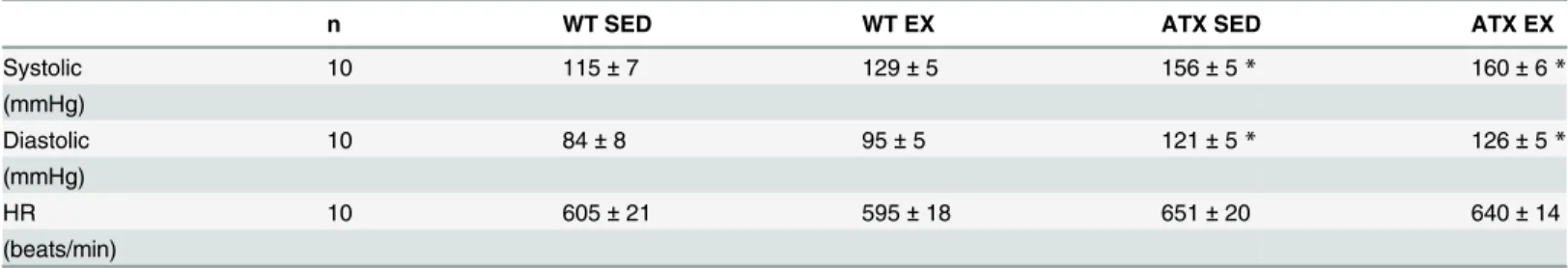
We previously reported that LDLr-/-; hApoB+/+atherosclerotic (ATX) mice exhibit severe dys-lipidemia: compared to wild-type (WT) mice, in 6 months old ATX mice plasma levels of total cholesterol, LDL-cholesterol and triglycerides are ~7, ~13 and ~10 folds higher, respectively [34,37,38]. In addition, 3 months of voluntary running had no influence on the observed severe dyslipidemia in ATX mice [34]. At 6-mo, as expected resting systolic and diastolic blood pressures were higher in ATX mice when compared to WT, and this was not affected by volun-tary exercise (Table 1). The heart rate was similar across all groups (Table 1). Severe dyslipide-mia did not interfere with the running capacity of ATX mice; they ran an average of 4.3 ± 0.4 km/d over the course of the study compared to 4.7± 1.0 km/d for WT mice (p>0.05) (Fig 1A). Both WT and ATX mice ran during the night (Fig 1B).
Dyslipidemia and exercise influence the expression of ROS generating
enzymes in the skeletal muscle
To evaluate the level of expression of endogenous producers of ROS, we quantified the expres-sion ofNOXand mitochondrialUCP.UCP2mRNA levels was significantly decreased in ATX mice when compared to WT for both sedentary and EX groups (Fig 2A).UCP3expression did not vary between groups (Fig 2B). We did not find differences in expression between WT and ATX sedentary mice for bothNOX2andNOX4. However,NOX2expression was significantly stimulated by exercise in both WT and ATX groups (Fig 2C), whileNOX4expression did not change with exercise (Fig 2D).
Dyslipidemia and exercise induce changes in antioxidant enzymes
expression in the skeletal muscle
compared to WT. This change in expression associated with dyslipidemia was prevented by exercise (Fig 3A and 3B).Prdx3was up-regulated in both sedentary and exercising ATX mice when compared to the corresponding WT group (Fig 3C).Gpx1mRNA expression was signifi-cantly lower in sedentary ATX mice in comparison to sedentary WT mice (Fig 3D).Gpx1 Table 1. Hemodynamic parameters in wild type and dyslipidemic mice exposed or not to a 3 months period of voluntary exercise.
n WT SED WT EX ATX SED ATX EX
Systolic 10 115±7 129±5 156±5* 160±6*
(mmHg)
Diastolic 10 84±8 95±5 121±5* 126±5*
(mmHg)
HR 10 605±21 595±18 651±20 640±14
(beats/min)
Systolic and diastolic blood pressure (mmHg) and heart rate (HR; beats/min) were measured by tail-cuff. Date are mean±SEM of 10 mice. WT: wild type; ATX: dyslipidemic mice; SED: sedentary; EX: exercise
*: p<0.05 vs. WT (Two-way ANOVA).
doi:10.1371/journal.pone.0151526.t001
Fig 2. Dyslipidemia and exercise-induced changes in ROS-generating enzymes expression.mRNA levels ofUCP2,UCP3,NOX2andNOX4in the skeletal muscle of wild type (WT) and dyslipidemic (ATX) mice in the sedentary (SED) and exercise (EX) groups. UCP2/3: uncoupling protein (mitochondrial proton carrier) 2/3; NOX2/4: NADPH oxidase 2/3. Data are mean±SEM, n = 9–10 mice.α: p<0.05vs. WT;β: p<0.05vs. SED (Two-way ANOVA).
mRNA expression was also higher in exercising WT and ATX mice when compared to seden-tary WT and ATX mice (Fig 3D). These changes in Gpx1 mRNA expression are also repro-duced in Gpx1 protein expression of WT, but not ATX mice (S1 Fig). Therefore, in ATX mice at this young age corresponding to the early stages of atherosclerosis, skeletal muscle antioxi-dant enzymes are up-regulated except forGpx1.
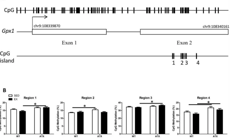
Differential DNA methylation of
Gpx1
in the skeletal muscle of ATX mice
We targeted CpG islands located in genes coding for the aforementioned antioxidant enzymes in order to test the hypothesis that epigenetic regulation contributes to the changes in gene expression. DNA methylation was below detection levels forTrx1andPrdx3, whileSod2 meth-ylation (>10%) did not vary between conditions (S2 Fig). Quantification ofGpx1gene methyl-ation by regions regrouping a single or more CpGs revealed differential methylmethyl-ation levels in the second exon (Fig 4A). Levels of methylation were significantly higher in ATX mice when compared to WT mice for both sedentary (regions 2 and 4) and EX conditions (regions 1, 3 and 4) (Fig 4B). Three months of voluntary exercise did not, however, alter the methylation of this gene, neither in WT nor in ATX mice (Fig 4B).
Fig 3. Dyslipidemia and exercise-induced changes in antioxidant enzymes expression.mRNA levels ofTrx1,Sod2,Prdx3andGpx1in the skeletal muscle of wild type (WT) and dyslipidemic (ATX) mice from the sedentary (SED) and exercise (EX) groups. Trx1: thioredoxin 1; Gpx1: glutathione peroxidase 1; Prdx3: peroxiredoxin 3; Sod2: superoxide dismutase 2, mitochondrial. Data are mean±SEM, n = 6–10 mice.α: p<0.05vs. WT;β: p<0.05vs. SED (Two-way ANOVA).
Transient changes in DNA methylation following exercise
To assess whether exercise could induce rapid changes in DNA methylation immediately after an exercise bout, we sacrificed WT mice during the last hour of their daily running activity (2:00 AM;Fig 1B), rather than during the day when they are inactive (Fig 1B). Interestingly, when methylation levels were measured in active WT mice (at t = 2:00), we observed that CpG regions 2 and 4 were demethylated during exercise, CpG region 1 tended to be demethylated, while region 3 remained unaffected (Fig 5). By comparison, methylation of none of these regions was affected in trained WT mice sacrificed during their inactive time (Fig 4B). This demonstrates the highly dynamic nature of epigenetic regulation.
In vitro
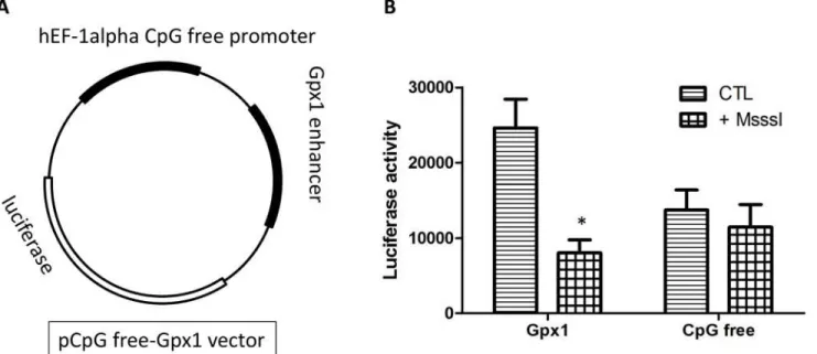
methylation decreases gene expression
To investigate the direct effect of DNA methylation on gene expression, the CpG-rich region of Gpx1, along with a CpG free promoter, was subcloned into the CpG-free-basic lucia vector. This new construction, CpG free-Gpx1 (Fig 6A), was methylated and transiently transfected in HEK293 cells to measure luciferase activity. The addition of the CpG-rich region significantly increases promoter activity (Fig 6B). Previously methylated vectors show no increase in pro-moter activity, similarly to empty vectors (Fig 6B). Therefore, methylation of this region is expected to inhibitGpx1expression.
Fig 4. Dyslipidemia induces methylation changes in theGpx1gene body.(A) Glutathione peroxidase 1 gene targeted for methylation quantification; representation of (top track) all CpG sites located inGpx1gene, (middle track) the coordinates of exons and (bottom track) the CpG islands containing the four regions covered by DNA methylation quantification. (B) Methylation percentage of four regions covering CpGs in the skeletal muscle of wild type (WT) or dyslipidemic (ATX) mice under sedentary (SED) or exercise condition (EX). Data are mean±SEM, n = 7–8 mice.*: p<0.05 (Two-way ANOVA).
No sign of oxidative stress in the skeletal muscle of dyslipidemic mice
Assessment of superoxide production by DHE staining and quantification of lipid peroxidation by 4-HNE immunostaining, in the skeletal muscle reveals similar oxidative stress between ATX and WT mice across all conditions (Fig 7), suggesting an efficient antioxidant compensa-tion at this age.
Discussion
Our findings indicate that the expression ofGpx1in the skeletal muscle is sensitive to the unstableredoxenvironment associated with severe dyslipidemia and exercise. We also provide evidence that its expression is associated with epigenetic regulation, namely DNA methylation of a novel locus located in a coding region of the gene.
To generate energy, mitochondria exert oxidative phosphorylation. This process needs to be tightly regulated since electrons leakage from the electron transport chain reacts to oxygen gen-erating superoxide. Uncoupling proteins (UCP) were first named as such for their ability to carry protons and steers the respiratory chain away from ATP synthesis in favour of thermo-genesis [39]. UCP1 is only expressed in the brown adipose tissue whereas UCP2 and UCP3 are also found in the skeletal muscle where they play a role in fatty acid metabolism [40]. Regard-ing their role in ROS production and oxidative stress regulation, knock-down experiments in Fig 5. Exercise induces a temporary decrease in DNA methylation.Methylation percentage of the four CpG groups covering the targeted CpG island found inGpx1comparing sedentary condition (SED, n = 7–8 mice) and exercise at time of sacrifice during their physically active time frame (EX (t = 2:00),
n = 3). For reference, see daily running activity onFig 1. Data are mean±SEM, n = 3–8 mice.*: p<0.05versusSED (Unpaired t-test).
mice have shown increases in ROS production linked to deficiency in UCP2 [41] and UCP3 [42]. Our study reveals that expression ofUCP2, but notUCP3, is lower in the skeletal muscle of ATX mice, suggesting that ROS production is favoured under dyslipidemia and that oxida-tive stress is more prone to happen.
Fig 6. DNA methylation ofGpx1decreases gene expression.In vitromethylation ofGpx1target region inhibited transcriptional activity, as measured by a luciferase reporter assay. (A) Schematic representation of the plasmid construction containing theGpx1CpG island region. (B) Luciferase activity ratio of methylated (M.SssI treated) to unmethylated control (CTL) plasmids containing a CpG-free promoter or theGpx1CpG island region. The assay was repeated 4 times and data are mean±SEM.*: p<0.05versusCTL (Unpaired t-test).
doi:10.1371/journal.pone.0151526.g006
Dysregulation of NOX activity is another well-known source of ROS in association with sys-temic pathological conditions including atherosclerosis [43] as well as diseases specific to the skeletal muscle [44,45]. Importantly, NOX are expressed in both arteries and skeletal muscle precursor cells [46]. NOX2 and NOX4 regulate basal ROS production and although they are generally associated with inflammation, there is a controversy regarding their role in cell sig-nalling, suggesting that there is an influence of the cell type and context [5]. In our study, we observed that exercise inducesNox2expression in the skeletal muscle of both WT and ATX mice without changes inNox4. Although we did not assess whether the overexpression ofNox2 is deleterious or beneficial to the skeletal muscle, this up-regulation ofNox2by exercise is somewhat surprising given the reports linking NOX2 to atherosclerotic lesions progression [47]. The role of NOX2 in the skeletal muscle is unclear, but recent reports have shown that the skeletal muscle contractile response triggers ROS production through a NOX2-dependent pathway [48] in addition to evidence for NOX2-mediated superoxide production in the aorta of rats following acute exercise [49]. Therefore, the contribution of ROS-generating enzyme to the level of oxidative stress in the presence of chronic severe dyslipidemia is only suggested by a change in their expression since non-specific DHE staining did not detect changes in ROS levels and 4-HNE immuno-histological staining did not show changes in lipid peroxidation. Antioxidant defense mechanisms are therefore expected to be up-regulated in these young dys-lipidemic mice.
Expression of antioxidant enzymes is very dynamic, and changes in both expression and activity have been linked to atherosclerosis [50–52] as well as exercise [53,54] and are determi-nant for preserving cellular functions [55–57]. In the present study, we found that expression of each antioxidant enzyme responded differently to the environmental context.Trx1,Sod2 andPrdx3are all up-regulated in the skeletal muscle of sedentary ATX mice, and this was pre-vented by exercise, except forPrdx3. The increase in these antioxidant enzymes suggests a pro-oxidative state requiring an antioxidant response from the organ to prevent pro-oxidative damage. These findings are consistent with other reports showing an increase in antioxidant enzymes expression during the onset and development of oxidative stress-related diseases [58,59]. Exer-cise seems to lessen the amplitude of the antioxidant response by preventing the overexpression ofTrx1andSod2only. The mechanistic origin of this regulation is however unknown. None-theless, because exercise normalized their level of expression, this is likely secondary to a reduc-tion in oxidative stress. If these two enzymes were responsible for the antioxidant effects of exercise, we would anticipate that their levels had increased above sedentary levels measured in WT mice.
In contrast toTrx1andSod2, we observed thatGpx1varies in an opposite manner: not only its expression is decreased in ATX mice, it is also increased by exercise above basal levels in both WT and ATX mice. A study has previously shown that low activity of Gpx1 is an indepen-dent risk factor of cardiovascular events [60]. The increase inGpx1with exercise is also coher-ent with the literature [61,62] and concords with the known antioxidant properties of chronic exercise [16,17]. Therefore, our data, in support of the current knowledge, suggest that overex-pression ofGpx1is one of the mechanisms responsible for the antioxidant protection by exercise.
these studies, we observed that hypermethylation of this novel CpG-rich region was associated with a reducedGpx1expression, in the context of dyslipidemia.
Thein vitromethylation assay allowed us to confirm that methylation of this specific locus causes an inhibition of gene expression. By itself, the CpG-rich region increases gene activity and when methylated, we observe transcriptional activity similar to that of aGpx1-free sequence. This suggests that methylation of the CpG island element blocks the binding and subsequent activity of a transcription co-factor. An interesting finding is that althoughGpx1 expression responds to both dyslipidemia and exercise, only changes in methylation are detected with dyslipidemia and not exercise, at least not in the long-term. This finding could contradict various studies that show changes of DNA methylation in response to exercise for various genes in the skeletal muscle [66–68]. Alternatively, changes inGpx1expression in response to physical training may be modulated by mechanisms other than epigenetics. Albeit this cannot be dismissed, a previous study in human skeletal muscle showed that acute exercise induces transient changes in the methylation of genes with changes in expression only
observed later, once methylation changes reverted to pre-exercise levels [69]. This mismatch in the timing of both events (changes in methylation and expression) could have occurred in our study. Exercise could induce temporary epigenetic changes that would have disappeared as lit-tle as minutes after the muscle work. In our first series of experiments, sacrifice of the animal was performed at 10 AM, several hours after the last running session. Hence, to assess the methylation profile in mice during physical activity, we sacrificed mice at 2 AM (t = 2:00) when they are still running (Fig 1B); in these conditions we observed methylation changes that were not detected in our previous experiments, demonstrating the temporary changes in meth-ylation induced by physical exercise. Altogether, these data suggest that epigenetic mechanisms could be responsible for the changes in expression in response to exercise that we observed for Gpx1in the long-term. Further investigation is required to understand the link between the ini-tial transient changes in DNA methylation that occurs during exercise bouts and lasting changes in gene expression observed in the long-term.
Exacerbation of oxidative stress in the skeletal muscle cells in the context of dyslipidemia is caused by a disturbance in the production and elimination of mitochondria-derived free radi-cals [13]. This, in turn, impairs the organ, and the excess of ROS can diffuse to the femoral vas-cular bed causing vasvas-cular endothelial dysfunction, consequently impeding blood flow
regulation to the muscle leading to sarcopenia [13]. This vicious circle has previously been observed in hypertensive rats [70]. In our experimental conditions, there is no evidence of oxi-dative stress-dependent damage in the skeletal muscle of our young, middle-age ATX mice, suggesting that oxidative stress is well compensated in skeletal muscle, supporting the adaptive responses of antioxidant gene expression reported above. This is confirmed by the demonstra-tion that in these mice, skeletal muscle funcdemonstra-tion was not altered as evidenced by their conserved running capacity.
Limitations of the study
discrepancies between dyslipidemia and exercise on epigenetics is that the former is a long-term and continuous stimulation, as opposed to a short-long-term and inlong-termittent stimulation for the latter. In other words, mice are constantly exposed to dyslipidemia fromintra uterobut they only practice intermittent voluntary bouts of exercise, starting at 3-month of age. Studies in the field of developmental plasticity have shown thatin uteroand early life exposure to envi-ronmental factors induces phenotypic adaptations that are reflected through adulthood and that these changes are deeply rooted in the epigenome [73,74]. Protein expressions of Gpx1 reveal yet another level of complexity and do not fully reflect the changes in mRNA levels in ATX mice. This is likely associated with differential post-translational regulation in dyslipide-mia beyond the scope of the present study. Finally, whether ROS are responsible for the epige-netic changes observed both with dyslipidemia and during exercise was not directly tested. The use of an antioxidant may help, although they most likely may directly impact on the
epigenome.
Conclusions
The overexpression of some pro- and anti-oxidant enzymes by severe dyslipidemia suggests a pro-oxidative state in the skeletal muscle where the chronic rise in ROS could damage skeletal muscle cells, to latter contribute to the damage of the neighbouring vascular cells. We show that one of the mechanisms by which the antioxidant enzyme Gpx1 is modulated is by DNA methylation; its lower expression is associated with the higher level of methylation of a novel CpG island region in ATX mice. We also reinforced the cause-effect relationship between DNA methylation of this loci and its mRNA expression. Our data suggests, therefore, that DNA methylation is a possible mechanism for regulating gene expression in dyslipidemia as well as following chronic voluntary exercise training. Whether ROS are directly involved remains debatable.
Supporting Information
S1 Fig. Gpx1 protein expression.
(PDF)
S2 Fig. CpG methylation analysis ofPrdx3,Sod2andTrx1. (PDF)
S1 Table. Primer sequences used for qPCR analysis.
(PDF)
S2 Table. Primer sequences used for DNA methylation analysis.
(PDF)
Author Contributions
Conceived and designed the experiments: AN MM ET. Performed the experiments: AN ND MM. Analyzed the data: AN ET. Contributed reagents/materials/analysis tools: MM. Wrote the paper: AN ET.
References
1. Siti HN, Kamisah Y, Kamsiah J. The role of oxidative stress, antioxidants and vascular inflammation in cardiovascular disease (a review). Vascul Pharmacol. 2015; 71:40–56. doi:10.1016/j.vph.2015.03.005
2. Montezano AC, Dulak-Lis M, Tsiropoulou S, Harvey A, Briones AM, Touyz RM. Oxidative stress and human hypertension: vascular mechanisms, biomarkers, and novel therapies. Can J Cardiol. 2015; 31 (5):631–41. doi:10.1016/j.cjca.2015.02.008PMID:25936489.
3. Droge W. Free radicals in the physiological control of cell function. Physiol Rev. 2002; 82(1):47–95. doi:
10.1152/physrev.00018.2001PMID:11773609.
4. Weber C, Noels H. Atherosclerosis: current pathogenesis and therapeutic options. Nat Med. 2011; 17 (11):1410–22. doi:10.1038/nm.2538PMID:22064431.
5. Lassegue B, San Martin A, Griendling KK. Biochemistry, physiology, and pathophysiology of NADPH oxidases in the cardiovascular system. Circ Res. 2012; 110(10):1364–90. doi:10.1161/CIRCRESAHA.
111.243972PMID:22581922; PubMed Central PMCID: PMC3365576.
6. Kim HS, Park KG, Koo TB, Huh S, Lee IK. The modulating effects of the overexpression of uncoupling protein 2 on the formation of reactive oxygen species in vascular cells. Diabetes Res Clin Pract. 2007; 77 Suppl 1:S46–8. doi:10.1016/j.diabres.2007.01.032PMID:17462780.
7. Victor VM, Rocha M, Sola E, Banuls C, Garcia-Malpartida K, Hernandez-Mijares A. Oxidative stress, endothelial dysfunction and atherosclerosis. Curr Pharm Des. 2009; 15(26):2988–3002. PMID:
19754375.
8. Katsiki N, Manes C. Is there a role for supplemented antioxidants in the prevention of atherosclerosis? Clin Nutr. 2009; 28(1):3–9. doi:10.1016/j.clnu.2008.10.011PMID:19042058.
9. Steinhubl SR. Why have antioxidants failed in clinical trials? Am J Cardiol. 2008; 101(10A):14D–9D.
doi:10.1016/j.amjcard.2008.02.003PMID:18474268.
10. Leblond F, Nguyen A, Bolduc V, Lambert J, Yu C, Duquette N, et al. Postnatal exposure to voluntary exercise but not the antioxidant catechin protects the vasculature after a switch to an atherogenic envi-ronment in middle-age mice. Pflugers Arch. 2013; 465(2):197–208. doi:10.1007/s00424-012-1206-8
PMID:23291710; PubMed Central PMCID: PMC3695887.
11. Ristow M, Zarse K, Oberbach A, Kloting N, Birringer M, Kiehntopf M, et al. Antioxidants prevent health-promoting effects of physical exercise in humans. Proc Natl Acad Sci U S A. 2009; 106(21):8665–70.
doi:10.1073/pnas.0903485106PMID:19433800; PubMed Central PMCID: PMC2680430.
12. Vuori IM, Lavie CJ, Blair SN. Physical activity promotion in the health care system. Mayo Clin Proc. 2013; 88(12):1446–61. doi:10.1016/j.mayocp.2013.08.020PMID:24290119.
13. Roden M. Future of muscle research in diabetes: a look into the crystal ball. Diabetologia. 2015; 58 (8):1693–8. doi:10.1007/s00125-015-3629-1PMID:26003326.
14. Palmefors H, DuttaRoy S, Rundqvist B, Borjesson M. The effect of physical activity or exercise on key biomarkers in atherosclerosis—A systematic review. Atherosclerosis. 2014; 235(1):150–61. doi:10.
1016/j.atherosclerosis.2014.04.026PMID:24835434.
15. Szostak J, Laurant P. The forgotten face of regular physical exercise: a 'natural' anti-atherogenic activ-ity. Clin Sci (Lond). 2011; 121(3):91–106. doi:10.1042/CS20100520PMID:21729002.
16. Seals DR. Edward F. Adolph Distinguished Lecture: The remarkable anti-aging effects of aerobic exer-cise on systemic arteries. J Appl Physiol (1985). 2014; 117(5):425–39. doi:10.1152/japplphysiol.
00362.2014PMID:24855137; PubMed Central PMCID: PMCPMC4157159.
17. Rengo G, Parisi V, Femminella GD, Pagano G, de Lucia C, Cannavo A, et al. Molecular aspects of the cardioprotective effect of exercise in the elderly. Aging Clin Exp Res. 2013; 25(5):487–97. doi:10.1007/
s40520-013-0117-7PMID:23949971.
18. Pedersen BK, Febbraio MA. Muscles, exercise and obesity: skeletal muscle as a secretory organ. Nat Rev Endocrinol. 2012; 8(8):457–65. doi:10.1038/nrendo.2012.49PMID:22473333.
19. Kim GH, Ryan JJ, Archer SL. The role of redox signaling in epigenetics and cardiovascular disease. Antioxid Redox Signal. 2013; 18(15):1920–36. doi:10.1089/ars.2012.4926PMID:23480168; PubMed
Central PMCID: PMC3624767.
20. Bird A. DNA methylation patterns and epigenetic memory. Genes Dev. 2002; 16(1):6–21. doi:10.1101/
gad.947102PMID:11782440.
21. Cedar H, Bergman Y. Linking DNA methylation and histone modification: patterns and paradigms. Nat Rev Genet. 2009; 10(5):295–304. doi:10.1038/nrg2540PMID:19308066.
22. Jones PA. Functions of DNA methylation: islands, start sites, gene bodies and beyond. Nat Rev Genet. 2012; 13(7):484–92. doi:10.1038/nrg3230PMID:22641018.
23. Jimenez-Chillaron JC, Diaz R, Martinez D, Pentinat T, Ramon-Krauel M, Ribo S, et al. The role of nutri-tion on epigenetic modificanutri-tions and their implicanutri-tions on health. Biochimie. 2012; 94(11):2242–63. doi:
10.1016/j.biochi.2012.06.012PMID:22771843.
25. Wilson AG. Epigenetic regulation of gene expression in the inflammatory response and relevance to common diseases. J Periodontol. 2008; 79(8 Suppl):1514–9. doi:10.1902/jop.2008.080172PMID:
18673005.
26. Ordovas JM, Smith CE. Epigenetics and cardiovascular disease. Nat Rev Cardiol. 2010; 7(9):510–9.
doi:10.1038/nrcardio.2010.104PMID:20603647; PubMed Central PMCID: PMC3075976.
27. Turunen MP, Aavik E, Yla-Herttuala S. Epigenetics and atherosclerosis. Biochim Biophys Acta. 2009; 1790(9):886–91. doi:10.1016/j.bbagen.2009.02.008PMID:19233248.
28. Zhu S, Goldschmidt-Clermont PJ, Dong C. Inactivation of monocarboxylate transporter MCT3 by DNA methylation in atherosclerosis. Circulation. 2005; 112(9):1353–61. doi:10.1161/CIRCULATIONAHA.
104.519025PMID:16116050.
29. Carrio E, Suelves M. DNA methylation dynamics in muscle development and disease. Front Aging Neu-rosci. 2015; 7:19. doi:10.3389/fnagi.2015.00019PMID:25798107; PubMed Central PMCID:
PMCPMC4350440.
30. Lakshmi SV, Naushad SM, Reddy CA, Saumya K, Rao DS, Kotamraju S, et al. Oxidative stress in coro-nary artery disease: epigenetic perspective. Mol Cell Biochem. 2013; 374(1–2):203–11. doi:10.1007/
s11010-012-1520-7PMID:23160801.
31. Archer SL, Marsboom G, Kim GH, Zhang HJ, Toth PT, Svensson EC, et al. Epigenetic attenuation of mitochondrial superoxide dismutase 2 in pulmonary arterial hypertension: a basis for excessive cell proliferation and a new therapeutic target. Circulation. 2010; 121(24):2661–71. doi:10.1161/
CIRCULATIONAHA.109.916098PMID:20529999; PubMed Central PMCID: PMC2914302.
32. Paneni F, Costantino S, Volpe M, Luscher TF, Cosentino F. Epigenetic signatures and vascular risk in type 2 diabetes: a clinical perspective. Atherosclerosis. 2013; 230(2):191–7. doi:10.1016/j.
atherosclerosis.2013.07.003PMID:24075743.
33. Gendron ME, Theoret JF, Mamarbachi AM, Drouin A, Nguyen A, Bolduc V, et al. Late chronic catechin antioxidant treatment is deleterious to the endothelial function in aging mice with established athero-sclerosis. Am J Physiol Heart Circ Physiol. 2010; 298(6):H2062–70. doi:10.1152/ajpheart.00532.2009
PMID:20382853; PubMed Central PMCID: PMC3701586.
34. Bolduc V, Drouin A, Gillis MA, Duquette N, Thorin-Trescases N, Frayne-Robillard I, et al. Heart rate-associated mechanical stress impairs carotid but not cerebral artery compliance in dyslipidemic athero-sclerotic mice. Am J Physiol Heart Circ Physiol. 2011; 301(5):H2081–92. doi:10.1152/ajpheart.00706.
2011PMID:21926346; PubMed Central PMCID: PMC3700878.
35. Ehrich M, Nelson MR, Stanssens P, Zabeau M, Liloglou T, Xinarianos G, et al. Quantitative high-throughput analysis of DNA methylation patterns by base-specific cleavage and mass spectrometry. Proc Natl Acad Sci U S A. 2005; 102(44):15785–90. doi:10.1073/pnas.0507816102PMID:16243968;
PubMed Central PMCID: PMC1276092.
36. Kent WJ, Sugnet CW, Furey TS, Roskin KM, Pringle TH, Zahler AM, et al. The human genome browser at UCSC. Genome Res. 2002; 12(6):996–1006. doi:10.1101/gr.229102Article published online before
print in May 2002. PMID:12045153; PubMed Central PMCID: PMC186604.
37. Drouin A, Bolduc V, Thorin-Trescases N, Belanger E, Fernandes P, Baraghis E, et al. Catechin treat-ment improves cerebrovascular flow-mediated dilation and learning abilities in atherosclerotic mice. Am J Physiol Heart Circ Physiol. 2011; 300(3):H1032–43. doi:10.1152/ajpheart.00410.2010PMID:
21186270; PubMed Central PMCID: PMC3702511.
38. Bolduc V, Baraghis E, Duquette N, Thorin-Trescases N, Lambert J, Lesage F, et al. Catechin prevents severe dyslipidemia-associated changes in wall biomechanics of cerebral arteries in LDLr-/-:hApoB+/+ mice and improves cerebral blood flow. Am J Physiol Heart Circ Physiol. 2012; 302(6):H1330–9. doi:
10.1152/ajpheart.01044.2011PMID:22268108; PubMed Central PMCID: PMCPMC3695886.
39. Argyropoulos G, Harper ME. Uncoupling proteins and thermoregulation. J Appl Physiol (1985). 2002; 92(5):2187–98. doi:10.1152/japplphysiol.00994.2001PMID:11960973.
40. Rousset S, Alves-Guerra MC, Mozo J, Miroux B, Cassard-Doulcier AM, Bouillaud F, et al. The biology of mitochondrial uncoupling proteins. Diabetes. 2004; 53 Suppl 1:S130–5. PMID:14749278. 41. Arsenijevic D, Onuma H, Pecqueur C, Raimbault S, Manning BS, Miroux B, et al. Disruption of the
uncoupling protein-2 gene in mice reveals a role in immunity and reactive oxygen species production. Nat Genet. 2000; 26(4):435–9. doi:10.1038/82565PMID:11101840.
42. Vidal-Puig AJ, Grujic D, Zhang CY, Hagen T, Boss O, Ido Y, et al. Energy metabolism in uncoupling protein 3 gene knockout mice. J Biol Chem. 2000; 275(21):16258–66. doi:10.1074/jbc.M910179199
PMID:10748196.
43. Sorescu D, Weiss D, Lassegue B, Clempus RE, Szocs K, Sorescu GP, et al. Superoxide production and expression of nox family proteins in human atherosclerosis. Circulation. 2002; 105(12):1429–35.
44. Whitehead NP, Yeung EW, Froehner SC, Allen DG. Skeletal muscle NADPH oxidase is increased and triggers stretch-induced damage in the mdx mouse. PLoS One. 2010; 5(12):e15354. doi:10.1371/ journal.pone.0015354PMID:21187957; PubMed Central PMCID: PMC3004864.
45. Sullivan-Gunn MJ, Campbell-O'Sullivan SP, Tisdale MJ, Lewandowski PA. Decreased NADPH oxi-dase expression and antioxidant activity in cachectic skeletal muscle. J Cachexia Sarcopenia Muscle. 2011; 2(3):181–8. doi:10.1007/s13539-011-0037-3PMID:21966644; PubMed Central PMCID:
PMC3177039.
46. Mofarrahi M, Brandes RP, Gorlach A, Hanze J, Terada LS, Quinn MT, et al. Regulation of proliferation of skeletal muscle precursor cells by NADPH oxidase. Antioxid Redox Signal. 2008; 10(3):559–74. doi:
10.1089/ars.2007.1792PMID:18092937.
47. Drummond GR, Sobey CG. Endothelial NADPH oxidases: which NOX to target in vascular disease? Trends Endocrinol Metab. 2014; 25(9):452–63. doi:10.1016/j.tem.2014.06.012PMID:25066192. 48. Diaz-Vegas A, Campos CA, Contreras-Ferrat A, Casas M, Buvinic S, Jaimovich E, et al. ROS
Produc-tion via P2Y1-PKC-NOX2 Is Triggered by Extracellular ATP after Electrical StimulaProduc-tion of Skeletal Mus-cle Cells. PLoS One. 2015; 10(6):e0129882. doi:10.1371/journal.pone.0129882PMID:26053483; PubMed Central PMCID: PMC4460042.
49. Tanaka LY, Bechara LR, dos Santos AM, Jordao CP, de Sousa LG, Bartholomeu T, et al. Exercise improves endothelial function: a local analysis of production of nitric oxide and reactive oxygen species. Nitric Oxide. 2015; 45:7–14. doi:10.1016/j.niox.2015.01.003PMID:25619203.
50. Fukai T, Ushio-Fukai M. Superoxide dismutases: role in redox signaling, vascular function, and dis-eases. Antioxid Redox Signal. 2011; 15(6):1583–606. doi:10.1089/ars.2011.3999PMID:21473702;
PubMed Central PMCID: PMC3151424.
51. Madrigal-Matute J, Martinez-Pinna R, Fernandez-Garcia CE, Ramos-Mozo P, Burillo E, Egido J, et al. Cell stress proteins in atherothrombosis. Oxid Med Cell Longev. 2012; 2012:232464. doi:10.1155/ 2012/232464PMID:22792412; PubMed Central PMCID: PMC3389727.
52. Bonomini F, Tengattini S, Fabiano A, Bianchi R, Rezzani R. Atherosclerosis and oxidative stress. Histol Histopathol. 2008; 23(3):381–90. PMID:18072094.
53. Powers SK, Lennon SL. Analysis of cellular responses to free radicals: focus on exercise and skeletal muscle. Proc Nutr Soc. 1999; 58(4):1025–33. PMID:10817171.
54. Brinkmann C, Brixius K. Peroxiredoxins and sports: new insights on the antioxidative defense. J Physiol Sci. 2013; 63(1):1–5. doi:10.1007/s12576-012-0237-4PMID:23055024.
55. Oelze M, Kroller-Schon S, Steven S, Lubos E, Doppler C, Hausding M, et al. Glutathione peroxidase-1 deficiency potentiates dysregulatory modifications of endothelial nitric oxide synthase and vascular dysfunction in aging. Hypertension. 2014; 63(2):390–6. doi:10.1161/HYPERTENSIONAHA.113.
01602PMID:24296279.
56. Ballinger SW, Patterson C, Knight-Lozano CA, Burow DL, Conklin CA, Hu Z, et al. Mitochondrial integ-rity and function in atherogenesis. Circulation. 2002; 106(5):544–9. PMID:12147534.
57. Jang YC, Van Remmen H. The mitochondrial theory of aging: insight from transgenic and knockout mouse models. Exp Gerontol. 2009; 44(4):256–60. doi:10.1016/j.exger.2008.12.006PMID:19171187. 58. Okuda M, Inoue N, Azumi H, Seno T, Sumi Y, Hirata K, et al. Expression of glutaredoxin in human
coro-nary arteries: its potential role in antioxidant protection against atherosclerosis. Arterioscler Thromb Vasc Biol. 2001; 21(9):1483–7. PMID:11557676.
59. Miao L, St Clair DK. Regulation of superoxide dismutase genes: implications in disease. Free Radic Biol Med. 2009; 47(4):344–56. doi:10.1016/j.freeradbiomed.2009.05.018PMID:19477268; PubMed
Central PMCID: PMC2731574.
60. Blankenberg S, Rupprecht HJ, Bickel C, Torzewski M, Hafner G, Tiret L, et al. Glutathione peroxidase 1 activity and cardiovascular events in patients with coronary artery disease. N Engl J Med. 2003; 349 (17):1605–13. doi:10.1056/NEJMoa030535PMID:14573732.
61. Alessio HM, Ansinelli H, Threadgill C, Hagerman AE. Comparison of gene and protein expressions in rats residing in standard cages with those having access to an exercise wheel. Biomed Res Int. 2014; 2014:950516. doi:10.1155/2014/950516PMID:24719897; PubMed Central PMCID: PMC3955688.
62. Sindler AL, Reyes R, Chen B, Ghosh P, Gurovich AN, Kang LS, et al. Age and exercise training alter signaling through reactive oxygen species in the endothelium of skeletal muscle arterioles. J Appl Phy-siol (1985). 2013; 114(5):681–93. doi:10.1152/japplphysiol.00341.2012PMID:23288555; PubMed
Central PMCID: PMC3615591.
63. Kulak MV, Cyr AR, Woodfield GW, Bogachek M, Spanheimer PM, Li T, et al. Transcriptional regulation of the GPX1 gene by TFAP2C and aberrant CpG methylation in human breast cancer. Oncogene. 2013; 32(34):4043–51. doi:10.1038/onc.2012.400PMID:22964634; PubMed Central PMCID:
64. Min SY, Kim HS, Jung EJ, Jung EJ, Jee CD, Kim WH. Prognostic significance of glutathione peroxidase 1 (GPX1) down-regulation and correlation with aberrant promoter methylation in human gastric cancer. Anticancer Res. 2012; 32(8):3169–75. PMID:22843889.
65. Jee CD, Kim MA, Jung EJ, Kim J, Kim WH. Identification of genes epigenetically silenced by CpG meth-ylation in human gastric carcinoma. Eur J Cancer. 2009; 45(7):1282–93. doi:10.1016/j.ejca.2008.12.
027PMID:19195878.
66. Laker RC, Lillard TS, Okutsu M, Zhang M, Hoehn KL, Connelly JJ, et al. Exercise prevents maternal high-fat diet-induced hypermethylation of the Pgc-1alpha gene and age-dependent metabolic dysfunc-tion in the offspring. Diabetes. 2014; 63(5):1605–11. doi:10.2337/db13-1614PMID:24430439. 67. Ronn T, Ling C. Effect of exercise on DNA methylation and metabolism in human adipose tissue and
skeletal muscle. Epigenomics. 2013; 5(6):603–5. doi:10.2217/epi.13.61PMID:24283873.
68. Nitert MD, Dayeh T, Volkov P, Elgzyri T, Hall E, Nilsson E, et al. Impact of an exercise intervention on DNA methylation in skeletal muscle from first-degree relatives of patients with type 2 diabetes. Diabe-tes. 2012; 61(12):3322–32. doi:10.2337/db11-1653PMID:23028138; PubMed Central PMCID:
PMC3501844.
69. Barres R, Yan J, Egan B, Treebak JT, Rasmussen M, Fritz T, et al. Acute exercise remodels promoter methylation in human skeletal muscle. Cell Metab. 2012; 15(3):405–11. doi:10.1016/j.cmet.2012.01.
001PMID:22405075.
70. Zhao W, Swanson SA, Ye J, Li X, Shelton JM, Zhang W, et al. Reactive oxygen species impair sympa-thetic vasoregulation in skeletal muscle in angiotensin II-dependent hypertension. Hypertension. 2006; 48(4):637–43. doi:10.1161/01.HYP.0000240347.51386.eaPMID:16940212.
71. Reul JM. Making memories of stressful events: a journey along epigenetic, gene transcription, and sig-naling pathways. Front Psychiatry. 2014; 5:5. doi:10.3389/fpsyt.2014.00005PMID:24478733; PubMed Central PMCID: PMC3897878.
72. Stankiewicz AM, Swiergiel AH, Lisowski P. Epigenetics of stress adaptations in the brain. Brain Res Bull. 2013; 98:76–92. doi:10.1016/j.brainresbull.2013.07.003PMID:23906660.
73. Hochberg Z, Feil R, Constancia M, Fraga M, Junien C, Carel JC, et al. Child health, developmental plasticity, and epigenetic programming. Endocr Rev. 2011; 32(2):159–224. doi:10.1210/er.2009-0039
PMID:20971919; PubMed Central PMCID: PMC3365792.
74. Hanson M, Godfrey KM, Lillycrop KA, Burdge GC, Gluckman PD. Developmental plasticity and devel-opmental origins of non-communicable disease: theoretical considerations and epigenetic mecha-nisms. Prog Biophys Mol Biol. 2011; 106(1):272–80. doi:10.1016/j.pbiomolbio.2010.12.008PMID: