512
Mattos BP
Cellular and biomolecular mechanisms in dilated cardiomyopathy
Arq Bras Cardiol volume 72, (nº 4), 1999
Hospital de Clínicas de Porto Alegre - UFRGS
Mailing address: Beatriz Piva e Mattos – Serviço de Cardiologia – Hospital de Clínicas de Porto Alegre – Rua Ramiro Barcelos, 2350 – 90035-003 – Porto Alegre, RS - Brazil
Beatriz Piva e Mattos
Porto Alegre, RS - Brazil
Cellular and Biomolecular Mechanisms in
Dilated Cardiomyopathy
Update
Dilated cardiomyopathy (DCM) is a primary myocar-dial disease, characterized by uni- or biventricular dilation associated with a generally progressive contractile dys-function 1. It is predominantly diagnosed in the advanced stage, in which it manifests itself by cardiomegaly and heart failure, causing significant morbidity and mortality 2. Less frequently, it is identified in the initial stage, in which the congestive signals are absent but ventricular dilation and dysfunction in a mild or moderate degree are evident 3,4.
At first, DCM was considered a disease of obscure etiology. Several decades after being identified as an entity, its complex pathogenesis has gradually become better understood. Systematic studies generated in immunology, genetics, and cellular and molecular biology have contri-buted to the understanding of the disease mechanism. However, the heterogeneous character of DCM, coupled with the multiplicity of pathologic processes involved, has made this task difficult.
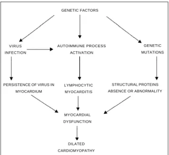
DCM is currently understood as a multifactorial disease, in which viral infections, immunologic mechanisms and genetic factors, acting individually or together, result in a definitive myocardial lesion (fig. 1).
Role of viral infection
Clinical and experimental evidence suggest the par-ticipation of a viral infection in the cellular injury process. Based on this concept, viral myocarditis is considered a precursor of DCM, leading to definitive heart disease in susceptible individuals. The concept of an evolutionary sequence is based on indirect clinical evidence and con-clusions from studies developed in experimental models.
Coxsackievirus B,from the Enteroviridaefamily, is considered the main etiological agent of human myocar-ditis 5 except in the endemic areas of Chagas’ disease. These viruses are related to lymphocytic myocarditis, which is a process characterized by diffuse cellular
infil-tration associated with myocytolysis 6, but in which the microorganism is not usually identified through conven-tional techniques. If viral participation in myocarditis is difficult to prove, viral action in DCM may still be consi-dered controversial.
Longitudinal studies of a small series of patients demonstrate an evolution to DCM in 40 to 52% of the cases with a histopathologic diagnosis of lymphocytic myo-carditis 7,8. However, the causal relationship between en-terovirus infections and further development of cardio-myopathy is difficult to confirm because of the virus’ endemic characteristics and the frequent subclinical cha-racter of the inflammatory manifestations in the acute phase. Serology of enterovirus infections is equally complex. Neutralizing antibodies to coxsackievirus B are detected at higher levels in DCM patients than in controls 9. The fact that most of the population has had contact with the virus, and the possibility of antibody levels remaining high after the acute phase of infection jeopardizes the evaluation of these results. Coxsackie B-specific immunoglobulin M is identified in patients with recent diagnosis and end-stage forms 10,11. Although this immunoglobulin is found more frequently in the DCM group than in nonselected controls 10, no difference is seen when serologic data are compared with that obtained from relatives sharing the same domicile 12. Sequential evaluation of serum specimens revealed antibodies persis-ting in some cases, as well as late seroconversion after reactions become negative in other cases 11,12.
Arq Bras Cardiol volume 72, (nº 4), 1999
Mattos BP Cellular and biomolecular mechanisms in dilated cardiomyopathy
513 possible association between enterovirus infections and
DCM 20. Interpretation of these data should take into ac-count methodological differences related to the technique applied and selection of patients and control groups. Small outbreaks of epidemics caused by enteroviruses may cause results to become positive in normal controls exposed to the same environmental factors, considering the cardio-tropic characteristics of these agents.
The presence of enteroviral genomes combined with the persistence of serologic reactions suggests the possi-bility of the virus remaining in the myocardium after the acute phase, with no evidence of protein synthesis but nonetheless interference with cellular functions 12. This hypothesis becomes questionable because the enteroviral RNA proportion usually detected through PCR in these hearts is small and localized 19. On the other hand, experi-mental myocarditis models show that some coxsackievirus B-3 strains have greater cardiovirulence 20. It is predicted that methods will be developed in the future that will be able to distinguish whether or not the identified genomes are related to strains with greater pathogenic potential.
Activation of immunologic mechanisms
It is estimated that, in one third of the cases, DCM is an autoimmune disease 21. Immunologic organ-specific reactions depend on genetic predisposition and are modu-lated by environmental factors. Enterovirus infections are thought to play a fundamental role in triggering autoimmu-nity in this disease. In an initial step, the virus may cause myocytolytic necrosis by direct action, depending on its capacity for intracellular replication. As this process evolves, activation of cytotoxic T lymphocytes occurs, which intensifies the injury of infected and intact cells during lysis 22. Persistence of infection could result in exacerbation of the cellular and humoral immunologic
response, which would perpetuate aggression against myocytes 12. Autoimmune reaction would result from m olecular m im esisbetween viral and host proteins or from neoantigen expression 23. Other factors may be responsible for triggering an autoimmune process, such as gestation and alcoholism.
Immunoregulatory disorders are seen in DCM. A decrease in the activity of natural killer cells 24 and reduction of the suppressor function of T lymphocytes 25 may indicate greater susceptibility to viral infections. Experimental models of viral and autoimmune myocarditis show the decisive participation of cellular immunity in the myocardial injury process through T lymphocyte activation and cytokine release 22. In DCM, a correlation of the expression of perforin and T cell intracellular antigen-1 with the degree of interstitial fibrosis revealed at endomyocardial biopsy 26 was observed.
Autoantibodies to cardiac proteins are identified in 30 to 40% of DCM cases 21. Organ-specific autoantibodies, which interact with beta-adrenergic receptors 27, cholinergic muscarinic receptors 28, mitochondrial antigens 29 and alpha and beta cardiac myosin heavy chains 30, have already been described elsewhere. The clinical importance of auto-antibodies and their pathogenic meaning are not fully understood, because they are usually identified in an evolutionary phase, in which the disease is established. It is possible that not all of them have the same clinical meaning. Negative results obtained in most of the patients could be justified by the fact that, similarly to other autoimmune processes, the autoantibodies become undetectable as the disease progresses 21.
There is no confirmation in regard to the ability of autoantibodies producing myocytolysis. They may interfe-re with cellular functions, as pinterfe-reviously shown for beta-adrenergic receptors 27 and calcium channels, by cross-reaction with autoantibodies to ADP/ATP carriers 29. Although there may be a correlation between disease severity and autoantibody presence, the autoantibodies may constitute mere cellular lesion markers. Their detection in 20% of asymptomatic individuals, 1st-degree relatives of DCM patients, indicates greater propensity to the disease in this subgroup 31.
Other evidence suggests the participation of two immunologic mechanisms in DCM: 1) presence of abnor-mal serologic concentrations of interleukin-2 and inter-leukin-10 32, and 2) abnormal expression of MHC class II antigens in endothelial cells 33.
Genetic factors and familiar forms
Recent studies introduced the concept that the deve-lopment of DCM depends on genetic factors, which modulate sensitivity to viral infections and subsequent activation of immunologic reactions. Multiple genes may be involved in this process. Myocardial lesion depends on the interaction between genetic and environmental factors. Clinical and experimental studies suggest the association GENETIC FACTORS
AUTOIMMUNE PROCESS ACTIVATION VIRUS
INFECTION
GENETIC MUTATIONS
LYMPHOCYTIC MYOCARDITIS
MYOCARDIAL DYSFUNCTION
DILATED CARDIOMYOPATHY PERSISTENCE OF VIRUS IN
MYOCARDIUM
STRUCTURAL PROTEINS ABSENCE OR ABNORMALITY
514
Mattos BP
Cellular and biomolecular mechanisms in dilated cardiomyopathy
Arq Bras Cardiol volume 72, (nº 4), 1999
with HLA genes, which modulate the immunologic res-ponse, and the genes of T cell receptors 34. HLA genes regulate the immunologic system and the predisposition to autoimmune disease, through the modulation of T cell receptors, and the selection and presentation of antigenic peptides. A higher frequency of class II HLA antigens, DR4 and, possibly, DQw-4 loci, is identified in DCM patients when compared with controls35, even though a further analysis restricted to patients in a heart transplant program did not confirm these results 36. Elevation of HLA-specific haplotypes is seen in the subgroup with deficiency in natu-ral-killer cells 37. Association of HLA-D alleles with auto-antibodies to beta-adrenergic receptors is also referred to 38. These findings indicate that the disease development may be related to immunologic response and genes controlling this process.
Studies performed during the past decade to determine the frequency of familialforms of DCM reveal that this entity, initially interpreted as an acquired process, has a ge-netic basis in a significant number of patients. In preliminary investigations, familial forms were sporadically identified. Further, prospective analyses reveal an increasing preva-lence of these forms, corresponding to 20% to 35% of the cases 39-42. These figures may be underestimated, consi-dering the difficulty in evaluating all members of an affected family and the reduced gene penetrance, which makes the carriers of the disease look apparently healthy.
In a recent prospective evaluation of 200 1st-degree re-latives of 100 patients, McKenna et al 41 identified familial nature and tendency in 25% and 45% of the cases, respecti-vely. HLA antigen was detected in 2/3 of the affected fa-milies and a greater prevalence of this antigen was noted in familial forms when compared to nonfamilial ones.
DCM has several forms of genetic transmission. Keeling et al 40, in a prospective study of 236 individuals from 40 affected families, identified the predominance of autosomal dominant inheritance with incomplete pene-trance. Early forms of the disease in asymptomatic members of the families were observed: 18% exhibited a slight in-crease of the left ventricle and 4% exhibited reduction in the fractional shortening when compared with controls. Other inherited forms are recognized: autosomal recessive, X chromosome-linked and mitochondrial 43.
From the genetic viewpoint, DCM also has hetero-geneous characteristics. Distinct genetic mutations produce multiple phenotypes, which determine similar clinical, histopathologic and hemodynamic manifestations. Familial forms are, however, diagnosed in earlier ages than sporadic ones 42. It is estimated that 20% of the 1st-degree relatives have a risk of developing the disease at more advanced ages, as gene penetrance is related to age range 40. Lately, the progress made in genetics and molecular biology has allowed great advances in the study of the familial forms of the disease. The autosomal dominant form manifests itself in the 2nd or 3rd decade of life, with ventri-cular dilation and dysfunction, expressed by heart failure or arrhythmias 43. The first genetic locus was identified in
chromosome 9q13-q22 44. Later, a second locus was descri-bed in chromosome 1q32 45. Forms associated with mitral valve prolapse were mapped to the short arm of chromo-some 10 46. These chromosomal regions are related to genes codifying regulatory and cytoskeletical proteins.
Rare forms, characterized by conduction system of the disease of the heart beginning in the 2nd or 3rd decade of life, followed by ventricular dilation and dysfunction some years after, are also transmitted by autosomal dominant inheritance through genes mapped to the centromere of chromosome 1 47 and to the short arm of chromosome 3 48. Association of DCM with complex diseases that affect the mitochondrial DNA and are transmitted by maternal inheritance has also been described 43.
So far, only two types of genes related to DCM have been identified 49: the 1st encodes dystrophin and associa-ted muscular proteins, among them LIM, and the 2nd en-codes transcription factors regulating gene expression of myocardial cell genes.
Dystrophin is a protein present in the sarcolemma; it establishes the linkage of the cytoskeleton to the extracellu-lar matrix. Anomalies affecting the gene codifying this substance cause Duchenne’s dystrophy and Becker type muscular dystrophy. In rare cases, mutations involving this gene produce myocytolysis and cardiomyopathy deve-lopment 50. In males, the disease develops during ado-lescence and evolves quickly, but in females it develops later and with a slow progression. These patients do not usually have neuromuscular compromise but lack dystro-phin in the myocardium and have generally high serum levels of creatine kinase 43. Inheritance is linked to the X chromosome and can be dominant or recessive 51. Sporadic cases are reported 50. Prevalence of disorders involving dystrophin in DCM patients is still unknown.
Mutations involving the gene encoding the muscle LIM protein produce severe cardiomyopathy in animal models. This protein is related to cellular maintenance and viability 49.
Identification of genetically transmitted DCM forms is of fundamental clinical importance. Systematic evaluation of affected families may establish the actual prevalence of familial forms and allow early diagnosis of the disease. The clinical meaning and evolutional potential of early manifes-tations, such as conduction disturbances, arrhythmias, incipient ventricular dilation and dysfunction, can be thus cleared up, as well as the effect of early pharmacological intervention.
Individualization of affected genes and their res-pective mutations represents the next step to the elucidation of genetic and molecular bases of familial forms of DCM. Applying techniques developed under the orientation of these concepts may make possible the pre-clinical diag-nosis in patients who still do not exhibit the disease phe-notype. Genetic therapy may allow, in the future, the correc-tion of molecular disorders through the substitucorrec-tion of anomalous structural proteins.
Arq Bras Cardiol volume 72, (nº 4), 1999
Mattos BP Cellular and biomolecular mechanisms in dilated cardiomyopathy
515
1. Richardson P, McKenna W, Bristow M, et al – Report of the 1995 World Health Organization/International Society and Federation of Cardiology task force on the definition and classification of cardiomyopathies. Circulation 1996; 93: 841-2. 2. Goodwin JF – The frontiers of cardiomyopathy. Br Heart J 1982; 48:1-18. 3. Kuhn H, Knierem HJ, Breithardt G, et al – Prognosis and possible
pre-symptomatic manifestations of congestive cardiomyopathy. Post Grad Med J 1978; 54: 451-9.
4. Mattos BP, Zettler CG, Pinotti AFF, Raudales JC, Zago AJ – Left ventricular function and endomyocardial biopsy in early and advanced dilated cardio-myopathy. Int J Cardiol 1998; 63: 141-9.
5. Woodruff JF – Viral myocarditis: a review. Am J Pathol 1980; 101: 425-84. 6. Aretz HT, Billingham ME, Edwards WD, et al – Myocarditis. A
histopa-thological definition and classification. Am J Cardiovasc Pathol 1986; 1: 3-14. 7. Billingham ME, Tazelaar HD – The morphological progression of viral
myo-carditis. Post Grad Med J 1986; 62: 581-4.
8. Quigley RJ, Richardson PJ, Many BT, et al – Long-term follow-up of acute myocarditis. Eur Heart J 1987; 8(suppl J): 39-42.
9. Cambridge G, MacArthur CGC, Waterson AP, Goodwin JF, Oakley CM – Antibody to coxsackie B viruses in congestive cardiomyopathy. Br Heart J 1979; 41: 692-6.
10. Keeling PJ, Lucaszyk A, Poloniecki J, et al – A prospective case-control study of antibodies to coxsackie B virus in idiopathic dilated cardiomyopathy. J Am Coll Cardiol 1994; 23: 593-8.
11. Muir P, Nicholson F, Tilzey AJ, Signy M, English TA, Banatvala JE – Chronic relapsing pericarditis and dilated cardiomyopathy: serological evidence of persistent enterovirus infection. Lancet 1989; i: 804-7.
12. Keeling PJ, Tracy S - Link between enteroviruses and dilated cardiomyopathy: serological and molecular data. Br Heart J 1994; 72(suppl): S25-S9. 13. Bowles NE, Rose Ml, Taylor P, et al – End-stage dilated cardiomyopathy.
Persistence of enterovirus RNA in myocardium at cardiac transplantation and lack of immune response. Circulation 1989; 80: 1128-36.
14. Archard LC, Bowles NE, Cunningham L, et al – Molecular probes for detection of persisting enterovirus infection of human heart and their prognostic value. Eur Heart J 1991; 12: 56-9.
15. Weiss LM, Movahed LA, Billingham ME, Claery ML – Detection of coxsackie virus B
3 RNA in myocardial tissues by the polymerase chain reaction. Am J Pathol 1991; 138: 497-503.
16. Grasso M, Arbustini E, Silini E, et al – Search for coxsackie B
3 RNA in idiopathic dilated cardiomyopathy using gene amplification by polymerase chain reaction. Am J Cardiol 1992; 69: 658-64.
17. Keeling PJ, Jeffery S, Caforio ALP, et al – Similar prevalence of enteroviral genome within the miocardium from patients with idiopathic dilated cardiomyopathy and controls by the polymerase chain reaction. Br Heart J 1992; 68: 554-9. 18. Petitjean JJ; Kopecka H, Freymuth F, et al – Detection of enterovirus in
endomyocardial biopsy by molecular approach. J Med Virol 1992; 37: 76-82. 19. Giacca M, Severini GM, Mestroni L, et al – Low frequency of detection by nested
polymerase chain reaction of enterovirus ribonucleic acid in endomyocardial tissue in patients with idiopathic dilated cardiomyopathy. J Am Coll Cardiol 1994; 24: 1033-40.
20. Baboonian C, Treasure T – Meta-analysis of the association of enteroviruses with human heart disease. Heart 1997; 78: 539-43.
21. Caforio ALP – Role of autoimmunity in dilated cardiomyopathy. Br Heart J 1994; 72(suppl): S30-S4.
22. Huber AS, Lodge PA – Coxsackie B-3 myocarditis in Balb/c mice. Evidence for autoimmunity to myocite antigens. Am J Pathol 1984; 116: 21-30. 23. Herskowitz A, Neumann DA, Ansari AA – Concepts of autoimmunity applied to
dilated cardiomyopathy. J Am Coll Cardiol 1993; 22: 1385-8.
24. Anderson JL, Carlquist JF, Higashikubo R – Quantitation of lymphocyte subset by immunofluoscence flow cytometry in idiopathic dilated cardiomyopathy. Am J Cardiol 1985; 55: 1550-4.
25. Eckstein R, Mempel W, Bolte HD – Reduced suppressor cell activity in congestive cardiomyopathy. Circulation 1982; 65: 1224-9.
26. Badorf C, Noutsias M, Kühl U, Schultheiss HP - Cell-mediated cytotoxicity in
hearts with dilated cardiomyopathy: correlation with interstitial fibrosis and foci of activated T lymphocytes. J Am Coll Cardiol 1997; 29: 429-34. 27. Limas JL, Goldenberg IF, Limas C – Effects of antireceptor antibodies in dilated
cardiomyopathy in cycling of cardiac beta-receptor. Am Heart J 1991; 122: 108-14. 28. Fu LX, Magnusson Y, Bergh CH, et al – Localisation of a functional autoimmune epitope on the muscarinic acetylcholine receptor-2 in patients with idiopathic dilated cardiomyopathy. J Clin Invest 1993; 91: 1964-8.
29. Schultheiss HP – Disturbance of the myocardial energy metabolism in dilated cardiomyopathy due to auto-immunological mechanism. Circulation 1993; 87(suppl IV): IV-43-IV-8.
30. Caforio ALP, Grazzini M, Mann JM, et al – Identification of α and β - cardiac myosin heavy chain isoforms as major auto-antigens in dilated cardiomyopathy. Circulation 1992; 85: 1734-42.
31. Caforio ALP, Keeling PJ, Zacchara E, at al – Evidence from family studies for autoimmunity in dilated cardiomyopathy. Lancet 1994; 344: 773-7. 32. Marriott JB, Goldman JH, Keeling PJ, Baig MK, Dalgleish AG, McKenna W –
Abnormal cytokine profiles in patients with idiopathic dilated cardiomyopathy and their asymptomatic relatives. Heart 1996; 75: 287-90.
33. Hufnagel G, Maisch B – Expression of MHC class I and II antigens and the IL-2 receptor in rejection, myocarditis and dilated cardiomyopathy. Eur Heart J 1991; 12(suppl D): 137-40.
34. Limas CJ – Autoimmunity in dilated cardiomyopathy and the major histocom-patibility complex. Int J Cardiol 1996; 54: 113-6.
35. Carlquist JF, Menlove RL, Murray MB, O’Connell JB, Anderson IL – HLA class II (DR and DQ) antigens associations in idiopathic dilated cardiomyopathy: validation study and meta-analyzis of published HLA – associations in idiopathic dilated cardiomyopathy. Circulation 1992; 83: 515-22. 36. Grant SCD, Sheldon S, Levy RD, Brooks NH – Do specific HLA antigens
predispose to ischaemic disease or idiopathic dilated cardiomyopathy? Br Heart J 1994; 71: 76-8.
37. Anderson JL, Carlquist JF, Hammond EH – Deficient natural killer cell activity in patients with idiopathic dilated cardiomyopathy. Lancet 1982; ii: 1124-7. 38. Limas CJ, Limas C – HLA-DR antigen linkage of anti- β receptors antibodies in
idiopathic dilated cardiomyopathy. Br Heart J 1992; 67: 402-5.
39. Michels VV, Moll PP, Miller FA, et al – The frequency of familial dilated cardiomyopathy in a series of patients with idiopathic dilated cardiomyopathy. N Engl J Med 1992; 326: 77-82.
40. Keeling PJ, Gang Y, Smith G, et al – Familial dilated cardiomyopathy in the United Kingdom. Br Heart J 1995; 73: 417-21.
41. McKenna CJ, Codd MB, McCann HA, Sugrue DD – Idiopathic dilated cardio-myopathy: familial prevalence and HLA distribution. Heart 1997; 77: 185-8. 42. Grünig G, Tasman JA, Kücherer H, Franz W, Kübler W, Katus HA – Frequency and
phenotypes of familial dilated cardiomyopathy. J Am Coll Cardiol 1998; 31: 186-94. 43. Mestroni L, Giacca M – Molecular genetics of dilated cardiomyopathy. Curr Op
Cardiol 1997; 12: 303-9.
44. Krajinovic M, Pinamonti B, Sinagra G, et al – Linkage of familial dilated cardiomyopathy to chromosome 9. Am J Hum Genet 1995; 57: 846-52. 45. Durand JB, Bachinski LL, Bieling LC, Czernuszewics GZ, Abchee AB, Yu QT –
Localization of a gene responsible for familial dilated cardiomyopathy to chromosome 1q32. Circulation 1995; 92: 3387-9.
46. Bowles KL, Gajarski R, Porter P, et al – Gene mapping of familial autosomal dominant dilated cardiomyopathy to chromosome 10q21-23. J Clin Invest 1996; 98: 1355-60.
47. Kass S, MacRae C, Graber HL, et al – A gene defect that causes conduction system disease and dilated cardiomyopathy. Nature Genet 1994; 7: 546-51. 48. Olson TM, Keating MT – Mapping a cardiomyopathy locus to chromosome
3p22-p25. J Clin Invest 1996; 97: 528-32.
49. Leinden JM – The genetics of dilated cardiomyopathy: emerging clues to the puzzle. N Engl J Med 1997; 337: 1080-1.
50. Muntoni F, Di Lenarda A, Porcu M, et al – Dystrophin gene abnormalities in two patients with idiopathic dilated cardiomyopathy. Heart 1997; 78: 608-12. 51. Berko BA, Swift M.X-linked dilated cardiomyopathy. N Engl J Med 1987; 316:
1186-91.
References
pathogenesis, justifying its multifactorial character. Eluci-dation of cellular and biomolecular mechanisms respon-sible for myocardial dysfunction will help with the