Abstract
Background: Pulmonary vein isolation (PVI) with balloon catheter has been used as the endpoint for AF ablation.
Objective: To determine the usefulness of intracardiac ultrasound (ICUS) to guide PVI using laser balloon catheter.
Methods: 59 PVs were ablated in 27 dogs. Doppler imaging was used to identify blood flow leaks between PV and balloon. After each energy delivery, the circular mapping catheter was repositioned to check if isolation had been achieved. The leak position was then correlated with the gap position at the pathological study. Multivariate logistic regression analysis was undertaken.
Results: 59 PV were ablated. Mean burn time was 279±177 sec, mean balloon diameter was 23±3 mm, and mean balloon length was 25±4 mm. Complete isolation was achieved in 38/59 (64%) cases, and it was significantly more common when there was no leak: [30/38 (79%) versus 8/23 (35%), p<0.001]. This occurred regardless of time of laser application (302±223 sec. vs. 266±148 sec., p=ns), laser power (3.5 W/cm, 4.5 W/cm, and 5.5 W/cm), balloon diameter (24± 3 mm vs. 22± 3 mm, p=ns) and length (27±4 mm vs. 24±4mm, p=ns). The positive predictive value for predicting incomplete isolation was 65% and the negative predictive value was 83%.
Conclusion: An identifiable leak between PV and the LBA device seen at the ICUS is predictive of lower PV isolation rates. ICUS may be useful for leak detection to avoid ineffective energy application during circumferential PV ablation. This could also be helpful when other types of energy are used. (Arq Bras Cardiol 2009; 93(6):616-621)
Key Words: Ultrasonography; catether ablation; pulmonary veins; balloon dilatation.
Mailing address: Luiz Leite •
SHIS QI 15 Conjunto G Bloco 3 - Subsolo, Lago Sul, Brasília, DF, Brazil. E-mail: [email protected]
Manuscript received June 22, 2008; revised manuscript received October 05, 2008; accepted October 22, 2008
Utility of Intracardiac Ultrasound Imaging to Guide Pulmonary Vein
Ablation Using Laser Balloon Catheter
Luiz Leite, Wilber Su, Susan B. Johnson, Mark Milton, Benhur Henz, Alvaro Sarabanda, Simone N. Santos,
Douglas L. Packer
Divisão de Doenças Cardiovasculares e Clínica Médica - Mayo Clinic, Rochester, Minnesota, USA
Introduction
Atrial Fibrillation (AF) is one of the most common cardiac arrhythmias in clinical practice. Recently, several authors have reported that most paroxysmal AF can be initiated by firing ectopic foci from the pulmonary veins (PV)1-3. Despite termination of AF by eliminating these PV foci with radiofrequency catheter ablation (RFCA), recurrence rates of AF are high and some complications are a cause of concern4-6.
In order to decrease recurrence of AF and avoid PV stenosis, PV isolation has been proposed as the endpoint for ablation, either with RF or alternative energy such as ultrasound and laser7-9. A novel laser balloon catheter ablation (LBCA) strategy was recently developed to electrically isolate the PV from the left atrium by creating circumferential lesions at the PV ostium10. Intracardiac ultrasound (ICUS) has been applied in the electrophysiology laboratory to guide RF ablation lesions, to perform transseptal puncture, and to guide catheter placement during AF ablation11-13. Although the usefulness of
ICUS is still unclear in LBCA, it may help to determine catheter position and adequate tissue contact. Therefore, the purpose of the present study was to determine the role of ICUS in guiding PV isolation with LBAC and to assess the role of direct tissue contact in lesion formation and PV isolation.
Methods
Animal preparation
Leite et al Intracardiac ultrasound for PV ablation
Original Article
Fr sheath was used to cannulate the right femoral artery for arterial pressure monitoring. Heparin was given to keep ACT > 300 seconds.
Intracardiac ultrasound imaging
Intracardiac ultrasound was performed using a 10-Fr 5.5-10 MHz, phased-array ultrasound catheter with a multi-directional steerable tip (Acunav; Acuson, Mountain View, CA, USA) with Doppler capabilities. Details of catheter design have been previously reported11,14,15. The ultrasound catheter was advanced via a 12-French sheath in the external jugular vein and positioned in the high right atrium, from where it was possible to visualize the left atrium and the PV, and the LBC position. The catheter was then coupled to a Sequoia ultrasound imaging platform (Acuson, Mountain View, CA, USA). Before laser energy application, ICUS with color Doppler was used to assess the presence of a blood flow leak between the targeted PV and the LBCA to optimize the balloon-tissue contact. Furthermore, diameter (anterior-posterior and medium-lateral), circumferentiality, and area of the left atrium and PV ostium diameter and Doppler flow velocities were measured before and after ablation using ICUS.
Electrophysiology study and laser balloon ablation
A 7 Fr, deflectable, circular mapping catheter was inserted via a transseptal sheath and positioned into the PV targeted for recording and pacing. A 7 Fr, 10-pole catheter was also inserted via the jugular access and positioned into the coronary sinus. Electrograms were displayed simultaneously with surface ECG and recorded on a multichannel system (Prucka Cardiolab System, GE Medical Systems, Waukesha, WI, USA). PV isolation was evaluated during normal sinus rhythm and distal coronary sinus pacing at twice the diastolic threshold using a circular mapping catheter (Figure 1).
Laser balloon catheter ablation was performed next to the PV orifice via a transseptal approach under fluoroscopic and ICUS guidance. Because of the initial experience with this new balloon catheter, superior PVs were preferably selected for ablation with this protocol. After positioning the laser balloon tip in the target PV, the ablation balloon was inflated to a pressure of 1 ATM with D2O/radiopaque contrast mix. Two different balloon ring sizes were available: 17 mm and 22 mm (CardioFocus). Both the balloon length and diameter were measured by ICUS after inflated. Laser energy was delivered at 3.5 W/cm, 4.5 W/cm, and at 5.5 W/cm for 120 to 720 sec. The ablation system was selectively coupled to the output of the laser generator at 980nm continuous wave and transferred to the ablation balloon via fiber optics. Power and time during ablation was recorded. The pulmonary veins were selected for ablation and the laser catheter was advanced through a 12 Fr sheath positioned at the ostium of the vein and the ablation surface was positioned against the tissue in the desired area of the pulmonary vein. The fluoroscopic view, light reflectance, contact sensed and ultrasound position of the catheter array was used to achieve the best position. An activation recording was made before and after each lesion
was created using a circumferential mapping catheter. If the fluoroscopic or ultrasound position information indicated that the contact was grossly sub-optimal, the catheter was repositioned. Laser energy was delivered according to established delivery paradigms. The delivery duration was 120 seconds. Following energy delivery, electrograms and pacing thresholds were repeated. This procedure was repeated up to 6 times or when PV isolation was observed. Longitudinal and circular mapping of the vein was used to assess entrance block during NSR, RA and DCS pacing.
Histological characterization of the ablation lesions After each ablation session, animals were returned to the vivarium for post-procedure monitoring and 30 days of the maturation phase. After that period, animals were again deeply anesthetized with nembutal. Ventricular fibrillation was then induced with high burst pacing and the animals were exsanguinated. A right lateral thoracotomy was performed and the entire heart-lung preparation was removed with the intact pericardium. A gross examination was performed, during which endocardial and epicardial tissue surfaces were reviewed for verification of lack of continuity of the ablation lesions (gap). The relationship between these sites and any apparent gaps in the ablation ring at the orifice of the pulmonary vein was also established. The gap location was then correlated with ultrasound findings.
Statistical analysis
Continuous data are presented as mean ± SD, unless otherwise stated. Differences between the presence and absence of PV isolation were performed with Student’s t test for continuous variables. For categorical variables, Chi-square or Fisher’s exact test were used when appropriate. Multivariate logistic regression analysis was performed to determine which of the following factors were independently associated with PV isolation: ablated PV, delivered power, and laser energy duration, presence of leak identified by ICUS, balloon diameter and length for successful PV isolation. A forward stepwise (likelihood ratio) method was used to determine the significant predictors of achieving complete PV isolation, considering the elimination criteria of P > 0.10). Confidence intervals at 95% were calculated for odds ratio following logistic regression. A P value ≤ 0.05 was considered statistically significant. The positive and negative predictive accuracy of the presence of leaks was calculated for the achievement of successful PV isolation.
Results
Laser balloon ablation and PV isolation
The mean total ablation time was 275 ± 88 minutes, and the mean fluoroscopy time was 83 ± 18 minutes. Of a total of 59 ablated PV, 18, 30% (4 RSPV, 7 LSPV, 6 LIPV, and 1 RIPV) were ablated at 3.5 W/cm. Twenty-four, 40% PV (11 RSPV, 12 LSPV, 1 and LIPV) were ablated at 4.5 W/cm, and 17, 30% (8 RSPV, 8 LSPV, and 1 LIPV) were ablated at 5.5 W/cm. In one dog 2 separate RSPV branches were ablated. The mean duration of energy applied was 279 ± 177 sec. Complete PV isolation was achieved in 38/59 (64%) of the ablated PV. There was no difference in mean duration of energy applied when PV isolation was achieved (266 ± 148 sec) compared to those in which isolation was not achieved (303 ± 223 sec). The rate of PV isolation was significantly higher (p=0.002) using a power of 5.5 W/cm (88.9%) and 4.5 W/cm (64.3%) than with 3.5 W/cm (30.8%). There was no difference between 4.5 and 5.5 W/cm.
Intracardiac ultrasound indings and pathological data With the ICUS catheter tip located at the right atrium (RA), near the junction of the superior vena cava (SVC) and the RA, it was possible to visualize the left atrium, determine location and diameter of all PV ostia, and measure PV Doppler flow velocities, LBCA measurements and leaks between PV and LBCA. Figure 2 depicts the LBCA deployment at the PV and how the length and diameter were measured. After being inflated, the mean LBCA diameter was 23.1 ± 3.0 mm, and the mean length was 25.6 ± 4.4 mm. There was no difference in the LBCA diameter and length whether PV isolation was achieved or not (24.1 ± 3.3 mm vs. 22.9 ± 3.2 mm; 27.2 ± 4.1 mm vs. 24.8 ± 4.3 mm, respectively). The mean PV diameter at the orifice was 14.1 ± 2.8 mm, and there was no difference between complete and incomplete isolation (14.4 ± 2.9 vs. 13.6 ± 2.5, p = 0.33). The difference between the PV diameter at the orifice and the balloon diameter after being
inflated shows that there was a significant distension of the PV wall during laser energy delivery.
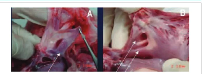
Color Doppler blood flow leak between the LBCA and the PV tissue was detected in 23 out of 59 (39%) targeted PV. Figure 3 shows a leak in the RSPV superiorly and in the LSPV inferiorly, which were correlated with the location of the gap in the pathological analysis (Figure 4). Achievement of complete PV isolation was significantly more common in the absence of blood flow leak: 30 out of 38 (79%) PV, compared to 8 of 23 (35%) PV when a leak was documented (p<0.001). The presence of blood flow leak criterion by ICUS had 86.8% specificity and 71.5% sensitivity for not achieving PV isolation. The positive value for predicting incomplete isolation was 75% and the negative predictive value was 84.6%.
All 4 leaks visualized in the RSPV were located inferiorly. Additionally, at the pathological analysis, a gap was present inferiorly in 3 RSPVs. Similarly, 11 leaks were visualized in the LSPV: 10 inferiorly and 1 laterally. The gross pathology assessment showed 9 gaps located inferiorly and no lateral gaps.
Multivariate logistic regression analysis
Leite et al Intracardiac ultrasound for PV ablation
Original Article
Discussion
Despite the emphasis given by several studies on the importance of catheter-tissue contact when radiofrequency current is used as energy source, this information has been recently studied when using laser energy, especially for circumferential ablation in the pulmonary veins. The main finding provided by the present study was to establish the usefulness of ICUS to guide PV ablation using a new fiber-optic balloon-catheter ablation with laser energy. We observed that complete electrical PV isolation was significantly more common when no blood flow leaks were identified between PV tissue and balloon catheter ablation, which determines the importance of catheter-balloon tissue contact also with laser energy. Good contact between the balloon and the PV orifice is necessary to obtain a circumferential lesion14-17. These requirements are probably related to photothermal and photomechanical interactions between laser energy on the tissue and heat transfer, which can be influenced by blood stream and tissue characteristics. Intracardiac ultrasound using
phased-array ultrasound catheter with a multi-directional steerable tip provides an excellent view of the pulmonary veins and of the catheters placed within these vessels18-20. The balloon catheter position in the targeted PV was confirmed before each single application, and the use of the color flow Doppler yielded the determination of the contact between the PV wall and the catheter balloon surface. The intracardiac ultrasound has shown to be very useful in interventional electrophysiology, including transseptal catheterization, visualization of targeted anatomical structures and evaluation of catheter-tissue contact. Mangrum et al20 reported the usefulness of ICUS for guiding PV isolation in humans, showing the possibility of visualizing the catheter positioning, tissue contact and lesion formation, but using the mechanical rotational system20. As new technologies for PV isolation using circumferential balloon catheter have been developed, we added the usefulness of color Doppler flow to this kind of procedure, and to the best of our knowledge, this is the first report showing the importance of tissue contact using laser ablation.
Figure 2 – A: Short-axis view showing the laser balloon inlated at the ostium of the RSPV (right superior pulmonary vein), its relationship with the right PA (pulmonary
artery) and the measurements of the balloon. Images were obtained with ICUS catheter positioned at SVC-RA junction; B: Fluoroscopic view of the Laser Balloon inlated
at RSPV; C: Circular mapping catheter positioned in the RSPV to check for PV isolation.
References
1. Chen SA, Tai CT, Tsai CF, Hsieh MH, Ding YA, Chang MS. Radiofrequency catheter ablation of atrial fibrillation initiated by pulmonary vein ectopic beats. J Cardiovasc Electrophysiol. 2000; 11: 218-27.
2. Arentz T, Blum T, von Rosenthal J, Peters K, Kalusche D. Focal paroxysmal atrial fibrillation: experiences with treatment using high frequency catheter ablation. Dtsch Med Wochenschr. 2000; 125: 479-83.
3. Haissaguerre M, Jais P, Shah DC, Takahashi A, Hocini M, Quinion G, et al. Spontaneous initiation of atrial fibrillation by ectopic beats originating in the pulmonary veins. N Engl J Med. 1998; 339: 659-66.
4. Robbins IM, Colvin EV, Doyle TP, Kemp WE, Loyd JE, McMahon WS, et al. Pulmonary vein stenosis after catheter ablation of atrial fibrillation. Circulation. 1998; 98: 1769-75.
Since the first report by Jais et al.21, of a focal origin of AF triggering by ectopic beats from the PV, several investigators have demonstrated that radiofrequency energy delivered at these foci eliminates the PV-initiated beats and paroxysmal AF21. However, recurrence rates of AF after successful ablation is still high and complications may ensue after this procedure22,23. Radiofrequency energy has also been used to isolate PV by creating a continuous circumferential lesion at the orifice. Alternatively, different energy sources have been used for circumferential ablation through radial energy delivery. Natale et al7 reported the use of ultrasound balloon catheter to isolate PV from the left atrium, which was accomplished in 60%7. In the present study, PV isolation was achieved in 64% after mean energy delivery duration of 280 ± 177 sec, which is very similar to that reported by Natale et al.7 However, our rates of PV isolation increased to 79% when no blood flow leaks were observed.
Study limitations
This study has several limitations to be considered. Although the ICUS imaging showed very well the left atrium and PV junction of all veins, the number of inferior veins ablated is very small when compared to the superior veins, and we were not able to prove the same usefulness for both. Finally, further studies are necessary to compare the use of the ICUS with other new technologies developed to establish catheter-tissue contact. Another potential limitation was the comparison between PV size and the balloon size, which did
not fit. However, as the mean balloon size diameter, after inflated, was significantly greater than the PV diameter at the orifice, this would actually improve the balloon catheter-tissue contact.
Clinical implications
The catheter-tissue contact showed to be important also for circumferential laser ablation using balloon catheter. For this purpose, intracardiac ultrasound may be useful in showing blood flow leaks during energy delivery, which decreases the possibility of achieving complete PV isolation. As a consequence, the use of ICUS would avoid ineffective energy application during circumferential PV ablation and would decrease the risk of PV stenosis.
Potential Conflict of Interest
No potential conflict of interest relevant to this article was reported.
Sources of Funding
There were no external funding sources for this study.
Study Association
This article is part of the thesis of post doctoral submitted by Luiz Roberto Leite da Silva, from Mayo Clinic Foundation.
5. Sohn RH, Schiller NB. Left upper pulmonary vein stenosis 2 months after radiofrequency catheter ablation of atrial fibrillation. Circulation. 2000; 101: E154-5.
6. Taylor GW, Kay GN, Zheng X, Bishop S, Ideker RE. Pathological effects of extensive radiofrequency energy applications in the pulmonary veins in dogs. Circulation. 2000; 101: 1736-42.
7. Natale A, Pisano E, Shewchik J, Bash D, Fanelli R, Potenza D, et al. First human experience with pulmonary vein isolation using a through- the-balloon circumferential ultrasound ablation system for recurrent atrial fibrillation. Circulation. 2000; 102: 1879-82.
8. Haissaguerre M, Jais P, Shah DC, Garrigue S, Takahashi A, Laverne T, et al. Electrophysiological end point for catheter ablation of atrial fibrillation initiated from multiple pulmonary venous foci. Circulation. 2000; 101: 1409-17.
9. Oral H, Knight BP, Tada H, Ozaydin M, Chugh A, Hassan S, et al. Pulmonary vein isolation for paroxysmal and persistent atrial fibrillation. Circulation. 2002; 105: 1077-81.
10. Su W, Johnson S, Packer D. First experience with circumferential pulmonary vein ablation using a laser energy balloon catheter. Circulation. 2001; 104: II-567.
11. Packer DL, Stevens CL, Curley MG, Bruce CJ, Miller FA, Khandheria BK, et al. Intracardiac phased-array imaging: methods and initial clinical experience with high resolution, under blood visualization: initial experience with intracardiac phased-array ultrasound. J Am Coll Cardiol. 2002; 39: 509-16.
12. Johnson SB, Seward JB, Packer DL. Phased-array intracardiac echocardiography for guiding transseptal catheter placement: utility and learning curve. Pacing Clin Electrophysiol. 2002; 25: 402-7.
13. Morton JB, Sanders P, Byrne MJ, Power J, Mow C, Edwards GA, et al. Phased-Array intracardiac echocardiography to guide radiofrequency ablation in the left atrium and at the pulmonary vein ostium. J Cardiovasc Electrophysiol 2001;12 (3): 343-8.
14. Reddy VY, Houghtaling C, Fallon J, Fisher G, Farr N, Clarke J, et al. Use of diode laser balloon ablation catheter to generate cincunferential
pulmonary venous lesions in a open-thoracotomy caprine model. Pacing Clin Electrophysiol. 2004; 27: 52-7.
15. Whelan WM, Wyman DR, Wilson B. Investigations of large vessel cooling during interstitial laser heating. Med Phys. 1995; 22 (1): 105-15.
16. Chen L, ter Haar G, Hill CR, Dworkin M, Carnochan P, Young H, et al. Effect of blood perfusion on the ablation of liver parenchyma with high-intensity focused ultrasound. Phys Med Biol. 1993; 38: 1661-173.
17. Liu Z, Ahmed M, Weistein Y, Yi M, Mahajan RL, Goldberg SN. Characterization of the RF ablation-induced “oven effect”: the importance of background tissue thermal conductivity on tissue heating . Int J Hyperthermia. 2006; 22: 327-42.
18. Bruce CJ, Packer DL, O’Leary PW, Seward JB. Feasibility study: transesophageal echocardiography with a 10F (3.2- mm), multifrequency (5.5- to 10-MHz) ultrasound catheter in a small rabbit model. J Am Soc Echocardiogr. 1999; 12: 596-600.
19. B r u c e C J, Pa c k e r D L , B e l o h l a v e k M , S e w a r d J B. I n t r a c a r d i a c echocardiography: newest technology. J Am Soc Echocardiogr. 2000; 13: 788-95.
20. Mangrum JM, Mounsey JP, Kok LC, De Marco JP, Haines DE. Intracardiac echocardiography-guided, anatomically based radiofrequency ablation of focal atrial fibrillation originating from pulmonary veins. J Am Coll Cardiol. 2002; 39: 1964-72.
21. Jais P, Haissaguerre M, Shah DC, Chouairi S, Gencel L, Hocini M, et al. A focal source of atrial fibrillation treated by discrete radiofrequency ablation. Circulation. 1997; 95: 572-6.
22. Oral H, Knight BP, Ozaydin M, Toda H, Chugh A, Hassan S, et al. Clinical significance of early recurrences of atrial fibrillation after pulmonary vein isolation. J Am Coll Cardiol. 2002; 40 (1): 100-4.
23. Cappato R, Calkins H, Chen SA, Davies W, Iesaka Y, Kalman J, et al. Worldwide survey on the methods, efficacy, and safety of catheter ablation for human atrial fibrillation. Circulation. 2005; 111: 1100-105.
Leite et al Intracardiac ultrasound for PV ablation