Prevalence of Coronary Risk Factors and Myocardial Perfusion
Scintigraphy Abnormalities in Asymptomatic Diabetic Outpatients
Francisco das Chagas Monteiro Júnior, Fhabyula da Silva Cunha, Natalino Salgado Filho, José Bonifácio Barbosa, João
Ribeiro Furtado, Pedro Antônio Muniz Ferreira, Vinícius Nina, Joyce Lages, Nilton Santana
Universidade Federal do Maranhão, São Luís, MA, Brazil
Summary
Objective: To determine the prevalence of coronary artery disease (CAD) risk factors (RF) and myocardial ischemia in a sample of asymptomatic diabetic patients treated on an outpatient basis.
Methods: From 80 type 2 diabetic patients initially recruited at an university outpatient endocrinology clinic, with no symptoms and/or CAD diagnosis, only 61 patients completed the study protocol, being 52,5% females, with a mean age of 56.3 ± 10.9 years. The patients were interviewed searching for RF and underwent electrocardiogram, echocardiogram and perfusional myocardial scintigraphy (PMS) at rest and under stress. According to the PMS results they were divided into two groups: an ischemic and a normal one .
Results: The RF identified were: male gender (48%), age ≥ 55 years (51%), family history of premature atherosclerotic
disease (16%), history of smoking (46%), hypertension (44%), sedentary lifestyle (62%), overweight / obesity (67%),
HDL- cholesterol < 45 mg/dl (69%), LDL- cholesterol ≥ 100 mg/dl (85%) and triglycerides ≥ 150 mg/dl (54%). Ischemic
MPS were diagnosed in 15% of the patients. The variables associated with this diagnosis were: male gender (p=0.007), low HDL levels (p=0.046), history of smoking (p=0.038), left ventricular hypertrophy (LVH) (p=0.043) and left ventricle ejection fraction (LVEF) < 60% (p=0.01).
Conclusion: A high prevalence of associated RF was observed, as well as a significant prevalence of 15% for myocardial ischemia. The variables identified as predictors of a myocardial ischemia diagnosis were: male gender, low HDL-cholesterol, past smoking, LVH and LVEF < 60%. (Arq Bras Cardiol 2007;88(5):277-282)
Key words: Diabetes mellitus, coronary risk factors, myocardial ischemia.
Mailing address: Francisco das Chagas Monteiro Junior •
Rua Santa Luzia, 27 QD.17 – Calhau - 65067-460 – São Luís, MA, Brazil E-mail: [email protected]
Manuscript received November 19, 2006; revised manuscript received April 18, 2007; accepted May 31, 2007.
Introduction
Diabetes mellitus (DM) is one of the most significant cardiovascular risk factors. In most study populations, cardiovascular disease rates among men and women with DM are two and three times higher, respectively, than in non-diabetic individuals.1 Nevertheless, cardiovascular disease
represents the greatest cause of morbidity and mortality in diabetics, as a result of both the diabetic state per se and the various associated risk factors, such as hypertension, overweight and dyslipidemia. In fact, it is currently known that type 2 diabetes mellitus is often one of the signs of a wider clinical condition commonly known as metabolic syndrome, that is primarily caused by insulin resistance and comprises various hemodynamic and metabolic abnormalities, including hypertension, lipid alterations, pro-thrombotic and pro-inflammatory state, which are all associated with increased cardiovascular risk. Cardiovascular disease, and more specifically coronary artery disease (CAD), accounts for roughly 80% of the deaths in type 2 diabetes mellitus patients.
In fact, the relative age-adjusted risk for diabetics to die as a result of cardiovascular events is three times greater than that of the general population.2
Owing to recognized high risk of cardiovascular events in diabetic patients, the American Heart Association (AHA) currently recommends classifying these patients in the high risk category, which was previously reserved for patients with known cardiovascular disease,3 and submitting them to equally
intensive preventive treatments. This recommendation was later endorsed by the 3rd Report of the National Cholesterol
Education Program – NCEP (2001) on detection, evaluation and treatment of hypercholesterolemia in adults.4
Diabetic patients are more likely to present silent myocardial ischemia and atypical chest pain, making early detection of coronary artery disease (CAD) more difficult in these individuals. In one study, it was confirmed that 75% of the diabetic patients with diagnosed CAD did not present typical symptomatology.5 Nevertheless, various studies have
severe CAD are the same in patients with silent ischemia and those with symptomatic ischemia.6
There are many methods that can be used to track coronary artery disease in populations at risk for CAD. For asymptomatic diabetic individuals in particular, even though it is believed that these patients have a greater prevalence of CAD than the general population, there is no consensus on whether or not systematic investigation of myocardial ischemia is needed, when it should be conducted or which tests should be used. Nevertheless, early diagnosis and treatment of CAD improve patient prognosis.
In Brazil, there are no studies that assess diabetic patients in relation to the presence of associated risk factors and silent myocardial ischemia. An established cost-benefit ratio of the systematic performance of diagnostic methods to investigate myocardial ischemic disease in this population is required, particularly for asymptomatic patients in order to adopt a routine diagnosis to treat these patients on a daily basis.
The objective of this study was to analyze a diabetic population sample treated at an endocrinology outpatient clinic at a university hospital, who did not appear to have any cardiovascular health problems and would not normally be submitted to routine cardiology assessments to determine the presence of other classic CAD risk factors or silent ischemia, in an attempt to identify the variables associated with these conditions.
Methods
This observational, cross sectional, descriptive and analytical study included a sample of 80 consecutive diabetic patients treated on an outpatient basis at the Endocrinology Clinic of our Institution between January and December 2003. The inclusion criteria were: established diagnosis of type 2 diabetes mellitus and the absence of symptoms and/or prior diagnosis of coronary artery disease (CAD). All participants were informed of the study objectives and methods and signed the consent form. The study was approved by the Institution’s Ethics Committee. Since the study involved a group of patients treated at an endocrinology clinic and while the attending physicians agreed to the participation of their patients, but not with their own direct involvement in the study, the Committee recommend that the researchers not request cinecardiographies in the cases indicated, due to the invasive nature, but just refer these patients back to their attending physicians, suggesting that they be submitted to the test. Exclusion criteria included intellectual incapacity and refusal to participate in the study.
Initially, a medical history was taken on the selected patients and they were given a clinical examination. The following CAD risk factors were studied: age greater than or equal to 55 years for both genders; family history of atherosclerotic cardiovascular disease in first degree relatives under the age of 55 (men) and 65 (women); systemic hypertension (prior record of blood pressure ≥140 per 90 mmHg on two or more occasions and/or history of anti-hypertensive medication); dyslipidemia (LDL cholesterol ≥100 mg/dl, triglyceride levels ≥150 mg/dl and/or HDL cholesterol <45 mg/dl); smoking (current or past); overweight/obesity (body mass index greater
than or equal to 25 kg/m2) and sedentary life style (less than 30
minutes of physical aerobic activity three times per week). Next, the patients were sent for the following complementary tests: electrocardiogram at rest (EKG), Doppler echocardiogram and myocardial perfusion scintigraphy (MPS) using Tc-99m- SESTAMIBI, at rest and under stress (physical or pharmacological), which was the standard method adopted in this study to diagnose myocardial ischemia. Interpretation criteria for the complementary tests are described below.
The electrocardiogram at rest was initially classified as normal or altered. The presence of pathological Q waves was considered indicative of a prior myocardial infarction. Alterations in ventricular repolarization were considered as suggestive of myocardial ischemia. Other significant abnormalities were also noted, such as cavity overloads, arrhythmia and intraventricular and atrioventricular conduction disturbances.
The echocardiogram was classified as normal or altered. The following measurements were analyzed: diastolic diameter of the left ventricle cavity, thickness of the interventricular septum and posterior wall, and left ventricle ejection fraction (EF). Left ventricular hypertrophy (LVH) was diagnosed when the thickness of the interventricular septum and the posterior wall were over 11mm without cavity dilation. EF was considered diminished when it was lower than 60% using the Teichholz method. Diastolic function was assessed by analyzing the mitral flow diagram on the pulsated Doppler which was classified as normal or altered (impaired left ventricle relaxation, pseudonormal or restrictive pattern). Left ventricle systolic function was analyzed in relation to overall and wall motion aspects.
The myocardial perfusion scintigraphy was performed using the single photon emission computed tomography (SPECT) technique, to obtain three dimensional and tomographic images of the heart (horizontal long axis, vertical long axis and short axis) after isotonic exercise on a treadmill or pharmacological stress with dipyridamole for patients who were unable to perform the exercise, and at rest, 60 minutes after the intravenous injection of radiotracer Tc-99m- SESTAMIBI. Transitory or persistent perfusion defects were considered criteria for the diagnosis of myocardial ischemia,7,8
and the scintigraphy was classified as positive in the presence of these defects.
The quantitative variables analyzed were: age, diabetes diagnosis period, HDL cholesterol serum levels, LDL cholesterol serum levels, triglyceride serum levels, body mass index (BMI), systolic blood pressure and diastolic blood pressure.
The qualitative (categorical) study variables analyzed were: age ≥ 55 years, gender, HDL cholesterol serum levels <45 mg/dl, LDL cholesterol serum levels ≥100 mg/dl and triglyceride serum levels ≥150 mg/dl, family history of early onset CAD, smoking, sedentary lifestyle, systemic hypertension, BMI ≥25 kg/m2, left ventricular hypertrophy,
Original Article
The data collected were submitted to statistical analysis using the program SPSS version 10.0. The qualitative variables were represented by absolute (n) and relative (%) frequency and the quantitative variables were expressed as mean and standard deviation. Differences between the quantitative variables were calculated using the Student’s t-test. Differences between the qualitative variables were calculated using the chi-square or Fisher exact test, when indicated. The level of significance adopted was p<0.05 (5%).
Results
From the 80 patients initially included in the study, only 61 completed the protocol and therefore comprised the sample for this study.
Mean patient age was 56.3 ± 10.9 years, and 32 were female (52.5%). All the patients had type 2 diabetes mellitus, with a diagnosis period of 9.1±7.7 years.
In regard to the prevalence of CAD risk factors, it was verified that 29 patients were male (48%); 10 had a family history of early onset cardiovascular disease (16%); 28 had a history of smoking (46%); 27 were hypertensive (44%); 38 were sedentary (62%) and 41 were either overweight or obese (67%). Deviations in the serum levels of HDL cholesterol, LDL cholesterol and triglycerides were observed in, respectively, 69%, 85% and 54% of the patients. Fifty-five patients (90%) presented two or more CAD risk factors in addition to diabetes mellitus (Tab. 1).
The EKG was considered normal in 48 patients (79%). Ventricular repolarization alterations suggestive of ischemia were found in seven patients (11%), and another six patients (10%) presented other abnormalities.
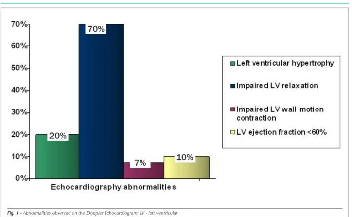
The following abnormalities were observed on the Doppler echocardiogram (fig. 1): left ventricular hypertrophy (LVH) in 12 patients (20%); left ventricle ejection fraction less than 60% in six patients (10%); impaired left ventricular relaxation
associated with diastolic abnormalities in 43 patients (70%); and impaired left ventricle wall motion contractibility in four patients (7%).
The myocardial perfusion scintigraphy (MPS) demonstrated alterations indicative of CAD (transitory and/or persistent low uptake of the radiotracer) in nine patients (15%).
Table 2 presents the quantitative variables analyzed according to their distribution in the two groups, complete with the respective statistical significance levels for the differences between the groups (MPS normal and MPS positive).
As demonstrated, there was no significant statistical difference between the groups in relation to any of these variables.
The qualitative (categorical) variables analyzed are shown in table 3, according to their distribution in the two groups.
As demonstrated there was a statistically significant difference between the two groups (p<0.05) for the variables male gender, history of smoking, HDL cholesterol <45 mg/dl, left ventricle hypertrophy and left ventricular ejection fraction <60%.
Discussion
In this study, radioisotope myocardial perfusion was chosen as the standard method to investigate myocardial ischemia since it is a non-invasive test with high sensitivity and specificity rates for diagnosing significant CAD,9,10 in addition to its well
established prognostic value,11 and the fact that the presence
and extension of perfusion abnormalities are regarded as independent predictors for adverse cardiac events.12 A
prevalence rate of roughly 15% was found for impaired myocardial perfusion, which is regarded as expressive, particularly when dealing with asymptomatic individuals that in accordance with the conventional practices of the Clinic where the study was conducted would not normally be submitted to complementary cardiology investigations. This rate is similar to that confirmed in other studies that tracked ischemia in asymptomatic diabetics, also using the myocardial scintigraphy as the standard test.13 Wackers,8 in his study
Detection of Silent Myocardial Ischemia in Asymptomatic Diabetic Subjects (DIAD), demonstrated positive tracking in 22% of the cases. However, in this study, patients with perfusion defects found on the myocardial scintigraphy and those with other abnormalities, even with normal perfusion, such as depression of the ST segment induced by adenosine, ventricular dilatation and ventricular dysfunction at rest were placed in the silent ischemia classification. If only the patients with perfusion defects had been considered, the positive tracking rate would have been 15.9%, which is similar to the findings of the present study. Considering the limitations of the ischemia research method that normally only detects significant obstructive lesions, it is easy to assume that the total CAD prevalence in this population is even greater.
The prevalence of other classic CAD risk factors in this study such as hypertension, dyslipidemia, overweight and sedentary lifestyle, as observed by other authors,2,14,15,16 was
elevated. This fact is very relevant, considering the additional effect of these factors for a population that already has Table 1 – CAD Risk Factors
Risk Factors n = 61 Frequency (%)
Male Gender 29 48
Age ≥55 years 31 51
HDL <45 mg/dl 42 69
LDL ≥100 mg/dl 52 85
Triglycerides ≥150 mg/dl 33 54 Family history of early onset
CAD 10 16
Smoking (history) 28 46
SH 27 44
Sedentary Lifestyle 38 62
IBMI ≥25 kg/m2 41 67
significantly greater cardiovascular risk due to the presence of DM when compared to the population as a whole. The percentage of patients satisfying the American Diabetes Association (1998) criteria to be indicated for CAD tracking, that is, the presence of two or more risk factors in addition to DM, was elevated and similar for both groups (roughly 90%), however, no significant statistical difference was found between them, indicating that this criteria is not valid for this population. Therefore, the elevated risk factor association rate for this study population as per the ADA criteria is irrelevant. In turn, the abovementioned DIAD study, that analyzed a much larger population, demonstrated that if only the patients
satisfying these criteria were selected, 41% of the patients with myocardial ischemia would go undetected. In fact, it is known that the ADA recommendations are based more on the clinical opinion of a group of experts than scientific evidence, and therefore further studies targeted at this issue raised by Wackers & Zaret17 are required.
By researching the risk factors with the greatest predictive values in the diagnosis of myocardial ischemia in this population of asymptomatic diabetics, it was demonstrated that only male gender, history of smoking, HDL <45 mg/dl, left ventricular hypertrophy and ejection fraction lower than 60% had a significant association with a positive myocardial
MPS
NORMAL POSITIVE
Factors n = 52 n= 9 valor do p
Age (years) 56,5±11,5 55,7±7,4 0,843
DM diagnosis period (years) 9,4±8 7,22±6,5 0,442
HDL-cholesterol (mg/dl) 41,2±9,1 39,8±1,1 0,442
LDL-cholesterol (mg/dl) 129±36,8 122,6±22,7 0,612
Triglycerides (mg/dl) 155,2±92,3 145,2±41,1 0,751
Systolic BP (mmHg) 145,8±36,8 139,4±34,5 0,628
Diastolic BP (mmHg) 84.6±19.5 84.4±13.1 0.978
BMI (kg/m2) 26.6±4.7 26±4.5 0.695
MPS – myocardial perfusion scintigraphy; DM – diabetes mellitus; HDL – high density lipoprotein; LDL – low density lipoprotein; BP – blood pressure; BMI – body mass index
Table 3 – Distribuição das variáveis qualitativas nos dois grupos
MPS / VARIABLES NORMAL POSITIVE p
n % n % VALUE
Male gender 21 40.4 8 88.9 0.007 *
Age ≥ 55 years 25 48.1 6 66.6 0.525
HDL-cholesterol <45 mg/dl 33 63.5 9 100 0.046 *
LDL-cholesterol ≥100 mg/dl 44 84.6 8 88.9 0.739
Triglycerides ≥150 mg/dl 26 50 7 77.8 0.123
Family history of early onset CAD 8 15.4 2 22.2 0.609
History of smoking 21 40.4 7 77.8 0.038 *
Systemic hypertension 22 42.3 5 55.6 0.460
Sedentary lifestyle 32 61.5 6 66.7 0.769
LV Hypertrophy 8 15.4 4 44.4 0.043 *
LV ejection fraction <60% 3 5.8 3 33.3 0.010 *
Impaired LV relaxation 35 67.3 8 88.9 0.190
LV overall / wall motion contraction abnormalities 3 5.8 1 11.1 0.550
BMI ≥ 25 kg/m2 36 69.2 5 55.6 0.420
Ventricular repolarization alterations on the EKG 5 9.6 2 22.3 0.185
2 or more additional RF 47 90.4 8 88.9 0.985
Original Article
References
1. Bloomgarden ZT. American Diabetes Association annual meeting 1999: more on cardiovascular disease. Diabetes Care. 2000; 23 (6): 845-52.
2. Schaan BD, Harzheim E, Gus I. Perfil de risco cardíaco no diabetes mellitus e na glicemia de jejum alterada. Rev Saúde Pública. 2004; 38 (4): 529-36.
scintigraphy test. On the other hand, it was revealed that some classic CAD risk factors such as elevated LDL cholesterol and triglyceride levels, hypertension, overweight and obesity, sedentary lifestyle and age ≥55 years were ineffective predictors of an ischemia diagnosis in this asymptomatic population.
Other authors such as Janand-Delenne and associates13 and
Wackers,8 also observed that a family history of early onset CAD
and male gender were the only statistically significant classic CAD risk factor predictors to detect ischemia. A significant finding in this study, which was not observed by the other authors, is that the variables, left ventricular hypertrophy and left ventricular ejection fraction less than 60% are predictors of a positive myocardial scintigraphy.
The relatively small sample in the present study, noting that more than 20% of the patients initially included did not complete the study and the fact that the study was conducted at a referral center, made it impossible to extrapolate these CAD results for the entire asymptomatic diabetic population. Nevertheless, the study could have been enhanced by the anatomic confirmation of obstructive CAD by means of a cinecardiography, if it had been approved by the Ethics Committee, or a computerized angiotomography
(angio-CT) of the coronary arteries, which was not available at the institution.
Therefore, in view of the abovementioned limitations and the limited number of other studies published on the subject, it is not possible to define recommendations based on the results of this study on the role of systematic tracking of asymptomatic diabetics for CAD or even diabetics who present some associated risk factors. However, the results clearly indicate the need for further studies on the subject and evidence based guidelines for the early detection of CAD in this high risk population.
Potential Conflict of Interest
No potential conflict of interest relevant to this article was reported.
Sources of Funding
There were no external funding sources for this study.
Study Association
This study is not associated with any graduation program.
3. Smith SCJ, Greenland P, Grundy SM. AHA Conference Proceedings:
Prevention conference V: beyond secondary prevention: identifying the high-risk patient for primary prevention: executive summary: American Heart
Association. Circulation. 2000; 101 (1): 111-6.
Fig. 1 - Abnormalities observed on the Doppler Echocardiogram; LV - left ventricular
20%
70%
Incremental prognostic value of myocardial perfusion single photon emission
computed tomography in patients with diabetes mellitus. Am Heart J. 1999;
138 (6 Pt 1): 1025-32.
13. Janand–Dellene B, Savin B, Habib G, Bory M, Vague P, Lassmann-Vague V.
Silent myocardial ischemia in patients with diabetes: who to screen. Diabetes
Care. 1999; 22 (9): 1396-400.
14. Alexander CM, Landsman PB, Teutsch SM, Hafner SM. NCEP-defined
metabolic syndrome, diabetes, and prevalence of coronary heart disease
among NHANES III participants age 50 years and older. Diabetes. 2003;
52 (5): 1210-4.
15. Laakso M, Lehto S. Epidemiology of risk factors for cardiovascular disease
in diabetes and impaired glucose tolerance. Atherosclerosis. 1998; 137
(Suppl.): S65-73.
16. Raza AJ, Mohaved A. Current concepts of cardiovascular diseases in diabetes
mellitus. Int J Cardiol. 2003; 89: 123-34.
17. Wackers FJ, Zaret BL. Detection of myocardial ischemia in patients with
diabetes mellitus. Circulation. 2002; 105: 5-7. 4. Expert Panel on Detection, Evaluation, and Treatment of High Blood
Cholesterol in Adults. Executive summary of the third report of the National Cholesterol Education Program. (NCEP). Evaluation, and Treatment of high blood cholesterol in adults (Adult Treatment Panel III). JAMA. 2001; 285 (19): 2486-97.
5. Gondim LGP, Oliveira WA, Grossi SAA. A diferenciação da dor do infarto do miocárdio entre pacientes diabéticos e não-diabéticos. Rev Latinoam Enf. 2003; 11 (6): 720-6.
6. Cooper S, Caldwell JH. Coronary artery disease in people with diabetes: diagnostic and risk factor evaluation. Clin Diabetes. 1999; 17 (2): 35-45.
7. Nesto RW. Screening for asymptomatic coronary artery disease in diabetes. Diabetes Care. 1999; 22 (9): 1393-5.
8. Wackers FT. Detection of silent myocardial ischemia in asymptomatic diabetic subjects: the DIAD study. Diabetes Care. 2004; 27 (8): 1954-61.
9. Taillefer R, Depuey EG, Udelson JE, Beller GA, Latour Y, Reeves F. Comparative diagnostic accuracy of Tl-201 and Tc-99m sestamibi SPECT imaging (perfusion and ECG-gated SPECT) in detecting coronary artery disease in women. J Am Coll Cardiol. 1997; 29 (1): 69-77.
10. Smanio PE, Watson DD, Segalla DL, Vinson EL, Smith WH, Beller GA. Value of gating of technetium-99m sestamibi single-photon emission computed tomographic imaging. J Am Coll Cardiol. 1997; 30 (7): 1687-92.
11. Brown KA. Prognosis in stable coronary artery disease. In: Zaret BL, Beller GA. Nuclear cardiology: state of the art and future directions. St Louis: Mosby; 1999. p. 331-45.