1 9 6 1 9 6
Barreto et al
Ascites in patients with endomyocardial fibrosis
Arq Bras Cardiol 2002; 78: 196-9.
Instituto do Coração do Hospital das Clínicas - FMUSP
Mailing address: Antonio Carlos Pereira Barretto - InCor - Av. Dr. Enéas C. Aguiar, 44 - 05403-000 - São Paulo, SP - Brazil - E-mail: [email protected] Received for publication on 16/11/00
Accepted on 14/2/01
Objective - To evaluate the clinical meaning of asci-tes and the main features of patients with asciasci-tes and endo-myocardial fibrosis.
Methods - We studied 166 patients with endomyocar-dial fibrosis (mean age 37 years, 114 women) treated over the last 20 years. Ventriculography findings, surgery or necropsy confirmed the diagnosis in all patients. Most pa-tients belonged to New York Heart Association Functional Class III/IV (134, 83.7%). Eighty-one (50.6%) had biven-tricular, 28 (17.5%) had right venbiven-tricular, and 51 (31.8%) had left ventricular involvement. During follow-up, 56 patients died.
Results - Ascites was present in 67 (41.8%) patients, and right ventricular involvement was present in 59 (88%). In the comparison between patients with or wi-thout ascites, those with ascites had higher mortality (49.2% and 24.7%, respectively). Patients with ascites had a higher incidence of edema (95% vs. 43%), hepatomega-ly (5.8cm vs. 4.1cm), mean right atrium pressure (19.3 vs. 12mmHg), and final right ventricle diastolic pressure (18.7 vs. 12.9mmHg). Also, patients with ascites had a lon-ger history of illness (5.1 and 3.9 years, respectively) and had atrial fibrillation more frequently (44.7% vs. 30.1%).
Conclusion - Ascites was observed in less than 50% of cases of endomyocardial fibrosis and was associated with greater involvement of the right ventricle and with a lon-ger duration of the disease, thus being a characteristic of a worse prognosis.
Key words: ascites, endomyocardial fibrosis, restrictive cardiomiopathy
Arq Bras Cardiol, volume 78 (nº 2), 196-9, 2002
Antonio Carlos Pereira Barretto, Charles Mady, Sergio Almeida Oliveira, Edmundo Arteaga, Creusa Dal Bó, José Antonio Franchini Ramires
São Paulo, SP - Brazil
Clinical Meaning of Ascites in Patients with
Endomyocardial Fibrosis
Original Article
Endomyocardial fibrosis is a restrictive cardiopathy that may evolve to cardiac failure, which is difficult to control 1-7.
Its detection enables, in most cases, recommendation for surgical treatment, with resection of fibrotic tissue and control of cardiac failure 2-4,8-10.
Ascites is a very important feature in the diagnosis of endomyocardial fibrosis. When it is associated with peri-pheral edema, and even in its absence, it is valued as an indi-cation of a restrictive syndrome and must be taken into ac-count as an etiological factor.
The objective of this study was to evaluate the impor-tance of ascites related to its clinical and laboratory charac-teristics, for the purpose of evaluating whether the presen-ce or absenpresen-ce of ascites identifies patients with different cli-nical courses and different prognoses.
Methods
We studied 166 patients with endomyocardial fibrosis followed at the Instituto do Coração-HC/FMUSP since 1978; 114 were women and 46 were men. Age ranged from 5 to 64 years (mean 34.3±13). Diagnosis was confirmed by he-modynamic and angiographic studies in 133 patients, by surgery in 88 patients, and by data obtained in necropsy in 30 patients.
Data obtained through angiographic study or necrop-sy revealed that 81 patients had biventricular, 28 had right ventricular, and 51 had left ventricular involvement. Based on the findings, involvement was classified as mild, mode-rate, or severe. Involvement of the right ventricle was consi-dered mild in 52 cases, moderate in 52 and severe in 56 ca-ses. Involvement of the left ventricle was mild in 28, mode-rate in 82 and severe in 49 cases.
Most patients were symptomatic, and 134 had New York Heart Association (NYHA) functional classes III/IV cardiac failure and 25 had NYHA classes I/II.
During follow-up, 56 (35%) patients died.
Arq Bras Cardiol 2002; 78: 196-9.
Barreto et al Ascites in patients with endomyocardial fibrosis
1 9 7 1 9 7
hepatomegaly. In 96 (65.4%), regurgitant systolic murmur was present. Proto-diastolic knock was described in 77 (48.4%) patients. Atrial fibrillation was verified in 58 (36.3%) cases. Ascites was detected by physical examination in 67 (42.1%) patients.
During follow-up, 96 patients underwent echocardio-graphic study. Because follow-up was long, echocardiogra-phies were performed using different machines and techni-ques; thus, we will only highlight the measures obtained by M-mode, common to all the cases. Mean left ventricle diame-ter was 51.9±9.6mm, left atrium diamediame-ter was 42.2±9.6mm, and left ventricle ejection fraction was 0.72±0.09.
Hemodynamic study was performed in 130 (31.2%) pa-tients. Mean right atrial pressure was 15.0±7.4mmHg, right ventricular end diastolic pressure was 15.3±7.1mmHg, left ventricular systolic pressure was 42.9±20.6mmHg, left ven-tricular end diastolic pressure was 21.8±10.4mmHg and left ventricular systolic pressure was 119.4±19.7mmHg. Tricus-pid regurgitation was observed in 98 (61.4%) patients and mitral regurgitation in 102 (63.7%) patients.
According to ventriculographic appearance, the degree of fibrosis was classified as mild, moderate or severe. Fibrosis was considered mild when small changes were observed in ventricular morphology, mainly a restriction in the inflow tract or apex. Moderate fibrosis was characterized by evident reduction in the diastolic ventricular dimension together with endocardial irregularities of the apical region. Severe fibrosis was characterized by a grossly abnormal appearance of the ventricular chamber in diastole (the chamber being reduced in size and acquiring a tubular form owing to the almost comple-te elimination of the inflow tract and oblicomple-teration of the apex). Patients were followed annually. Those who failed to attend their scheduled follow-up visit were contacted by te-lephone or letter or by both tete-lephone and letter at least three times. The mean follow-up period was 4.2±4 years. Se-venteen patients were lost to follow-up during the first year. Deaths occurred in 56 (34%) patients, 27 among patients treated clinically and 28 among patients treated surgically.
Patients were divided into 2 groups according to the presence or the absence of ascites. We evaluated whether differences existed between the 2 groups regarding clinical symptoms, or in echocardiographic and hemodynamic fin-dings. Kaplan-Meier survival curves were constructed for those with and without ascites. Differences between the curves were analyzed with the log-rank test. Chi-squared analysis was used to test the association between several variables. A p value < 0.05 was considered significant. Sta-tistical analysis was performed using SAS (StaSta-tistical Ana-lyses Systems) software.
Results
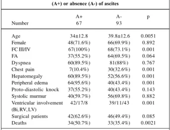
Ascites was observed during clinical examination in 67 (42.1%) patients treated at our institution. Table I shows clinical characteristics of patients with or without ascites and table II shows echocardiographic, hemodynamic and ventriculographic data. Patients with ascites were younger than those without it.
On physical examination, patients with ascites fre-quently had more hepatomegaly and peripheral edema and less chest pain than the patients without ascites. The inci-dence of dyspnea, proto-diastolic knock, and regurgitant systolic murmurs was the same for both groups.
Patients with ascites had left ventricles with lower dia-meters on echocardiographic examination (48.3mm vs 53.54mm, p=0.0131). In the hemodynamic study, patients with ascites had higher mean right atrial pressure and right ventricular end diastolic pressure, and lower left ventricular systolic pressure. In ventriculography, a greater number of patients had tricuspid regurgitation (p=0.001), and the pro-portion of patients with a greater intensity of right ventri-cular fibrosis was higher (p=0.001).
Table I – Clinical profile of patients according to the presence (A+) or absence (A-) of ascites
A+ A- p
Number 67 93
Age 34±12.8 39.8±12.6 0.0051
Female 48(71.6%) 66(69.9%) 0.892 FC III/IV 67(100%) 68(73.1%) 0.001
FA 37(55.2%) 64(69.5%) 0.064
Dyspnea 60(89.5%) 81(88%) 0.767
Chest pain 7(10.4%) 30(32.6%) 0.001 Hepatomegaly 60(89.5%) 52(56.6%) 0.001 Peripheral edema 64(95.6%) 40(43.4%) 0.001 Proto-diastolic knock 37(55.2%) 40(43.4%) 0.143 Systolic murmur 40(59.7%) 56(69.8%) 0.882 Ventricular involvement 42/17/8 39/11/43 0.001 (Bi, RV, LV)
Surgical patients 42(62.6%) 46(49.4%) 0.085 Deaths 34(50.7%) 33(35.4%) 0.0021
FC- NYHA functional class; AF- atrial fibrillation; Bi- biventricular involvement; RV- prevalent involvement of right ventricle; LV- prevalent involvement of left ventricle.
Table II – Data from echocardiographic, hemodynamic and ventriculographic study, according to the presence (A+) or absence
(A-) of ascites
A+ A- p
Left atrium diameter (mm) 42.7±9.2 42.0+8.6 0.7482 Diastolic diameter of LV (mm) 48.3±10 53.5±9.1 0.0131 Ejection fraction of LV (%) 71.8±8.5 72.5±9.4 0.1342 Pressure of left atrium (mmHg) 19.2±6.5 12.0±6.5 <0.0001 Final diastolic pressure
of RV (mmHg) 18.6±6.4 12.8±6.6 <0.0001 Systolic pressure
of RV (mmHg) 38.7±21.6 46.0±19.5 0.454 Diastolic pressure
of LV (mmHg) 20.7±9.3 22.6±11.1 0.3091 Systolic pressure
of LV (mmHg) 113.1±16.4 124.1±20.6 0.0010 Tricuspid regurgitation 48(88.9%) 49(59.7%) 0.001 Mitral regurgitation 43(76.8%) 58(69%) 0.317 Fibrosis intensity
RV (Mi, Mo, I) 9/23/35 43/29/21 0.001 Fibrosis intensity
LV (Mi, Mo, I) 18/33/16 11/49/33 0.037
1 9 8 1 9 8
Barreto et al
Ascites in patients with endomyocardial fibrosis
Arq Bras Cardiol 2002; 78: 196-9.
The proportion of patients who underwent surgery was slightly greater among those with ascites (62.6% vs 49.4%), but this difference did not reach statistical signifi-cance (p=0.085).
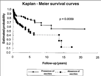
Mortality was higher among patients with ascites (p=0.0021). Analysis of the Kaplan-Meier survival curves revealed that patients with ascites reached 30% mortality at two years, while those without ascites had a mortality rate of 15% at eight years (p=0.00009) (fig. 1).
Discussion
The main findings of this study include: 1) ascites is frequent in patients with endomyocardial fibrosis (inciden-ce of 42.1%), 2) ascites is usually associated with hepatome-galy and peripheral edema, 3) ascites is related to a greater hemodynamic and morphological involvement of the right ventricle, 4) ascites is associated with the presence of tricus-pid regurgitation in the hemodynamic study, 5) presence of ascites is associated with a worse prognosis.
Ascites appears to be a sign frequently found in res-trictive cardiomyopathies and is considered an important factor for diagnosis of those entities. It may be present both in constrictive pericardites and in endomyocardial fibrosis, which suggests the need for confirming the diagnosis.
The presence of ascites in patients with endomyocar-dial fibrosis is mentioned in the majority of articles that dis-cuss the disease 1-7. It is worth noting that, generally, no
special point is made in these articles about stressing its im-portance; it is presented as another clinical sign of right ventricular involvement. Its incidence ranged between 28% and 100% of reported cases. No concern, in most articles, is shown about verifying whether ascites is important in the follow-up of patients. When we compare our analysis with those in the literature, we observe that in the literature the reported frequency of ascites is greater, because in these studies most patients had biventricular involvement or isolated right ventricular involvement. Analyses of patients with prevalent left ventricular involvement, such as ours,
are rare, because in the majority of the cases, endomyocar-dial fibrosis is diagnosed because of its congestive manifes-tations and because of the presence of ascites.
In our data, we have a great number of cases with pre-valent involvement of the left ventricle. These patients had chest pain and underwent cineangiography with ventri-culography, which facilitates the diagnosis of endomyocar-dial fibrosis. Ascites was more frequent among patients in whom the disease involved the right ventricle, and the greater the involvement the more frequent the presence of ascites. Thus, in patients with severe fibrosis of the right ventricle, the incidence of ascites was 52%. In those with moderate fibrosis, it was 34%, and in those with mild fibro-sis, it was 13%, indicating how important chamber oblitera-tion (fibrosis intensity) is in the restrictive syndrome and in hemodynamic changes that lead to the genesis of ascites.
When we analyzed ascites in relation to the chamber involved, we observed that biventricular involvement was present in 52.5% of patients, right ventricular involvement was present in 60.7% and prevalent involvement of the left ventricle was present in 15.6%. These data show the impor-tance of right ventricular involvement.
If right ventricle involvement is the cause of ascites, why do some patients with endomyocardial fibrosis of the left ventricle have ascites? One hypothesis is that at autop-sy we can usually detect involvement of both chambers. In many cases, this involvement is not important enough to determine distortions in the chambers and we cannot iden-tify right ventricular involvement in the clinical or laboratory examinations. These cases are identified as dominant invol-vement of the left ventricle, but, probably, minimal changes in the right ventricle are enough to explain alterations res-ponsible for the appearance of ascites 11. In fact, this disease
is seldom exclusively univentricular. Another possibility is that ascites is due to an inflammatory process of the perito-nious. Researchers who have observed the high level of proteins and leptocytosis in ascites effusion defend this hypothesis 12.
Patients with ascites more often had signs related to the involvement of the right chambers, such as hepatome-galy and peripheral edema, usually present in cases of con-gestive heart failure. Patients with ascites were younger, what could be related to the characteristics of right heart in-volvement, because since the right ventricle was a smaller muscular mass, it results in earlier symptoms when compa-red to the left ventricular involvement.
Atrial fibrillation, which is also associated with a worse prognosis, was slightly more frequent among the patients with ascites, but no statistically significant difference exis-ted between the two 13. The presence of murmur, identifying
mitral and tricuspid regurgitation, was more frequent among patients with ascites. The presence of tricuspid regurgitati-on increases clinical manifestatiregurgitati-on in cases of cardiac res-triction because the ventricles lose the ability to adapt to the volume of regurgitation, as in cases of endomyocardial fibrosis, which explains the higher incidence of ascites in these patients.
Fig. 1 - Kaplan-Meier survival curves for patients with and without ascites. Kaplan - Meier survival curves
E
s
ti
m
a
te
d
p
ro
b
a
b
il
it
y
Follow-up (years)
Presence of ascites
Absence of ascites .
. . . . . . . . . .
Arq Bras Cardiol 2002; 78: 196-9.
Barreto et al Ascites in patients with endomyocardial fibrosis
1 9 9 1 9 9 1. Somers K, Brenton PP, Sood NK. Clinical features of endomyocardial fibrosis of
the right ventricle. Br Heart J 1968; 30: 309-21.
2. Bertrand E, Chauvet J, Assamoi MO, et al. Results, indications and contra-indi-cations of surgery in restrictive endomyocardial fibrosis: comparative study on 31 operated and 30 non-operated patients. E Afr Med J 1985; 62: 151-60. 3. Metras D, Coulibaly AO, Ouattara K. The surgical treatment of endomyocardial
fibrosis: results in 55 patients. Circulation 1985;72: II-274-9.
4. Valiathan MS, Balakrishnan KG, Sankarkumar R, Kartha CC. Surgical treatment of endomyocardial fibrosis. Ann Thorac Surg 1987; 43: 68-73.
5. Fujita PN, Valiathan MS, Balakrishnan KG, Kartha C, Ghosh MK. Clinical co-urse of endomyocardial fibrosis. Br Heart J 1989; 62: 450-4.
6. Dubost C, Chapelon C, Deloche A, et al. Chirurgie des fibroses endomyocardi-ques. A propos de 22 cases. Arch Mal Coeur 1990; 83: 481-6.
7. Pereira Barretto AC, Luz PL, Oliveira SA, et al. Determinants of survival in en-domyocardial fibrosis. Circulation 1989; 80: I-177-82.
References
8. Oliveira SA, Pereira Barretto AC, Mady C, et al. Surgical treatment of endomyo-cardial fibrosis: A new approach. J Am Coll Cardiol 1990; 16: 1246-51. 9. Cherian G, Vijayaraghavam G, Krishnaswami S, et al. Endomyocardial fibrosis:
Report on the hemodynamic data in 29 patients and review of the results of surge-ry. Am Heart J 1983; 105: 659-66.
10. Moraes CR, Buffolo E, Lima R, et al. Surgical treatment of endomyocardial fibrosis. J Thoracic Cardiovasc Surg 1983; 85: 738-45.
11. Lopes EA, Pereira-Barretto AC. Endomiocardiofibrose: estudo morfológico ba-seado em 25 casos necropsiados no InCor. Arq Bras Cardiol 1988; 51: 94. 12. Freers J, Mayanja-Kizza H, Rutakingirwa M, Gerwing E. Endomyocardial
fi-brosis: why is there striking ascites with little or no peripheral oedema? Lancet 1996; 347: 197.
13. Pereira-Barretto AC, Mady C, Nussbacher A, et al. Atrial fibrillation in en-domyocardial fibrosis is a marker of worse prognosis. Int J Cardiol 1998; 67: 19-25.
Data from hemodynamic examinations confirmed this assessment. Patients with ascites had greater elevation in mean pressure of the right atrium and in the final diastolic pressure of the right ventricle, which indicates greater in-volvement of the right ventricle. They also show that no re-lation exists between the presence or absence of ascites and the intensity of the involvement and restriction of the left ventricle, because the final diastolic pressures in the left ventricles were similar in both groups.
Mortality was higher in patients with ascites, which may be because it is a clinical manifestation of right ventri-cular involvement, which is associated with a worse prog-nosis, as is reported in the literature 1-7. Right ventricular
in-volvement characterizes a group with greater cardiac oblite-ration and a higher frequency of tricuspid regurgitation, both of which are clinical situations that aggravate the evol-vement of patients with this disease.
We verified that ascites was slightly more frequent in patients referred for surgery, a finding that was probably related to the greater difficulty in clinically controlling these patients, a condition in which surgery is recom-mended.
Kaplan-Meier survival curves (fig. 1) show that the prognosis of patients with ascites is poorer than the prognosis of those without ascites. These curves differ from the beginning of the study and separate progressi-vely.