283
Focal nodular hyperplasia of the liver: a case report
Radiol Bras 2007;40(4):283–285 Case Report
FOCAL NODULAR HYPERPLASIA OF THE LIVER: A CASE REPORT
AND REVIEW OF THE LITERATURE*
Marise Silva Teixeira1
, Francisca Teresa Veneziano Faleiros2
, Beatriz Lotufo Griva3 , Yoshio Kiy3
, Sonia Maria Moriguchi4
, Altamir Santos Teixeira5
, Bonifácio Kategawa6 , Kunie Iabuki Rabello Coelho7
, Cláudio Antonio Rabello Coelho8
In this case report we discuss a focal nodular hyperplasia diagnosed in a female, six-year old patient, as well as her follow-up from the diagnosis to the present time. Imaging techniques, particularly hepatosplenic scin-tigraphy and computed tomography, are essential for the diagnosis. Also, a literature review is presented. Keywords: Focal nodular hyperplasia; Scintigraphy; Computed tomography; Ultrasonography.
Hiperplasia nodular focal do fígado: apresentação de um caso e revisão da literatura.
Neste trabalho apresentamos um caso de hiperplasia nodular focal que foi diagnosticado aos seis anos de idade e que está sendo acompanhado até o momento presente. Para o diagnóstico foram imprescindíveis as técnicas de imagem, tendo importância de realce a cintilografia hepatoesplênica e a tomografia computado-rizada. Apresentamos, também, revisão da literatura sobre o assunto.
Unitermos: Hiperplasia nodular focal; Cintilografia; Tomografia computadorizada; Ultra-sonografia. Abstract
Resumo
* Study developed at Hospital das Clínicas de Botucatu – Uni-versidade Estadual Paulista Júlio de Mesquita Filho (Unesp), Botucatu, SP, Brazil.
1. MD at Department of Pediatrics, Faculdade de Medicina de Botucatu – Universidade Estadual Paulista Júlio de Mesquita Filho (Unesp), Botucatu, SP, Brazil.
2. Doctor Professor, Discipline of Pediatrics, Faculdade de Medicina de Botucatu – Universidade Estadual Paulista Júlio de Mesquita Filho (Unesp), Botucatu, SP, Brazil.
3. Doctor Professors, Discipline of Nuclear Medicine, Facul-dade de Medicina de Botucatu – UniversiFacul-dade Estadual Paulista Júlio de Mesquita Filho (Unesp), Botucatu, SP, Brazil.
4. Assistant Professor, Discipline of Nuclear Medicine, Facul-dade de Medicina de Botucatu – UniversiFacul-dade Estadual Paulista Júlio de Mesquita Filho (Unesp), Botucatu, SP, Brazil.
5. Assistant Professor, Discipline of Tropical Diseases and Imaging Diagnosis, Faculdade de Medicina de Botucatu – Uni-versidade Estadual Paulista Júlio de Mesquita Filho (Unesp), Botucatu, SP, Brazil.
6. Assistant Professor, Discipline of Pediatric Surgery, Facul-dade de Medicina de Botucatu – UniversiFacul-dade Estadual Paulista Júlio de Mesquita Filho (Unesp), Botucatu, SP, Brazil.
7. Doctor Professor, Discipline of Pathology, Faculdade de Medicina de Botucatu – Universidade Estadual Paulista Júlio de Mesquita Filho (Unesp), Botucatu, SP, Brazil.
8. Doctor Professor, Discipline of Pediatrics and Hepathology, Faculdade de Medicina de Botucatu – Universidade Estadual Paulista Júlio de Mesquita Filho (Unesp), Botucatu, SP, Brazil.
Mailing address: Dra. Marise Silva Teixeira. Rua Francisco Lyra Brandão, 312, Vila Sônia. Botucatu, SP, Brazil, 18607-000. E-mail: [email protected]
Received January 12, 2006. Accepted after revision Novem-ber 11, 2006.
INTRODUCTION
Focal nodular hyperplasia (FNH) is the second most frequent benign tumor of the liver, surpassed in prevalence only by he-patic hemangiomas, and corresponding to 8% of all the primary hepatic tumors(1).
Most of times, FNH is found as a solitary nodule smaller than 5 cm in diameter, al-though larger lesions with more than 15 cm
in diameter have already been found(1).
Usually, the course of the disease is asymp-tomatic, and, most of times, the lesion is incidentally found either at a clinical ex-amination, as an imaging finding, or dur-ing autopsy (2). Macroscopically, FNH
pre-sents as a well circumscribed, lobulated and non-encapsulated lesion(3). Typical
his-tological alterations include a dense, cen-tral star-like scar with radiating fibrous septa dividing the tumor into several nod-ules(3). Microscopically, these fibrous septa
are composed of biliary structures sur-rounded by inflammatory cells, Kupffer cells and vascular malformations including arteries and capillaries(3).
Developments in imaging studies have made the diagnosis easier without necessity of laparotomy or surgical, besides the dif-ferentiation between FNH and other benign diseases of the liver and hepatocellular carcinoma(2).
CASE REPORT
The present study reports the case of a black, female, 19-year-old patient who, at six year of age, had presented complaining of mesogastric pain and abdominal disten-tion. Clinically, the patient presented a good general condition, flushed and hy-drated. Weight and height were near the 25th percentile, according to her
chrono-logical age (weight: 20.4 kg, height: 116 cm). The liver was palpable at 6.5 cm from the right costal border, and at 11.5 cm from the xiphoid appendix, painless, with an ir-regular surface and hardened consistency. Liver function tests were normal.
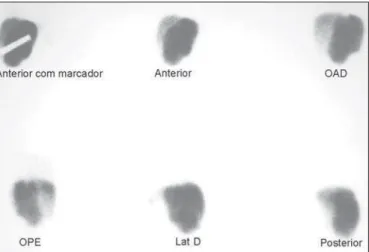
Ultrasound evidenced hepatomegaly with lobulated margins (Figure 1). Hepato-splenic scintigraphy was suggestive of FNH, since it has shown the left lobule increased in volume and with increased uptake (Figure 2). Laparotomy has evi-denced a large mass measuring 20 cm × 15 cm × 10 cm in the anterior portion of the liver, presenting a nodular aspect, reddish color, solid consistency and central venous irrigation, suggesting a tumor-like process (Figure 3). Open biopsy was performed, confirming the diagnosis suggested by the hepatosplenic scintigraphy (Figure 4). These studies were performed at the time of the diagnosis.
284
Teixeira MS et al.
Radiol Bras 2007;40(4):283–285 Figure 1. Abdominal ultrasound demonstrating a liver increased in volume
because of a lesion in the left lobe, with heterogeneous, nodular images with clearly defined margins.
Figure 2. Hepatosplenic scintigraphy with colloidal sulfur (99mTc). Planar images
demonstrating an increased left lobe with increased uptake.
Figure 3. Abdominal laparotomy showing a large mass measuring 20 cm × 15 cm × 10 cm in the anterior portion of the liver, presenting a nodular as-pect, reddish color, solid consistency and central venous irrigation.
Figure 4. Nodular areas corresponding to focal nodular hyperplasia. Anatomo-pathological study: Hematoxilin-eosin staining demonstrating fibrosis with for-mation of incomplete septa (arrow).
at 16 years of age, also demonstrated find-ings suggestive of FNH (Figure 5).
DISCUSSION
Developments in imaging techniques have contributed to the increase in the num-ber of diagnosis of benign hepatic tumors, the differential diagnosis between FNH and hepatic adenoma being extremely impor-tant in the clinical practice, considering the risk of rupture and bleeding of hepatic ad-enomas(1). The apparent increase in the
in-cidence of FNH starting in 1960, although coinciding with the time where oral contra-ceptives were made available in the United
States, may be just a result of the develop-ment of imaging diagnosis techniques and the improvement in the quality of ultra-sound studies(4,5).
The FNH pathogenesis is still to be de-fined, despite a study reporting a simulta-neous occurrence of hemangioma, FNH and hepatic adenoma, suggesting that these three lesions are different manifestations of a same malformation(6). A pre-existing
vas-cular anomaly could cause a local hyper-plastic response of hepatocytes(7). The
pos-sible role of sexual hormones, including oral contraceptives, in the FNH develop-ment has been suggested due to the higher prevalence in women (80% to 95% of
cases)(8) and potential spontaneous
regres-sion of the FNH as a result of an interrup-tion in the use of oral contraceptives(10).
However, the assumption that a FNH may not be associated with the use of oral con-traceptives is evidenced by the occurrence of this type of lesion even before the intro-duction of oral contraceptives, and by the presence of FNH in children, men and women who have never utilized them(5,8).
Actually, the use of oral contraceptives may be associated with the development of FNH, even accelerating its growth, but their possible implication in the occurrence of this lesion has not been demonstrated(3,8).
Nevertheless, new studies are necessary to
285
Focal nodular hyperplasia of the liver: a case report
Radiol Bras 2007;40(4):283–285 clarify the role of oral contraceptives in the occurrence of FNH(2).
The definition of the type of lesion is es-sential, and, among the lesions more eas-ily confused with FNH, hepatic adenoma should be highlighted because of its un-equivocal relationship with the use of oral contraceptives, especially in high doses and during long periods of time(2). In contrast
to the usually conservative approach adopted for FNH, in case of hepatic ad-enoma a surgical excision is performed be-cause of the risk of bleeding and, mainly, malignization(2). In cases of FNH surgical
intervention is reserved for those which present with symptoms, complications, progressive lesions or adjacent organs com-pression(2,9).
FNH complications such as abdominal discomfort, intratumoral hemorrhage and intraperitoneal rupture are rare, although some cases of intraperitoneal rupture have been described in the literature and, in this circumstance the surgical approach have been necessary(10,11).
Potential malignant transformation of FNH has not been reported, although FNH coexistence with hepatocellular carcinoma has rarely been described(8).
The following non-invasive diagnostic techniques are increasingly utilized(3,8,12): a)
ultrasound; b) computed tomography, al-lowing high accuracy in the differentiation between the most frequent types of cen-trally scarred hepatic tumors, including FNH, fibrolamellar hepatocelullar carci-noma and giant cavernous hemangioma; c) hepatosplenic scintigraphy; d) magnetic resonance imaging. An useful finding in the diagnosis of FNH is the evidence of increase in the hepatic uptake at hepato-splenic scintigraphy, allowing the differen-tial diagnosis with other hepatic masses. Ultrasound, computed tomography and magnetic resonance imaging may demon-strate benign hepatic masses, with typical, although not universal, the presence of a central scar. Although less frequently, a characteristic radiating hypervascular mal-formation may be found(3).
Notwithstanding the great usefulness of imaging techniques for the diagnosis of this tumor, if doubts persists about the diagno-sis, a hepatic biopsy becomes mandatory, considering its significance as a highly specific diagnostic method(3).
REFERENCES
1. Biecker E, Fischer HP, Strunk H, Sauerbruch T. Benign hepatic tumours. Z Gastroenterol 2003; 41:191–200.
2. Choi BY, Nguyen MH. The diagnosis and man-agement of benign hepatic tumors. J Clin Gastro-enterol 2005;39:401–412.
3. Kehagias D, Moulopoulos L, Antoniou A, et al. Focal nodular hyperplasia: imaging findings. Eur Radiol 2001;11:202–212.
4. Cherqui D, Rahmouni A, Charlotte F, et al. Man-agement of focal nodular hyperplasia and hepa-tocellular adenoma in young women: a series of 41 patients with clinical, radiological, and patho-logical correlations. Hepatology 1995;22:1674– 1681.
5. Kerlin P, Davis GL, McGill DB, Weiland LH, Adson MA, Sheedy PF. Hepatic adenoma and focal nodular hyperplasia: clinical, pathologic, and radiologic features. Gastroenterology 1983; 84(5 Pt 1):994–1002.
6. Di Carlo I, Urrico GS, Ursino V, Russello D, Puleo S, Latteri F. Simultaneous occurrence of ad-enoma, focal nodular hyperplasia, and heman-gioma of the liver: are they derived from a com-mon origin? J Gastroenterol Hepatol 2003;18: 227–230.
7. Ohmoto K, Honda T, Hirokawa M, et al. Sponta-neous regression of focal nodular hyperplasia of the liver. J Gastroenterol 2002;37:849–853. 8. Vilgrain V. Focal nodular hyperplasia. Eur J
Radiol 2006;58:236–245.
9. Okada T, Sasaki F, Kamiyama T, et al. Manage-ment and algorithm for focal nodular hyperpla-sia of the liver in children. Eur J Pediatr Surg 2006;16:235–240.
10. Koch N, Gintzburger D, Seelentag W, Denys A, Gillet M, Halkic N. Rupture of hepatic focal nodular hyperplasia. About two cases. Ann Chir 2006;131:279–282.
11. Demarco MP, Shen P, Bradley RF, Levine EA. Intraperitoneal hemorrhage in a patient with he-patic focal nodular hyperplasia. Am Surg 2006; 72:555–559.
12. Machado MM, Rosa ACF, Herman P, et al. Ava-liação dos tumores hepáticos ao Doppler. Radiol Bras 2004;37:371–376.
Figure 5. Abdominal helical computed tomography. In the center of the lesion there is a hypointense central scar with 1 cm in diameter (A), that becomes hyperintense (B) on delayed phases. There are tortuous and dilated vessels (C) in the preserved right lobe (arteriovenous malformation) “feeding” the hypervascularized hepatic mass.
A B