Introduction
Nutrition, combined with physical training, is an important tool in sports practice, in an attempting to reduce fatigue and to give the athletes better recovery and yield. Indeed, many athletes try to enhance physical performance training by different ap-proaches and supplementation is one of a nutritional strategy. 1 Beta-hydroxy-beta-methylbutyrate (HMB) has been con-sidered as an agent of empowerment to elevate strength levels, enhancing size skeletal muscle and preventing its collapse when combined with exercise training. It may even attenuate the loss of muscle mass induced by Acquired Immunodeiciency Syndrome – HIV2. In addition, HMB improves strength in-creases fat metabolism, suppress muscle proteolysis, activates myogenic cell proliferation and differentiation, as well as pro-vides an adequate amount of cholesterol precursor3-7. HMB is a byproduct of the metabolism of leucine which is synthesized from a-ketoisocaproate (KIC) in the liver and in the cytosol of hepatocytes, and also in muscle cells8. It is irst converted to ß-hydroxy-ß-methylglutaryl coenzyme A (HMG-CoA) and can follow two distinct metabolic pathways: the irst occurs through the action of the enzyme HMG-CoA reductase, which
is a limiting factor for the synthesis of cholesterol when there is a large demand for the formation of cell membranes (such as occurs in periods of cell growth and muscle repair), which converts HMG-CoA to cholesterol. The second occurs by the action of the enzyme HMG-CoA synthetase which converts HMG-CoA to acetyl-CoA, which is a substrate for power gen-eration8. Adaptations to exercise training and improvement in physical performance are highly speciic to the type of exercise. Concurrent training refers to a program that combines strength with aerobic endurance training in the same session, as well as the possible antagonistic adaptations resulting from these two abilities9,10. The reason for choosing concurrent training is due to the possibility of achievements of the beneits of strength training and endurance simultaneously in a single training session11. Thus, HMB supplementation might be an important factor to minimize the opposing effects of different metabolism recruited by concurrent training.
A previous study reported that aerobic exercise training per-formed primarily as part of a concurrent training program may inhibit the strength as consequence of the anaerobic training when compared with strength training performed alone12. It is well-known that aerobic training increases maximal oxygen
Original article (full paper)
The effect of β-hydroxy-β-methylbutyrate (HMB)
on the morphology of skeletal muscle after
concurrent training
Giovana Rampazzo Teixeira Luis Alberto Gobbo Nilton José dos Santos Rafael Gavassa de Araújo Carolina Cabral dos Santos Olga Cristina de Mello Malheiro
Robson Chacon Castoldi Jose Carlos Silva Camargo-Filho
Universidade Estadual Paulista, Presidente Prudente, SP, Brasil
Marcelo Papoti
Universidade de São Paulo, Ribeirão Preto, SP, Brasil
Abstract–– The aim of the present study was to investigate the effects of β-hydroxy-β-methylbutyrate (HMB) supplementation in association with concurrent training on morphological soleus muscle of rats. Wistar male rats were divided randomly into four groups: Control (C), Supplemented (S), Training (T) and Training + Supplemented (TS). Groups S and TS received 76mg/kg/day of HMB and the training groups (T and TS) were inserted into concurrent training program 3 times/week for 8 weeks. HMB had positive effects either on body composition of the animals or in type II muscle ibers. The concurrent exercise training was able in reducing the total fat mass as well as in increasing the diameter of muscle ibers. Our indings shows that HMB had an anti-catabolic effect with reference to the parameters of volume, weight and morphology of the soleus muscle, and there was a positive interaction between HMB supplementation and concurrent exercise training.
consumption, thereby improving cardiorespiratory itness and in -creasing mitochondrial density in muscle, among other beneits13. On the other hand, strength training promotes an increase in lean body mass and increases the cross-sectional area of skeletal mus-cle ibers12-14. Both, resistance and endurance exercise, stimulate the rate of mixed muscle protein synthesis, an aggregate measure of all muscle proteins. Moreover, resistance training increases myoibrillar proteins (actin and myosin), whereas endurance training increases mitochondrial proteins 15,16.
Therefore, we hypothesized that HMB supplementation may modify the metabolism of muscle ibers, preventing loss of lean body mass, increasing muscular ibers diameter and contribut -ing to the adjustment of oxidative/glycolytic metabolism of the muscle ibers promoted by concurrent training. Considering the importance of supplementation and its impact on the sports ield, the aim of this study was to investigate the effects of HMB sup-plementation on the anthropometric, morphological and physio-logical parameters of Wistar rats undergoing concurrent training.
Methods
Animals and experimental procedure
Twenty-two male Wistar adult rats from the Central Animal Laboratory, UNESP, Botucatu - SP, who were approximately 60 days of age, were used. The animals were maintained in poly-ethylene, solid bottomed cages (40x30x15cm) covered with a coarse sawdust substrate under controlled light and temperature conditions (12 hours of light and 12 hours of darkness) and 20 to 25ºC, respectively, and provided with iltered water and Labina of Purina - Food LTDA Alisul Ind. ad libitum. After the period of environmental adaptation, the animals were randomly divided into four groups: Control (C) - the animals remained in their cages, with water, were fed ad libitum and received no physical stimulation (n=6); Supplementation with HMB (S) - the animals remained in their cages, with water, were fed ad libitum, received no physical stimulus, but they were sup-plemented daily with HMB (n=6); training (T) - the animals were submitted to the protocol of concurrent training (n=5); training + HMB supplementation (TS) - the animals underwent the concurrent training protocol and were supplemented daily with HMB (n=5). The experimental design followed the rules and ethics of experimentation on animals adopted by the Ethics Committee for animal research of FCT/UNESP - Presidente Prudente, Protocol 03/2011.
HMB supplementation
The oral protein supplementation HMB (Trade Mark Arnold Nutrition Inc., Hollywood, FL) was initiated simulataneously with the concurrent training period (81 days of age). HMB supplementation was given by gavage (30 minutes before the concurrent training protocol) in a single dose comprising 76mg/ kg/day, a dosage equivalent to that used in studies involving HMB supplementation in humans, which corresponds to about
3 to 6 g / day of HMB for a subject of 80 kilograms 7. Both sup-plemented groups (S and TS) received the dose at the same time.
Concurrent training program
At 60 days of age, the animals of all groups began their ad-aptation to the water environment with the purpose of reducing stress from the exercise. For the groups assigned to concurrent training (T and TS) the adaptation period took place over three weeks (21 days) with swimming sessions with increasing in-tensities in individual PVC cylindrical tanks (120x60cm) with a controlled temperature (31 ± 1ºC), 30 minutes per session, frequency of three times a week. After the last adaptation sec-tion, the animals had interval the 48h for physical tests.
At 81 days of age, the animals in the training groups began receiving sessions of concurrent training, starting with aerobic training, followed by anaerobic training with no break between them. The aerobic endurance training consisted of a swimming session for thirty-eight minutes conducted in PVC cylindrical tanks. A vest was placed on the anterior chest region of the animal with a constant overload, equivalent to 50% of the body weight of each animal, as previously established in the lactate minimum test, corresponding to 70% of the anaerobic threshold17. In the strength training, the animals were subjected to sessions of jumps performed in a cylindrical PVC container (90x60cm) containing water at 30º ± 1ºC with a depth of 40 cm18. The physical training program occurred for eight con-secutive weeks. The jump sessions in the liquid medium were composed of four sets of ten jumps with an interval equivalent to 1 minute between sets.
Determination of Aerobic and Anaerobic Performances
The aerobic and anaerobic performances were admitted as intensity timeout (Tlim). For the determination of these variables the protocol validated by Tegtbur19, for humans, applied to rats by Voltarelli20 and adapted for these animals by Araujo21 was used. This procedure for assessment of anaerobic performance served as a method to induce hyperlactemia with a load corresponding to 13% of body weight (BW). The animals performed two efforts in the irst phase of the test, the irst lasting 30s, and after an interval of 30s, a second effort with the same load until exhaustion (Tlim). Blood samples (25μL) were collected from a cut at the tip of the tail with the aid of graded capillary tubes during the exercise tests and placed in minutes 1, 3, 5, 7 and 9 after Tlim for determining the peak of lactate concentration (mmol/L).
the overload used for physical training was identiied (percent body weight) to each animal.
Anthropometric, histological and histochemical analyses
During the experiment, the animals were subjected to mea-surement of body weight. Body weight was measured using an electronic scale (Shimadzu BL3200H, with a precision of 0.01 g). To evaluate the weight gain, the calculation of body mass gain was adopted, using the formula22:
Δ = Final weight - Initial weight.
Body mass index was calculated as body weight (g)/length2, in cm2.
At 140 days of age the animals were euthanized with an overdose of sodium pentobarbital (100mg/kg) applied intraper-itoneally, 48 h after the last training session. After laparotomy, the abdominal-pelvic liver, renal fat and epididymal fat were removed for weighing. Samples of the ventral portion of the soleus muscle were collected and weighed. A portion of the soleus was ixed by immersion in buffered formaldehyde solu -tion (PBS + 10% formaldehyde) and then soaked in paraplastic (Paraplast Plus, McCormick, St. Louis, MO, USA). Cuts, 4 mm in thickness, were made and used for general morphology analysis with H/E.
Histological analysis focused on the cell morphology of the soleus muscle tissue: shape, size, color sarcoplasm and nuclei using a light microscope (AxioCam - 2 with digital camera, ERC 5S model AxioCam, Carl Zeiss, Department of Physical Education / FCT UNESP).
For the histochemical analysis another portion of the muscle was frozen in liquid nitrogen at -70ºC. After freezing the soleus muscles were cut on a cryostat microtome with a thickness of 5μm. The sections were incubated in the histochemical reac -tion Nicotinamide adenine dinucleotide tetrazolium reductase (NADH-TR) a technique modiied by Dubowitz23 to indicate the presence of oxidative activity, and detect the types of mus-cle ibers in cross section: slow oxidative type I, fast oxidative glycolytic type IIb and fast glycolytic type IIa. Slides were analyzed and 10 cross sections/animal was photographed un-der a light microscope (Axiocam - 2, Carl Zeiss, Department of Physical Education FCT/UNESP), and the amount of iber were determined.
For stereology tissue volume (iber core and connective tissue) the reticle 160 points of Weibel24 was used on the muscle cross-section stained with H/E, 5 animals per group two blades per animal and 10 pictures per slide with a 40x objective using a light microscope AxioCam - 2 model AxioCam HR, Zeiss Department of Physical Education FCT / UNESP.
Statistical Analysis
Statistical analysis was performed using a 2-way analysis of variance (ANOVA) with factors of supplementation and exercise
group and its interaction, complemented with the Tukey multiple comparison test to contrast comparisons between parametric data in Tlim, weight and relative weight (g/100g) for total fat, soleus and liver. To compare body weight throughout the inter-vention and group, a 3-way factor ANOVA (supplement group x exercise group x time) with repeated measures on the last factor were performed. Results were expressed as mean ± standard deviation. Variables not normally distributed were treated for non-parametric statistics (i.e.: median absolute deviation based on the median, conidence interval for the median and coeficient of variation based on the median), using the Kruskal Wallis analysis related to the percentage used in the stereology mus-cle. The statistical conclusions were made at 5% signiicance. Details about the methodology can be found in Banzatto and Kronka25. The statistical software used was IBM SPSS Statistics
version 22, GraphPad Instat version 4 and Sigma Plot version 11.0 used for the graphics.
Results
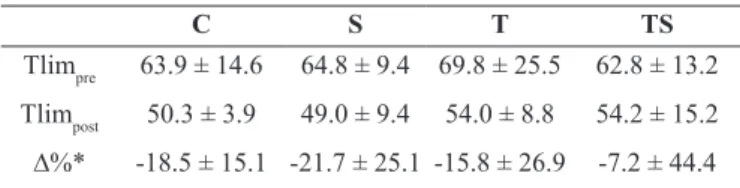
The swimming time to fatigue (Tlim) was used in this study as the anaerobic performance parameter and these values did not change signiicantly over the phases among groups, with a tendency for the post-training Tlim values to be lower than the pre-training Tlim (Table 1).
Table 1. Mean values of Tlim pre and post training and variation
(∆%) per group of rats.
C S T TS
Tlimpre 63.9 ± 14.6 64.8 ± 9.4 69.8 ± 25.5 62.8 ± 13.2 Tlimpost 50.3 ± 3.9 49.0 ± 9.4 54.0 ± 8.8 54.2 ± 15.2
∆%* -18.5 ± 15.1 -21.7 ± 25.1 -15.8 ± 26.9 -7.2 ± 44.4 Values expressed as mean ± standard deviation. C = control group; S = supplementation group; T = trained group; TS = supplementation plus trained group.
* Non-signiicant (p=0.866) 2-way ANOVA model (with factors of supplementation and exercise); r2=0.039. Insert here Table 1
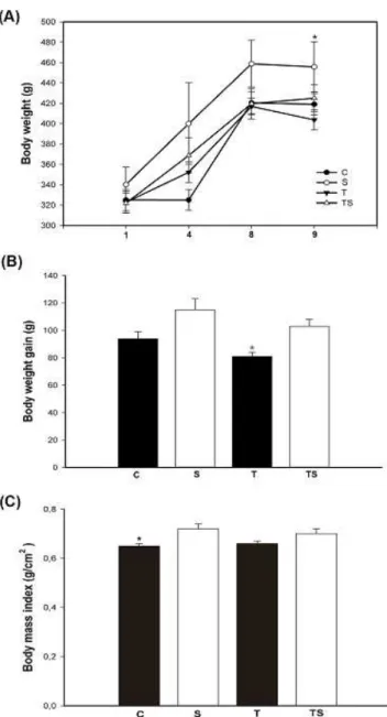
Body weight did not show signiicant differences between the groups throughout the intervention (0=0.678); however, signiicant differences (p<0.001) were veriied from week 1 to
9 (Figure 1a). As expected, the rats that practiced concurrent training showed signiicant lower body weight as compared with control group, approximately 16% in percentage weight gain (Figure 1b). The S group, HMB supplementation only, presented increased body weight as compared with other groups (Figure 1b). The interaction between HMB supple-mentation and concurrent training promoted an increase in weight gain when compared with exercise animals without supplementation, approximately 21%, (Figure 1b). When analyzing BMI, it was veriied signiicant difference only for the control group, compared with group that received supple-mentation (p<0.05). Regarding to body mass index, signiicant
Figure 1: (A) Time-course of total body mass (g) during the 8 weeks
of training and supplementation: (●) Control, (○) Supplemented, (▼) Trained and (▲) Trained Supplemented.*Signiicant difference with
respect to T. (B) Effect of supplementation and concurrent training on weight gain (g) after 8 weeks of treatment. b p<0.05 versus
supplement-ed. (C) Effects of supplementation with HMB and concurrent training on body mass index (g/cm2) of animals. b p<0.05 versus supplemented.
The weight and relative weight (g/100g) of whole-body fat mass of animals submitted to physical training (with or with-out supplementation) exhibited signiicant lower values when compared with animals that received only supplementation (p=0.016). In addition, relative weight of the soleus was signii -cantly higher for the animals submitted to physical training and to supplementation only (p=0.026). HMB supplementation and concurrent training did not change (p>0.05) soleus and the liver weights, and relative weight of the liver (Table 2).
The values of the soleus muscle stereology expressed in cm2, showed that concurrent exercise training with supplementation promoted a signiicant increase in the ibers of the soleus muscle as compared with the C group (Table 3). The volume of tissue was signiicantly lower in S and T groups, comparing with control group. There was a signiicant increase of nuclei in the S, T and TS groups as compared with control group (Table 3).
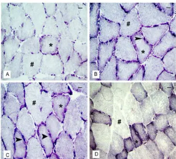
Morphological analysis of the control animals showed nor-mal-looking ibers arranged in parallel and peripheral nuclei (Figure 2). The supplementation resulted in a more rounded aspect than the polygonal characteristic of muscle ibers, i.e., an increase in the diameter of muscle ibers in a cross section of the soleus muscle ibers in group S, compared with group C (Figure 2). Furthermore, the S group had a higher quantity of peripheral nuclei distributed per iber than the C group (Figure 2). The occurrence of micro lesions, characterized by the incidence of leukocyte iniltration into the ibers, or immune cells iniltrating the muscle iber hypertrophy characteristic of inlammation was observed in groups T, TS and S. The connec -tive tissue (endomysium and perimysium) was lower in trained animals than in control group, suggesting a possible increase in protein synthesis of muscle ibers (Figure 2).
Examining the enzymatic reaction (NADH-TR) we found an increase in cross sections of the ibers IIa and IIb when compared with the number of ibers I in animals from the S, T and TS groups, compared with group C (Figure 3). The enzymatic reaction of muscle ibers in the S and TS groups showed an increase in the quantity of IIa and IIb ibers (totalizing 72% and 75%, respective -ly) in relation to I ibers (28% and 25%, respective-ly) compared with other groups (30% for ibers I - control and trained) (Figure 3). The characteristics presented by the muscle ibers showed that training, supplementation and the association of both modiied the morphological and metabolic state of the muscle ibers.
Table 2. Total weight (g) and relative weights (g/100g) of total fat, soleus muscle and liver of the Wistar trained rats supplemented with HMB. ANOVA model*
Varibles C S T TS p r2
Total fat (g) 9.3±2.1 10.3±3.4 5.8±0.8 6.3±0.9 0.016a 0.446
Relative weight of total fat 2.2±0.6 2.3±0.6 1.4±1. 6 1.5±0.3 0.015a 0.450
Weight of the soleus (g) 0.20±0.02 0.23±0.04 0.20±0.02 0.20±0.02 0.163 0.254
Relative weight of the soleus 0.048±0.002 0.051±0.003 0.051±0.003 0.046±0.002 0.026b 0.411
Weight of the liver (g) 15.6±3.7 16.9±3.6 12.8±1.7 15.9±2.1 0.207 0.242
Relative weight of the liver 3.7±0.8 3.8±0.3 3.2±0.4 3.7±0.4 0.306 0.197
The values are expressed as mean ± standard deviation. C = control group; S = supplementation group; T = concurrent trained group; TS = supplementation and concurrent trained group. All relative weights are expressed in g/100g.
* 2-way ANOVA with factors of supplementation and exercise; a effect on trained groups (p=0.002); b effect on trained and supplementation groups (p=0.005).
Table 3. Median stereology of the soleus muscle in cm2.
C S T TS
Area of
muscle iber 108.7(105.7-112.6)d
113.8(105.3-119.8)
113.3(109.9-114.0)
112.6(112.6-114.4) Area of
connective tissue
20.4 (15.2-22.9)c,d
13 (9.6-20.2)
13.9 (12.4-17.6)
14.9 (12.4-20.4)
Nuclear area
2.1 (1.8-3.1)b,c,d
3.3 (2.3-5.4)
3.8 (2.6-4.5)
3.8 (3.3-4.3) The values are expressed as median (minimum;maximum). The Kruscal Wallis test, bp<0.05 versus supplemented; cp<0.05 versus trained group; d
p<0.05 versus trained and supplemented group;
C = control group; S = supplementation group; T = exercise group; TS = supplementation and exercise group.
Figure 2: Morphological distribution in cross section (a,c,e,g) and longitudinal (b,d,f,h) soleus muscle. Images (A) and (B) refer to the C group having regular architecture. Images (C) and (D) are the group S, arrow indicating peripheral nuclei and reduction of adjacent
con-nective tissue. * Indicates micro lesions in the longitudinal ibers. Im -ages (E) and (F) the group T, arrow indicating the increase of nuclei
wrapped ibers and reduction of tissue. Images (G) and (H) for the
TS group. “E” = Endomysium. “P” = Perimysium. = Nuclei. ¢ =
Blood vessel. Bar= 20μm.
Figure 3: Morphological distribution of a cross section of soleus mus-cles incubated in the enzymatic reaction NADH-TR. Image (A) is related to group C; Image (B) to the S group; Image (C) the T group
and image (D) the TS group. The symbols “*” in the images represent the type I ibers. = Peripheral mitochondria.
Discussion
In the present study, we investigated the effects of HMB supplementation on anthropometric, morphological and his-tochemical parameters in male Wistar rats submitted to eight weeks of concurrent training. The major indings of this study were that HMB promoted changes in body composition and muscle cellular dimensions.
lactate minimum. In agreement, it was observed a reduction of anaerobic capacity after monotonous training for 12 weeks, because high training volume reduces anaerobic adaptations29. HMB is converted into ß-hydroxy-ß-methylglutaryl coen-zyme A (HMG-CoA), a limiting factor for cholesterol synthesis activated by the formation of cell membranes in cell growth and muscle repair30. The highest expression of body fat in the supplemented group corresponds to the increased cholesterol promoted by HMB synthesis with muscle and fatty tissue as the two main receptors31. According to a previous study, cholesterol from the HMG-CoA, a co-factor of the pathways of HMB may have actions in the muscle tissue, immune and tissue cells in the mammary gland, promoting responses to differentiation and synthesis of muscle membrane and organelles, increasing cell growth and increasing function32. Differently, our indings did not observe changes in body weight. However, HMB was able to alter body composition both acting alone or in association with physical training. Thus, we can conclude that there were signiicant physiological adjustments in the regulatory pathways of the metabolism, favoring smaller storage of triglycerides in visceral tissues when combined with concurrent training.
Previous research has shown that progressive resistance training for 8–21 weeks caused fast-to-slow iber-type transitions within the fast iber subtypes and considerable iber hyper -trophy33,34. In contrast, endurance training for a similar period typically induces iber atrophy and, depending on the intensity and volume of repeated contractions, may also enhance the pro-portion of type I ibers35. Morphological data from the present study demonstrate that concurrent training increased the diameter of the muscle ibers and reduced the adjacent connective tissue. The increase in cross section is an indicator of protein synthesis and/or muscle hypertrophy. During the concurrent training, the animals performed activities at high intensities, resulting in high energy applications as well as the recruitment of muscle groups; thus, several biochemical adaptations at different levels (such as in the liver and muscle tissues) facilitate the mobilization and oxidation triacylglycerol and lead to a conservative effect on lean body mass36. The greater number of myonuclei as consequence of satellite cell proliferation would result in an elevated potential for transcription, leading to muscle hypertrophy and an increased nuclear protein, showing higher proliferation of markers and inhibition of differentiation-2 and cyclin A37. Possibly, HMB may have been more effective at suppressing apoptotic pathways associated with muscle loss by attenuating proapoptotic proteins as observed previuosly38.We also observed that HMB supple-mentation alone was able to increase muscle iber, muscle iber damage and nuclear areas, decrease area of connective tissue, and causes hypertrophy due to micro adjustments indicative of protein. Recently, it was reported that phosphatidic acid (PA), which is a glycoprotein and Ras homolog enriched in brain (Rheb) appears to play a fundamental role in the mechanical regulation of mus-cle mass enabled through late endosome/lysosome (LEL) in the regulation of mTOR by various growth regulatory inputs such as amino acids, growth factors and mechanical stimuli39. Resistance-like exercise speciically increased the phosphorylation of the an -abolic Akt/mTOR signaling pathway, along with the activation of the translation initiation regulators p70 S6k, 4E-BP1, and eIF2B,
but had little effect on the AMPK/PGC-1 pathway. In contrast, endurance-like exercise increased AMPK phosphorylation and PGC-1 protein levels39. Accordingly, it was proposed that selective activation of either the Akt–mTOR or AMPK–PGC-1 signaling pathways can explain speciic adaptive responses to resistance or endurance-like exercise responses40. From a regulatory per-spective, the notion of an AMPK–Akt master switch is attractive. More recently, however, the hypothesis that HMB might directly stimulate muscle protein anabolism has been proposed. In this respect, a crucial step in the anabolic response is phosphorylation and activation of the mTOR which in turn activates p70S6 kinase (p70S6K) and inhibits 4E-BP141. Phosphorylation of 4E-BP1 induced by growth factors such as insulin and IGF-I has been shown to depend on the Akt/PI3K/mTOR pathway, whereas an Akt-unrelated, mTOR-dependent activation has been proposed to result from increased intracellular amino acid availability41-43. Therefore, it seems that the mTOR responds to various stimuli, including mechanical stimuli (physical training) and nutrition (supplementation), promoting cell growth44. HMB reduces pro-teolysis in muscle iber after strength and endurance exercises, and increases lean body mass and strength levels45-47.
In the present study, NADH-TR oxidative staining was used for the differentiation of three muscle iber types. The results suggest that HMB supplementation and concurrent training, individually or in association, promoted muscular adaptations, converting ibers into slow twitch or fast type I ibers in iber type IIb and type IIa promoting the remodeling of muscle ibers. Skeletal muscle in obese individuals exhibits reduced oxi-dative capacity, increased glycolytic capacity, and a decreased percentage of type I ibers48,49. A prevalence of type II ibers may thus result in the partitioning of lipid towards storage in skeletal muscle (i.e., intramuscular triglyceride) or adipose tissue rather than oxidation within skeletal muscle, resulting in a positive fat balance. Indeed, it was reported that rodents that gained the most mass with high-fat feedings possessed signiicantly fewer type I ibers than their littermates that gained little or no weight50.
In conclusion, concurrent training in association with HMB supplementation showed positive effects on body composition as well as on skeletal muscle ibers in rats. HMB prevented the loss of fat mass and increased the diameter of the muscle ibers in cross-section, contributing to a change in oxidative metabolism / glycolytic ibers.
References
1. Maughan RJ, Burke LM. Practical nutritional recommendations for the athlete. Nestle Nutrition Institute Workshop Series, 2011; 69:131-149.
2. Clark RH, Feleke G, Din M, Yasmin T, Singh G, Khan FA et al. Nutritional Treatment for Acquired Immunodeiciency
Virus-Associated Wasting Using β-Hydroxy-β-Methylbutyrate,
Glutamine, and Arginine: A Randomized, Double-Blind, Placebo-Controlled Study. J Parenter Enteral Nutr. 2000; 24:133-139. 3. Wilson J, Wilson GJ. Contemporary issues in protein requirements
4. Ransone J, Neighbors K, Lefavi R, Chromiak J. The effect of beta-hydroxy beta-methylbutyrate on muscular strength and body composition in collegiate football players. J Strength Cond Res, 2003;17:34-39.
5. Eley HL, Russell ST, Baxter JH, Mukerji P, Tisdale MJ. Signaling pathways initiated by beta-hydroxy-beta-methylbutyrate to atten-uate the depression of protein synthesis in skeletal muscle in re-sponse to cachectic stimuli. Am J Physiol. 2007; 293:E923- E931. 6. Kornasio R, Riederer I, Butler-Browne G, Mouly V, Uni Z,
Halevy O. Beta-hydroxy-beta-methylbutyrate (HMB) stimulates myogenic cell proliferation, differentiation and survival via the MAPK/ERK and PI3K/Akt pathways. Biochim Biophys Acta, 2009; 1793:755-763.
7. Gallagher PM, Carrithers JA, Godard MP, Schulze KE, Trappe SW. Beta-hydroxy-beta-methylbutyrate ingestion, Part I: effects on strength and fat free mass. Med Sci Sports Exerc, 2000; 32:2109-2115.
8. Pimentel GD, Rosa JC, Lira FS, Zanchi NE, Ropelle ER, Oyama LM et al. b-Hydroxy-b-methylbutyrate (HMb) supplementation stimulates skeletal muscle hypertrophy in rats via the mTOR pathway. Nutr Metabol, 2011; 8:11.
9. McCarthy JP, Pozniak MA, Agre JC. Neuromuscular adaptations to concurrent strength and endurance training. Med Sci Sports Exerc, 2002; 34:511-519.
10. Gomes RV, Aoki MS. Suplementação de Creatina Anula o Efeito Adverso do Exercicio de Endurance sobre o subsequente desem-penho de Força. Rev Bras Med Esporte, 2005; 11:131-134. 11. Leveritt M, Abernethy PJ, Barry BK, Logan PA. Concurrent
Strength and Endurance Training: the inluence of dependent
variable selection. J Strength Cond Res. 2003; 17:503-508. 12. Collins MA, Snow TK. Are adaptations to combined endurance
and strength training affected by sequence of training? J Sports Sci. 1993; 11:485-491.
13. Hakkinen K, Alen M, Kraemer WJ, Gorostiaga E, Izquierdo M, Rusko H et al. Neuromuscular adaptations during concurrent strength and endurance training versus strength training. Eur J Appl Physiol. 2003; 89:42-52.
14. Dudley GA, Fleck SJ. Strength and Endurance Training: are they mutually exclusive? Sports Med. 1987; 4:79-85.
15. Carraro F, Stuart CA, Hartl WH, Rosenblatt J, Wolfe RR. Effect of exercise and recovery on muscle protein synthesis in human subjects. Am J Physiol. 1990; 259:E470– E476.
16. Chesley A, MacDougall JD, Tarnopolsky MA, Atkinson SA, Smith K. Changes in human muscle protein synthesis after resis-tance exercise. J Appl Physiol. 1992; 73:1383–1388.
17. Teixeira GR, Fávaro WJ, Pinheiro PFF, Chuffa LGA, Amorim JPA, Mendes LO et al. Physical exercise on the rat ventral pros-tate: Steroid hormone receptors, apoptosis and cell proliferation. Scand J Med Sci Sports. 2012; 22:86-92.
18. Harri M, Kuusela P. Is swimming exercise or cold exposure for rats? Acta Physiol Scand. 1986; 126:189–197.
19. Tegtbur U, Busse MW, Braumann KM. Estimation of an individual equilibrium between lactate production and catabolism during exercise. Med Sci Sports Exerc. 1993; 25:620-627.
20. Voltarelli FA, Gobatto CA, Mello MAR. (2002). Determination of anaerobic threshold in rats using the lactate minimum test. Braz J Med Biol Res. 2002; 35: 1389-1394.
21. Araujo GG, Papoti M, Manchado FB, Mello MA, Gobatto CA. Protocols for hyperlactatemia induction in the lactate minimum test adapted to swimming rats. Comp Biochem Physiol, 2007; 148:88-92.
22. Novelli ELB, Diniz YS, Galhardi CM, Ebaid GMX, Rodrigues HG, Mani F. et al. Anthropometrical parameters and markers of obesity in rats. Lab Animal. 2007; 41:111-119.
23. Dubowitz V, Sewry CA, Oldfors A. Muscle biopsy: a practical approach. London: Elsevier Health Sciences, 2013; pp 492. 24. Weibel ER, Kistler GS, Scherle WF. Pratical stereologigal
meth-ods for morfhometric cytology. J Cell Biol. 1996; 30:23-38. 25. Banzatto DA, Kronka SN. Experimentação agrícola. Jaboticabal:
FUNEP, 1989; p.247.
26. Bosquet L, Leger L, Legro SP. Methods to determine aerobic endurance. Sports Med. 2002; 32:675-700.
27. Araujo GG, Papoti M, Manchado-Gobatto FB, Mello MA, Gobatto CA. Padronização de um Protocolo Experimental de Treinamento Periodizado em Natação Utilizando Ratos Wistar. Rev Bras Med Esporte. 2010; 16:51-6.
28. Voltarelli FA, Mello MAR, Gobatto CA. Limiar anaeróbio deter-minado pelo teste do lactato mínimo em ratos: efeito dos estoques de glicogênio muscular e do treinamento físico. Rev Port Ciências Desporto. 2004; 4;16-25.
29. Araújo GG, Papoti M, Manchado-Gobatto FB, Mello MA, Gobatto CA. (2013). Monitoring chronic physical stress using biomarkers, performance protocols and mathematical functions to identify physiological adaptations in rats. Lab Animals. 2013; 47:36-42.
30. Nissen SL, Abumrad NN. Nutritional role of the leucine me-tabolite b-hydroxy-h-methylbutyrate (HMB). J Nutr Biochem. 1997; 8:300-311.
31. Durstine JL, Grandjean PW, Cox CA. Thompshom PD. Lipids, lipoproteins and exercise. J Cardiopulm Rehab Prev. 2002; 22:385-398.
32. Wilson JM, Grant SC, Lee SR, Masad IS, Park YM, Henning PC et al. Beta-hydroxy-beta-methyl-butyrate blunts negative
age-re-lated changes in body composition, functionality and myoiber
dimensions in rats. J Int Society Sports Nutr. 2012; 9:18. 33. Staron RS, Karapondo DL, Kraemer WJ, Fry AC, Gordon SE,
Falkel JE et al. Skeletal muscle adaptations during early phase of heavy-resistance training in men and women. J Appl Physiol. 1994; 76:1247–1255.
34. Kadi F, Thornell LE. Training affects myosin heavy chain phenotype in the trapezius muscle of women. Histochem Cell Biol. 1999; 112:73–78.
35. Fitzsimons DP, Diffee GM, Herrick RE, Baldwin KM. Effects of endurance exercise on isomyosin patterns in fast- and slow-twitch skeletal muscles. J Appl Physiol. 1990; 68:1950–1955.
36. Hawley JA. Molecular responses to strength and endurance training: Are they incompatible? Appl Physiol Nutr Metab. 2009; 34:355–361.
37. Always SE, Pereira SL, Edens NK, Hao Y, Bennett BT. β-Hy
-droxy-β-methylbutyrate (HMB) enhances the proliferation of
satellite cells in fast muscles of aged rats during recovery from disuse atrophy. Exp Gerontolol. 2013; 48:973-84.
38. Hao Y, Jackson JR, Wang Y, Edens N, Pereira SL, Always SE.
during recovery from hind limb suspension-induced muscle iber
atrophy in aged rats. Am J Physiol Regul Integr Comp Physiol. 2001; 301:R701-715.
39. Jacobs BL, Goodman CA, Hornberger TA. The mechanical activation of mTOR signaling: an emerging role late endosome/ lysosomal targeting. J Muscle Res Cell Motil. 2014; 35:11-21. 40. Atherton PJ, Babraj J, Smith K, Singh J, Rennie MJ, Wackerhage
H. Selective activation of AMPK-PGC-1alpha or
PKB-TSC2-mTOR signaling can explain speciic adap-tive responses to
endurance or resistance training-like electrical muscle stimulation. FASEB J. 2005; 19:786–98.
41. Gingras AC, Raught B, Sonenberg N. eIF4 initiation factors: effectors of mRNA recruitment to ribosomes and regulators of translation. Ann Rev Biochem. 1999; 68: 913-63.
42. Shen W, Boyle DW, Wisniowski P, Bade A, Liechty A. Insulin and IGF-I stimulate the formation of the eukariotik initiation factor 4F complex and protein synthesis in C2C12 myotubes indepen-dent of availability of external amino acids. J Endocrinol. 2005; 185:275-289.
43. Gerlinger-Romero F, Guimarães-Ferreira L, Giannocco G, Nunes MT. Chronic supplementation of beta-hydroxy-beta
methylbutyr-ate (HMβ) increases the activity of the GH/IGF-I axisand induces
hyperinsulinemia in rats. Growth Horm IGF Res. 2011; 21:57-62. 44. Zanchi NE, Nicastro H, Gualano B, Costa AS, Lancha-Junior
AH. Suplementação de HMB: relevância clínica e mecanismos de ação. Rev Mackenzie Educação Física Esporte. 2009; 8:123-133. 45. Nissen S, Sharp RL, Panton L, Vukovich M, Trappe S, Fuller JC.
β-Hydroxy-β-Methylbutyrate (HMB) Supplementation in Humans
Is Safe and May Decrease Cardiovascular Risk Factors. J Nutr, 2000; 130:1937-1945.
46. Panton LB, Rathmacher JA, Baier S, Nissen SL. Nutritional Supplementation of the Leucine Metabolite b-Hydroxy-b-Meth-ylbutyrate (HMB) During Resistance Training. Nutrition. 2000; 16:734- 739.
47. Park BS, Henning PC, Grant SC, Lee WJ, Lee SR, Arjmandi BH et al. HMB attenuates muscle loss during sustained energy
deicit induced by calorie restriction and endurance exercise.
Metabolism. 2013; 62:1718-1729.
48. Tanner CJ, Barakat HA, Dohm GL, Pories WJ, MacDonald KG, Cunningham PRG, et al. Muscle fiber type is associated with obesity and weight loss. Am J Physiol Endocrinol Metab., 2002; 282:E1191- E1196.
49. Hickey MS, Carey JO, Azevedo JL, Houmard JA, Pories WJ, Israel RG et al. Skeletal muscle iber composition is related to adiposity and in vitro glucose transport rate in humans. Am J Physiol Endocrinol Metab. 1995; 268:E453–457.
50. Mrad JA, Yakubu F, Lin D, Peters JC, Atkinson JB, Hill JO. Skeletal muscle composition in dietary obesitysusceptible and dietary obesity-resistant rats. Am J Physiol Regul Integr Comp Physiol. 1992; 262:684–688.
Acknowledgments
The authors would like to thank The Laboratory of Histology and Histochem-istry for the collaboration on histochemical techniques, and PIBIC - Proc. No. 106235/2012-5, for inancial support.
Corresponding author
Giovana Rampazzo Teixeira Department of Physical Education, Center of Science and Technology, UNESP, Presidente Prudente, SP, Brazil.
Email: [email protected]
Manuscript received on November 27, 2015 Manuscript accepted on April 10, 2016