Protective microcirculatory and anti-inflammatory
effects of heparin on endotoxemic hamsters
Marcos L. Miranda,ILuiz Felipe M. Prota,IMaria Ju´lia B. Silva,IIFernando L. Sicuro,IEliane S. Furtado,I Ana Olimpia M.T. Santos,IIEliete BouskelaI
ILaboratory for Clinical and Experimental Research in Vascular Biology – BioVasc, Biomedical Center, Rio de Janeiro State University, Rio de Janeiro, RJ, BrazilIIOswaldo Cruz Foundation – Fiocruz, Rio de Janeiro, RJ, Brazil
OBJECTIVE:Apart from its anticoagulant properties, heparin has vasodilator and anti-inflammatory effects that could assist in the reversal of septic microcirculatory changes. This paper investigates the effects of heparin on endotoxemia-related microcirculatory changes and compares them to those observed with the use of recombinant human activated protein C.
METHODS: After skinfold chamber implantation procedures and endotoxemia induction by intravenous Escherichia colilipopolysaccharide administration (2 mg.kg21), male golden Syrian hamsters were treated with intravenous unfractionated heparin (0.2 mg.kg21). Intravital microscopy of skinfold chamber preparations
allowed quantitative analysis of microvascular variables and venular leukocyte rolling and adhesion. Macro-hemodynamic parameters were also analyzed. Endotoxemic hamsters treated with recombinant human activated protein C and non-treated animals served as controls.
RESULTS:Heparin decreased lipopolysaccharide-induced leukocyte rolling and arteriolar vasoconstriction; it also increased survival when compared with non-treated animals, while recombinant human activated protein C decreased leukocyte adhesion. Administration of heparin plus recombinant human activated protein C was associated with a significant attenuation of lipopolysaccharide-induced capillary perfusion deficits.
CONCLUSIONS:Heparin yields protective effects on endotoxemic animals’ microcirculation. Those benefits were potentiated when heparin was administered in conjunction with recombinant human activated protein C.
KEYWORDS: sepsis; endotoxemia; microcirculation; heparin; recombinant human activated protein C.
Miranda ML, Prota LFM, Silva MJB, Sicuro FL, Furtado ES, Santos AOMT, Bouskela E. Protective microcirculatory and anti-inflammatory effects of heparin on endotoxemic hamsters. MEDICALEXPRESS. 2014 June;1(3):127-134.
Received for publication onMarch 3 2014;First review completed onMarch 9 2014;Accepted for publication onMarch 30 2014
E-mail: [email protected]
B INTRODUCTION
Sepsis is an infection-related systemic inflammatory syndrome associated with a massive activation of the coagulation cascade, consumption of clotting factors, and disruption of the normal balance between coagulation and fibrinolysis.1,2This altered coagulant state is associated with uncontrolled formation of intravascular platelet/fibrin clots in the microcirculation. The so-called microthrombosis is responsible for physical occlusion of capillaries, being an important cause of microcirculatory dysfunction.3As sepsis progresses, microcirculatory abnormalities ultimately result in tissue hypoxia, organ failure, and death.4,5
Seeing that levels of antithrombotic factors are frequently very low in patients with severe sepsis and septic shock, it could be speculated that correction of those low levels could assist in reversal of the septic procoagulant state, attenuating
microvascular perfusion deficits and improving patients’ outcome.6In this way, numerous clinical trials have already examined the use of antithrombotic drugs such as recombinant human activated protein C (rhAPC), tissue factor pathway inhibitor, and antithrombin III in septic patients. Unfortunately, none of these agents consistently improved the outcome of these patients, but all significantly increased the risk of bleeding.7
Unfractionated heparin is the most widely used antic-oagulant in clinical practice and is recommended for thromboembolism prophylaxis in septic patients; a vast experience regarding its safety has accumulated.8,9Besides its anticoagulant property, heparin has other less studied properties, such as its vasodilator and anti-inflammatory effects.8,9 Taken together, these properties could be beneficial for microcirculatory function. So, we have hypothesized that heparin could assist in the reversal of septic microcirculatory changes, a decisive step in sepsis treatment, but this subject has been poorly explored in the literature. Strengthening our hypothesis, Dobosz et al.10have
DOI:10.5935/MedicalExpress.2014.03.06
already demonstrated that heparin has a positive influence on organ microcirculatory disturbances accompanying non-infectious experimental systemic inflammatory syndrome. Additionally, in a meta-analysis involving septic patients, heparin was associated with improved 28-day survival.11
Aiming to investigate the effects of heparin on endotox-emia-related microcirculatory changes and to compare its effects with those observed with rhAPC (an extensively studied antithrombotic and anti-inflammatory drug), we have carried out this controlled experimental study.
B MATERIALS AND METHODS
Experiments were performed on 28 male golden Syrian hamsters (Mesocricetus auratus, ANILAB, Animais de Laborato´rio, Paulı´nia, SP, Brazil) weighing between 60 and 80 g. Animals were housed one per cage under controlled conditions of lighting (12:12 hours light/dark cycle) and temperature (21.0^1.08C), with free access to water and standard chow (NUVILAB CR1, Quimtia S/A, Colombo, PR, Brazil). All procedures were approved by the Rio de Janeiro State University Animal Care and Use Committee (protocol number CEUA/060/2010) and are consistent with the United States National Institutes of Health Guide for the Care and Use of Laboratory Animals (National Research Council, 1996) and ethical rules specified by the Basel Declaration.
Animal preparation
The chamber implantation procedure has been described previously by Endrich et al.12 in details. Briefly, under anesthesia with sodium pentobarbital (90 mg.kg21 intraper-itoneal injection; Hypnol 3%, Syntec, Cotia, SP, Brazil) animals’ dorsal hair was shaved and depilated with a commercial hair-removing solution. After that, the dorsal skin of the back was lifted away from the animal, creating a skinfold. Then, this skinfold was sandwiched between two titanium frames and one of its layers was microsurgically excised in a circular area of 15 mm in diameter. The remaining layer, consisting of epidermis, subcutaneous tissue, and thin striated skin muscle (panniculus carnosus muscle) was covered with a removable circular cover glass incorporated into one of the metal frames, creating the window chamber. After a recovery period of 6 days, animals were re-anesthetized and the left carotid artery catheterized (polyethylene-50 catheter) allowing continuous hemo-dynamic monitoring and blood sampling. The left jugular vein was also catheterized (polyethylene-10 catheter) for fluid infusion and drug injection. These catheters were tunneled under the skin, exteriorized at the dorsal side of the neck, filled with heparinized saline solution (40 IU.ml21), and attached to the chamber frame with tape. Experiments were performed on awake animals after 24 hours of catheter implantation.
Hemodynamic monitoring
Mean arterial blood pressure (MAP) was continuously monitored during the experimental period through the arterial catheter and a pressure transducer. Analog pressure signals were digitized (MP100 Data Acquisition System, BIOPAC Systems, Goleta, CA, USA) and processed using data acquisition software for hemodynamic experiments (AcqKnowledge Software v. 3.5.7, BIOPAC Systems, Goleta,
CA, USA). Heart rate (HR) was determined from the pressure trace and expressed as beats per minute (bpm).
Intravital microscopy
The unanesthetized animal was placed in a restraining plexiglass tube attached to the stage of an intravital microscope (Ortholux, Leitz, Wetzlar, Germany) equipped with an epifluorescence assembly (100-W HBO mercury lamp with filter blocks, Leitz, Wetzlar, Germany). The body temperature of the hamsters was maintained with a heating pad placed near the animal, which was controlled by a rectal thermistor (LB750, Uppsala Processdata AB, Uppsala, Sweden). Moving images of the microcirculation were obtained using a 20x objective (CF SLWD Plan EPI 20x/0.35 Achromat Objective WD 20.5 mm, Nikon, Tokyo, Japan) and a charge-coupled device digital video camera system (SBC-320P B/W Camera, Samsung, Seoul, South Korea) resulting in a total magnification of 800-fold at the video monitor. Microcirculatory acquired images were recorded as video files in digital media for later evaluation. Quantitative off-line analysis of videos was performed using Cap-Image 7.2, a computer-assisted image analysis system (Dr. Zeintl Biomedical Engineering, Heidelberg, Germany13) by an investigator blinded to drug treatment. In each animal, 2 arterioles, 2 venules, and 10 capillary fields were chosen, taking into account the absence of inflammation or bleeding in the microscopic field. The presence of histological landmarks that could facilitate the subsequent return to the same field was also noted, because the same vessels and capillary fields were studied throughout the experiment. Arteriolar and venular mean internal diameters were measured as perpendicular distance (in micrometers) between the vessel walls. Functional capillary density (FCD) was considered to be the total length (in centimeters) of spontaneously red blood cell-perfused capillaries per square centimeter of tissue surface area (cm.cm22).
Evaluation of leukocyte-endothelial interactions After in vivo staining of leukocytes with rhodamine 6G (0.15 mg.kg21 intravenously [IV]; 0.4 ml; Sigma-Aldrich, St. Louis, MO, USA), leukocyte-endothelial interactions were assessed by intravital fluorescence microscopy. According to their interaction with the microvascular endothelium, leukocytes were classified as passing, rolling, or adhered. Passing leukocytes were defined as white blood cells traversing an observed venular segment without sticking contact (adherence) to the endothelium lining. Rolling leukocytes were defined as white blood cells moving along the endothelial lining with a velocity significantly slower than surrounding erythrocytes. The number of rolling leukocytes was expressed as a percentage of the number of passing leukocytes. A leukocyte was considered to be adherent to the venular endothelium lining if it remained stationary for more than 30 seconds. Adherent cells were counted in a 100mm venular segment and the number of adherent leukocytes was expressed as the number of adherent cells per field. Cell counting was performed off-line by an investigator blinded to treatment. One venule was studied in each animal, and a single period of 60 seconds was analyzed for all cell counts.
Experimental protocol
Animals with signs of inflammation and/or bleeding in the chamber were excluded from the study.
At the beginning of the experiment, animals were given 30 minutes to adapt to restraining plexiglass tube before baseline variables were measured. Immediately after baseline determination of hemodynamic and microcirculatory para-meters and evaluation of leukocyte-endothelial interactions, endotoxemia was induced by an IV injection of 2 mg.kg21of Escherichia coli serotype 055:B5 lipopolysaccharide (LPS; Sigma-Aldrich, St. Louis, MO, USA) diluted in normal saline (total volume of 0.2 ml). After endotoxemia induction, hamsters were randomly allocated to one of four study groups: (1) LPS (n¼7) – no further treatment after LPS injection; (2) rhAPC (n¼7) – one hour after LPS injection, a continuous IV infusion of rhAPC solution (24mg.kg21.h21) was initiated and maintained at a 0.05 ml.h21infusion rate for 5 hours; (3) HEP (n¼7) – one hour after LPS injection, a single IV bolus of unfractionated heparin (0.2 mg.kg21; 0.1 ml) was administered; (4) rhAPC/HEP (n¼7) – one hour after LPS injection, a single IV bolus of unfractionated heparin (0.2 mg.kg21; 0.1 ml) was administered and a continuous IV infusion of rhAPC (24 mg.kg21.h21) was initiated and maintained at a 0.05 ml.h21
infusion rate for 5 hours.
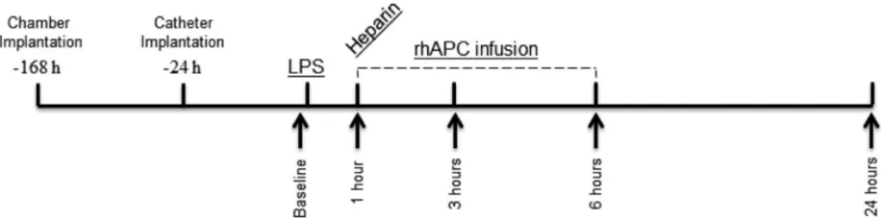
As shown in Figure 1, sequential measurements of hemodynamic and microcirculatory parameters were per-formed at five time points: at baseline and after 1, 3, 6, and 24 hours of LPS injection. Sequential evaluations of leuko-cyte-endothelial interactions were performed at two time points: at baseline and after six hours of LPS injection.
Survival analysis
After the intravital microscopy phase of the experiments, animals were returned to their individual cage in the vivariumwith free access to water and standard chow and monitored for survival three times per day for 7 days. After 7 days, surviving animals were euthanized by a lethal dose of pentobarbital.
Statistical analysis
Results are expressed as means^standard deviation of the mean (SD) for each group, unless otherwise noted. Microvascular diameters and FCD data are presented as ratios relative to baseline values. All hemodynamic and microcirculatory measurements were compared with the baseline of the same group and between groups at the same
time point. Statistical differences within and between groups were determined by Friedman and Kruskal-Wallis tests, followed, when appropriate, by Dunn’s multiple-compari-sons test forpost hocanalysis. Survival curves were obtained using the Kaplan-Meier procedure, and the Mantel-Cox log-rank test was applied for determination of significant differences between study groups. All statistical analyses were performed using GraphPad Prism 6.03 (GraphPad Software, La Jolla, CA, USA) and the significance level was set asp,0.05.
Ethical adherence:All procedures were approved by the Rio de Janeiro State University Animal Care and Use Committee (protocol number CEUA/060/2010) and are consistent with the United States National Institutes of Health Guide for the Care and Use of Laboratory Animals (National Research Council, 1996) and ethical rules specified by the Basel Declaration.
B RESULTS
The average body weight of the hamsters did not differ significantly between study groups. All animals survived the intravital microscopy phase of the experimental protocol and underwent survival analysis.
Hemodynamic alterations
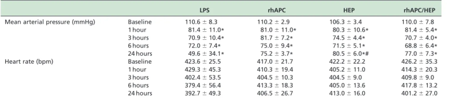
Hemodynamic parameters are presented in Table 1. MAP and HR basal values were not significantly different among experimental groups and were comparable to control values for reported data on healthy hamsters.14Systemic adminis-tration of LPS elicited statistically similar reductions of MAP levels in all study groups (Table 1; 1 hour). MAP was comparable among study groups until 24 hours. At this time point, MAP was significantly higher in the HEP group than in the LPS group. HR was comparable among study groups at each time point (Table 1).
Microcirculatory parameters
.Arteriolar and venular diameters: At baseline, there were no significant differences in arteriolar and venular mean internal diameters between study groups. Arteriolar mean internal diameter was significantly reduced by endotoxemia in LPS and rhAPC groups at 6 and 24 hours when compared to baseline. These groups had compar-able arteriolar diameters at each time point (Figure 2). Arteriolar diameter was wider in heparin treated groups
(HEP and rhAPC/HEP groups) than in the other groups; at 3, 6, and 24 hours, a significant difference was observed between the HEP group and both LPS and rhAPC groups (Figure 2). In terms of venular mean internal diameter, LPS caused slight venodilation, evident at 3 hours (p ,0.05vs. baseline). Venular diameters tended to be wider in HEP group and smaller in rhAPC group. These differences faded away in such a manner that at 24 hours there were no significant differences between the venular diameters of the study groups (Figure 2).
.Capillary perfusion (FCD): At baseline, FCD did not
significantly differ between study groups. LPS adminis-tration decreased FCD in all study groups. FCD was comparable among study groups until 3 hours. From this time point, FCD was significantly higher in the rhAPC/HEP group than in the LPS group (Figure 3).
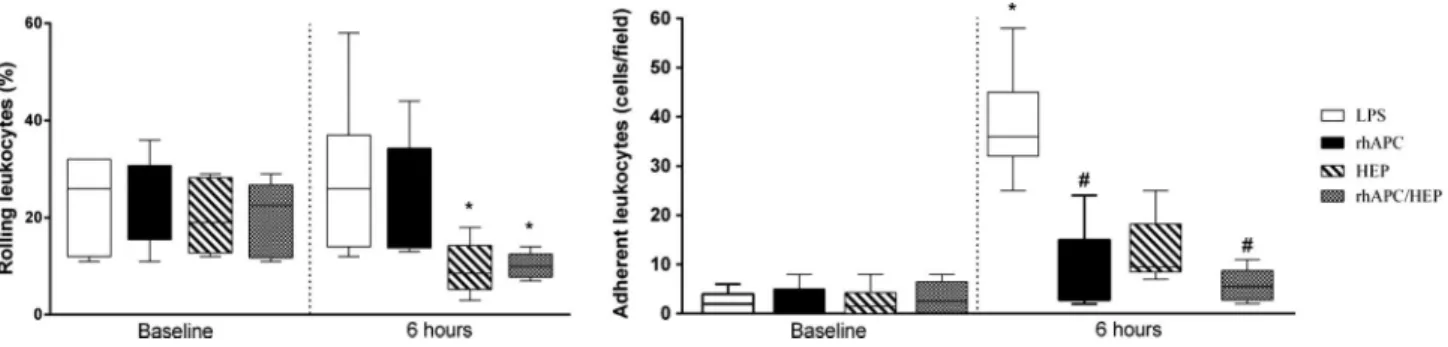
Evaluation of leukocyte-endothelial Interactions Treatment with heparin or heparin plus rhAPC reduced the percentage of rolling leukocytes compared with baseline (Figure 4). Endotoxemia significantly increased the number of adhered leukocytes in LPS group. At 6 hours, a smaller number of adherent leukocytes was observed in rhAPC, HEP, and rhAPC/HEP groups compared with the LPS group, although a significant difference was only observed
between LPS and both rhAPC and rhAPC/HEP groups (Figure 4).
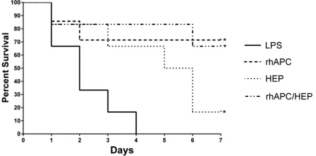
Seven-day survival
Median survival time after experiments was 2.2, 5.4, 4.7, and 5.8 days for LPS, rhAPC, HEP, and rhAPC/HEP groups, respectively (p,0.05 for LPSvs.all other groups; Figure 5).
B DISCUSSION
In few pathological states are the connections between coagulation and inflammatory cascades so evident as in sepsis. In this syndrome, inflammatory cytokines, including tumor necrosis factor a, interleukin-1b, and interleukin-6, are capable of activating coagulation and inhibiting fibrinolysis, whereas the procoagulant thrombin is capable of stimulating multiple inflammatory pathways.15 Heparin may play a positive role in sepsis treatment due to its anticoagulant and anti-inflammatory properties. In fact, in the present study, these effects proved to be beneficial in a validated endotoxemia rodent model that allows in vivo evaluation of inflammation and perfusion dysfunction.
The heparin dose used in our study was similar to that capable of preventing ferric chloride-induced venous thrombosis in a murine model of deep vein thrombosis (unpublished data of our group). At this dose, heparin did
Table 1 -Mean arterial pressure and heart rate evolution during the experimental period
LPS rhAPC HEP rhAPC/HEP
Mean arterial pressure (mmHg) Baseline 110.6^8.3 110.2^2.9 106.3^3.4 110.0^7.8
1 hour 81.4^11.0* 81.0^11.0* 80.3^10.6* 81.4^5.4*
3 hours 70.9^10.4* 81.7^7.2* 74.5^4.4* 70.7^4.0*
6 hours 72.0^7.4* 75.0^9.4* 71.5^5.1* 68.8^6.4*
24 hours 49.6^34.1* 75.2^3.7* 80.5^6.0*# 77.0^7.3*
Heart rate (bpm) Baseline 423.6^25.5 417.0^21.7 422.2^22.2 426.2^35.3
1 hour 429.3^45.3 410.3^19.4 405.2^11.0 414.3^20.3
3 hours 402.4^53.5 404.5^10.3 404.5^9.0 409.8^9.0
6 hours 379.4^56.4 413.3^18.3 405.0^13.6 417.8^13.2
24 hours 392.7^49.3 406.5^26.7 413.0^16.0 401.2^27.0
Data are presented as means^SD for each group. LPS, endotoxemic animals (n¼7); rhAPC, endotoxemic and rhAPC treated animals (n¼7); HEP, endotoxemic and heparin treated animals (n¼7); rhAPC/HEP, endotoxemic and rhAPCþheparin treated animals (n¼7). * p,0.05 vs. group baseline; # p,0.05 vs. LPS group at the same time point.
Figure 2 - Evolution of arteriolar and venular mean internal diameters during the experimental period. Values are presented as ratios relative to baseline values. Sequential measurements were performed after one (1 hour), three (3 hours), six (6 hours), and twenty-four (24 hours) hours of LPS injection. LPS, endotoxemic animals (n¼7); rhAPC, endotoxemic and rhAPC treated animals (n¼7); HEP, endotoxemic and heparin treated animals (n¼7); rhAPC/HEP, endotoxemic and rhAPCþheparin treated animals (n¼7). *p,0.05vs.
not result in bleeding complications. Whereas rhAPC also has anti-inflammatory and anticoagulant properties and its use has been extensively studied in sepsis syndrome, we considered this drug as a control treatment in our experiment. rhAPC was administered at a dose rate similar to that clinically given in the PROWESS study15to patients with severe sepsis (24 mg.kg21.h21). In a previous experimental study of Hoffmann et al.2, at this dose, rhAPC demonstrated beneficial microcirculatory and anti-inflammatory effects in endotoxemic hamsters without increasing bleeding complications. The endotoxin dose used in our study was adjusted to affect microcirculatory parameters without the induction of severe hypotension. During the initial hours of the experiment, this simulates the hyperdynamic phase of sepsis. After this initial period, non-treated animals (LPS group) were considered to be in endotoxemic shock. Because LPS-induced hypotension is dependent on activation of the inflammatory cascade, it is understandable that substances with anti-inflammatory
properties (heparin and rhAPC) could hamper the hypoten-sive effect of LPS. Some of the anti-inflammatory effects of heparin have already been described: TNF-a inhibition, protection against oxygen free radicals, and blockade of complement activity.9,10
Although heparin is largely used for the prevention of blood clotting, it produces additional vascular effects independent of its anticoagulant activity. Tasatargil et al.8 have shown that heparin causes concentration-dependent vasodilatation in human arteries, and that this action seems to be linked to endothelium-dependent mechanisms, includ-ing changes in the generation of nitric oxide and endothelium derived hyperpolarizing factor. Furthermore, Tangphao et al.16 have shown that heparin is an endothelium-dependent venodilator. The mechanism of heparin-induced venous relaxation involves increased availability of nitric oxide, possibly related to local release of histamine. Sternbergh et al.17 have demonstrated that heparin has dose-dependent protective effects on vascular endothelial
Figure 4 - Sequential evaluation of leukocyte-endothelial interactions. The number of rolling leukocytes is expressed as a percentage of the number of passing leukocytes. The number of adherent leukocytes is expressed as the number of adherent cells per field. Sequential measurements were performed at baseline and after six hours of LPS injection. LPS, endotoxemic animals (n¼7); rhAPC, endotoxemic and rhAPC treated animals (n¼7); HEP, endotoxemic and heparin treated animals (n¼7); rhAPC/HEP, endotoxemic and rhAPCþheparin treated animals (n¼7). *p,0.05vs.group baseline; #p,0.05vs.LPS group at the same time point.
Figure 3 - Functional capillary density evolution during the experimental period. Values are presented as ratios relative to baseline values. Sequential measurements were performed after one (1 hour), three (3 hours), six (6 hours), and twenty-four (24 hours) hours of LPS injection. LPS, endotoxemic animals (n¼7); rhAPC, endotoxemic and rhAPC treated animals (n¼7); HEP, endotoxemic and heparin treated animals (n¼7); rhAPC/HEP, endotoxemic and rhAPCþheparin treated animals (n¼7). *p,0.05vs.group baseline; #p,0.05
cell function distinct from its anticoagulant, antiplatelet, or anticomplement activity. Thus, heparin can preserve endo-thelial-dependent vasodilatation in states of oxidative stress.17An inhibitory effect on biosynthesis and release of endothelin-1, a potent endogenous vasoconstrictor, has also been shown.18 In our study, we have observed that the vasodilator effect of heparin also occurs in venules and arterioles, even during endotoxemia. Surprisingly, rhAPC administration prevented LPS-induced venular vasodilata-tion but could not prevent endotoxin-mediated decrease of arteriolar mean internal diameter. Apparently, apart from an anti-inflammatory action, an endothelium-mediated vasodi-lator effect is also necessary to maintain arteriolar diameters during endotoxemia.
Because endotoxemia is a known up-regulator of leukocyte-endothelial interactions and because increased leukocyte-endothelium interactions are related to micro-circulatory impairment5, we have performed intravital fluorescence microscopy to study leukocyte kinetics. Leukocyte migration involves a step-wise process: first leukocytes roll along the vascular endothelium, then they firmly adhere and finally emigrate from the microcirculation to inflamed tissues. Thus, the quantitation of rolling and adherent leukocytes can reliably reflect the extent of the inflammatory response.
To understand the results related to the number of rolling leukocytes, it is important to know that as early as 30 minutes after LPS administration there is a massive decrease in venular leukocyte rolling fraction (about 12% of non-adherent leukocytes are rolling); baseline conditions are reached again 3 hours after LPS (20 –30% of non-adherent leukocytes are rolling) and a profound increase in the number of rolling leukocytes occurs 8 hours after LPS administration (about 60% of the non-adherent leukocytes are rolling).14When leukocyte-endothelial interactions were assessed (6 hours after LPS administration) there was sufficient time for recovery of the basal number of rolling
leukocytes as observed in LPS group. The maintenance of a small fraction of rolling leukocytes in HEP and rhAPC/HEP groups may be related to an anti-inflammatory action of heparin. In our study, rhAPC was not capable of preventing the induction of leukocyte rolling. An opposite behavior was observed regarding leukocyte adhesion. This result might be related to different adhesion molecules involved; while rolling involves the selectin family of adhesion molecules, firm adhesion involves b2-integrins (expressed in leuko-cytes) and the endothelial intercellular adhesion molecule-1 (ICAM-1).19,20So, the discrepancy between our observations could be attributed to complex cellular interactions found in in vivostudies. Heparin and rhAPC may have different and additional actions on these adhesion molecules. The improved results found in rhAPC/HEP group regarding both leukocyte rolling and adhesion favor this hypothesis.
Considering the temporal evolution of functional capillary perfusion, our study showed that administration of rhAPC plus heparin was associated with significant attenuation of capillary perfusion deficits induced by LPS administration. Several factors are related to the microcirculatory impairment observed after endotoxemia induction, such as systemic hypotension, vasoconstriction, stiffness of RBC, increased leukocyte-endothelium interactions, and platelet/fibrin clot formation.5 Because differences in capillary perfusion between groups cannot be entirely explained by changes in arteriolar mean internal diameter or by macro-hemodynamic changes, we can speculate that anti-inflammatory and antithrombotic activities of both heparin and rhAPC may have been crucial to capillary perfusion differences observed between groups seeing that capillary obstruction by micro-thrombi and an increased presence of rolling and adherent leukocytes in venules may hamper adequate capillary flow.2,21,22,23 Thus, reduction of microvascular platelet aggregation and leukocyte plug formation facilitates capil-lary blood flow, improving tissue perfusion and reducing organ failure.3
Figure 5 - Kaplan-Meier survival curves. LPS, endotoxemic animals (n¼7); rhAPC, endotoxemic and rhAPC treated animals (n¼7); HEP, endotoxemic and heparin treated animals (n¼7); rhAPC/HEP, endotoxemic and rhAPCþheparin treated animals (n¼7). *p,0.05vs.
Heparin or/and rhAPC treated animals had increased survival compared with animals in LPS group. This is in consonance with prior studies regarding human sepsis.11,15 Unfortunately, recent studies concerning the use of antithrombotic agents in septic patients ended in contro-versial and/or inconclusive results: although benefits were not consistently observed, these agents were associated with significant increase in the risk of bleeding.7,24The interest in antithrombotic drugs decreased after the publication of the PROWESS-SHOCK trial in which rhAPC failed to show survival benefit for patients with severe sepsis or septic shock.24 This resulted in the announcement of rhAPC’s manufacturer of a worldwide voluntary market withdrawal, leaving many important unanswered questions about the role of antithrombotics in sepsis treatment. The expressive benefit in mortality observed in our study may be related to the LPS model: endotoxemic animals might be a population with high risk of death or, in other words, those animals that would most benefit from antithrombotic drug therapy.25 Another hypothesis is that LPS-induced endotoxemia is an inflammation-dependent state in which anti-inflammatory drugs have important benefits on survival rates. Given the number of mechanisms involved, a similar response is not found in human sepsis.26Finally, and differently from what happened in human studies, no hemorrhagic events were observed in our study, which may have decisively contributed to our results.
Taken together, our results may suggest that heparin has beneficial microcirculatory and anti-inflammatory effects in endotoxemic hamsters. These benefits were potentiated when heparin was administered in conjunction with rhAPC. It is possible that each of these drugs has different, but additive anti-inflammatory effects, making treatment with both drugs more effective in controlling the inflammatory response. Our results may indicate a path to be followed in development of new drugs for sepsis treatment: a drug or a combination of drugs with potent anti-inflammatory effects and capable of preserving endothelial-dependent vasodila-tation could attenuate sepsis-related capillary perfusion deficits, possibly improving patient outcome. Drugs with pleiotropic effects such as antithrombotics have greater therapeutic potential than compounds directed against a single target.9Clinical use of these drugs for sepsis treatment has been held back by fear of bleeding, but the development of non-anticoagulant drugs or of agents with mild anti-coagulation properties could mitigate this concern.
We are aware that our study has some limitations. Initially, we recognize that the study of the skin and subcutaneous muscle microcirculation may not be representative of microcirculatory changes in splanchnic organs. Given the crucial importance of the splanchnic perfusion in the pathophysiology of sepsis, this could be considered a limitation of the skinfold window chamber model. However, the first reactions after endotoxin administration seem to be comparable in different organs.14 In the second place, although we know that fluid therapy is recommended for the early management of severe sepsis and septic shock, in our study, animals were not fluid resuscitated because this study was designed to evaluate effects of heparin on microcircula-tion independently of fluid therapy. Finally, the use of recombinant human proteins (rhAPC) in hamsters may be limited by their even shorter half-lives.2However, because we found systemic effects long after drug discontinuation, it could be speculated that rhAPC worked well in this study.
B CONCLUSIONS
In our study, heparin was effective in attenuating the inflammatory response, as depicted by decreased leukocyte rolling, reducing LPS-induced arteriolar vasoconstriction at the hamster skinfold window microcirculation, and improved survival. These benefits were potentiated when heparin was administered in conjunction with rhAPC, because treatment with both drugs was associated with marked anti-inflammatory effects and significant attenu-ation of capillary perfusion deficits induced by LPS administration. Further studies in experimental models closer to human sepsis are required to confirm these results.
B ACKNOWLEDGEMENTS
The authors wish to thank Dr. Maria das Gracas Coelho de Souza for her technical assistance, and Mr. Cla´udio Natalino Ribeiro and Mr. Paulo Jose´ Ferreira Lopes for their help with animal care.
This study was supported by grants from CNPq (National Council for Scientific and Technological Development, Brasilia, Brazil) and FAPERJ (State of Rio de Janeiro Agency for Research Support, Rio de Janeiro, Brazil).
Author Contributions: Miranda ML analyzed the data and wrote the manuscript. Prota LFM contributed with data analysis and manuscript drafting. Silva MJB designed the study and analyzed the data. Sicuro FL analyzed the data. Furtado ES performed the experiments. Santos AOMT designed the study and performed the experiments. Bouskela E designed and supervised the study and critically revised the manuscript.
Conflicts:Authors report no conflicts of interest.
B RESUMO
OBJETIVO: Ale´m de suas propriedades anticoagulantes, a heparina apresenta efeitos vasodilatadores e anti-inflamato´rios que podem ajudar na reversa˜o das alteraco˜es se´pticas da microcirculaca˜o. Este artigo investiga os efeitos da heparina sobre alteraco˜es da microcirculaca˜o relacionadas com endotoxemia e compara-os com os observados com o uso de proteı´na C humana recombinante ativada.
ME´ TODOS:Apo´s os procedimentos de implantaca˜o da caˆmara de dobras cutaˆneas e induca˜o de endotoxemia por administraca˜o intravenosa de lipopolissacarı´deo da Escherichia coli (2 mg.kg21), hamsters sı´rios dourados
machos foram tratados com heparina na˜o fracionada intravenosa (0,2 mg.kg21). A microscopia intravital de preparaco˜es de caˆmara de dobras cutaˆneas permitiu a ana´lise quantitativa de varia´veis microvasculares e venulares, bem como de rolamento e adereˆncia de leuco´citos. Paraˆmetros micro-hemodinaˆmicos tambe´m foram analisados. Hamsters endotoxeˆmicos tratados com proteı´na C humana recombinante ativada e animais na˜o tratados serviram como controles.
RESULTADOS: A heparina diminuiu o rolamento de leuco´citos e a vasoconstricca˜o arteriolar induzida por lipopolissacarı´deo e produziu um aumento de sobrevida em comparaca˜o com animais na˜o tratados, enquanto proteı´na C humana recombinante ativada diminuiu a adesa˜o de leuco´citos. A administraca˜o de heparina mais proteı´na C humana recombinante ativada resultou numa significativa atenuaca˜o dos de´ficits de perfusa˜o capilar induzida por lipopolissacarı´deo.
CONCLUSO˜ ES:A heparina produz efeitos protetores sobre a microcircu-laca˜o dos animais endotoxeˆmicos. Estas vantagens foram potenciados quando a heparina foi administrada em combinaca˜o com proteı´na C humana recombinante ativada.
B REFERENCES
1. Dempfle CE. Coagulopathy of sepsis. Thromb Haemost. 2004; 91(2):213-24.
2. Hoffmann JN, Vollmar B, Laschke MW, Inthorn D, Fertmann J, Schildberg FW, et al., Microhemodynamic and cellular mechanisms of activated protein C action during endotoxemia. Crit Care Med. 2004;32(4):1011-7. 3. Mariscalco MM. Unlocking (perhaps unblocking) the microcirculation in
4. Trzeciak S, Cinel I, PhillipDellinger R, Shapiro NI, Arnold RC, Parrillo JE, et al. Resuscitating the microcirculation in sepsis: the central role of nitric oxide, emerging concepts for novel therapies, and challenges for clinical trials. Acad Emerg Med. 2008;15(5):399-413.
5. Santos AO, Furtado ES, Villela NR, Bouskela E. Microcirculatory effects of fluid therapy and dopamine, associated or not to fluid therapy, in endotoxemic hamsters. Clin Hemorheol Microcirc. 2011;47(1):1-13. 6. Koestenberger M, Cvirn G, Gallistl S, Baier K, Leschnik B, Muntean W.
Drotrecogin alfa (activated, Xigris) in combination with heparin or melagatran: an in vitro investigation. J Thromb Thrombolysis. 2004; 18(1):5-10.
7. Minneci PC, Deans KJ, Cui X, Banks SM, Natanson C, Eichacker PQ. Antithrombotic therapies for sepsis: a need for more studies. Crit Care Med. 2006;34(2):538-41.
8. Tasatargil A, Golbasi I, Sadan G, Karasu E. Unfractioned heparin produces vasodilatory action on human internal mammary artery by endothelium-dependent mechanisms. J Cardiovasc Pharmacol. 2005; 45(2):114-9.
9. Young E. The anti-inflammatory effects of heparin and related compounds. Thromb Res. 2008;122(6):743-52.
10. Dobosz M, Mionskowska L, Hac S, Dobrowolski S, Dymecki D, Wajda Z. Heparin improves organ microcirculatory disturbances in caerulein-induced acute pancreatitis in rats. World J Gastroenterol. 2004; 10(17):2553-6.
11. Davidson BL, Geerts WH, Lensing AWA. Low-dose heparin for severe sepsis. N Engl J Med. 2002;347(13):1036-7.
12. Endrich B, Asaishi K, Go¨tz A, Messmer K. Technical report – a new chamber technique for microvascular studies in unanesthetized hamsters. Res Exp Med. 1980;177(2):125-34.
13. Klyscz T, Ju¨nger M, Jung F, Zeintl H. Cap image – a new kind of computer-assisted video image analysis system for dynamic capillary microscopy. Biomed Tech. 1997;42(6):168-75.
14. Hoffmann JN, Vollmar B, Inthorn D, Schildberg FW, Menger MD. A chronic model for intravital microscopic study of microcirculatory disorders and leukocyte/endothelial cell interaction during normo-tensive endotoxemia. Shock. 1999;12(5):355-64.
15. Bernard GR, Vincent JL, Laterre PF, LaRosa SP, Dhainaut JF, Lopez-Rodriguez A, et al. Efficacy and safety of recombinant human activated protein C for severe sepsis. N Engl J Med. 2001;344(10):699-709. 16. Tangphao O, Chalon S, Moreno HJ, Abiose AK, Blaschke TF, Hoffman BB. Heparin-induced vasodilation in human hand veins. Clin Pharmacol Ther. 1999;66(3):232-8.
17. Sternbergh WC, Makhoul RG, Adelman B. Heparin prevents postis-chemic endothelial cell dysfunction by a mechanism independent of its anticoagulant activity. J Vasc Surg. 1993;17(2):318-27.
18. Imai T, Hirata Y, Emori T, Marumo F. Heparin has an inhibitory effect on endothelin-1 synthesis and release by endothelial cells. Hypertension. 1993;21(3):353-8.
19. Chandra A, Enkhbaatar P, Nakano Y, Traber LD, Traber DL. Sepsis: emerging role of nitric oxide and selectins. Clinics. 2006;61(1):71-6. 20. Li X, Klintman D, Weitz-Schmidt G, Schramm R, Thorlacius H.
Lymphocyte function antigen-1 mediates leukocyte adhesion and subsequent liver damage in endotoxemic mice. Br J Pharmacol. 2004; 141(4):709-16.
21. Yeh YC, Sun WZ, Ko WJ, Chan WS, Fan SZ, Tsai JC, et al., Dexmedetomidine prevents alterations of intestinal microcirculation that are induced by surgical stress and pain in a novel rat model. Anesth Analg. 2012;115(1):46-53.
22. Turek Z, Sykora R, Matejovic M, Cerny V. Anesthesia and the microcirculation. Semin Cardiothorac Vasc Anesth. 2009;13(4):249-58. 23. Cepinskas G, Wilson JX. Inflammatory response in microvascular
endothelium in sepsis: role of oxidants. J Clin Biochem Nutr. 2008; 42(3):175-84.
24. Ranieri VM, Thompson BT, Barie PS, Dhainaut JF, Douglas IS, Finfer S, et al., Drotrecogin alfa (activated) in adults with septic shock. N Engl J Med. 2012;366(22):2055-64.
25. Abraham E, Laterre PF, Garg R, Levy H, Talwar D, Trzaskoma BL, et al. Drotrecogin alfa (activated) for adults with severe sepsis and a low risk of death. N Engl J Med. 2005;353(13):1332-41.