379
Araujo Júnior E et al. Beckwith-Wiedemann syndrome: 2D and 3DUS
Radiol Bras. 2013 Nov/Dez;46(6):379–381
Prenatal diagnosis of Beckwith-Wiedemann syndrome
by two- and three-dimensional ultrasonography
*
Diagnóstico pré-natal da síndrome de Beckwith-Wiedemann pela ultrassonografia bidimensional e tridimensional
Edward Araujo Júnior1, Christiane Simioni2, Luciano Marcondes Machado Nardozza1, Antonio Fernandes Moron3
Beckwith-Wiedemann syndrome is a genetic syndrome characterized by macroglossia, omphalocele, fetal gigantism and neonatal hypoglycemia. The authors report a case of Beckwith-Wiedemann syndrome diagnosed in a 32-year-old primigravida in whom two-dimensional ultrasonography revealed the presence of abdominal wall cyst, macroglossia and polycystic kidneys. Three-dimensional ultrasonography in rendering mode was of great importance to confirm the previous two-dimensional ultrasonography findings.
Keywords: Prenatal diagnosis; Beckwith-Wiedemann syndrome; Two-dimensional ultrasonography; Three-dimensional ultrasonography.
A síndrome de Beckwith-Wiedemann é uma síndrome genética caracterizada por macroglossia, onfalocele, gigantismo fetal e hipoglicemia neonatal. Apresentamos um caso de síndrome de Beckwith-Wiedemann em uma primigesta de 32 anos, em que a ultrassonografia bidimensional evidenciou presença de cisto de parede abdominal, macroglossia e rins policísticos. A ultrassonografia tridimensional modo de renderização foi de grande importância ao confirmar os achados da ultrassonografia bidimensional.
Unitermos: Diagnóstico pré-natal; Síndrome de Beckwith-Wiedemann; Ultrassonografia bidimensional; Ultrassonogra-fia tridimensional.
Abstract
Resumo
* Study developed in the Department of Obstetrics at Escola Paulista de Medicina – Universidade Federal de São Paulo (EPM-Unifesp), São Paulo, SP, Brazil.
1. Private Docents, Associate Professors of Fetal Medicine, Department of Obstetrics, Escola Paulista de Medicina – Universi-dade Federal de São Paulo (EPM-Unifesp), São Paulo, SP, Brazil.
2. Master, Volunteer Physician, Division of Fetal Medicine, Department of Obstetrics at Escola Paulista de Medicina – Uni-versidade Federal de São Paulo (EPM-Unifesp), São Paulo, SP, Brazil.
3. Private Docent, Full Professor, Department of Obstetrics, Escola Paulista de Medicina – Universidade Federal de São Paulo (EPM-Unifesp), São Paulo, SP, Brazil.
Mailing Address: Dr. Edward Araújo Júnior. Rua Carlos We-ber, 956, ap. 113, Visage, Vila Leopoldina. São Paulo, SP, Bra-zil, 05303-000. E-mail: [email protected]
Received March 15, 2013. Accepted after revision April 26, 2013.
Araujo Júnior E, Simioni C, Nardozza LMM, Moron AF. Prenatal diagnosis of Beckwith-Wiedemann syndrome by two- and three-dimen-sional ultrasonography. Radiol Bras. 2013 Nov/Dez;46(6):379–381.
0100-3984 © Colégio Brasileiro de Radiologia e Diagnóstico por Imagem CASE REPORT
agnosis. Three-dimensional ultrasonogra-phy (3DUS) in rendering mode allows a better assessment of the fetal surface, con-tributing to a more comprehensive under-standing of the malformation by the par-ents. Only one report of prenatal diagnosis of BWS by 3DUS is found in the litera-ture(3).
The authors present a case of BWS di-agnosed by 2DUS at the 28th gestational week, and emphasize the main findings of such disorder at 3DUS in rendering mode.
CASE REPORT
A 32-year-old, white, primigesta was referred to the Department of Obstetrics at Universidade Federal de São Paulo (Unifesp), São Paulo, Brazil, with a diag-nosis of renal dysplasia and polyhydram-nios. The patient presented gestational age of 28 weeks and five days, based on the date of her last menstruation, and denied previous history of familial genetic dis-eases.
ivity and gene deregulation in the chromo-somal region 11p15.5(2).
Generally, the diagnosis of this condi-tion is made in the postnatal period by means of findings of macroglossia, ompha-locele, gigantism, visceromegaly, advanced bone age, renal dysplasia, facial nevi and ear creases(1). The prenatal diagnosis of such condition is of paramount importance to the perinatal follow-up as well as for de-termining the way of delivery, pediatric care due to neonatal hypoglycemia, supe-rior airways obstruction, congestive heart failure, risk for malignancy and genetic tests for family members. The prenatal di-agnosis of BWS by means of two-dimen-sional ultrasonography (2DUS) may be based on two major criteria or on one ma-jor and one minor criterion. Mama-jor criteria include macroglossia, macrosomia (esti-mated weight > 90th percentile for the ges-tational age), and defective abdominal wall. Minor criteria include polyhydramnios, nephromegaly, renal dysplasia and adrenal cytomegaly confirmed by pathological
di-INTRODUCTION
express-380
Araujo Júnior E et al. Beckwith-Wiedemann syndrome: 2D and 3DUS
Radiol Bras. 2013 Nov/Dez;46(6):379–381
2DUS demonstrated biometry compat-ible with 29 weeks, polyhydramnios (am-niotic fluid index 255 mm), estimated fetal weight 2,661 g (90th percentile = 1,416 g), increased placental thickness, multicystic left kidney. Abdominal cystic mass measur-ing 5.4 × 5.7 × 5.9 cm and macroglossia (Figure 1). Based on such findings, the au-thors raised the hypothesis of BWS. Echocardiography revealed a normal heart, while karyotyping by amniocentesis dem-onstrated normal male karyotype (46,XY). In order to supplement the 2D diagnosis. 3DUS was performed with a Voluson 730 Expert equipment (General Electric Health-care; Zipf, Austria) coupled to a multifre-quency convex transducer (RAB 4-8L). 3DUS in the rendering mode clearly dem-onstrated the remarkable fetal abdominal
distension and macroglossia (Figure 2). The patient remained under prenatal follow-up in the authors’ institution and presented preterm labor at 30 weeks and one day, which was inhibited with intravenous terbutaline. During her hospital stay, fetal hemodynamic centralization was identified at Doppler US. A single corticoid cycle (betamethasone, 12 mg/day for two days) was performed and the patient underwent Cesarean section three days afterwards and gave birth to a male child weighting 2,450 g (above the 97th percentile for the gesta-tional age). Immediately after birth, the neonate presented severe hypoglycemia, and progressed to respiratory insufficiency and death after three hours of life. Anato-mopathological study confirmed the diag-nosis of BWS.
DISCUSSION
BWS was first described by Beckwith in 1963 and by Wiedemann in 1964(4). Ac-cording to Eliott et al.(5) who followed-up 74 pediatric patients with BWS, the most frequent findings were the following: mac-roglossia (97%), gigantism (88%), abdomi-nal wall defect (80%), ear creases (76%), hypoglycemia (63%), facial nevi (62%), multicystic dysplasia or nephromegaly (59%) and hemihypertrophy (24%). Hemi-hypertrophy is the only sign significantly associated with the development of embry-onic tumors, particularly Wilms’ tumor.
In the present case, the authors observed the presence of macrosomia and macroglo-ssia, which represent the two major signs for the diagnosis of BWS, besides polyhy-dramnios and renal dysplasia (minor signs). O’Connor et al.(6) have described a case of BWS diagnosed by 2DUS at the 22nd week, in association with omphalocele and placentomegaly with cystic areas. In the present case, the authors have also ob-served placentomegaly, but without cystic areas. Prenatal and neonatal ultrasonogra-phy has been utilized for many years in the assessment of fetal malformations(7–9). 3DUS in rendering mode has clearly dem-onstrated the remarkable fetal abdominal distension and macroglossia. For many years, 3DUS in rendering mode has been utilized in the assessment of fetal malfor-mations, allowing a better understanding of the parents in relation to the fetal anoma-lies and aiding in the definition of the ap-proach to be adopted during the prenatal period(10,11). In spite of not being essential for the diagnosis of BWS, 3DUS was ex-tremely important for a better understand-ing on the disease by the parents and by the involved multidisciplinary team. In the lit-erature, there is only one report about pre-natal diagnosis of BWS by means of 3DUS, in a study developed by Eckmann-Scholz et al.(3). Such authors have described a case that was also diagnosed at the 28th. gesta-tional week, where, like in the present case, 3DUS has clearly demonstrated the pres-ence of macroglossia.
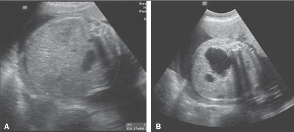
In summary, the authors believe that 3DUS in association with 2DUS can con-tribute to a better prenatal assessment of some fetal malformations such as BWS, Figure 1. 2DUS findings in a fetus with BWS at the 28th gestational week. A: Abdominal circumference
compatible with a gestational age of 37 weeks and six days (> the 98th percentile). B: Cystic dysplasia in the right kidney.
A B
Figure 2. 3DUS rendering mode findings in a fetus with BWS at the 28th gestational week. A: Remark-able fetal abdominal distension. B: Macroglossia.
381
Araujo Júnior E et al. Beckwith-Wiedemann syndrome: 2D and 3DUS
Radiol Bras. 2013 Nov/Dez;46(6):379–381
allowing a better understanding of this dis-ease by the parents as well as by the multi-disciplinary clinical team.
REFERENCES
1. Harker CP, Winter T 3rd, Mack L. Prenatal diag-nosis of Beckwith-Wiedemann syndrome. AJR Am J Roentgenol. 1997;168:520–2.
2. Tilghman SM. The sins of the fathers and moth-ers: genomic imprinting in mammalian develop-ment. Cell. 1999;96:185–93.
3. Eckmann-Scholz C, Jonat W. 3-D ultrasound im-aging of a prenatally diagnosed Beckwith-Wiede-mann syndrome. Arch Gynecol Obstet. 2011;284: 1051–2.
4. Wiedemann HR. Complexe malformatif familial avec hernie ombilicale et macroglossie: un “syn-drome nouveau”? J Genet Hum. 1964;13:223–32. 5. Elliott M, Bayly R, Cole T, et al. Clinical features and natural history of Beckwith-Wiedemann syn-drome: presentation of 74 new cases. Clin Genet. 1994;46:168–74.
6. O’Connor C, Levine D. Case 49: Beckwith-Wiede-mann syndrome. Radiology. 2002;224:375–8. 7. Baldisserotto M, Fiori H, Fiori R, et al. Neonato
com ascite urinária e ruptura de cálice renal secun-dárias a válvula de uretra posterior: diagnóstico ultrassonográfico. Radiol Bras. 2011;44:68–70. 8. Barros LM, Fernandes DA, Melo EV, et al.
Mal-formações do sistema nervoso central e
malfor-mações associadas diagnosticadas pela ultrasso-nografia obstétrica. Radiol Bras. 2012;45:309– 14.
9. Rios LT, Araújo Júnior E, Nardozza LM, et al. Rânula congênita: diagnóstico ultrassonográfico antenatal. Radiol Bras. 2012;45:300–1. 10. Araujo Júnior E, Kawanami TE, Nardozza LM,
et al. Prenatal diagnosis of bilateral anoph-thalmia by 3D “reverse face” view ultrasound and magnetic resonance imaging. Taiwan J Obstet Gynecol. 2012;51:616–9.