ABSTRACT
Even though differentiated thyroid carcinoma is a slow growing and usu-ally curable disease, recurrence occurs in 20–40% and cellular dedifferen-tiation in up to 5% of cases. Conventional chemotherapy and radiotherapy have just a modest effect on advanced thyroid cancer. Therefore, dediffer-entiated thyroid cancer represents a therapeutic dilemma and a critical area of research. Targeted therapy, a new generation of anticancer treat-ment, is planned to interfere with a specific molecular target, typically a protein that is believed to have a critical role in tumor growth or progres-sion. Since many of the tumor-initiation events have already been identi-fied in thyroid carcinogenesis, targeted therapy is a promising therapeutic tool for advanced thyroid cancer. Several new drugs are currently being tested in in vitro and in vivostudies and some of them are already being used in clinical trials, like small molecule tyrosine kinase inhibitors. In this review, we discuss the bases of targeted therapies, the principal drugs already tested and also options of redifferentiation therapy for thyroid car-cinoma. (Arq Bras Endocrinol Metab 2007;51/4:612-624)
Keywords:Thyroid cancer; Redifferentiation therapy; Targeted therapy; Tyrosine kinase inhibitors
RESUMO
Novas Perspectivas no Tratamento do Carcinoma Diferenciado da Tireóide.
Apesar de o carcinoma diferenciado da tireóide ser considerado uma doença de curso indolente e geralmente curável, recorrência tumoral ocorre em aproximadamente 20 a 40% e desdiferenciação celular, em até 5% dos casos. A quimioterapia convencional e a radioterapia apresentam apenas um modesto efeito sobre o câncer de tireóide avançado. Dessa forma, o carcinoma da tireóide desdiferenciado representa um dilema terapêutico e uma importante área de pesquisa. A terapia direcionada, uma nova geração de tratamento para o câncer, tem como objetivo interferir com um alvo molecular específico, geralmente uma proteína considerada fundamental para o crescimento e progressão tumoral. Como muitos eventos iniciadores do processo de carcinogênese tireoideana já foram identificados, a terapia direcionada representa uma promissora opção terapêutica para o carcinoma da tireóide avançado. Várias drogas novas estão em estudos in vitro e in vivo e algumas já estão sendo testadas em estudos clínicos, como as pequenas moléculas inibidoras de tirosina cinase. Nesta revisão, as bases moleculares da terapia direcionada, as principais drogas utilizadas e as opções terapêuticas de rediferenciação do carcinoma da tireóide serão discutidas. (Arq Bras Endocrinol Metab 2007;51/4:612-624)
Descritores: Câncer da tireóide; Rediferenciação tumoral; Terapia dire-cionada; Inibidor de tirosina cinase
perspectiva
SABRINAMENDESCOELHO DENISEPIRES DECARVALHO MÁRIO VAISMAN
Serviço de Endocrinologia do Hospital Universitário Clementino Fraga Filho (SMC & MV) e Labo-ratório de Fisiologia Endócrina da Universidade Federal do Rio de Janeiro (DPC), Rio de Janeiro, RJ.
T
HYROID CARCINOMA IS the most prevalentendocrine malignancy, and accounts for just 1% of all human cancers. Approximately 90% of non-medullary thyroid malignancies are well-differentiated thyroid carcinomas, which are classified as papillary or follicular based on histopathological criteria.
Even though differentiated thyroid carcinomas (DTC) are slow growing and usually curable by the combination of surgery, radioiodine ablation and thyroid stimulating hormone (TSH) suppressive therapy, recur-rence occurs in 20–40% of patients (1,2). During tumor progression, cellular dedifferentiation occurs in up to 5% of cases and is usually accompanied by more aggressive growth, metastatic spread and loss of iodide uptake abil-ity, making the tumor resistant to the traditional thera-peutic modalities. Conventional chemotherapy and radiotherapy have modest, if any effect on advanced thy-roid cancer (3), which is responsible for the vast majori-ty of deaths attributed to thyroid cancer. Therefore, advanced thyroid cancer represents a therapeutic dilem-ma and is considered a critical area of research.
Several new drugs are currently being tested in
in vitro and in vivo studies and some of them are
already being used in clinical trials (4,5). In this review, we discuss the bases of targeted therapies for differenti-ated thyroid carcinoma, which correspond to drugs that act on molecules involved in neoplastic transfor-mation and tumor progression (table 1). Other types of drugs are being tested for their ability to impair further loss of cell differentiation during tumor progression and reprogram cell differentiation. Special interest on redifferentiation therapy for thyroid cancers has been given, since the re-induction of functional NIS protein expression allows the use of radioiodine, a well-known efficient therapy for thyroid cancer.
OVERVIEW OF GENETIC ALTERATIONS IN DIFFERENTIATED THYROID CANCER
Like other cancers, thyroid carcinomas are character-ized by genetic alterations that result in deregulated cell proliferation and death, together with tissue inva-sion ability. The different morphologic subtypes of thyroid cancer correlate with specific genetic alter-ations. However, mutations in different genes can be involved in the development of a specific histopatho-logical tumor subtype. As a result, the microscopic phenotype of a tumor does not necessarily correspond to a specific genetic alteration.
In papillary thyroid carcinoma, the most preva-lent thyroid cancer subtype, mutations in two protein
kinases, RET and BRAF, are responsible for the vast majority of the cases (6,7). Also, activating point mutations in RAS occur in about 10% of cases, mainly in the follicular variant (8). Rarely, papillary carcinoma is associated with NTRK1 (neurotrophic tyrosine receptor kinase) rearrangement (9) or germline muta-tion in PTEN (phosphatase and tensin homologue deleted on chromosome ten), which is related to familial non-medullary thyroid carcinoma (10). PTEN has also a possible role in sporadic thyroid cancer, since a high frequency PTEN promoter hypermethyla-tion is detected in sporadic tumors (11).
Follicular thyroid cancer accounts for about 10% of all thyroid cancers and 10 to 50% (12-14) of cases are associated with mutations in RAS oncogene. Chromosomal imbalances are frequent in follicular neoplasm, with gains at chromosomes 7 and 5 and deletions at 3p. In some of these tumors, somatic rearrangement results in the fusion of PAX-8 to PPARγ1 (peroxisome proliferator-actived receptor gamma 1). The PAX-8 PPARγ1 fusion oncogene appears to act through a dominant negative effect on the transcriptional activity of the wild-type PPARγ1. The frequency of rearrangement in follicular thyroid carcinoma is estimated in 30% and is not found in clas-sic papillary carcinomas (15). However, this rearrange-ment has also been recently described in the follicular variant of papillary carcinoma (16). PAX-8 PPARγ1 fusion and RAS gene activation rarely occur in the same tumor.
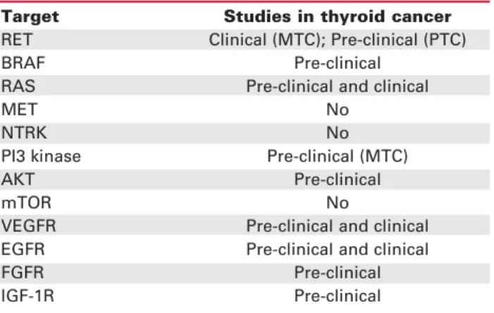
Table 1.Possible targets in thyroid cancer.
Target Studies in thyroid cancer
RET Clinical (MTC); Pre-clinical (PTC)
BRAF Pre-clinical
RAS Pre-clinical and clinical
MET No
NTRK No
PI3 kinase Pre-clinical (MTC)
AKT Pre-clinical
mTOR No
VEGFR Pre-clinical and clinical EGFR Pre-clinical and clinical
FGFR Pre-clinical
IGF-1R Pre-clinical
In addition to these well-established genetic causes of thyroid cancer, overexpression and activation of a variety of tyrosine kinase receptors, mutations in the p53 gene, DNA hypermethylation, leading to silencing of tumor suppressor genes, activation of PI3K (phosphatidylinositol-3-kinase), Wnt signalling and expression of angiogenic factors and receptors are found in thyroid cancer progression.
TARGETED THERAPY
Conventional chemotherapy acts through inducing toxic effects on dividing cells, resulting in damage of tumoral cells, but also of normal tissues. Therefore, side effects like myelosuppression, alopecia and gas-trointestinal symptoms are frequent. The optimum goal of anticancer therapy is the discovery of drugs that specifically kill malignant cells and cause no or lit-tle side effects. Targeted therapy refers to a new gen-eration of cancer drugs designed to interfere with a specific molecular target, typically a protein that is believed to have a critical role in tumor growth or pro-gression. Therefore, the objective of this therapy is to disrupt pathways that are inappropriately activated in cancer cells. This type of treatment has been applied specially to oncogenic protein kinases (17).
Antisense drugs, monoclonal antibodies and small-molecule drugs are examples of therapy intercept-ing important molecules in tumors and are beintercept-ing used in clinical trials. Targeted therapy is a promising thera-peutic tool for thyroid cancer because many of the tumor-initiation events have already been identified.
Antisense drugs are small synthetic single-stranded DNA sequences of 13-25 oligonucleotides complementary to a particular targeted mRNA. When hybridised to the corresponding mRNA, RNAse H recognises the complex and cleaves the mRNA, leav-ing the antisense drug intact. These substances also interfere with ribosomal assembly, blocking gene expression and inhibiting protein synthesis. Systemic treatment with antisense drugs is generally well toler-ated. Side effects are dose-dependent and include thrombocytopenia, hypotension, fever, complement activation, prolonged partial thromboplastin time, asthenia, and increased concentration of hepatic enzymes (18).
In 1998, the first antisense drug, Fomivirsen (Vitravene®), was approved by the US Food and Drugs Administration (FDA) for the treatment of retinitis by cytomegalovirus in patients with AIDS. The first generation of antisense drugs is
2’-deoxyri-bophosphorothioate antisense oligonucleotides. More recently, second-generation drugs, in which several nucleotides on each end are chemically modified to 2’-methoxyethoxyribophosphorothioate nucleotides, are in clinical trials. These new drugs are designed to enhance resistance to degradation by exonucleases and improve tissue pharmacokinetics, allowing less fre-quent dosing schedules. Second generation drugs are also more amenable to subcutaneous and oral delivery than first generation ones (19).
Small-molecules inhibitors and monoclonal antibodies are designed to intercept protein kinases in tumors. Most small-molecule kinase inhibitors obstruct the binding of ATP to the ATP pocket with-in the catalytic domawith-in and are known as ATP mimet-ics. Other compounds target regions outside the ATP-biding site of the enzyme, for example the substrate-biding domain. These drugs obstruct autophosphory-lation and signal transduction downstream from the targeted kinase. The most notable initial success of small molecule kinase inhibitor has been obtained with Imatinib (Gleevec®) in chronic myelogenous leukemia (CML), where BCR-ABL translocation results in expression of a fusion protein with constitutive activa-tion of Abl kinase. This event promotes unregulated proliferation of haematopoietic cell clone. Imatinib is a relative selective inhibitor of Abl kinase and induces remission in up to 90% of patients with CML in chron-ic phase and a durable response in a high proportion of patients (20). This drug is also effective in patients with gastrointestinal stromal tumors and dermatofi-brosarcoma protuberans, which are associated with activating mutation of tyrosine kinase receptors: C-KIT and platelet-derived growth factor receptor-β
(PDGFR-β). Recent clinical studies suggest that Ima-tinib might also be effective in glioblastoma multiform (21) and malignant gliomas (22) by inhibiting PDGFR tyrosine kinase.
THERAPEUTIC TARGETS IN DIFFERENTIATED THYROID CANCER
RET oncogene
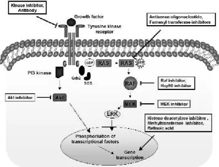
RET encodes a transmembrane tyrosine kinase recep-tor, member of a cell-surface complex that binds lig-ands of the glial-derived neutrophic factor (GDNF) family (24). These ligands bind RET in conjunction with co-receptors, trigging autophosphorylation and intracellular signalling with stimulation of the Ras/ERK and PI3 kinase/V-Akt cascades (figure 1).
In papillary thyroid cancer, the genetic hall-marks are chromosomal inversions or translocations that cause recombination of the intracellular kinase-encoding domain of RET with the 5’ end of heterolo-gous genes. The resulting chimeric sequence is called RET/PTC and is found in around 30% of cases and in over 60% of post-Chernobyl thyroid cancers (9). There are 12 rearrangements described and RET/PTC1 and 3 are the most prevalent variants.
RET/PTC recombination promotes RET expression in the cytoplasm of follicular cells, deletion of negative regulatory domains and a ligand-indepen-dent dimerization, resulting in a constitutive activation of RET. The finding that RET-PTC transgenic mice develop papillary thyroid cancer confirms that this oncogene can initiate thyroid carcinogenesis (25). In humans, it has also been demonstrated that
RET/PTC is an early event of thyroid tumorigenesis, since it is frequently found in microcarcinoma (26). Therefore, RET is a logical target for selective inhibi-tion in both medullary and papillary thyroid cancer that present RET oncogenic activation.
Several drugs have already been tested in pre-clinical studies (table 2). One of these compounds, the 4-anilinoquinazoline ZD6474 (Zactima), is being tested in patients with medullary thyroid cancer (27). This drug is an orally available small molecule inhibitor, originally developed as an anti-angiogenic agent that acts through the inhibition of the vascular endothelial growth factor receptor (VEGFR). Zactima also inhibits the epidermal growth factor receptor (EGFR), TIE-2 and RET kinase. Phase II assessment of Zactima is now in progress in a variety of tumor types in single and combination regimens.
In papillary thyroid cancer, it was demonstrated that RET/PTC3-transformed cells treated with Zacti-ma lose the proliferative autonomy and show morpho-logical reversion. Zactima also prevented the growth of human PTC cell lines that carry RET/PTC1 rearrangements and blocked anchorage-independent growth of RET/PTC3-transformed NIH3T3 fibrob-lasts and the formation of tumors after injection of NIH-RET/PTC3 cells into nude mice (28). More recently, it was observed that the administration of ZD6474 led to 90% reduction of cell number in
illary thyroid cancer cell lines (TPC1). Thus, targeting RET oncogene with Zactima might offer a potential treatment strategy for papillary carcinomas sustaining oncogenic activation of RET.
Other inhibitors of RET kinase have been test-ed in thyroid cancer cell lines, like pyrazolopyridines, PP1 and PP2. These compounds prevented the growth of two human papillary thyroid carcinoma cell lines that carry spontaneous RET/PTC1 rearrange-ments (29,30). Indolocarbazole derivatives, CEP-701 and CEP-751, inhibit RET in medullary thyroid
can-cer cells, but these compounds have not been tested in papillary thyroid cell lines (31).
BAY 43-9006 (Sorafenib), a multikinase inhibitor, was found to be a potent RET kinase inhibitor and was tested in medullary (TT) and papillary (TPC1) thyroid cancer cell lines (32). Recently, the inhibitory effect of SU11248, another multikinase inhibitor, was evaluated by quantification of RET/PTC autophosphorylation. This compound effectively inhibited the kinase activity of RET/PTC3 and represents another potential therapeutic tool for RET-positive thyroid tumors (33).
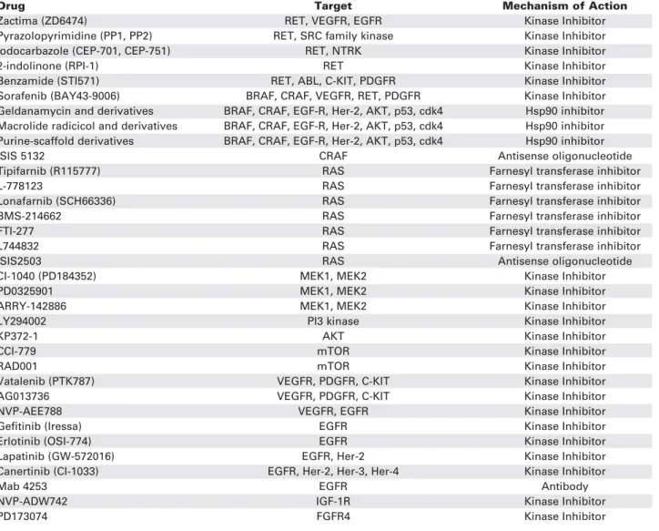
Table 2. Targeted therapy for thyroid cancer.
Drug Target Mechanism of Action
Zactima (ZD6474) RET, VEGFR, EGFR Kinase Inhibitor
Pyrazolopyrimidine (PP1, PP2) RET, SRC family kinase Kinase Inhibitor
Iodocarbazole (CEP-701, CEP-751) RET, NTRK Kinase Inhibitor
2-indolinone (RPI-1) RET Kinase Inhibitor
Benzamide (STI571) RET, ABL, C-KIT, PDGFR Kinase Inhibitor
Sorafenib (BAY43-9006) BRAF, CRAF, VEGFR, RET, PDGFR Kinase Inhibitor Geldanamycin and derivatives BRAF, CRAF, EGF-R, Her-2, AKT, p53, cdk4 Hsp90 inhibitor Macrolide radicicol and derivatives BRAF, CRAF, EGF-R, Her-2, AKT, p53, cdk4 Hsp90 inhibitor Purine-scaffold derivatives BRAF, CRAF, EGF-R, Her-2, AKT, p53, cdk4 Hsp90 inhibitor
ISIS 5132 CRAF Antisense oligonucleotide
Tipifarnib (R115777) RAS Farnesyl transferase inhibitor
L-778123 RAS Farnesyl transferase inhibitor
Lonafarnib (SCH66336) RAS Farnesyl transferase inhibitor
BMS-214662 RAS Farnesyl transferase inhibitor
FTI-277 RAS Farnesyl transferase inhibitor
L744832 RAS Farnesyl transferase inhibitor
ISIS2503 RAS Antisense oligonucleotide
CI-1040 (PD184352) MEK1, MEK2 Kinase Inhibitor
PD0325901 MEK1, MEK2 Kinase Inhibitor
ARRY-142886 MEK1, MEK2 Kinase Inhibitor
LY294002 PI3 kinase Kinase Inhibitor
KP372-1 AKT Kinase Inhibitor
CCI-779 mTOR Kinase Inhibitor
RAD001 mTOR Kinase Inhibitor
Vatalenib (PTK787) VEGFR, PDGFR, C-KIT Kinase Inhibitor
AG013736 VEGFR, PDGFR, C-KIT Kinase Inhibitor
NVP-AEE788 VEGFR, EGFR Kinase Inhibitor
Gefitinib (Iressa) EGFR Kinase Inhibitor
Erlotinib (OSI-774) EGFR Kinase Inhibitor
Lapatinib (GW-572016) EGFR, Her-2 Kinase Inhibitor
Canertinib (CI-1033) EGFR, Her-2, Her-3, Her-4 Kinase Inhibitor
Mab 4253 EGFR Antibody
NVP-ADW742 IGF-1R Kinase Inhibitor
PD173074 FGFR4 Kinase Inhibitor
BRAF
BRAF belongs to the RAF family of serine/threonine kinases, and corresponds to the predominant isoform found in thyroid follicular cells. Raf proteins (A, B and CRAF) are components of the RAF-MAPK kinase-ERK (RAF-MEK-ERK) intracellular signalling pathway.
Point mutations in BRAF gene are the most common genetic lesions found in papillary thyroid cancer, 36–69% of the cases (7,34), and there is prac-tically no overlap between RET/PTC, BRAF and RAS mutations. This suggests that mutations at more than one of theses sites are unlikely to confer additional bio-logic advantage.
BRAF represents an attractive target for treat-ment of papillary thyroid cancer due to the relative high prevalence of activating point mutations in this gene, its association with more aggressive cancer behavior and mainly because BRAF mutation is also a possible tumor initiating event in thyroid carcinogenesis (35). Several RAF small molecule kinase inhibitors or anti-sense oligonucleotides are being tested (table 2).
Sorafenib (Bay 43-9006) is a novel oral multi-target inhibitor that reached clinical testing. Sorafenib inhibits RAF kinase and tyrosine kinase activity of PDGFRα, VEGFR-2, VEGFR-3, FLT-3 and c-KIT. This drug has antitumor effects in colon, pancreas and breast cell lines and in colon, breast and non-cell lung xenograft models. More recently, Salvatore et al. (35) investigated the effect of chemical BRAF blockade by Sorafenib on anaplastic and poorly differentiated thy-roid carcinoma cell lines. Sorafenib reduced the phos-phorylation of MEK1/2, p44/p42 MAPK and p90RSK and proliferation rate in these cells. Nude mice injected with anaplastic cells (ARO) and treated with Sorafenib had significantly smaller tumors than control mice, with large areas of necrosis. This last effect is probably related to the block of VEGFR, pre-venting tumor neovascularization.
Therapy with Sorafenib is licensed for advanced renal cell carcinoma. Phase III trials in melanoma and advanced hepatocellular carcinoma, and phase II trials in multiple tumor types, including thyroid cancer, are currently ongoing (23). Sorafenib and other similar small molecule inhibitors represent a promising mole-cular therapy for advanced papillary and anaplastic thy-roid carcinoma with BRAF mutation.
The antisense oligonucleotides designed to inhibit c-RAF, ISIS 5132, has been demonstrated to inhibit the growth of several malignant cells. Initial phase I studies demonstrated minimal side effects, with more than 50% reduction of Raf activity. In phase II trials in patients with colorectal, lung, prostate and
ovarian cancer no objective clinical response was observed (18). Although this compound may be active against thyroid cancer, this has not been tested yet.
The multichaperone heat shock protein (Hsp) 90 complex mediates the maturation and stability of a variety of proteins, many of which are crucial in oncogenesis, including epidermal growth factor receptor (EGF-R), Her-2, AKT, BRAF, CRAF, p53, and cdk4. Inhibition of Hsp90 function disrupts the complex and leads to degra-dation of these proteins in a proteasome-dependent man-ner. This results in interruption of many signal transduc-tion pathways essential for tumor progression and survival (36). Numerous classes of Hsp90 inhibitors have recent-ly been developed, such as the geldanamycin and deriva-tives 17-AAG (17-allylamino-17-demethoxygel-danamycin) and 17-DMAG (17-dimethylaminoethy-lamino-17-demethoxygeldanamycin); the macrolide radi-cicol and derivatives; purine-scaffold derivatives; pyra-zoles; and shepherdins that bind to the N-terminal high-affinity ATP-binding domain of Hsp90. Other inhibitors have recently been shown to bind to the C-terminal dimerization domain of Hsp90, such as cisplatin and novobiocin, or modify Hsp90 postranslationally, such as histone deacetylase or proteasome inhibitors (37). The most advanced compound, 17-AAG, is in phase I/II clinical trials. Hsp90 inhibitors represent a novel target for cancer therapy and might be beneficial in the molec-ular treatment of thyroid cancer (4).
RAS
Ras is a GTP-binding protein involved in prolifera-tion, differentiation and cell survival (figure 1). In thyroid cancer, activation of Ras occurs through acti-vating mutations in genes encoding RAS or through activation of upstream regulators. Mutations in all the three RASoncogenes (H-RAS, K-RAS, and N-RAS) have being reported in thyroid cancer since
1988.
The presence of mutant RAS in microfollicular adenomas supported the concept that RASoncogene
activation could be an early event in follicular thyroid tumorigenesis. Therefore, Ras may be considered a target for anti-cancer therapy for follicular and also papillary thyroid carcinoma (figure 1).
Translocation of Ras to the cytoplasmatic mem-brane is an important step in its activation. Studies have shown that farnesylation of Ras is the first oblig-atory step in a series of post-translational modifications leading to membrane association, which, in turn, determines the switch from an inactive to an active form. Farnesyl transferase inhibitors are anticancer agents that were designed to block the post-transla-tional attachment of the prenyl moiety to the C-termi-nal cysteine residue of Ras and thus inactivate it. Cur-rently, several inhibitors have been developed and are in clinical trials, like R115777 (Tipifarnib), L-778123, SCH66336 (Lonafarnib), BMS-214662, FTI-277 and L744832. L-778123 and BMS-214662 have been evaluated in a phase I study that involved patients with advanced solid carcinomas, including patients with thyroid cancer (table 2). Although phase I clinical tri-als confirmed low toxicity (except for L778123), no improvement in overall survival has been reported in phase II and III trials in patients with malignant glioma, advanced colorectal, urothelial, lung and pancreatic can-cer. Therefore, farnesyl transferase inhibitors failed as sin-gle anticancer agent for most solid cancers, but are promising in hematological malignancies (38).
MEK
Given the central role of the ERK/MAPK pathway in mediating growth-promoting signals for a diverse group of upstream stimuli, inhibitors of MEK, as a key central mediator, could have significant clinical benefit in the treatment of several cancers, including thyroid cancer (figure 1). CI-1040 (PD184352) is an orally active, highly specific, small-molecule inhibitor of MEK1/ MEK2, and thereby effectively blocks the phosphoryla-tion of ERK and the continued signal transducphosphoryla-tion through this pathway (table 2). Antitumor activity has been reported in preclinical models, particularly for pan-creas, colon and breast cancers. In phase I studies, this drug has been shown to be well tolerated, with safety and pharmacokinetic profiles that permit continuous daily dosing. Recently, it was demonstrated that muta-tion of BRAF is associated with enhanced and selective sensitivity to MEK inhibition when compared to either “wild-type” cells or cells harbouring a RAS mutation (39). New inhibitors, like PD0325901 and ARRY-142886, have reached the clinical trial stage (40).
VEGF receptor
Angiogenesis is a crucial step in tumor progression and is greatly dependent on angiogenic factors pro-duced by cells undergoing hypoxia or mechanical compression. Vascular endothelial growth factor
(VEGF) appears to be the most prominent growth fac-tor involved in tumor angiogenesis and presumably in tumor growth and haematogeneous spread of tumor cells. VEGF belongs to the platelet-derived growth factor (PDGF) superfamily and consists of VEGFA, -B, -C, -D, -E and the placenta growth factor (PIGF).
Studies have demonstrated that VEGFR is over-expressed in thyroid cancer (41) and VEGF-C immunoreactive protein is correlated with papillary lymph node metastases (42). Serum VEGF is signifi-cantly elevated in patients with papillary thyroid cancer compared with the control group (43), especially in patients with metastatic differentiated thyroid cancer but not in those with poorly differentiated thyroid cancer metastases (44).
Most VEGFR kinase inhibitors under investiga-tion block multiple kinases not involved in angiogene-sis, resulting in diverse side effects. Newer drugs are being developed to selectively inhibit a small group of protein kinases, with fewer side effects (table 2).
NVP-AEE788 (AEE788), a novel dual specific EGFR and VEGFR kinase inhibitor, reduced follicular thyroid cancer cell growth in vitroand the
phosphory-lation status of EGFR, VEGFR, and two downstream targets, AKT and mitogen-activated protein kinase. AEE788 alone and, to a greater extent, AEE788 plus paclitaxel suppressed tumor growth in nude mice (45). PTK787/ZK222584 (PTK/ZK, Vatalanib), a specific oral blocker of VEGF-receptor tyrosine kinases, caused 41.4% reduction in volume of human follicular thyroid tumor xenografts implanted into nude mice. Immuno-histochemistry revealed a significant decrease in neoangiogenesis, expression of extracellular matrix protein and no compensatory overexpression of VEGF protein was detectable (46). These results showed that VEGF receptor blockade is a rational approach to the therapy of thyroid cancer alone or in combination with external radiation for poorly differentiated and radioiodine-resistant thyroid cancers.
AG013736 is an oral inhibitor of the tyrosine kinase portion of the VEGF, PDGF receptors and c-kit. This inhibitor of angiogenesis is undergoing phase II study for thyroid cancer unresponsive to radioiodine therapy (47).
Epidermal growth factor receptor
activa-tion of EGFR tyrosine kinase triggers pathways that lead to cell cycle progression and apoptosis. The major downstream signaling route of EGFR family is Ras-Raf-MAPK cascade. Another important route is the phosphatidylinositol-3-kinase pathway.
EGFR and Her2/neu have been implicated in thyroid cancer and overexpression of EGFR by papil-lary thyroid carcinoma has been associated with a worse prognosis (48). Therefore, EGFR seems to be an adequate therapeutic target. Two main anti-EGFR strategies are currently in clinical development: mono-clonal antibodies that are directed to the ligand-bind-ing extracellular domain, causligand-bind-ing receptor internaliza-tion, and low molecular tyrosine kinase inhibitors that compete with ATP at the tyrosine kinase portion.
Mab 4253, an anti-EGFR-antibody, was tested in papillary thyroid carcinoma cell line (ONCO-DG-1); however, this drug alone was not effective enough for therapeutical use (49). More recently, papillary and follicular carcinoma cell lines were treated with EGF and the EGFR tyrosine kinase inhibitor AG1478. EGF stimulated invasion by thyroid cancer cells up to sev-enfold, a process that was antagonized completely by AG1478, suggesting that this drug may be effective for aggressive thyroid carcinomas treatment (50). Gef-tinib (ZD1839), the first commercially available EGFR tyrosine kinase inhibitor, which is now regis-tered for use in second and third line therapy for advanced lung cancer (51), is undergoing phase II testing in patients with iodine-refractory advanced thy-roid carcinoma (47). However, in another study, the presence of EGFR-activating tyrosine kinase domain mutations was detected in just two out of 62 histolog-ical specimens (3.2%). Therefore, just a small minority of thyroid cancer patients may benefit from EGFR inhibitors, but additional preclinical evidence of effica-cy is needed (52).
Other kinases as potential therapeutic targets
The NTRK1 gene encodes the high affinity receptor for Nerve Growth Factor. Somatic rearrangements of NTRK1 generate TRK oncogenes with constitutive tyrosine kinase activity and are detected in 3 to 12% of papillary thyroid carcinomas (9,53).
Tyrosine kinase inhibitor CEP-701 blocks the NTRK1/NGF receptor and limits the invasive capa-bility of prostate cancer cells in vitro(54). This drug
might be beneficial in the treatment of patients with papillary thyroid cancer with rearrangements of NTRK1, although it has not been tested.
Fibroblast growth factor (FGF) comprises a large family of heparin-binding growth factors. These
ligands signal through four tyrosine kinases, FGFR1-4. Overexpression of FGFR has been identified in var-ious malignancies, including thyroid cancer. FGFR2 is the only receptor consistently expressed in normal thy-roid tissue and reduced in thythy-roid cancer. FGFR1 and 3 are expressed in most well-differentiated thyroid cancers and FGFR4 is expressed predominantly in advanced thyroid cancer. The administration of PD173074, an inhibitor of FGFR4 tyrosine kinase, resulted in significant reduction of aggressive cell line (MRO) growth in xenografted mice (55).
Insulin-like growth factor-I (IGF-I) is a potent growth factor. It was observed that IGF-I receptor stain-ing was more intense in aggressive than indolent thyroid tumors (56) and that the addiction of NVP-ADW742, a small molecule inhibitor of IGF-I receptor type 1, caused a cytotoxic effect in thyroid cancer cell lines (57).
The activation of the Akt protein kinase B (Akt/PKB), a serine/threonine kinase, appears to play an important role in apoptosis, proliferation, cell cycle pro-gression, cytoskeleton stability and motility and energy metabolism. Akt is either constitutively activated by the PI3 kinase pathway or the genetic loss of PTEN expres-sion. The role of Akt in thyroid cancer was first recog-nized when loss of PTEN expression was identified as the cause of Cowden syndrome, an autosomal disease in which more than 50% of patients develop thyroid neo-plasia. Subsequently, the importance of Akt in sporadic follicular thyroid cancer was demonstrated by the finding of increased expression of Akt in thyroid tumor sample compared to normal tissue. KP372-1, an inhibitor of Akt, suppressed Akt activity, cell proliferation and induced apoptosis in thyroid cancer cell line (58).
Several phase I and phase II clinical studies with rapamycin-like drugs have been conducted to inhibit mTOR (mammalian target of rapamycin) and have demonstrated antitumor activity in various types of refractory neoplasms. Inhibition of the PI3 kinase with LY294002 may also lead to a reduction in tumor growth. However, until now this inhibitor was only tested in medullary thyroid carcinoma cell line, and a dose-dependent decrease in cellular proliferation was observed (59).
TRIGGERS OF APOPTOSIS AS ANTICANCERTHERAPY
Tumor necrosis factor-related apoptosis-inducing ligand (TRAIL) is a cytokine member of the TNF family that triggers apoptosis in many human cancer cells, but not in normal cells. TRAIL activates the caspase pathway through two of its receptors: TRAIL-R1 and TRAIL-R2. Park et al. (60) observed that the majority of thyroid can-cer cell lines tested were resistant to TRAIL, and growth inhibition was less than 20%. However, pretreatment with troglitazone, cycloheximide, and paclitaxel enhanced TRAIL-induced cell death significantly.
TRM-1 and HGS-ETR2 are human monoclon-al agonistic antibodies specific for TRAIL-R1 and TRAIL-R2, respectively. These antibodies can induce cell death in a variety of cultured cells and may have therapeutic value (61,62). However, these compounds have not yet been tested in thyroid cancer.
RE-INDUCING TUMOR IODIDE UPTAKE – DIFFERENTIATING THERAPY
The ability of thyroid to concentrate iodine permits the use of radioactive isotopes of iodine for diagnosis and therapy of benign and malignant thyroid diseases. In the case of DTC, the ability of tumor cells to accu-mulate iodine allows investigation of cancer relapse by whole body scanning and treatment of cervical remnant, loco regional and distant metastasis with radioiodine.
After the cloning of NIS gene (63), the under-standing of mechanisms underlying iodine uptake modu-lation in thyroid neoplasia was facilitated. Although reduced expression of NIS has been demonstrated and suggested to be responsible for the impaired iodine uptake ability (64-66), immunohistochemistry study ver-ified that, instead, in some thyroid cancer samples NIS is over expressed (67,68). However, in these tumors NIS localization was mostly intracellular, highlighting the importance of understanding the molecular mechanism involved in targeting NIS to plasma membrane (69).
The crucial role of radioiodine therapy in the course of thyroid carcinoma stimulated studies to investigate possible drugs that could act by enhancing functional NIS expression and iodine accumulation.
Lithium has been used for many years for mood disorders therapy. This drug can cause goiter in some patients and inhibit the release of iodine from the thyroid cells. Studies in DTC suggest that lithium therapy (300 mg three times a day or 10 mg/kg/day) may be a poten-tial adjuvant to radioiodine therapy, since it can increase the uptake of iodine and prolong its retention in follicu-lar cells (70-72). However, its possible role as adjuvant to redifferentiating agents has not been evaluated so far.
Histone deacetylase inhibitors can alter transcrip-tion of DNA into mRNA, via modificatranscrip-tion of the level of relaxation of histona-DNA complex. By unclear mecha-nisms, these inhibitors can induce cell cycle arrest and dif-ferentiation. The use of FR901228 in 2 follicular and 2 anaplastic carcinoma cell lines increased not only histona acetylation and expression of both Tg and NIS mRNA, but also iodine accumulation, suggesting induction of functional NIS (73). Study with suberoylanilide hydrox-amic acid, another histone deacetylase inhibitor, demon-strated that this drug can inhibit the growth of several anaplastic and papillary thyroid cancer cell lines (74). Val-proic acid, a widely used anticonvulsant, is also able to inhibit histone deacetylase, promoting differentiation, NIS expression, iodide uptake, and growth suppression of poorly differentiated thyroid cancer cell lines (75,76). Nevertheless, no clinical trials have been published to confirm safety and efficacy of these drugs.
Aberrant methylation of gene promoter regions, resulting in loss of gene expression, plays an important role in human tumorigenesis, including thyroid tumor. Several tumor suppressor genes are aberrantly methylat-ed in thyroid cancer, like PTEN and RASSF1A genes in follicular thyroid carcinomas (77). In papillary thyroid carcinoma, methylation of TIMP3, SLC5A8 and DAPK are significantly associated with several aggressive fea-tures, including extrathyroidal invasion, lymph node metastasis, multifocality and advanced tumor stages. Methylation of these genes was also significantly associ-ated with BRAF mutation (78). Methylation of thyroid-specific genes, such as NIS and TSH receptor, is also common in thyroid cancer. Methylation, and hence silencing of these thyroid-specific genes, may be a cause for the failure of clinical radioiodine treatment of thyroid cancer. Blockers of methyltransferase have been used to induce reexpression of tumor suppressor genes and other genes important for facilitating therapy or reducing pro-liferation. In 7 human thyroid carcinoma cell lines lack-ing NIS mRNA, treatment with 5-azacytine or sodium butyrate was able to restore NIS expression in 4 and also iodide transport in 2 cell lines (79).
phenylac-etate decreases TSH and non-TSH induced growth and increases radioiodine concentration (81). Phenylacetate also inhibited the secretion of vascular endothelial growth factor from the thyroid cancer cell lines (82).
Retinoic acids (RAs) are biologically active metabolites of vitamin A, which regulate growth and differentiation of many cell types by binding to specif-ic nuclear receptors (83). Different analogues of RA have been used for treatment and chemoprevention of some malignancies, such as acute promyelocytic leukemia (84) and skin carcinoma (85,86). Recent studies with RAs have shown that these drugs can induce in vitrore-differentiation of thyroid carcinoma
cell lines, as suggested by increased expression of some thyroid specific proteins (87-91), and by increment of cellular radioiodine uptake (91,92). More recently, RA was shown to decrease in vitro VEGF accumulation and reduce microvessels density in experimental undif-ferentiated thyroid cancer cell line, suggesting that reduced angiogenesis may be an important mechanism responsible for the therapeutic effect of RA in thyroid carcinoma (93).
In different clinical studies, the administration of RA was able to re-stimulate iodide uptake in 20 to 50% of cases (94-97). The dose recommended is 1.0 to 1.5 mg/kg/day of isotretinoin for at least 5 weeks. Initially, RA was considered a promising option for de-differentiated thyroid cancer, with low rate of side effects, especially when compared with cytotoxic drugs. However, tumor regression or, at least, its sta-bilization is seen in just 20% (95,97) or less (98,99). Therefore, an indiscriminate use of isotretinoin in all patients with untreatable thyroid cancer cannot be rec-ommended. It is crucial to determine a possible pre-dictive factor of response to this therapy. Cell lines expressing both RARβand RXRγdemonstrated signif-icant growth suppression when treated with retinoids, whereas cell lines lacking these isoforms were unaffect-ed. So, these isoforms seem to predict response to retinoid therapy in thyroid cancer cell lines (100). In a preliminary clinical study, the possible correlation between clinical response to RA and tumor expression of RARβwas also identified (101).
OTHER THERAPIES
Cyclooxigenase-2 inhibitor
Activation of cyclooxigenase-2, which is overexpressed in many cancers, including thyroid carcinoma (102), pro-motes tumor initiation and progression. A phase II trial of high dose of celecoxib was conducted in 32 patients
with advanced thyroid cancer. This selective COX-2 inhibitor failed to stop the progression of the disease and 3 patients were off the study due to toxicities (103).
Thiazolidinedione and derivatives
Actually, PPARγis considered a tumor suppressor gene. In follicular thyroid carcinomas, the PAX8/PPARγ
fusion oncogene appears to suppress the activity of the wild-type gene. The antitumor effect of PPARγagonists is likely to be through transactivating genes that regulate cell proliferation, apoptosis, and differentiation. Park et al. (104) investigated the antiproliferative and redifferentia-tion effects of troglitazone in 6 human thyroid cancer cell lines: TPC-1 (papillary), FTC-133, FTC-236, FTC-238 (follicular), XTC-1 (Hurthle cell), and ARO82-1 (anaplastic) cell lines. Troglitazone inhibited cell growth, downregulated expression of CD97, a dedifferentiation marker, in FTC-133 cells, and upregulated NIS mRNA in TPC-1 and FTC-133 cells. PPARγoverexpression was not a prerequisite for treatment response in this study.
A novel high-affinity PPARγ agonist (RS5444) demonstrated growth inhibition of anaplastic thyroid carcinoma cell line. RS5444 plus paclitaxel demon-strated additive antiproliferative activity in cell culture and minimal ATC tumor growth in vivo(104).
CONCLUSIONS
REFERENCES
1. Mazzaferri EL, Massoll N. Management of papillary and fol-licular (differentiated) thyroid cancer: new paradigms using recombinant human thyrotropin. Endocr Relat Cancer 2002;9:227-47.
2. Schlumberger M. Papillary and follicular thyroid carcinoma.
N Engl J Med 1998;338:297-306.
3. Haugen BR. Management of the patient with progressive radioiodine non-responsive disease. Semin Surg Oncology 1999;16:34-41.
4. Braga-Basaria M, Ringel MD. Beyond radioiodine: a review of potential new therapeutic approaches for thyroid cancer. J Clin Endocrinol Metab 2003;88:1947-60.
5. Graf H. Poorly differentiated thyroid carcinomas: new thera-peutic considerations. Arq Bras Endocrinol Metab 2005;49:711-8.
6. Fagin JA. Genetics of papillary thyroid cancer initiation: implications for therapy. Trans Am Clin Climatol Assoc 2005;116:259-71.
7. Kimura ET, Nikiforova MN, Zhu Z, Knauf JA, Nikiforov YE, Fagin JA. High prevalence of BRAF mutations in thyroid can-cer: genetic evidence for constitutive activation of the RET/PTC-RAS-BRAF signaling pathway in papillary thyroid carcinoma. Cancer Res 2003;63:1454-7.
8. Di Cristofaro, Marcy M, Vasko V, Sebag F, Fakhry N, Wynford-Thomas D, et al. Molecular genetic study comparing follicular variant versus classic papillary thyroid carcinomas: associa-tion of N-ras mutaassocia-tion in codon 61 with follicular variant.
Hum Pathol 2006;37:824-30.
9. Rabes HM, Demidchik EP, Sidorow JD, Lengfelder E, Beim-fohr C, Hoelzel D, et al. Pattern of radiation-induced RET and NTRK1 rearrangements in 191 post-chernobyl papillary thy-roid carcinomas: biological, phenotypic, and clinical implica-tions. Clin Cancer Res 2000;6:1093-103.
10. Liaw D, March DJ, Li J, Dahi PL, Wang SI, Zheng Z, et al. Germline mutations of the PTEN gene in Cowden disease, an inherited breast and thyroid cancer syndrome. Nat Genet 1997;16:64-7.
11. Alvarez-Nunez F, Bussaglia E, Mauricio D, Ybarra J, Vilar M, Lerma E, et al. PTEN promoter methylation in sporadic thy-roid carcinomas.Thyroid 2006;16:17-23.
12. Vasko V, Ferrand M, Di Cristofaro J, Carayon P, Henry JF, de Micco C. Specific pattern of RAS oncogene mutations in fol-licular thyroid tumors. J Clin Endocrinol Metab 2003;88:2745-52.
13. Garcia-Rostan G, Zhao H, Camp RL, Pollan M, Herrero A, Pardo J, et al. Ras mutations are associated with aggressive tumor phenotypes and poor prognosis in thyroid cancer J Clin Oncol 2003;21:3226-35.
14. Fukushima T, Takenoshita S. Roles of RAS and BRAF muta-tions in thyroid carcinogenesis. Fukushima J Med Sci 2005;51:67-75.
15. Reddi HV, McIver B, Grebe SK, Eberhardt NL. The PAX8/PPARγoncogene in thyroid tumorigenesis. Endocrinol 2007;148(3):932-5.
16. Castro P, Roque L, Magalhães J, Sobrinho-Simões M. A sub-set of the follicular variant of papillary thyroid carcinoma har-bors the PAX8-PPARg translocation. Int J Surg Pathol 2005;13:235-8.
17. Fagin JA. How thyroid tumors start and why it matters: kinase mutants as targets for solid cancer therapy. J Endocrinol 2004;183:249-56.
18. Tamm I, Dörken B, Hartmann G. Antisense therapy in oncol-ogy: new hope for an old idea? Lancet 2001;358:489-97. 19. Holmlund JT. Applying antisense technology – Affinitak and
other antisense oligonucleotides in clinical development.
Ann N Y Acad Sci 2003;1002:244-51.
20. Druker BJ, Guilhot F, O’Brien SG, Gathmann I, Kantarjian H, Gattermann N, et al; IRIS investigators. Five-year follow-up of patients receiving imatinib for chronic myeloid leukemia. N Engl J Med 2006;355:2408-17.
21. Reardon DA, Egorin MJ, Quinn JA, Rich JN, Gururangan S, Vredenburgh JJ, et al. Phase II study of imatinib mesylate plus hydroxyurea in adults with recurrent glioblastoma mul-tiforme. J Clin Oncol 2005;23:9359-68.
22. Stupp R, Hegi ME, van den Bent MJ, Mason WP, Weller M, Mirimanof RO, et al. Changing paradigms — an update on the multidisciplinary management of malignant glioma. Oncolo-gist 2006;11:165-80.
23. Steeghs N, Nortier JW, Gelderblom H. Small molecule tyro-sine kinase inhibitors in the treatment of solid tumors: an update of recent developments. Ann Surg Oncol 2007;14(2):942-53.
24. Santoro M, Melillo RM, Carlomagno F, Vecchio G, Fusco A. RET: normal and abnormal functions. Endocrinology 2004;145:5448-51.
25. Jhiang SM, Cho JY, Furminger TL, Sagartz JE, Tong Q, Capen CC, et al. Thyroid carcinomas in RET/PTC transgenic mice. Recent results Cancer Res 1998;154:265-70.
26. Sugg SL, Ezzat S, Rosen IB, Freeman JL, Asa SL. Distinct mul-tiple RET/PTC gene rearrangements in multifocal papillary thy-roid neoplasia. J Clin Endocrinol Metab 1998;83:4116-22. 27. Wells S, You YN, Lakhani V, Hou J, Langmuir P, Headley D, et
al. A phase II trial of ZD6474 in patients with hereditary metastatic medullary thyroid cancer. 2006 ASCO Annual Meeting.
28. Carlomagno F, Vitagliano D, Guida T, Ciardiello F, Tortora G, Vecchio G, et al. ZD6474, an orally available inhibitor of KDR tyrosine kinase activity, efficiently blocks oncogenic RET kinases. Cancer Res 2002;62:7284-90.
29. Carlomagno F, Vitagliano D, Guida T, Napolitano M, Vecchio G, Fusco A, et al. The kinase inhibitor PP1 blocks tumorigene-sis induced by RET oncogenes. Cancer Res 2002;62:1077-82. 30. Carlomagno F, Vitagliano D, Guida T, Basolo F, Castellone MD, Melillo RM, et al. Efficient inhibition of RET/papillary thy-roid carcinoma oncogenic kinases by 4-amino-5-(4-chloro-phenyl)-7-(t-butyl)pyrazolo[3,4-d]pyrimidine (PP2). J Clin Endocrinol Metab 2003;88:1897-902.
31. Strock CJ, Park JI, Rosen M, Dionne C, Ruggeri B, Jones-Bolin S, et al. CEP-701 and CEP-751 inhibit constitutively activated RET tyrosine kinase activity and block medullary thyroid car-cinoma cell growth. Cancer Res 2003;63:5559-63.
32. Carlomagno F, Anaganti S, Guida T, Salvatore G, Troncone G, Wihelm SM, et al. BAY 43-9006 inhibition of oncogenic RET mutants. J Natl Cancer Inst 2006;98:326-34.
33. Kim DW, Jo YS, Jung HS, Chung HK, Song JH, Park KC, et al. An orally administered multitarget tyrosine kinase inhibitor, SU11248, is a novel potent inhibitor of thyroid oncogenic RET/papillary thyroid cancer kinases. J Clin Endocrinol Metab 2006;91:4070-6.
34. Vasko V, Hu S, Wu G, Xing JC, Larin A, Savchenko V, et al. High prevalence and possible de novo formation of BRAF mutation in metastasized papillary thyroid cancer in lymph nodes. J Clin Endocrinol Metab 2005;90:5265-9. 35. Salvatore G, De Falco V, Salerno P, Nappi TC, Pepe S,
Tron-cone G, et al. BRAF is a therapeutic target in aggressive thy-roid carcinoma. Clin Cancer Res 2006;12:1623-9.
36. Kamal A, Thaol L, Sensintaffar J, Zhang L, Boehm MF, Fritz LC. A high-affinity conformation of Hsp90 confers tumor selectivity on Hsp90 inhibitors. Nature 2003;425:407-10. 37. Xiao L, Lu X, Ruden DM. Effectiveness of hsp90 inhibitors as
anti-cancer drugs. Mini Rev Med Chem 2006;6:1137-43. 38. Apples NM, Beijnen JH, Schellens JH. Development of
farne-syl transferase inhibitors: a review. Oncologist 2005;10:565-78.
39. Solit DB, Garraway LA, Pratilas CA, Sawai A, Getz G, Basso A, et al. BRAF mutation predicts sensitivity to MEK inhibition.
Nature 2006;439:358-62.
40. Kohno M, Pouyssegur J. Targeting the ERK signaling path-way in cancer therapy. Ann Med 2006;38:200-11.
42. Yu XM, Lo CY, Chan WF, Lam KY, Leung P, Luk JM. Increased expression of vascular endothelial growth factor C in papil-lary thyroid carcinoma correlates with cervical lymph node metastases.Clin Cancer Res 2005;11:8063-9.
43. Pasieka Z, Stepien H, Komorowski J, Kolomecki K, Kuzdak K. Evaluation of the levels of bFGF, VEGF, sICAM-1, and sVCAM-1 in serum of patients with thyroid cancer. Recent Results Cancer Res 2003;162:189-94.
44. Tuttle RM, Fleisher M, Francis GL, Robbins RJ. Serum vascular endothelial growth factor levels are elevated in metastatic dif-ferentiated thyroid cancer but not increased by short-term TSH stimulation. J Clin Endocrinol Metab. 2002;87:1737-42. 45. Younes MN, Yazici YD, Kim S, Jasser SA, El-Naggar AK,
Myers JN. Dual epidermal growth factor receptor and vascu-lar endothelial growth factor receptor inhibition with NVP-AEE788 for the treatment of aggressive follicular thyroid can-cer. Clin Cancer Res 2006;12:3425-34.
46. Schoenberger J, Grimm D, Kossmehl P, Infanger M, Kurth E, Eilles C. Effects of PTK787/ZK222584, a tyrosine kinase inhibitor, on the growth of a poorly differentiated thyroid car-cinoma: an animal study. Endocrinology 2004;145:1031-8. 47. Santoro M, Carlomagno F. Drug insight: small-molecule
inhibitors of protein kinases in the treatment of thyroid can-cer. Nat Clin Pract Endocrinol Metab 2006;2:42-52. 48. Kebebew E, Peng M, Reiff E, Duh QY, Clark OH, McMillan A.
Diagnostic and prognostic value of angiogenesis-modulating genes in malignant thyroid neoplasms. Surgery 2005;138:1102-9.
49. Gabler B, Aicher T, Heiss P, Senekowitsch-Schmidtke R. Growth inhibition of human papillary thyroid carcinoma cells and multicellular spheroids by anti-EGF-receptor antibody.
Anticancer Res 1997;17:3157-9.
50. Yeh MW, Rougier JP, Park JW, Duh QY, Wong M, Werb Z, et al. Differentiated thyroid cancer cell invasion is regulated through epidermal growth factor receptor-dependent activa-tion of matrix metalloproteinase (MMP)-2/gelatinase A.
Endocr Relat Cancer 2006;13:1173-83.
51. Steeghs N, Nortier JW, Gelderblom H. Small molecule tyro-sine kinase inhibitors in the treatment of solid tumors: an update of recent developments. Ann Surg Oncol 2007;14(2):942-53.
52. Mitsiades CS, Kotoula V, Poulaki V, Sozopoulos E, Negri J, Charalambous E, et al. Epidermal growth factor receptor as a therapeutic target in human thyroid carcinoma: mutational and functional analysis. J Clin Endocrinol Metab 2006;91:3662-6.
53. Musholt TJ, Musholt PB, Khaladj N, Schulz D, Scheumann GF, Klempnauer J. Prognostic significance of RET and NTRK1 rearrangements in sporadic papillary thyroid carcinoma.
Surgery 2000;128:984-93.
54. Festuccia C, Muzi P, Gravina GL, Millimaggi D, Speca S, Dolo V, et al. Tyrosine kinase inhibitor CEP-701 blocks the NTRK1/NGF receptor and limits the invasive capability of prostate cancer cells in vitro. Int J Oncol 2007;30:193-200. 55. St Bernard R, Zheng L, Liu W, Winer D, Asa SL, Ezzat S.
Fibroblast growth factor receptors as molecular targets in thyroid carcinoma. Endocrinology 2005;6:1145-53. 56. Gydee H, O’Neill JT, Patel A, Bauer AJ, Tuttle RM, Francis GL.
Differentiated thyroid carcinomas from children and adoles-cents express IGF-I and the IGF-I receptor (IGF-I-R). Cancers with the most intense IGF-I-R expression may be more aggressive. Pediatr Res 2004;55:709-15.
57. Mitsiades CS, Mitsiades NS, McMullan CJ, Poulaki V, Shringarpure R, Akiyama M, et al. Inhibition of the insulin-like growth factor receptor-1 tyrosine kinase activity as a thera-peutic strategy for multiple myeloma, other hematologic malignancies, and solid tumors. Cancer Cell 2004;5:221-30. 58. Mandal M, Kim S, Younes MN, Jasser SA, El-Naggar AK, Mills GB, et al. The Akt inhibitor KP372-1 suppresses Akt activity and cell proliferation and induces apoptosis in thyroid cancer cells. Br J Cancer 2005;92:1899-905.
59. Kunnimalaiyaan M, Ndiye, Chen H. Apoptosis-mediated medullary thyroid cancer growth suppression by the PI3K inhibitor LY294002. Surgery 2006;140:1009-15.
60. Park JW, Wong MG, Lobo M, Hyun WC, Duh QY, Clark OH. Modulation of tumor necrosis factor-related apoptosis-induc-ing ligand-induced apoptosis by chemotherapy in thyroid cancer cell lines. Thyroid 2003;13:1103-10.
61. Zeng Y, Wu XX, Fiscella M, Shimada O, Humphreys R, Albert V, et al. Monoclonal antibody to tumor necrosis factor-relat-ed apoptosis-inducing ligand receptor 2 (TRAIL-R2) induces apoptosis in primary renal cell carcinoma cells in vitro and inhibits tumor growth in vivo. Int J Oncol 2006;28:421-30. 62. Georgkis GV, Li Y, Humphreys R, Andreeff M, O’Brien S,
Younes M, et al. Activity of selective fully human agonistic antibodies to the TRAIL death receptors R1 and TRAIL-R2 in primary and cultured lymphoma cells: induction of apoptosis and enhancement of doxorubicin- and bortezomib-induced cell death. Br J Haematol 2005;130:501-10. 63. Ryu KY, Tong Q, Jhiang SM. Promoter characterization of the
human Na+/I- symporter. J Clin Endocrinol Metab 1998;83:3247-51.
64. Smanik PA, Ryu K-Y, Theil KS, Mazzaferri EL, Jhiang SM. Expression, exon-intron organization, and chromosome mapping of the human sodium iodide symporter.
Endocrinology 1997;138:3555-8.
65. Arturi F, Russo D, Schlumberger M, du Villard JA, Caillou B, Vigneri P, et al. Iodide symporter gene expression in human thyroid tumors. J Clin Endocrinol Metab 1998;83:2493-6. 66. Lazar V, Bidart JM, Caliou B, Mahe C, Lacroix L, Filetti S, et al.
Expression of Na+/I- symporter gene in human thyroid tumors: a comparison study with other thyroid-specific genes. J Clin Endocrinol Metab 1999;84:3228-34. 67. Satio T, Endo T, Kawaguchi A, Ikeda M, Katoh R, Kawaoi A, et
al. Increased expression of the sodium/iodide symporter in pap-illary thyroid carcinomas. J Clin Invest 1998;101:1296-300. 68. Dohan O, Baloch Z, Banrevi Z, Livolsi V, Carrasco N.
Predom-inant intracellular overexpression of the Na+/I- symporter (NIS) in a larger sampling of thyroid cancer cases. J Clin Endocrinol Metab 2001;86:2697-700.
69. Tonacchera M, Viacava P, Agretti P, de Marco G, Perri A, di Cosmo C, et al. Benign nonfunctioning thyroid adenomas are characterized by a defective targeting to cell membrane or a reduced expression of the sodium iodide symporter protein.
J Clin Endocrinol Metab 2002;87:352-7.
70. Gershengorn MC, Izumi M, Robbins J. Use of lithium as an adjuvant to radioiodine therapy of thyroid carcinoma. J Clin Endocrinol Metab 1976;42:105-11.
71. Koong S-S, Reynolds JC, Movius EG, Keenan AM, Ain KB, Lakshmanan MC, et al. Lithium as a potential adjuvant to 131I
therapy of metastatic, well differentiated thyroid carcinoma.
J Clin Endocrinol Metab 1999;84:912-6.
72. Sapienza MT, Endo IS, Campos Neto GC, Tavares MGM, Marone MMS. Radioiodine therapy for differentiated thyroid carcinoma: methods used to increase the radiation-absorbed dose. Arq Bras Endocrinol Metab 2005;49:341-9. 73. Kitazono M, Robey R, Zhan Z, Sarlis NJ, Skarulis MC, Aikou T, et
al. Low concentration of the histona deacetylase inhibitor, dep-sipeptide (FR901228), increase expression of the Na+/I- sym-porter and iodine accumulation in poorly differentiated thyroid carcinoma cells. J Clin Endocrinol Metab 2001;86:3430-5. 74. Luong QT, O'Kelly J, Braunstein GD, Hershman JM, Koeffler
HP. Antitumor activity of suberoylanilide hydroxamic acid against thyroid cancer cell lines in vitro and in vivo. Clin Cancer Res 2006;12:5570-7.
75. Catalano MG, Fortunati N, Pugliese M, Costantino L, Poli R, Bosco O, et al. Valproic acid induces apoptosis and cell cycle arrest in poorly differentiated thyroid cancer cells. J Clin Endocrinol Metab 2005;90:1383-9.
76. Fortunati N, Catalano MG, Arena K, Brignardello E, Piovesan A, Boccuzzi G. Valproic acid induces the expression of the Na+/I-symporter and iodine uptake in poorly differentiated
thyroid cancer cells. J Clin Endocrinol Metab 2004;89:1006-9.
78. Hu S, Liu D, Tufano RP, Carson KA, Rosenbaum E, Cohen Y, et al. Association of aberrant methylation of tumor suppres-sor genes with tumor aggressiveness and BRAF mutation in papillary thyroid cancer. Int J Cancer 2006;119:2322-9. 79. Venkataraman GM, Yatin M, Marcinek R, Ain KB. Re-toration
of iodide uptake in dedifferentiated thyroid carcinoma: rela-tionship to human Na+/I- symporter gene methylation status.
J Clin Endocrinol Metab 1999;84:2449-57.
80. Camacho LH, Olson J, Tong WP, Young CW, Spriggs DR, Malkin MG. Phase I dose escalation clinical trial of phenylbu-tyrate sodium administered twice daily to patients with advanced solid tumors. Invest New Drugs 2007;25:131-8. 81. Eigelberger MS, Wong MG, Duh QY, Clark OH. Phenylacetate
enhances the antiproliferative effect of retinoic acid in follicu-lar thyroid cancer. Surgery 2001;130:931-5.
82. Kebebew E, Wong MG, Siperstein AE, Duh QY, Clark OH. Phenylacetate inhibits growth and vascular endothelial growth factor secretion in human thyroid carcinoma cells and modulates their differentiated function. J Clin Endocrinol Metab 1999;84:2840-7.
83. Marcus M, Coulton AM. Fat-soluble vitamins A, K, E. In: Had-man JG, Limbird LE, GilHad-man AG, eds. Goodman and
Gilmamn’s The Farmacologic Basis of Therapeutics. Mc Graw Hill, 2000. pp. 1773-92.
84. Castaigne S, Chomienne C, Daniel MT, Ballerini P, Berger R, Fenaux P, et al. All-trans retinoic acid as a differentiation ther-apy for acute promyelocytic leukemia. I. Clinical results.
Blood 1990;76:1704-9.
85. Lippman SM, Meyskens FL. Treatment of advanced squa-mous cell carcinoma of the skin with isotretinoin. Ann Int Med 1987;107:499-501.
86. Saade M, Debahy NE, Houjeily S. Clinical remission of xero-derma pigmentosum-associated squamous cell carcinoma with isotretinoin and chemotherapy: a case report. J Chemother 1999;11:313-7.
87. Kurebayashi J, Tanaka K, Otsuki T, Moriya T, Kunisue H, Uno M, et al. All-trans-retinoic acid modulates expression levels of thyroglobulin and cytokines in a human poorly differentiated papillary thyroid carcinoma cell line, KTC-1. J Clin Endocrinol Metab 2000;85:2889-96.
88. Schmutzler C, Winzer R, Maissner-Weigl J, Köhrle J. Retinoic acid increases sodium/iodide symporter mRNA levels in human thyroid cancer cell lines and suppresses expression of function symporter in nontransformed FRTL-5 rat thyroid cells. Biochem Biophys Res Commun 1997;240:832-8. 89. Schreck R, Schnieders F, Schmutzler C, Köhrle J. Retinoids
stimulate type I iodothyronine 5’-deiodinase activity in human follicular thyroid carcinoma cell lines. J Clin Endocrinol Metab 1994;79:791-8.
90. Schmutzler C, Brtko J, Bienert K, Köhrle J. Effects of retinoids and role of retinoic receptor in human thyroid carcinomas and cell lines derived therefrom. Exp Clin Endocrinol Dia-betes 1996;104(suppl 4):16-9.
91. Jeong H, Kim YR, Kim KN, Choe JG, Chung JK, Kim MK. Effect of all-trans retinoic acid on sodium/iodide symporter expression, radioiodine uptake and gene expression profiles in a human anaplastic thyroid carcinoma cell line. Nucl Med Biol 2006;33:875-82.
92. Van Herle AJ, Agatep ML, Padua DN, Totanes TL, Canlapan DV, Van Herle HML, et al. Effects of 13-cis-retinoic acid on growth and differentiation of human follicular carcinoma cells (UCLA RO 82 W-1) in vitro. J Clin Endocrinol Metab 1990;71:755-63.
93. Hoffmann S, Rockenstein A, Ramaswamy A, Celik I, Wunder-lich A, Lingelbach S, et al. Retinoic acid inhibits angiogenesis and tumor growth of thyroid cancer cells. Mol Cell Endocrinol 2007;264(1-2):74-81.
94. Grünwald F, Menzel C, Bender H, Palmedo H, Otte R, Fim-mers R, et al. Redifferentiation therapy-induced radioiodine uptake in thyroid cancer. J Nucl Med 1998;39:903-6. 95. Simon D, Körber C, Krausch M, Segering J, Groth P, Görges
R, et al. Clinical impact of retinoids in redifferentiation thera-py of advanced thyroid cancer: final results of a pilot study.
Eur J Nucl Med 2002;29:775-82.
96. Grüning T, Tiepolts C, Zöphel K, Bradow J, Kropp J, Franke W-G. Retinoic acid for redifferentiation of thyroid cancer – does it hold its promise? Eur J Endocrinol 2003;148:395-402. 97. Coelho SM, Corbo R, Buescu A, Carvalho DP, Vaisman M.
Retinoic acid in patients with radioiodine non-responsive thy-roid carcinoma. J Endocrinol Invest 2004;27:334-9. 98. Short SC, Suovuori A, Cook G, Vivian G, Harmer C. A phase II
study using retinoids as redifferentiation agents to increase iodine uptake in metastatic thyroid cancer. Clin Oncol (R Coll Radiol) 2004;16:569-74.
99. Gruning T, Tiepolt C, Zophel K, Bredow J, Kropp J, Franke WG. Retinoic acid for redifferentiation of thyroid cancer — does it hold its promise? Eur J Endocrinol 2003;148:395-402. 100.Haugen BR, Larson LL, Pugazhenthi U, Hays WR, Klopper JP,
Kramer CA, et al. Retinoic acid and retinoid X receptors are differentially expressed in thyroid cancer and thyroid carci-noma cell lines and predict response to treatment with retinoids. J Clin Endocrinol Metab 2004;89:272-80. 101.Coelho SM, Vaisman M, Carvalho DP. Tumor re-differentiation
effect of retinoic acid: a novel therapeutic approach for advanced thyroid cancer. Curr Pharm Des 2005;11:2525-31. 102.Lo CY, Lam KY, Leung PP, Luk JM. High prevalence of cyclooxygenase 2 expression in papillary thyroid carcinoma.
Eur J Endocrinol 2005;152:545-50.
103.Mrozek E, Kloos RT, Ringel MD, Kresty L, Snider P, Arbogast D, et al. Phase II study of celecoxib in metastatic differentiat-ed thyroid carcinoma. J Clin Endocrinol Metab 2006;91:2201-4.
104.Park JW, Zarnegar R, Kanauchi H, Wong MG, Hyun WC, Ginzinger DG, et al. Troglitazone, the peroxisome prolifera-tor-activated receptor-gamma agonist, induces antiprolifera-tion and redifferentiaantiprolifera-tion in human thyroid cancer cell lines.
Thyroid 2005;15:222-31.
105.Copland JA, Marlow LA, Kurkat S, Fujiwara K, Wong AK, Kreisnest PA, et al. Novel high-affinity PPARγagonist alone and in combination with paclitaxel inhibits human anaplastic thyroid carcinoma tumor growth via p21WAF1/CIP1. Onco-gene 2006;25:2304-17.
Endereço para correspondência:
Mário Vaisman