© 2014 Sociedade Brasileira de Hemodinâmica e Cardiologia Intervencionista. Published by Elsevier Editora Ltda. All rights reserved.
Outcomes of the Use of a Superflexible Nitinol
Stent in the Popliteal Artery
Thiago Osawa Rodrigues, Patrick Bastos Metzger, Fabio Henrique Rossi, Samuel Martins Moreira,
Mohamed Hassan Saleh, Nilo Mitsuru Izukawa, Bruno Lorenção de Almeida,
Antonio Massamitsu Kambara
ABSTRACT
Background: The long-term primary patency rates for percutane-ous transluminal angioplasty using irst and second generation stents for the treatment of the popliteal artery have been disap-pointing. However, results with the new nitinol stents seem promising. Our objective was to evaluate short-term clinical outcomes using the superlexible nitinol stent in the treatment
of atherosclerotic lesions in popliteal segments. Methods:
Retrospective longitudinal study conducted from April to De-cember 2013. Population characteristics, procedure-related data and imaging tests were assessed at 6 months. Stent patency
and limb salvage rates were obtained. Results: A total of 14
patients with mean age of 73 ± 11 years were included, of
which 50% were male and 64.3% diabetic. All patients had trophic lesions in the treated limbs. The arteriographic lesions were classiied according to the criteria of the Trans-Atlantic Inter-Society Consensus (TASC) criteria as TASC B and C in-equal proportions. In the assessment of below-the-knee runoff, 78.6% of the patients had only one distal pervious artery, of which the ibular artery was the most frequently observed. The stent landing zone was the mid segment of the popliteal artery in 57.1% of the cases and the distal segment, crossing the knee joint, in the remaining patients. During the 6 month follow-up there were no stent fractures. The primary patency
rate was 85.7% and the limb salvage rate was 100%.
Conclu-sions: In our study, angioplasty using the super lexible nitinol stent demonstrated to be safe and effective for the treatment of atherosclerotic lesions of the popliteal artery.
DESCRIPTORS: Stents. Angioplasty. Popliteal artery.
Instituto Dante Pazzanese de Cardiologia, São Paulo, SP, Brazil. Correspondence to: Patrick Bastos Metzger. Avenida Dr. Dante Pazza-nese, 500 – Vila Mariana – CEP: 04012-180 – São Paulo, SP, Brazil E-mail: [email protected]
Received: 3/5/2014 • Accepted: 5/23/2014 RESUMO
Resultados do Uso de Stent de Nitinol Superflexível em Artérias Poplíteas
Introdução: As taxas de patência primária no longo prazo para a angioplastia transluminal percutânea, com implante de stents de primeira e segunda geração, no tratamento da artéria poplítea, têm sido desapontadoras. No entanto, re-sultados com novos stents de nitinol parecem promissores. Nosso objetivo foi avaliar desfechos clínicos no curto prazo do uso de stents de nitinol superlexíveis no tratamento de
lesões ateroscleróticas nos segmentos poplíteos. Métodos:
Estudo retrospectivo, longitudinal, realizado no período de abril a dezembro de 2013. Foram avaliados as característi-cas populacionais, os dados do procedimento e os exames de imagem aos 6 meses, sendo obtidas as taxas de patência
do stent e de salvamento de membro. Resultados: Incluímos
nesta análise, 14 pacientes, com idade de 73 ± 11 anos, 50%
do sexo masculino e 64,3% diabéticos. Todos os pacientes apresentavam lesão tróica nos membros tratados. As lesões
arteriograias foram classiicadas pelo critério Trans-Atlantic
Inter-Society Consensus (TASC) em B e C em igual proporção. Na avaliação do leito de escoamento, 78,6% dos pacientes possuíam apenas uma artéria pérvia, sendo a artéria ibular a mais frequentemente observada. As zonas de aterrissagem dos stents foram o segmento médio da artéria poplítea em 57,1% dos casos e, nos demais, o segmento distal da artéria, cruzando a articulação do joelho. Durante o seguimento de 6 meses, não foram observadas fraturas dos stents. A taxa de patência primária foi de 85,7% e a de salvamento do membro foi de
100%. Conclusões: A angioplastia com uso de stent de nitinol
superlexível demonstrou ser segura e efetiva no tratamento das lesões ateroscleróticas da artéria poplítea.
DESCRITORES: Stents. Angioplastia. Artéria poplítea.
L
ower-limb revascularization with the use of stents for the treatment of peripheral obstructive arterial disease (POAD) is usually restricted to segments not submitted to external compression, or to regions without mobility, due to the complex mechanisms of forces acting on the vessels, with generation of consid-erable biomechanical stress, especially in the popliteal artery.1 Even today, the results are not ideal, with stents of irst and second generation showing patency rates below those of conventional surgical treatment. The rates of stent fracture, when the device is implanted in the popliteal artery, range from zero to 65% in dif-ferent cohorts.2A new generation of more lexible and resistant stents3,4 has shown comparable results, in the short-and medium-term, versus surgical treatment, which is still considered the standard technique for the treatment of these lesions.1 With the development of third-generation superlexible stents, endoluminal therapy has increas-ingly replaced surgical revascularization.4-6
This study aimed to evaluate the clinical outcomes in the short-term use of superlexible nitinol stents in the treatment of atherosclerotic lesions of popliteal segments.
METHODS
Study type
This was a retrospective, longitudinal, observational study in a referral center for cardiovascular diseases, conducted in the period from April to December 2013. A total of 66 patients underwent angioplasty with stenting of the lower limbs, with 14 cases in the popliteal artery.
Inclusion and exclusion criteria
Patients of both genders, with limiting intermittent claudication, pain at rest in the affected limb or with ipsilateral trophic lesion, and with lesions restricted to the popliteal artery and the presence of at least one leg artery for distal run-off, were treated. Patients with creatinine clearance < 30 mL/kg/min, history of severe allergy to iodinated contrasts, and those with signiicant atherosclerotic disease in aortoiliac and/or femoral territories were excluded from the procedures.
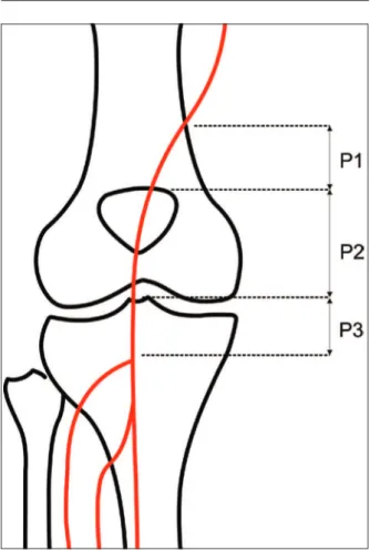
Preoperative arteriography was used to classify the lesions according to: (1) the Trans-Atlantic Inter-Society Consensus II (TASC-II) 7 criteria: A, B, C, or D (Box); (2) type of lesion: stenosis, occlusion, dissection, or restenosis; and (3) location relative to the articular line: proximal, middle, or distal (Figure 1).
Endovascular procedure
All procedures were performed by the same team at the Hemodynamics Laboratory, Center for Endovascular Interventions, Instituto Dante Pazzanese de Cardiologia.
Clopidogrel (75 mg/day) and acetylsalicylic acid (100 mg) were started at least three days before the procedure. Clopidogrel was maintained for at least 30 days and acetylsalicylic acid was maintained indeinitely.
BOX
Classification of lesions according to the Trans-Atlantic Inter-Society Consensus II (TASC II)
A Lesions that produce the best results and that should be treated by endovascular route.
B Lesions that produce suficiently good results with endovascular methods, so that this is still the preferred approach, unless surgical
revascularization is required to treat other lesions in the same anatomic area.
C Lesions exhibiting superior long-term results with surgery, so that endovascular methods should be used only in patients at high surgical risk. D Lesions that do not produce good enough results
with endovascular methods to justify them as primary treatment.
The participants were treated with local anesthetic, and antimicrobial prophylaxis was performed with 1.5 g of cefuroxime immediately before the beginning of the procedure. Radiographic control was obtained with an AXIOM Artis Flat Panel device (Siemens Healthcare Sector, Forchheim, Germany) or in a hybrid room with a Zeego Artis device (Siemens Healthcare Sector , Forchheim, Germany).
The approach was preferentially performed through the ipsilateral common femoral artery, with anterograde puncture, using a Prelude
(Merit Medial Systems, South Jordan, United States) 6 F valved sheath. In the case of impossibility of using this access route, or when it was not possible to cross the target lesion, a retrograde ac-cess by puncturing one of the leg arteries was chosen, with a Prelude
4 F valved sheath. 5 F MPA-1 and/or 4 F STR diagnostic catheters (Cordis Corporation, Warren, United States) were used, and the crossing of target lesions was performed by luminal or sub-intimal route, with the help of a Radiofocus
(Terumo Interventional Systems, Somerset, United States) hydrophilic guide wire (wire size, 0.035” × 150 cm). Pre-dilation was conducted in cases of occlusion, or when a proper positioning of the stent was not possible. In all cases, the sinus-SuperFlex
(Optimed, Ettlingen, Germany) nitinol stent was used (Figure 2).
In all cases, the immediate postoperative period oc-curred in the ward, and local hemostasis was performed with manual compression for 30 minutes.
Postoperative follow-up
The follow-up was performed with outpatient evalua-tion, consisting of physical examination and ankle-brachial index (ABI) determination at 15, 30, 90, and 180 days after angioplasty. The control with Doppler ultrasound (USG-D) was performed at 30, 90, and 180 days after surgery, with the aim of identifying restenoses (Figure 3). Radiographs of the knee joint in posteroanterior (PA) and lateral views were performed at 30 and 180 days, with the aim of identifying stent fractures (Figure 4).
Outcomes and definitions
The analyzed outcomes were: (1) immediate technical success, when the target lesion was treated in the man-ner previously planned, with residual lesion < 30% in the angiographic control; (2) primary patency, indicating an uninterrupted permeability after the revascularization procedure; and assisted primary patency, indicating cases where a new percutaneous intervention was applied, in order to prevent the impending occlusion or the progres-sion of stenosis; (3) perioperative morbidity/mortality, for complications and deaths recorded for up to 30 days postoperatively; (4) major amputation: transfemoral and transtibial amputations; (5) restenoses, for in-stent lesions > 50% detected with USG-D, with peak systolic veloc-ity > 200 cm/s or a pre-and post-stenosis rate ≥ 2; (6) fractures, for disconnection or twisting of stent meshes; and (7) rate of limb salvage.
RESULTS
Fourteen patients with symptomatic POAD, treated with primary angioplasty (without employing other adjuvant methods of arterial unblocking) with stent implantation in popliteal arteries, were included in the study.
Characteristics of the population
The demographic characteristics, comorbidities, and indications for treatment are described in Table 1. Mean age was 73 ± 11 years, 50% of patients were male, and approximately two-thirds were diabetic. The left leg was the most frequently treated limb (57.1%) and all patients had trophic lesions with tissue loss. Patients with intermittent claudication or pain at rest were not treated.
Characteristics of lesions and of procedure
The lesions were classiied into TASC B (50%) and C (50%). Regarding their location, 71.4% of cases were in the proximal segment of popliteal artery (P1), and in
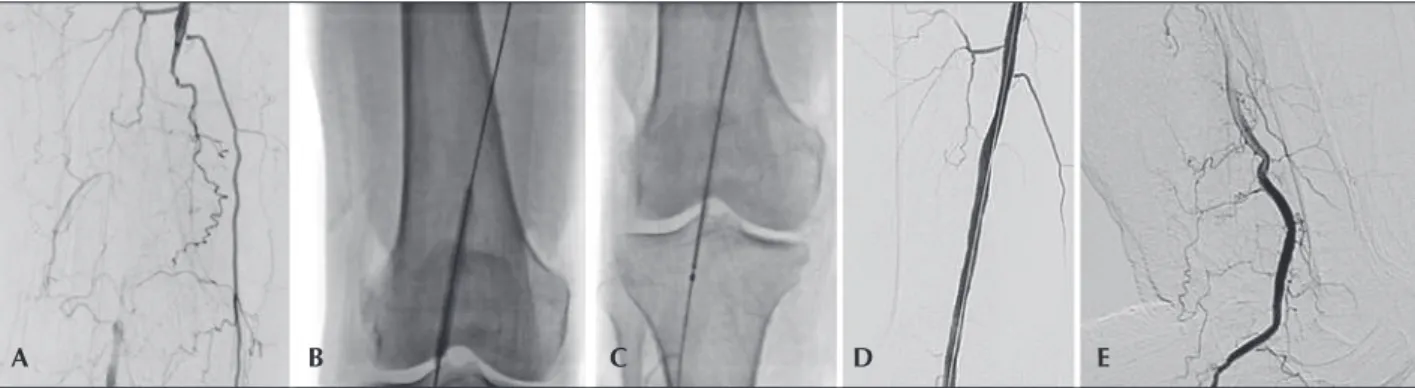
Figure 2 – Angioplasty of popliteal artery with landing zone in the distal segment of this artery. (A) Digital subtraction arteriogram showing occlusion of popliteal artery with distal illing in the interline. (B) Pre-dilation. (C) Positioning of the stent in the distal segment of popliteal artery. (D) Final arteriogram with return of axial low. (E) Arteriography in leg lexion, without exhibiting kinking or blood low-limiting injuries.
28.6% of the cases were in middle or distal segments, in equal proportion (P2 and P3). In eight cases (57.1%), the stents were implanted in previously occluded ves-sels, with pre-dilation performed in all lesions.
In the evaluation of the run-off bed, the majority of patients had only one patent artery (78.6%) and the ibular artery was the most frequently observed. A mean of 1.3 ± 0.4 arteries by treated limb was obtained (Table 2).
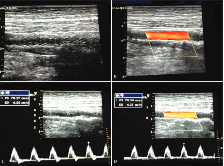
It was possible to achieve the target lesion re-vascularization in all cases with the use of one stent, obtaining technical success in 100%. The average extent of lesion coverage was 177 ± 18 mm (120-200 mm). Ten stents (71.4%) with a diameter of 6 mm and four stents (28.6%) with a diameter of 5 mm were used. There were no cases of intraoperative dissection or embolization. The areas of more frequent stent land-ing were the middle segment of popliteal artery (P2) in eight cases (57.1%) and the distal segment (P3) of the artery, crossing the joint knee, in six cases (42.9%). No implants occurred in the proximal segment (P1). Mean procedure times and of radioscopy were, re-spectively, 55 ± 43 minutes (range 20-240 min) and Figure 3 – Ultrasonographic control at 6 months of stenting in the middle segment of popliteal artery. (A) Color-mode image without signs sugges-tive of stent fracture. (B) Color-mode image, showing laminar low without vortices and no signs of neointimal hyperplasia. (C) Doppler Ultrasound demonstrating spectral curve of speed in native artery. (D) Doppler Ultrasound demonstrating spectral curve of intra-stent speed with a velocity of 70 cm/s and a speed ratio of 1.
Figure 4 – (A) Radiography of knee in semi-lexion, with no signs of stent twisting or fracture. (B) Radiography of knee in posteroanterior view and with no signs of fracture. (C) Radiography of knee without evidence of compression at the level of adductor canal.
A
C D
B
19 ± 14 minutes (range 5-66 min). The mean volume of iodinated contrast used was 88 ± 41 mL. In one case, a distal retrograde access by the dorsalis pedis artery was performed, thanks to the impossibility of crossing the target lesion by the anterograde technique. No complications with respect to the puncture site (Table 3) were observed.
Patient follow-up
The mean follow-up was 6.4 ± 2.1 months (range 3.2 to 8.4 months). There were no intraoperative or procedure-related deaths. Ninety days post-procedure, one elderly patient died due to cardiac complications.
The limb salvage rate was 100%. There was no major amputation during the follow-up. In three pa-tients (21.4%), there was a development of their trophic lesions; two of these lesions were caused by in-stent stenoses in the 60th and 90th day of follow-up, and were treated by balloon dilation. The third injury occurred due to infectious complications, which resulted in a minor amputation. There was an increase of ABI, from 0.4 ± 0.15 preoperatively to 0.8 ± 0.13 at the time of hospital discharge.
The primary patency rate was 85.7% at 6 months. In two cases, it was necessary to perform a new balloon angioplasty for treatment of in-stent restenosis, with therapeutic success in only one case. This resulted in an assisted primary patency rate of 92.8% at follow-up. There were no stent fractures documented by radiographs of the knee joint.
DISCUSSION
The management of atherosclerotic lesions in the segments of the popliteal artery near the knee joint represents a dificult endovascular approach, because of the biomechanical stresses occurring in this region. Complex rotational, tensile, compressive, and stretching forces act simultaneously on a short arterial segment
TABLE 1
Baseline clinical characteristics
Variable n = 14
Age, years 73 ± 11
Male gender, n (%) 7 (50)
Body mass index, kg/m2 25.3 ± 5.5
Hypertension, n (%) 14 (100)
Diabetes mellitus, n (%) 9 (64.3)
Dyslipidemia, n (%) 14 (100)
Smoking, n (%) 7 (50)
Previous acute myocardial infarct, n (%)
2 (14.3)
Alcohol abuse, n (%) 4 (28.6)
Laterality, n (%)
Right 6 (42.9)
Left 8 (57.1)
Indication for the procedure, n (%)
Trophic lesion 14 (100)
Pain at rest 0
Intermittent claudication 0
Creatinine, mg/dL
Preoperative 0.8 ± 0.6
Postoperative 0.9 ± 0.5
Ankle-brachial index
Preoperative 0.4 ± 0.15
Postoperative 0.8 ± 0.13
TABLE 3
Characteristics of the procedure
Variable n = 14
Procedure time, min 55 ± 43
Radioscopy time, minutes 19 ± 14
Contrast volume, mL 88 ± 41
Stent characteristic
Diameter, mm 7 ± 0.5
Length, mm 177 ± 18
Stent location
P1 0
P2 8 (57.1)
P3 6 (42.9)
TABLE 2
Angiographic characteristics of lesions
Variable n = 14
TASC, n (%)
A 0
B 7 (50)
C 7 (50)
D 0
Characteristic, n (%)
Stenosis 6 (42.9)
Occlusion 8 (57.1)
Location, n (%)
Popliteal and femoral 10 (71.4)
Popliteal 4 (28.6)
Arteries for low, n (%)
1 11 (78.6)
2 2 (14.3)
3 1 (7.1)
and on the stent.8,9 For many years, these lesions were treated by surgical grafting using autologous vein, with rates of primary and secondary patency after 5 years of 63% to 75% and 80% to 83%, respectively, and with a perioperative mortality of 1% to 3%.10-12 However, these high patency and low mortality rates are accompanied by a considerable perioperative morbidity, as are the subsequent multiple surgical procedures and hospital readmissions.13
Currently, with the development and improvement of endovascular materials, the number of minimally invasive procedures for the treatment of popliteal lesions has increased. Generally, balloon angioplasty, or mechani-cal or laser arterectomy, are the preferred choices for the treatment of arterial lesions behind the knee, due to the lack of metallic devices capable of withstanding the forces exerted on this arterial segment.14,15 However, the eficacy of these techniques is limited.15
The irst large study comparing treatment with bal-loon angioplasty versus stenting in the femoropopliteal segment was RESILENT2 (Randomized Study Comparing the Edwards Self-ExpandIng Lifestent versus Angioplasty Alone In LEsions INvolving The SFA and/or Proximal Popliteal Artery). During the irst phase of this study, patients with intermittent claudication because of femo-ropopliteal occlusive lesions, with length < 15 cm and diameter of the target vessel between 4 to 6.5 mm, were included. The results showed the superiority of the LifeStent
(Bard Peripheral Vascular, Tempe, United States) over balloon angioplasty in the femoropopliteal segment. In its second phase, 206 patients were included, when the treatment of more than one lesion was al-lowed, provided they did not exceed the length of 15 cm. Its primary endpoint was the revascularization of the target vessel within 12 months. The result was a primary patency of 80% vs. 38% for the LifeStent
vs. balloon angioplasty, respectively.3
The choice of the stent depends on the anatomic characteristics of the lesion, such as its location and the extent of calciication, as well as the characteristics of the stent – its diameter, length, proile, lexibility, long-term durability, and resistance to fracture. The limi-tations to stent implant in the popliteal artery include insuficient radial force to maintain the patency of the vessel, and the possibility of folding and fractures of the device. Stent fracture is associated with high incidence of restenoses and occlusions. Scheinert et al.16 detected radiographically 64 fractures in 261 stents (24.5%) used in the treatment of femoropopliteal lesions. The primary patency rate obtained in the 12 months’ follow-up was signiicantly lower in patients with fracture (41.1% vs. 84.3%, p < 0.0001). In the present study, stent fractures detected by the same method were not observed.
Regarding the rate of primary patency, the results of several studies in the literature considered hetero-geneous populations, also differing in the location of
the segments treated. Chan et al.4 reported primary patency rates of the femoropopliteal stent implanted in the segment after six and 12 months of 83.5% and 78.6%, respectively, in 82 limbs treated with Supera
stent (IDEV Technologies Inc., Webster, United States). In the Durability-200 study, Bosiers et al.17 analyzed 100 patients, 71% of whom were treated for intermittent claudication and 29% treated for critical ischemia, with their injuries classiied as TASC C and D, and used the devices in the femoropopliteal segment. These authors obtained a primary patency of 85.4% at 6 months and of 64.8% at 12 months. As for stents implanted in the popliteal segment, Kickuth et al.18 studied 35 patients with intermittent claudication and critical limb ischemia, in which auto-expandable nitinol stents were implanted in the popliteal artery. In 22 cases, stents were implanted in the distal portion of the popliteal artery, and in the other cases, in the tibioibular trunk, with a primary patency rate of 82% and a limb salvage rate of 100%. In the present study, most stents were implanted in the middle segment, and in only six cases (42.9%), stents were implanted in the distal segment, crossing the joint line. A primary patency and an assisted primary patency of 85.7% and 92.8% were obtained, respectively, at the six-month follow-up, in a population consisting of patients with critical ischemia of Rutherford classes 5 and 6, and in arteriographic lesions classiied as TASC B and C, with a similar rate of limb salvage.
During the clinical follow-up, there was improve-ment in ABI, from 0.4 ± 0.15 preoperatively to 0.8 ± 0.13 at the time of hospital discharge, as well as an improvement of trophic lesions in 78.6% of cases. The measurement of ABI may have been overestimated, due to the high prevalence of diabetes in our population (64.3%), considering that the predominance of arte-rial calciications in the distal portions of leg arteries of diabetic people overestimates the mensuration of this index.
Study limitations
The small number of cases, the heterogeneous group of segments treated in the popliteal artery, the short follow-up period, and the absence of an established algorithm for clinical follow-up may have inluenced the results of this study. Finally, the accuracy of the results may have been affected by the retrospective data analysis.
CONCLUSIONS
CONFLICTS OF INTEREST
The authors declare no conlicts of interest.
FUNDING SOURCE
None.
REFERENCES
1. Iida O, Soga Y, Hirano K, Suzuki K, Yokoi H, Nobuyoshi M, et al. Long-term outcomes and risk stratiication of patency following nitinol stenting in the femoropopliteal segment: retrospective multicenter analysis. J Endovasc Ther. 2011;18(6):753-61. 2. Neil N. Stent fracture in the supericial femoral and proximal
popliteal arteries: literature summary and economic impacts. Perspect Vasc Surg Endovasc Ther. 2013;25(1-2):20-7. 3. Laird JR, Katzen BT, Scheinert D, Lammer J, Carpenter J,
Bu-chbinder M, et al. Nitinol stent implantation versus balloon angioplasty for lesions in the supericial femoral artery and proximal popliteal artery: twelve-month results from the RESIL-IENT randomized trial. Circ Cardiovasc Interv. 2010;3(3):267-76. 4. Chan Y, Cheng S, Ting A, Cheung G. Primary stenting of
femoropopliteal atherosclerotic lesions using new helical interwoven nitinol stents. J Vasc Surg. 2014;59(2):384-91. 5. Schillinger M, Sabeti S, Loewe C, Dick P, Amighi J,
Mle-kusch W, et al. Balloon angioplasty versus implantation of nitinol stents in the supericial femoral artery. N Engl J Med. 2006;354(18):1879-88.
6. Arena FJ. Arterial kink and damage in normal segments of the supericial femoral and popliteal arteries abutting nitinol stents: a common cause of late occlusion and restenosis? A single-center experience. J Invasive Cardiol. 2005;17(9):482-6. 7. Norgren L, Hiatt WR, Dormandy JA, Nehler MR, Harris KA,
Fowkes FG, et al: Inter-Society Consensus for the Management of Peripheral Arterial Disease (TASC II). Eur J Vasc Endovasc Surg. 2007;33 Suppl 1:S1-S75.
8. Kroger K, Santosa F, Goyen M. Biomechanical incompatibility of popliteal stent placement. J Endovasc Ther. 2004;11(6):686-94.
9. Goltz JP, Ritter CO, Petritsch B, Kellersmann R, Hahn D, Kickuth R. Endovascular treatment of acute limb ischemia secondary to fracture of a popliteal artery stent. J Vasc Interv Radiol. 2010;21(11):1739-45.
10. Taylor LM Jr, Edwards JM, Porter JM. Present status of reversed vein bypass grafting: ive-year results of a modern series. J Vasc Surg. 1990;11(2):193-205.
11. Bergamini TM, Towne JB, Bandyk DF, Seabrook GR, Schmitt DD. Experience with in situ saphenous vein bypasses during 1981 to 1989: determinant factors of long-term patency. J Vasc Surg. 1991;13(1):137-47.
12. Donaldson MC, Mannick JA, Whittemore AD. Femoral-distal bypass with in situ greater saphenous vein. Long-term results using the Mills valvulotome. Ann Surg. 1991;213(5):457-64. 13. Goshima KR, Mills JL Sr, Hughes JD. A new look at outcomes
after infrainguinal bypass surgery: traditional reporting standards systematically underestimate the expenditure of effort required to attain limb salvage. J Vasc Surg. 2004;39(2)330-5. 14. Goltz JP, Ritter CO, Kellersmann R, Klein D, Hahn D, Kickuth
R. Endovascular treatment of popliteal artery segments P1 and P2 in patients with critical limb ischemia: initial experience using a helical nitinol stent with increased radial force. J Endovasc Ther. 2012;19(2):450-6.
15. Karnabatidis D, Katsanos K, Siablis D. Infrapopliteal stents: overview and unresolved issues. J Endovasc Ther. 2009;16 Suppl 1:I153-62.
16. Scheinert D, Scheinert S, Sax J, Piorkowski C, Bräunlich S, Ulrich M, et al. Prevalence and clinical impact of stent fractures after femoropopliteal stenting. J Am Coll Cardiol. 2005;45(2):312-5.
17. Bosiers M, Deloose K, Callaert J, Moreels N, Keirse K, Verbist J, et al. Results of the Protégé EverFlex 200-mm-long nitinol stent (ev3) in TASC C and D femoropopliteal lesions. J Vasc Surg.2011;54(4):1042-50.