637
Taxonomic assessment of
Leptodoras
(Siluriformes: Doradidae)
with descriptions of three new species
Mark Henry Sabaj
The genus Leptodoras Boulenger is a monophyletic assemblage of at least ten species distributed in large, predominantly lowland rivers throughout the northern half of cis-Andean South America. Leptodoras is diagnosed in Doradidae (thorny catfishes) by unique morphologies of the oral hood (upper labial extensions distinct with comparatively smooth ventral surface, lateral margins of extensions entire, weakly scalloped or fimbriate and without marginal papillae, and interlabial membranes of narrow or moderate width), first gill arch (enlarged accessory lamellae extend well onto medial face of gill filaments), and gas bladder (reduced size and modified cordiform shape, two distinct horn-like diverticula project from posterior walls of posterior chambers, and a pair of bulbous diverticula project from lateral walls of anterior chamber). Examination of specimens of Leptodoras from throughout its range verified the distinctiveness of the seven nominal species (L. acipenserinus, L. copei,L. hasemani,L. juruensis,L. linnelli,L. myersi and L. praelongus) and revealed three new species described herein (Leptodoras nelsoni,L. rogersae, and L. cataniai). A lectotype for L. hasemani is designated.
O gênero Leptodoras Boulenger é um agrupamento monofilético com pelo menos dez espécies, distribuídas em rios grandes e predominantemente de terras baixas na metade norte da região cis-Andina da América do Sul. Leptodoras é diagnosticado em Doradidae pelas morfologias únicas da região bucal (extensões labiais superiores distintas com superfície ventral comparativamente lisa, margens laterais destas extensões inteiras, fracamente franjadas ou fimbriadas e sem papilas marginais, e membranas interlabiais estreitas a moderadamente largas), do primeiro arco branquial (lamellas acessórias aumentadas, extendendo-se pela face medial dos filamentos branquiais), e da bexiga natatória (tamanho reduzido e formato cordiforme modificado, dois divertículos em forma de guampas projetando-se das paredes posteriores das câmaras posteriores, e um par de divertículos bulbosos projetando-se das paredes laterais da câmara anterior). O exame de exeplares de Leptodoras em sua área de distribuição permitiu a distinção de sete espécies nominais (L. acipenserinus,L. copei,L. hasemani,L. juruensis,L. linnelli, L. myersi e L. praelongus) e revelou três espécies novas, descritas aqui (Leptodoras nelsoni,L. rogersae, e L. cataniai). É designado um lectótipo para L. hasemani.
Key words: Barbel morphology, Longlip thornycat, Taxonomic key, Thorny catfishes.
Department of Ichthyology, The Academy of Natural Sciences, 1900 Benjamin Franklin Parkway, Philadelphia, Pennsylvania, USA 19103. e-mail: sabaj@acnatsci.org
Introduction
Leptodoras Boulenger (longlip thornycats) is a monophyl-etic genus of the Doradidae (Order Siluriformes) comprised of seven previously described and three new species distrib-uted in large, predominantly lowland rivers throughout the northern half of cis-Andean South America. Leptodoras is easily recognized by its long conical snout and well-devel-oped oral hood formed by the membranous union of maxillary barbels, paired jaw barbels and labial structures. The oral hood presumably facilitates the detection and suction-feeding of shallowly buried invertebrates. Stomach contents typically include chironomid larvae, sand and detritus (pers. obs.).
PROOFS
mouths of the Orinoco and Amazon. Leptodoras is not knownfrom trans-Andean drainages (e.g., Maracaibo and Magdalena) or Atlantic-slope drainages south of the rio Tocantins (e.g., rios São Francisco and Paraná-Paraguay).
The taxonomic history of Leptodoras dates back to Günther’s (1868a) description of Oxydoras acipenserinus from the Peruvian Amazon. Boulenger (1898) subsequently pro-posed the genus Leptodoras, designated L. acipenserinus the type, and described a second species, L. juruensis, from the rio Jurua, Brazil. Boulenger (1898) distinguished Leptodoras as having a longer body and longer anal fin (15 to 17 rays) than Oxydoras Kner. Eigenmann (1912) doubted the diagnosability of Leptodoras based on anal-fin ray counts, but retained Leptodoras as distinct because of its lack of teeth. Eigenmann (1912) introduced a third species, L. linnelli, described from the Essequibo river basin, Guyana. Steindachner (1915) described Hemidoras hasemani based on 18 syntypes from the rio Branco and one specimen, re-identified here as Hemidoras stenopeltis (Kner), from the mouth of the rio Negro, Brazil. Eigenmann (1925) transferred hasemani to Leptodoras and provided the first detailed
de-scription of the genus. Myers & Weitzman (1956) described Hassar praelongus from the rio Negro, Brazil, and Fernández-Yépez (1968) described Anduzedoras copei from the Orinoco Basin, Venezuela. Most recently, L. myersi was described by Böhlke (1970) from specimens collected in a deep-water trawl of the río Amazonas near Iquitos, Peru.
Although all seven nominal Leptodoras are valid spe-cies, their identification in the literature (L. juruensis excepted) is often inconsistent. Taxonomic confusion has been the prod-uct of several factors: 1) several species (L.praelongus,L. copei,L. hasemani) closely resemble one another or species in other genera (e.g.,Hassar), 2) the distributions and identi-ties of two species (acipenserinus and linnelli) have been misunderstood, 3) all seven species were described from small geographic areas (usually a single locality) with no informa-tion on their potential distribuinforma-tions, and 4) specimens (espe-cially adults) have been rare in museums until very recently. Increased sampling efforts over the past decade, particu-larly those utilizing bottom trawls (e.g., the Calhamazon Project led by J.G. Lundberg), have yielded large numbers of Leptodoras specimens. These recent collections and a sur-vey of the primary types (all extant) made this study possible. Perhaps the most surprising discovery resulted from the ex-amination of specimens routinely identified as L. acipenserinus or L. linnelli. This revealed three undescribed species distributed in the main channel of the río Orinoco, the Colombian and Venezuelan llanos (Orinoco drainage) and Amazon basin, respectively.
The objectives of this paper are to diagnose the genus Leptodoras and its nominal species and to describe three new species.
Material and Methods
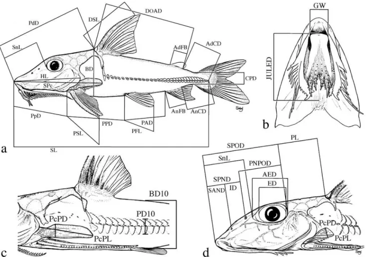
Institutional abbreviations follow Leviton et al. (1985) with the addition of UNT for Universidade do Tocantins, Porto Nacional, Brazil. Standard terminology for features of barbels and labial structures was developed to facilitate their descrip-tion (Fig. 1). Measurements were made to the nearest 0.1 mm using digital calipers (<150 mm), dial calipers (150 to 180 mm) or a beam compass (>180 mm). Measurements and correspond-ing landmarks (Fig. 2) coincided in part with those of Böhlke (1970) and Higuchi et al. (1990). Some measurements were changed to improve repeatability (via better landmarks) or to synchronize the terminus of one with that of another. Poorly defined landmarks (e.g., those requiring physical manipula-tion of specimen to visualize) were marked with insect pins.
Point-to-point straight-line measurements (Fig. 2) are de-fined as follows with the first set reported as percentages of standard length (SL)-snout tip to point on midlateral side of caudal peduncle level with ventroposterior corner of hypural 3+4 (determined by gently flexing the caudal fin and/or back-lighting); head length (HL)-snout tip to posterior-most ex-tremity of fleshy opercular flap; predorsal distance (PdD)-snout tip to posterior margin of second nuchal plate coincid-ing with median sagittal plane; dorsal origin-adipose distance
Fig. 1. Standard terminology for features of barbels and labial
PROOFS
(DOAD)-from groove between posterior margin of secondnuchal plate (anterior to base of dorsal-locking spine) to pos-terior-most base of adipose fin; (AdCD)-pospos-terior-most base of adipose fin to point coinciding with posterior terminus of SL; prepectoral distance (PpD)-snout tip to point between notch formed by margin of cleithrum and extreme base of pectoral spine (spine positioned at a 30-45° angle with the long axis of body); pectoral-pelvic distance (PPD)-from base of pectoral spine (terminus of PpD) to base of first (anterior-most) pelvic-fin ray (best visualized by abducting pelvic fin); pelvic-anal distance (PAD)-from base of first (anterior-most) pelvic-fin ray to base of first anal-fin ray; anal-caudal dis-tance (AnCD)-from base of posterior-most anal-fin ray to point coinciding with posterior terminus of SL; dorsal spine length (DSL)-from groove between posterior margin of second nuchal plate and base of dorsal locking spine to bony tip of dorsal spine (spine at a 30-45° angle with the long axis of the body, soft break-away tip excluded if present); pectoral spine length (PSL)-from base of pectoral spine (terminus of PpD) to bony tip (soft break-away tip excluded if present); pelvic fin length (PFL)-from base of first (anterior-most) ray to distal-most tip of anterior fin (not the measurement of an individual ray); anal-fin base (AnFB)-distance between anterior-most and
posterior-most bases of anal fin insertion; body depth (BD)-greatest distance in median sagittal plane between shallow crest of posterior margin of second nuchal plate (anterior to dorsal-fin origin) and midventral contour of body; caudal peduncle depth (CPD)-least depth of caudal peduncle.
The following set is reported as percentages of predorsal distance (preferred to head length because of ease and accu-racy of measurement): horizontal adipose eye diameter (AED)-from anterior-most margin (often level with the weakly de-fined posteroventral corner of the lateral ethmoid forming anterodorsal portion of bony orbit) to posterior-most margin (usually level with bony margin of orbit coinciding specifi-cally with the anteroventral corner of sphenotic) of adipose eyelid; snout length (SnL)-snout tip to anterior-most margin of adipose tissue covering eye (adipose eyelid often becomes opaque during preservation and its margin is usually distin-guishable from pigmented skin on snout); snout-anterior nares distance (SAND)-snout tip to center of opening of an-terior nares (membranous flap ignored); snout-posan-terior nares distance (SPND)-snout tip to center of opening of posterior nares (flap ignored); snout-posterior orbit distance (SPOD)-snout tip to posterior-most bony margin of orbit coinciding specifically with anteroventral corner of sphenotic; anterior
Fig. 2. Landmarks and measurements used for morphometric analysis (shown on generalized doradids). See Material and
PROOFS
nares-posterior orbit distance (ANPOD)-from center ofopen-ing of anterior nares to posterior-most bony margin of orbit coinciding specifically with anteroventral corner of sphenotic; posterior nares-posterior orbit distance (PNPOD)-from cen-ter of opening of poscen-terior nares to poscen-terior-most bony mar-gin of orbit coinciding specifically with anteroventral corner of sphenotic; internares distance (ID)-between centers of openings of anterior and posterior nares; postorbital length (PL)-from posterior-most bony margin of orbit coinciding specifically with the anteroventral corner of the sphenotic to posterior margin of second nuchal plate coinciding with me-dian sagittal plane; postcleithral (humeral) process length (PcPL)-from posterior-most tip of process to point along anterodorsal margin of exposed process where the cleithral bone is deflected medially (exposed process often appears textured compared to the smooth face of medially deflected portion of the cleithrum and this deflection often coincides with or lies just beneath the posterior-most margin of the fleshy opercular flap); jaw-upper labial extension distance (JULED)-straight-line measurement along median sagittal plane from point level with anterior jaw margin to point level with posterior-most tip of shorter of paired upper labial exten-sions lying (or pinned) flat against ventral surface of head (performed only on specimens with well preserved labial struc-tures not desiccated or twisted); head width (HW)-greatest transverse distance between lateral contours of head (i.e., opercula compressed to normal position if flared) anterior to cleithra; cleithral width (CW)-greatest transverse distance between lateral contours of cleithra; interorbital width (IW)-shortest transverse distance between orbital (lateral) margins of bony frontals; gape width (GW)-horizontal measurement between gape corners of closed mouth (in specimens pre-served with mouth open, this measurement coincided with the width of the anterior margin of the lower jaw).
One measurement is reported as percentage of body depth taken in the same transverse plane (adapted from Böhlke, 1970): depth of tenth midlateral plate-vertical depth orthogo-nal to horizontal line formed by medial thorns of plates, from dorsal-most exposed margin of tenth plate to ventral-most margin of corresponding plate.
Counts of fin rays follow Hubbs & Lagler (1958), Böhlke (1970) and Higuchi et al. (1990). Counts in dorsal, anal, and paired fins are separated into anterior spine (capital roman numeral) or unbranched soft ray (lower-case roman numeral) and posterior branched soft rays (arabic numerals). The small locking bone anterior to dorsal spine is not counted. The last (posterior-most) pectoral-fin ray may appear unbranched (par-ticularly in juveniles). It is counted if clearly segmented with base separate from penultimate ray. In rare instances the last pectoral-fin ray may be followed by a much smaller and rather inconspicuous sliver-like element that is clearly unsegmented. This bony element is not included in the count. The anterior-most anal-fin ray may be extremely small and closely adhered to the second ray. The last anal ray may be simple or com-posed of two branches with bases joined or in very close proximity (counted as one in either case). Counts of midlateral
plates begin with the vertically expanded infranuchal plate that dorsally contacts the posterior nuchal plate and ven-trally contacts or approaches the distal tip of the postcleithral process. Though conspicuous and sometimes bearing a me-dial thorn, the small plate immediately anterior to the
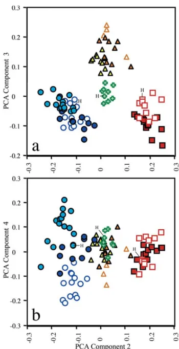
Fig. 3. Scatterplots of scores factored from covariance matrix
PROOFS
infranuchal plate (in the tympanal region) is not included incounts of midlateral plates.
Principal Components Analysis (PCA) was used to inves-tigate morphometric variation among Leptodoras acipenserinus,L. linnelli and the three new species. One of the three new species (L. cataniai) was further parsed into three groups: L. cataniai sensu stricto (rio Negro and Casiquiare canal), L. cf. cataniai Amazon form (Amazonas basin excluding Nanay and Negro) and L. cf. cataniai Nanay form (specimens largely from río Nanay, Peru). Leptodoras linnelli also was geographically divided into Essequibo, Orinoco and eastern Guiana Shield (northeastern Brazil) speci-mens. A total of 34 measurements (Tables 1 & 2) was taken on 96 juvenile and adult specimens representing a comparable range of sizes: L. acipenserinus (n=12, SL 107.5-196.5 mm), L. linnelli (25, SL 84.6-200.6 mm), Leptodoras nelsoni (11, SL 92.3-162 mm), Leptodoras rogersae (9, SL 89.2-177.9 mm), L. cataniai sensu stricto (15, SL 97.7-171 mm), L. cf. cataniai Amazon form (10, SL 77.4-194 mm), and L. cf. cataniai Nanay form (14, SL 89.7-181 mm). Specimens used in PCA analyses are denoted by an asterisk in material examined.
Principal components analysis was performed on the co-variance matrices of 34 log-transformed measurements. The
resulting first principal axis (PC I) explained a large propor-tion of the total variance (88.6%) and all variable loadings were positive and varied little in magnitude. PC I was there-fore interpreted as a general size factor (Jolicoeur & Mosimann, 1960; Jolicoeur, 1963; McElroy & Douglas, 1995). Scores were plotted for PC II, III and IV, interpreted to repre-sent “general-size-allometry-free shape” (Bookstein, 1989).
Results
Morphometric analysis. Plots of factor scores of principal components II (PCII) vs. III (PCIII) grouped specimens into four non-overlapping clusters corresponding to: Leptodoras linnelli,L. rogersae,L. cataniai + L. cf. cataniai (Nanay and Amazon forms) and L. acipenserinus + L. nelsoni (Fig. 3a). PC II and III accounted for 4.2 and 2.2% of the total variance, respectively. Measurements loading most heavily on PC II are, in decreasing order: length of upper labial extension (-0.086), length of postcleithral process (0.052), adipose-eye diameter (-0.043), interorbital width (0.031), pelvic-fin length 0.028) and distance from posterior nares to posterior orbit (-0.024). Standard length had a loading of 0 on PCII. Measure-ments loading most heavily on PCIII are, in decreasing order:
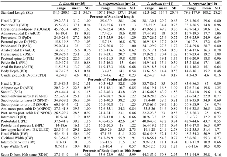
Table 1. Morphometrics of Leptodoras linnelli,L. acipenserinus,L. nelsoni, and L. rogersae. Measured specimens denoted with asterisk in material examined.
L. linnelli (n=25) L. acipenserinus (n=12) L. nelsoni (n=11) L. rogersae (n=10) range mean SD range mean SD range mean SD range mean SD Standard Length (SL) 84.6–200.6 121.1 34.78 107.5-196.5 137.4 29.68 92.3-162 114.7 20.49 79.9-177.9 108.2 28.24
Percents of Standard length
Head L (HL) 29.2-33.1 31.2 1.09 25.6-30 28.1 1.26 28.1-30.1 29.2 0.63 28.1-30.5 29.6 0.80 Predorsal D (PdD) 35.5-38.7 37.1 0.94 31.6-35.6 33.9 1.13 33-35.2 34.4 0.75 33.1-36.5 34.8 0.96 Dorsal origin-adipose D (DOAD) 45.7-53.4 48.5 1.63 48.9-52.4 50.6 1.03 47.9-51.2 49.8 0.86 48.7-54.3 50.6 1.61 Adipose-caudal D (AdCD) 16-19.4 18 0.87 17.4-20 18.6 0.88 17.4-19.2 18 0.54 15.7-19.5 17.7 1.06 Prepectoral D (PpD) 24.9-28.6 27.2 0.96 21.7-25.9 24.4 1.29 23.7-26.2 25.4 0.72 23.6-25.9 24.9 0.64 Pectoral-pelvic D (PPD) 15.8-19.8 17.9 1.05 15.7-18 16.8 0.78 16.9-18.8 17.7 0.68 16-17.8 17.2 0.59 Pelvic-anal D (PAD) 25.0-31.4 28 1.27 27.9-30.8 29 1.00 24.1-29.9 27.3 1.72 27.4-29.8 28.7 0.80 Anal-caudal D (AnCD) 14.2-17.5 15.6 0.76 15.5-17.6 16.5 0.62 15.7-17.1 16.4 0.50 15.4-17.6 16.3 0.78 Dorsal spine L (DSL) 20.9-26.3 23.1 1.65 17.5-23.1 20.7 1.70 17.9-21.5 20.4 1.08 15.7-23.4 19.2 1.88 Pectoral spine L (PSL) 19.9-26.2 22.6 1.65 18.6-21.3 19.8 0.88 16.7-21 19.1 1.37 17.6-20.9 18.8 0.96 Pelvic fin L (PFL) 13.9-17.4 15.6 0.88 14.2-16.3 15 0.64 14.9-16.1 15.4 0.39 15.2-18.6 17.1 1.03 Anal fin Base (AnFB) 11.5-15.4 13.7 1.02 14.9-17.3 15.8 0.68 13.9-18.5 16.1 1.40 14.1-16.2 15.1 0.64 Body Depth (BD) 14.7-17.5 16.1 0.81 13.4-15 14.1 0.54 13.6-15.6 14.4 0.58 14.6-16.1 15.2 0.48 Caudal peduncle Depth (CPD) 4.2-4.8 4.6 0.17 3.9-4.6 4.2 0.23 4.2-4.7 4.4 0.19 4.3-4.9 4.6 0.16
Percents of Predorsal distance
Head L (HL) 81.9-86.3 84.2 1.31 80.3-84.5 82.8 1.30 83.7-86.2 85 0.97 83.6-86.3 85 0.89 Adipose eye D (AED) 20.3-24.8 22.5 0.93 15.4-18.1 16.7 0.85 15.6-19.1 16.8 1.09 17.6-21.6 19.8 1.25 Snout L (SnL) 39.4-44.4 41.6 1.15 42.3-46.3 43.8 1.39 41.6-46.5 43.9 1.58 37.8-41.8 39.6 1.16 Snout-anterior nares D (SAND) 22.3-27.4 25.1 1.14 24.9-28.7 26.4 1.22 24.9-28.3 26.7 1.25 21.6-24.2 22.9 0.77 Snout-posterior nares D (SPND) 34.9-39.2 36.9 1.04 36.1-40.3 38.2 1.33 37.4-40 38.5 0.81 33.8-35.9 34.9 0.69 Snout-posterior orbit D (SPOD) 60.1-64.4 62 1.02 56.5-60.8 59 1.25 57.8-61.6 59.7 1.10 56.8-58.9 58 0.74 Ant. nares-post. orbit D (ANPOD) 37.3-41.8 39.3 1.00 32.6-35.1 34.3 0.84 33.5-36 34.6 0.89 34.6-38.5 36.5 1.30 Post. nares-post. orbit D (PNPOD) 26.5-29.7 28.5 0.97 21.4-24.1 23.1 0.86 22.2-23.8 23 0.53 23.3-26.3 25 0.93 Internares D (ID) 10.5-14 11.9 0.85 10.7-13.0 11.6 0.66 10.9-13.8 12 0.97 11-13.2 12.2 0.72 Postorbital L (PL) 37.6-41.8 39.8 1.16 40.6-45.5 42.6 1.47 40.8-43.6 42.2 0.84 42.9-44.6 43.7 0.55 Postcleithral process L (HPL) 14-18.6 16.1 1.15 16.2-20.5 18 1.22 16.4-19.2 17.7 0.94 14.6-17.8 16.3 0.97 Jaw-upper labial ext. D (JULED) 25.5-34.6 29.1 2.09 20.9-29 25.5 2.73 19.1-28 24.9 2.78 29.2-35.5 31.4 1.71 Head Width (HW) 45.4-54.1 50.6 1.97 47.1-55 51.1 2.22 48.6-54.8 52.1 1.59 48.2-54.2 50.9 1.97 Cleithral Width (CW) 51.3-54.8 52.8 0.98 54.3-59.9 55.9 1.91 53.8-56.1 54.8 0.77 53.6-56.1 55.3 0.73 Interorbital Width (IW) 8.3-13 10.3 1.36 8.7-13.5 11.5 1.32 9.9-12.1 11.1 0.74 10.1-11.9 10.9 0.66 Gape Width (GW) 8.7-11.9 10.4 0.83 8.3-10.4 9 0.57 8.5-12.5 10.2 1.23 8.6-11.6 10.5 0.85
Percents of Body depth at 10th Scute
PROOFS
tenth midlateral plate depth (-0.039), adipose eye diameter(0.035), anal-fin base (-0.03), distance from posterior nares to posterior orbit (0.029), and distance from anal fin to terminus of SL (-0.028).
Plots of PCII vs. PCIV grouped specimens into three clus-ters along the x-axis (PCII), one distinct (L. acipenserinus + L. nelsoni) and two weakly overlapping corresponding to L. cataniai + L. cf. cataniai (Nanay and Amazon forms) and L. linnelli + L. rogersae, respectively (Fig. 3b). PCIV contrib-uted very little to the resolution of individual species. How-ever, PCIV (y-axis) did separate L. cataniai and L. cf. cataniai into three weakly overlapping clusters with specimens from the Amazon (L. cf. cataniai Amazon form) intermediate to those from the Negro (L. cataniai sensu stricto) and Nanay (L. cf. cataniai Nanay form). Measurements loading most heavily on PCIV are, in decreasing order: snout-anterior nares distance (-0.039), snout length (-0.029), length of upper labial extension (-0.025), adipose-eye diameter (0.024), internares distance (0.022), and body depth at 10th plate (0.021).
Leptodoras Boulenger, 1898
Leptodoras Boulenger 1898:478. Type species: Oxydoras acipenserinus Günther 1868a, by subsequent designa-tion of Eigenmann, 1910:395.
Diagnosis.Leptodoras is diagnosed from other genera of the Doradidae by unique modifications of the oral hood (upper labial extensions distinct, elongate and comparatively smooth with entire, scalloped or fimbriate lateral margins and interlabial membranes narrow or of moderate width), first gill arch (en-larged accessory lamellae on medial face of arch extend well onto medial face of gill filaments), and gas bladder (reduced size, modified cordiform shape, two distinct horn-like diver-ticula project from posterior walls of posterior chambers and pair of bulbous diverticula project from lateral walls of ante-rior chamber).
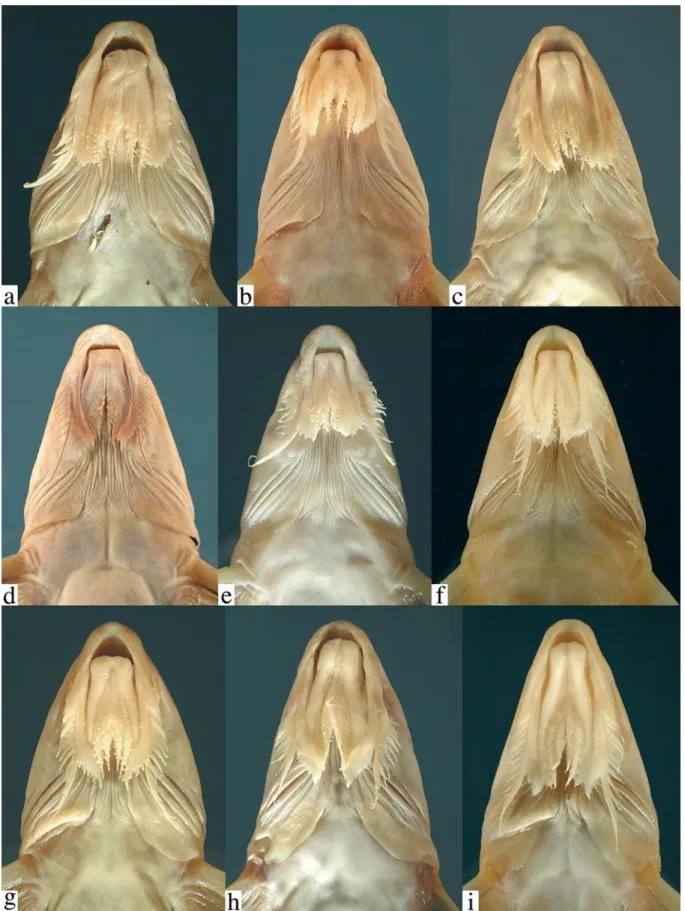
InLeptodoras the labial skin covering the jaws at the corners of the mouth continues posteriorly as a pair of long
Table 2. Morphometrics of Leptodoras cataniai, L. cf. cataniai (Nanay form) and L. cf. cataniai (Amazon form). Measured specimens denoted with asterisk in material examined.
L. cataniai (n=15) L. cf. cataniai, Nanay form (n=13)
L. cf. cataniai, Amazon form (n=10) range mean SD range mean SD range mean SD Standard Length (SL) 97.7-171 138.5 21.63 89.7-181 122.3 28.86 77.4-194 122.8 36.70
Percents of Standard length
Head L (HL) 26.9-31.9 28.5 1.36 29.3-32.9 30.9 1.26 27.9-31.4 29.2 0.99 Predorsal D (PdD) 31.1-35.5 32.6 1.13 32.7-37 34.6 1.43 31.6-35.6 33.1 1.23 Dorsal origin-adipose D (DOAD) 47.7-53.8 51.3 1.96 45.9-50.8 48.6 1.56 47.4-51.6 49.7 1.63 Adipose-caudal D (AdCD) 17.2-20.5 18.8 1.02 16.9-20.2 19.2 0.96 18.7-20.9 19.6 0.58 Prepectoral D (PpD) 22.6-26.8 24.2 1.23 24.9-28.5 26.5 1.18 24.0-26.5 24.9 0.66 Pectoral-pelvic D (PPD) 14.3-17.6 16.2 0.97 14.1-16.2 15.2 0.67 15.1-16.9 16 0.65 Pelvic-anal D (PAD) 29.2-31.4 30.1 0.60 27-29.9 28.6 0.99 28.4-30.7 29.4 0.82 Anal-caudal D (AnCD) 14.7-18.1 16.6 1.02 14.3-18.5 17.3 1.24 16.9-18.2 17.5 0.47 Dorsal spine L (DSL) 21.3-27.4 24.2 1.82 19.7-23.7 21.8 1.23 18.7-23 20.7 1.49 Pectoral spine L (PSL) 19-21.1 19.9 0.64 18.3-20.7 19.5 0.79 17.9-20.8 19.3 0.90 Pelvic fin L (PFL) 15.7-18.5 17.6 0.74 16.9-19.7 18.3 0.73 16.9-18.6 17.6 0.59 Anal fin Base (AnFB) 13.7-16.9 14.9 0.96 13-15.1 14.3 0.63 12.9-15.3 14.5 0.71 Body Depth (BD) 14.2-15.6 14.7 0.52 13.7-15 14.2 0.44 13.5-15.1 14.1 0.46 Caudal peduncle Depth (CPD) 4-4.6 4.3 0.16 3.4-4.2 3.9 0.23 3.7-4.4 4 0.19
Percents of Predorsal distance
Head L (HL) 85-90.6 87.5 1.61 87.8-91.8 89.5 1.05 86.1-90.2 88.4 1.36 Adipose eye D (AED) 23.1-27.8 25.5 1.49 18-21.5 19.7 0.99 19.8-23.2 21.4 1.14 Snout L (SnL) 35.4-40.2 37.7 1.28 42.4-46.6 44.1 1.26 37.6-41.3 39.1 1.19 Snout-anterior nares D (SAND) 19.9-24.6 22.2 1.61 25.3-28.8 27 1.14 21.6-25.7 23.3 1.25 Snout-posterior nares D (SPND) 33.6-37.6 35.6 0.97 36.2-39.4 37.9 0.94 32-36.7 34.6 1.23 Snout-posterior orbit D (SPOD) 60.2-63.5 61.9 0.90 60.7-63.9 62.5 0.86 56.8-61.9 59.3 1.52 Ant. nares-post. orbit D (ANPOD) 39-43.6 41.3 1.48 34.9-38.9 37.3 1.24 35.7-38 37.1 0.75 Post. nares-post. orbit D (PNPOD) 28.2-30 28.9 0.52 25.7-28.5 26.8 0.74 25.1-28.4 26.4 1.09 Internares D (ID) 11.6-16.4 13.5 1.38 10-13 11 0.84 9.4-13.6 11.7 1.06 Postorbital L (PL) 39.5-41.6 40.7 0.76 38-40.8 39.6 0.74 40.8-45.1 42.8 1.25 Postcleithral process L (HPL) 9.9-16.3 13.9 1.63 11.9-15.3 13.7 1.15 12.6-17.3 14.2 1.24 Jaw-upper labial ext. D (JULED) 33-44.8 40.8 2.84 39.3-47.8 44.2 2.38 36.3-43.8 39.8 2.52 Head Width (HW) 49.6-56.2 52.4 1.81 47.8-54.2 51.3 2.24 48.8-54.6 52.2 1.67 Cleithral Width (CW) 52.7-57 55 1.25 51.2-56.1 54 1.18 52.5-58.5 56 2.09 Interorbital Width (IW) 7.8-10.6 9.3 0.82 9.5-11.5 10.5 0.67 8.8-12.2 10.6 1.08 Gape Width (GW) 9.6-12.8 10.8 0.80 9.3-12 10.9 0.72 8.4-11.2 9.8 1.02
Percents of Body depth at 10th Scute
PROOFS
PROOFS
PROOFS
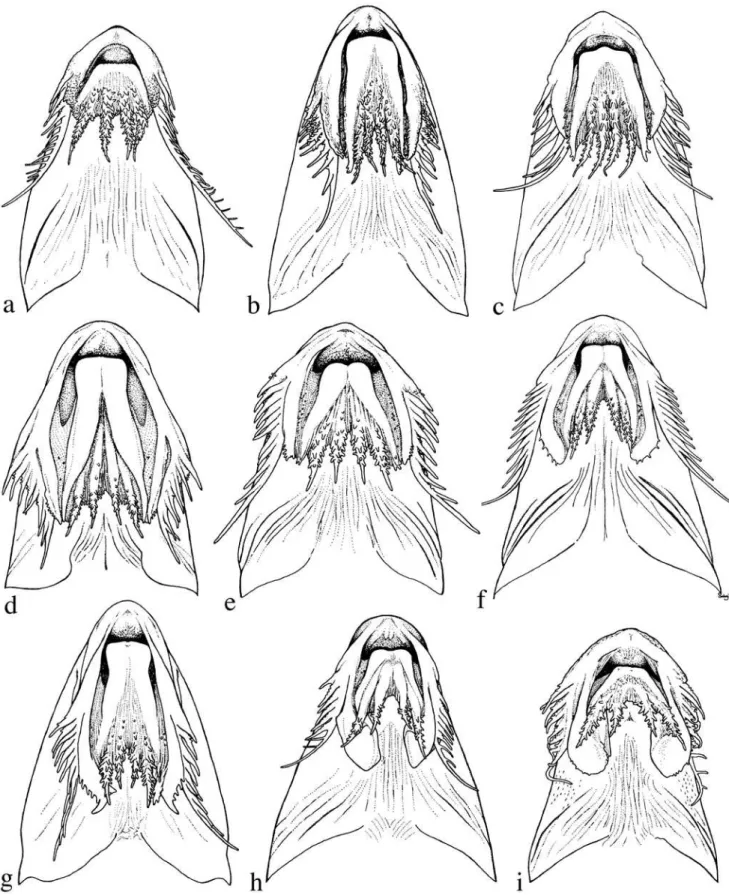
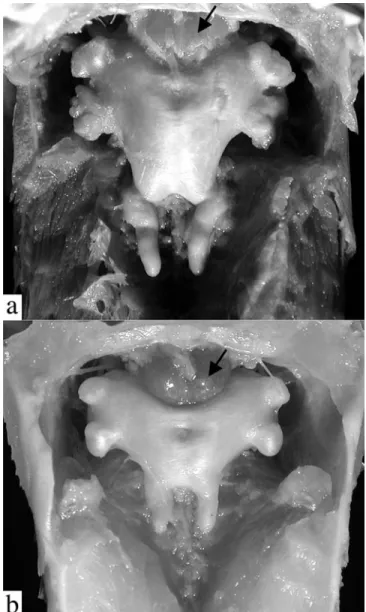
flap-like extensions (Figs. 4b-i, 5). These extensions arevari-ously united by a thin and weakly papillate interlabial mem-brane. A thin membrane also joins the medial margin of lower labial extension to the outer jaw barbel. The lateral margin of upper labial extension is free from maxillary barbel and its origin roughly coincides with that of anteriormost 2° maxil-lary barbel. In all Leptodoras except L. praelongus and L. copei, the dorsalmedial surface of the lower labial extension is joined to a membranous fold (dorsolabial membrane, Fig. 1b) that originates on ventrolateral corner of snout posterior to base of maxillary barbel. Higuchi (1992:220) also consid-ered the oral hood to be uniquely derived among Leptodoras. InLeptodoras enlarged accessory lamellae (each appear-ing as a column of soft lappets) occur along the medial face of the first gill arch and extend well onto the medial face of the gill filaments (Fig. 6). In some Leptodoras (cataniai, juruensis, myersi), smaller accessory lamellae extend well onto the lat-eral face of the gill filaments and the gill rakers are absent or inconspicuous (Fig. 6b). Higuchi (1992:221) previously noted the morphology of the first gill arch as diagnostic of Leptodoras.
InLeptodoras the gas bladder has a modified cordiform shape and is reduced in overall size (i.e., shortened in particu-lar; width greater than or equal to length) (Fig. 7). Further-more, in all Leptodoras there are: two distinct and relatively large horn-like diverticula that project from the posterior wall of the gas bladder, and discrete pairs of bulbous diverticula that project from the anterolateral walls on either side. The uniqueness of gas-bladder morphology in Leptodoras was first suggested by Eigenmann (1925).
Comparisons. The oral hood in Leptodoras is most closely approximated among other doradids by Anduzedoras oxyrhynchus (Valenciennes) (Fig. 4a). In this monotypic ge-nus the labial skin extends posteriorly beyond the corners of the mouth as flap-like structures united by an interlabial mem-brane. However, the labial extensions are relatively short and thickened, their ventral surfaces are rugose and papillate (vs. relatively smooth in Leptodoras), and the interlabial
mem-brane is relatively thick with numerous papillae (vs. thin and few papillae in Leptodoras). Some authors have suggested that the labial extensions (the upper in particular) are derived from the barbels (e.g., Günther 1868b:230; Boulenger 1898:478, Eigenmann 1925:357; Böhlke 1970:57). Based on their forma-tion in Leptodoras and related taxa these structures appear to be modifications of the labial skin as indicated by Higuchi (1992:220).
The accessory lamellae on the gill arches in Leptodoras resemble to those of certain other doradids (e.g., Doras, Anduzedoras,Hassar,Hemidoras). However, in these taxa the accessory lamellae are restricted to the medial face of the gill arch itself and do not extend onto the gill filaments.
In most other doradids the gas bladder is relatively large and often has an elongate cordiform shape. Other
fimbriate-Fig. 6. Lateral (left) and medial (right) views of 1st gill arch. a. Leptodoras copei, ANSP 162466 (SL 141.5 mm). b. Leptodoras cataniai, ANSP 161532 (SL 148 mm).
PROOFS
barbel doradids have a single medial posterior diverticula thatmay be simple (e.g.,Doras carinatus), split into two finger-like projections joined at the base (e.g.,Trachydoras), or or-namented with small bulbous swellings or thin finger-like pro-jections (e.g.,Anduzedoras,Hassar). Also unlike Leptodoras, the anterolateral walls of the gas bladder in other fimbriate-barbel doradids are smooth (e.g.,Doras) or variously orna-mented with many bulbous (e.g.,Anduzedoras) or finger-like (e.g.,Hassar,Hemidoras) diverticula. Rhynchodoras (genus with simple barbels) also has a reduced gas bladder and one species (R. woodsi) has two posterior horn-like diverticula. However, in R. woodsi the lateral walls of the anterior cham-bers are smooth (vs. with a pair of bulbous diverticula in Leptodoras).
Key to the species of Leptodoras.
1. Distal half of dorsal fin with distinct black spot or blotch ... Leptodoras hasemani [p.653] 1’. Distal half of dorsal fin without black spot or blotch ... 2 2. Dorsal spine thin and greatly extended as long flexible fila-ment; predorsal distance relatively short, 26-28% of SL; bases of dorsal and paired fins with intense black pig-ment ... Leptodoras juruensis [p.671] 2’. Dorsal spine rarely extended as a long flexible filament; predorsal distance longer, >30% of SL; pigmentation on bases of dorsal and paired fins variable ... 3 3. Broad dusky nuchal saddle extending ventrally across tympa-num and humeral process, finishing on the lower side level with insertions of paired fins ...Leptodoras myersi [p.675] 3’. No dark nuchal saddle and bar on side of body ... 4 4. Flap-like posterior extensions at corners of lower lip narrow and long, finishing beyond tips of similar extensions at corners of upper lip (Figs. 4b,c); black melanophores con-centrated on basal portions of dorsal spine and, sometimes, fin; sum of midlateral plates usually <75 (range 70-76) ... 5 4’. Flap-like posterior extensions at corners of lower lip rela-tively short, finishing even with or before tips of similar extensions of upper lip (Figs. 4e-g); base of dorsal fin without intense black pigmentation (may appear dusky, but never black); sum of midlateral plates usually >75 (range 74-92) ... 6 5. Anterior midlateral plates shallow, height of 2nd midlateral plate less than or equal to vertical diameter of eye; ante-rior nuchal plate usually narrow, permitting suture between supraoccipital and middle nuchal plate; profile of snout weakly concave ... Leptodoras praelongus [p.647] 5’. Anterior midlateral plates deeper, height of 2nd midlateral plate distinctly greater than vertical diameter of eye; ante-rior nuchal plate usually expanded laterally and sutured to epioccipital; profile of snout rounded (convex) ... ... Leptodoras copei [p.649] 6. Upper labial extension relatively long with narrow base and expanded distal portion that curves medially and has a distinctly pointed or triangular-shaped tip (Fig. 4g);
PROOFS
Leptodoras praelongus (Myers & Weitzman, 1956)
Figs. 4b, 7a and 8
Hassar praelongus Myers & Weitzman 1956: 2, fig. 1 (type locality: São Gabriel Rapids of the rio Negro (Amazonas dr.), Amazonas, Brazil).
Diagnosis. Uniquely distinguished among Leptodoras by
shal-low midlateral plates covering < 1/4 body depth (vs. >1/4 in all other species), and anterior nuchal plate usually of moderate width (longer than wide) permitting weak to firm suture be-tween supraoccipital and middle nuchal plate (vs. anterior nuchal plate laterally expanded, sharing broad suture with epioccipital or forming dorsal border of nuchal foramina). Also distinguished from all Leptodoras except L. copei by having distal tip of lower labial extension extending beyond that of upper labial extension.
PROOFS
Description. Overall shape (excluding caudal fin) lenticular,
weakly compressed, ventrally flattened from snout to vent; head deep with prominent conical snout; body elongate, cau-dal peduncle short, depressed. Eye large with well-developed adipose eyelid.
Mouth subterminal, jaws edentulous in adults, juveniles < 55 mm SL often with one or two small acicular teeth on distal-medial surface of each dentary (aside symphysis). Maxillary barbel of moderate length, usually finishing short of ventromedial extent of gill opening. Secondary maxillary barbels 7-10 (modally 9), flattened, largely overlapping; proxi-mal secondary maxillary barbels with fimbriate anterior and posterior margins; distal ones smooth. Upper labial exten-sion lanceolate (tapered distally), moderately elongate, com-paratively thick, straight to weakly curved medially, ventral surface smooth or weakly rugose (sometimes with small pa-pillae medially), lateral margin scalloped or with triangular fimbriae distally. Lower labial extension long (extends beyond upper labial extension), narrow, attenuate and straight; me-dial margin and tip with small papillae or fimbriae. Interlabial membrane of moderate width, slightly expanded distally (la-bial extensions divergent), comparatively thin with small pa-pillae and free distal margin. Dorsolabial membrane absent. Two pairs of jaw barbels well-ornamented with elongate pa-pillae and cojoined by basal membrane; outer pair only slightly longer than inner pair, shorter than maxillary barbel, and cojoined with lower labial extension via narrow membrane. Branchiostegal membrane with thin fleshy margin moderately overlapping ventral gill opening; fleshy inner flap along anteroventral face of cleithrum incomplete, not reaching cleithral notch for pectoral spine insertion. First gill arch with 15-20 moderately developed gill rakers (length about 4 times width); accessory lamellae on medial face of arch continuing well onto medial face of gill filaments (present on every third or fourth filament); each accessory lamella appears as a col-umn of lappets with first one (opposite rakers) enlarged and deflected medially; accessory lamellae absent on lateral faces
of gill arch and filaments.
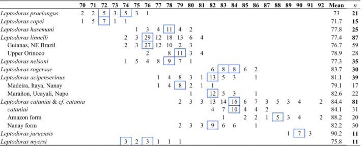
Sum of midlateral plates 70-76 (modally 72 or 74, Table 3). Anterior midlateral plates shallow, covering less than 1/4 of corresponding body depth; posterior margins of dorsal and ventral wings rounded with small serrae. Tympanum without superficial ossifications or occasionally with 1 to 3 very small, weak spines along postotic laterosensory canal. Postcleithral process of moderate length and nearly uniform width, some-what trapezoidal in shape. Middorsal groove on nuchal shield weakly defined. Nuchal foramina absent. Skin relatively smooth.
Dorsal-fin rays I,6; pectoral-fin rays I,8-10 (modally 9); pelvic-fin rays i,6; total anal-fin rays 14-15 (first 4 or 5 un-branched); fin rays i,8/9,i; dorsal procurrent caudal-fin rays 14-17, ventral procurrent caudal-caudal-fin rays 13-16. Dor-sal-fin spine long, moderate width, weakly curved and evenly attenuate with slender distal tip; anterior denticulations antrorse, small and crowded basally, absent from distal quar-ter; posterior denticulations retrorse, separated and well formed nearly to tip. Pectoral-fin spine sturdy, of moderate length, gently bowed with sharp tip; anterior denticulations antrorse, small and absent from distal quarter; posterior den-ticulations retrorse, decreasing in size from midlength to tip; last posterior denticulation subterminal. Pelvic fin short with straight distal margin. Anal fin triangular, tip of longest branched ray more or less even with vertical through base of last ray, distal margin shallowly concave. Caudal fin forked with rounded to weakly pointed lobes.
Coloration in alcohol. Head and body weakly countershaded,
upper sides relatively uniform tan or gray-brown, lower sides (below midlateral plates) occasionally dusky with scattered melanophores (particularly in juveniles wherein the depigmented midlateral plates form white midlateral stripe), undersurfaces white to cream. Melanophores sometimes weakly concentrated in skin between and above dorsal wings of midlateral plates, effecting appearance of faint dusky stripe.
70 71 72 73 74 75 76 77 78 79 80 81 82 83 84 85 86 87 88 89 90 91 92 Mean n Leptodoras praelongus 2 2 5 3 5 3 1 73 21
Leptodoras copei 1 5 7 1 1 71.7 15
Leptodoras hasemani 1 3 4 11 4 2 77.8 25
Leptodoras linnelli 2 3 29 12 18 13 6 4 77.4 87
Guianas, NE Brazil 2 3 27 12 10 2 3 76.7 59
Upper Orinoco 2 8 11 3 4 78.9 28
Leptodoras nelsoni 1 5 4 8 9 7 1 77.3 35
Leptodoras rogersae 6 8 8 6 2 83.7 30
Leptodoras acipenserinus 1 4 8 3 1 13 5 3 1 81.1 39
Madeira, Itaya, Nanay 1 4 8 2 1 1 79.1 17
Marañon, Ucayali, Napo 1 12 5 3 1 82.6 22
Leptodoras cataniai &cf. catania 2 3 3 13 14 16 6 7 3 5 3 4 2 84.4 81
cataniai 4 7 10 4 4 2 84.1 31
Amazon form 1 2 2 1 5 3 4 2 88.2 20
Nanay form 2 3 3 9 6 6 1 82.2 30
Leptodoras juruensis 1 7 3 90.2 11
PROOFS
Dorsal fin with large sub-basal black blotch on dorsal spine,first fin ray and intervening membrane; blotch often expanded posteriorly as dark wedge-shaped band across entire base of fin. Pectoral fin with melanophores concentrated along base and forming radiate streaks on distal membranes (fin rays relatively depigmented). Pelvic fin occasionally with similar but lighter pattern (fewer melanophores). Anal fin cream, hya-line, occasionally with few scattered melanophores. Caudal fin with melanophores forming black streaks on lower mem-branes of upper lobe and upper memmem-branes of lower lobe, remaining portion cream, hyaline.
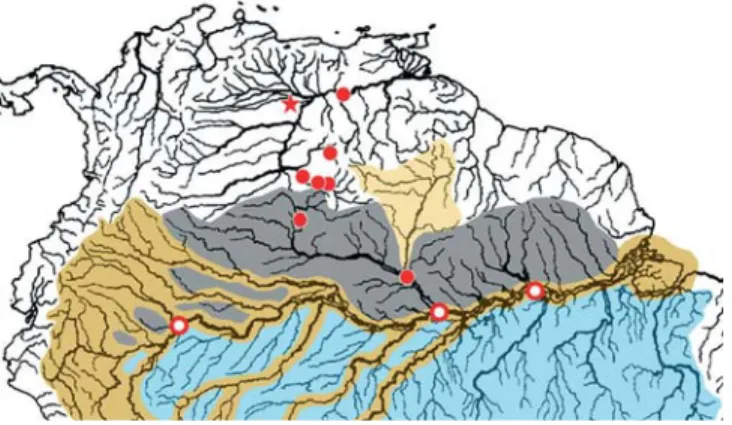
Distribution and habitat.Leptodoras praelongus is known from Brazil and Venezuela in the upper río Orinoco (upstream of Puerto Ayacucho), rio Negro, rio Tocantins, several locali-ties along the rio Amazonas (between the rios Jutaí and Ma-deira), and one site in the Madeira basin (Fig. 9). Many col-lecting sites are associated with large river cataracts. Most records are from blackwaters. However, a few are from whitewater rivers (e.g., rios Branco and Madeira), and one is from a clearwater river (rio Tocantins).
Type-material examined.Leptodoras praelongus: holotype (unique), CAS 148679 [ex. SU 48679] (1, 112.25 mm), rio Ne-gro, Amazonas dr., São Gabriel rapids, Amazonas, Brazil, 1 Feb 1925, C. Ternetz.
Non-type material. Brazil: Amazonas: ANSP 178538 (1), rio
Madeira (Amazonas dr.), 9 km upstream from Vila Urucarituba (3°35’28"S, 58°56’53"W), 6 Aug 1996, CCF-96-78; ANSP 180908 (3), rio Jutaí (Solimões dr.), between Pto. Antunes and Foz do Jutaí (2°50’6"S, 66°55’39"W), 13 Nov 1993, JPS-93-049; ANSP 180909 (1), rio Jutaí (Solimões dr.), between Pto. Antunes and Foz do Jutaí (2°49’39"S, 66°54’55"W), 13 Nov 1993, OTO-93-033; ANSP 180910 (3), rio Negro (Amazonas dr.), 37.8 km upstream from
Carvoeiro (1°12’27"S, 62°15’3"W), 10 Dec 1993, MG-93-037; ANSP 180911 (2), rio Negro (Amazonas dr.), 10 km downstream from Carvoeiro (1°22’8"S, 61°54’22"W), 9 Dec 1993, JGL-93-190; ANSP 180912 (3), rio Negro (Amazonas dr.), 3.8 km down-stream from S. Francisco (1°41’10"S, 61°27’3"W), 6 Dec 1993, JPF-93-158; ANSP 180913 (1, 40.2 mm), rio Negro (Amazonas dr.), 5 miles downstream from Santa Maria (3°0’49"S, 60°24’31"W), 18 Jul 1996, MTP-96-007; ANSP 180916 (1), rio Jutaí (Solimões dr.), downstream of Pto. Antunes and upstream of Copatana (2°56’52"S, 67°00’32"W), 16 Nov 1993, OTO-93-057; INPA ex. 17709 (4), rio Demini (Negro dr.), near mouth of rio Aracá, 22 Jun 1993; INPA 17727 (15), rio Negro (Amazonas dr.), near mouth of rio Branco, 13 Nov 1996; INPA 17761 (1, 112.1 mm), INPA 17764 (3, 65.6-158 mm), rio Negro (Amazonas dr.), igarapé Zamula, near Barcelos, 19 Nov 1996; INPA 17907 (1, 111.1 mm), rio Negro (Amazonas dr.), at mouth of rio Branco, 4 Feb 1998. Para: ANSP 180914 (2), rio Amazonas (Atlantic dr.), 24 km upstream from Gurupa, 6 km downstream of mouth of rio Xingu (1°28’12"S, 51°49’43"W), 14 Nov 1994, AMZ-94-100. Rondônia: INPA un-cataloged (3, 156-185 mm), rio Jamari (Madeira dr.), at and a little upstream of Ariquemes, downstream of mouth of rio Camaã, 15 Nov 1983. Roraima: ANSP 178534 (1), rio Branco (Negro dr.), between Atauba and Caruna, 9.3 km above confluence with rio Negro (1°19’34"S, 61°52’13"W), 8 Dec 1993, JGL-93-171; ANSP 180915 (3), rio Branco (Negro dr.), downstream of Atauba and upstream of Caruna (1°17’55"S, 61°51’21"W), 8 Dec 1993, JGL-93-174; INPA 2004 (1, 147.3 mm), rio Tacutu (Branco dr.), 17 Mar 1988; USNM 332438 (2), rio Branco (Negro dr.), 25 Sep 1992. Tocantins: UNT 153 [ex. 3080] (1), rio Tocantins (Atlantic dr.), fazenda Traçadal, near Paranã City (12°29’S, 48°12’W), 3 Oct 2000. Venezuela: Amazonas: ANSP 162463 (3, 141.4-173 mm), río Pamoni (Casiquiare dr.), ca. 0.5 km from confluence with Casiquiare (2°49’N, 65°55’W), 18-19 Mar 1987, V87-34; ANSP 162464 (4, 120.24-127.9 mm), río Casiquiare, main channel, ca. 1.5 hrs from its confluence with Orinoco (3°5’N, 65°55’W), 16 Mar 1987, V87-24; ANSP 162465 (3, 118-166.8 mm), río Casiquiare, from mouth of río Pamoni to 4 km below (2°48’N, 65°57’W), 17 Mar 1987, V87-27; ANSP 165791 (1, 31.25 mm), río Sipapo (Orinoco dr.), along playas of sand and rock ca. 1-4 km above Pendare (4°51’N, 67°44’W), 12 Nov 1985, V85-8; ANSP 165793 (6, 34.76-49.2 mm), río Sipapo (Orinoco dr.), ca. 3-4 km upstream from Pendare (4°52’N, 67°43’W), 12 Nov 1985, V85-5; MBUCV-V 25602 (1, 151.0 mm), río Canucunuma (Orinoco Dr.), desde campamento 2 km rio abajo, 40 km sobre boca en río Orinoco, 23 Mar 1987; MBUCV-V 25603 (2), río Casiquiare, canal principal ca. 1.5 hrs des de boca en río Orinoco, ca. 1 hr rio abajo, 23 Mar 1987; MBUCV-V 25664 (1, 134.7 mm), same data as ANSP 162465.
Leptodoras copei (Fernández-Yépez, 1968)
Figs. 4c, 6a and 10
Anduzedoras copei Fernández-Yépez 1968: 31, fig. 10 (type locality: río Capanaparo, Orinoco dr., Apure, Venezuela).
Diagnosis. Uniquely distinguished among Leptodoras by the following combination: anterior nuchal plate usually wide (lat-erally expanded) and sharing broad suture with epioccipital, nuchal foramina absent, distal tip of lower labial extension reaching beyond that of upper labial extension, anterior midlateral plates moderately deep (covering a little more than
PROOFS
1/3 of body depth), and pectoral-fin spine short, not reachinganterior origin of pelvic fin when depressed.
Comparisons. Among Leptodoras, only L. hasemani also has a laterally expanded anterior nuchal plate with no nuchal fo-ramina and it is distinguished by having a dark black blotch in distal half of dorsal fin (vs. absent in L. copei).Leptodoras praelongus also has a long lower labial extension, but is dis-tinguished by having a narrower anterior nuchal plate (su-praoccipital usually sharing suture with middle nuchal plate vs. like suture absent in copei), shallower anterior plates
(cov-ering at most 1/4 of body depth vs. 1/3 in copei), and longer pectoral-fin spine (reaching or extending beyond anterior ori-gin of pelvic fin when depressed vs. falling short of pelvic-fin origin in copei).
Description. Somewhat torpedo-shaped; body elongate,
nearly symmetrical in cross-section, predorsal region bluntly cone-shaped, caudal peduncle short, depressed. Eye large with well-developed adipose eyelid.
Mouth subterminal, jaws edentulous in adults and juve-niles as small as 31 mm SL. Maxillary barbel of moderate length,
PROOFS
finishing short of ventromedial extent of gill opening.Sec-ondary maxillary barbels 7-10 (modally 8), flattened, largely overlapping; proximal secondary maxillary barbels with fim-briate anterior margins; distal ones smooth. Remaining fea-tures of buccal hood very similar to L. praelongus except lateral margin of upper labial extension entire or weakly scal-loped. Branchiostegal flap and morphology of first gill arch also as in L. praelongus.
Sum of midlateral plates 70-74 (modally 72, Table 3). Ante-rior midlateral plates of moderate depth, covering slightly more than 1/3 of corresponding body depth; posterior margins of dorsal and ventral wings rounded with small serrae. Tympa-num usually with 3 small ossifications (plates or weak spines) along postotic laterosensory canal; small fixed spine usually visible along posterior margin of neurocranium where postotic laterosensory canal exits supracleithrum. Postcleithral pro-cess short to moderate in length, nearly uniform width or slightly expanded posteriorly. Middorsal groove on nuchal shield weakly defined. Nuchal foramina absent. Skin some-times with very small fleshy papillae scattered over snout and dorsolateral flanks; papillae on snout variously cojoined to form irregular longitudinal ridges especially in juveniles (<100 mm SL).
Dorsal-fin rays I,6; pectoral-fin rays I,8-9 (modally 9); pel-vic-fin rays i,6; total anal-fin rays 13-16 (first 4 or 5 un-branched); fin rays i,8/9,i; dorsal procurrent caudal-fin rays 16-19, ventral procurrent caudal-caudal-fin rays 15-17. Dor-sal-fin spine of moderate length, slender and weakly curved for much of length with short, sharp divergent tip; anterior denticulations antrorse, very small, crowded basally, largely absent from distal half; posterior denticulations retrorse, small but regularly spaced nearly to tip. Pectoral-fin spine sturdy, short, gently bowed with sharp tip; anterior denticulations antrorse, very small, absent from distal third; posterior den-ticulations retrorse, gradually decreasing in size from midlength to tip; last posterior denticulation subterminal. Pelvic fin of moderate length, rounded to weakly pointed. Anal fin triangular, tip of longest branched ray more or less even with vertical through base of last ray, distal margin shal-lowly concave. Caudal fin forked with rounded to weakly pointed lobes.
Coloration in alcohol. Similar to L. praelongus except lower sides more often depigmented. Head and body weakly countershaded, upper sides relatively uniform tan or gray-brown, lower and ventral surfaces white to cream. Base of dorsal spine with black anterior margin and dusky sides, re-maining dorsal fin usually depigmented except for scattered melanophores on skin covering insertion. Pectoral fin with faint concentration of melanophores along base forming light streaks on distal membranes. Pelvic, anal and paired fins cream, hyaline. Caudal fin with melanophores forming black streaks on lower membranes of upper lobe and upper membranes of lower lobe, remaining fin cream. Juveniles often with two dusky blotches on base of caudal fin, one above and one below midlateral plates (blotches overlie aforementioned streaks).
Specimens from the rio Amazonas (all juveniles, hereafter referred to as Leptodoras cf. copei) exhibit a color pattern distinguishable from that typical of juvenile L. copei from the rios Negro and Orinoco. In Amazon specimens the base of the dorsal fin is rather darkly pigmented and the dorsolateral sides beneath the dorsal and adipose fins are often dusky with scattered melanophores. In dorsal view, these markings appear as a pair of opposing chevrons.
Distribution and habitat.Leptodoras copei sensu lato is known from the upper río Orinoco, rio Negro and río Amazonas in Venezuela, Brazil and Peru (Fig. 11). Most collections are from large river habitats in the upper río Orinoco, rio Negro and the mouths of their larger tributaries. Leptodoras copei was described on the basis of a juvenile from the río Capanaparo, tributary to the middle Orinoco. Another collec-tion of small juveniles is from a downstream site on the Orinoco near the mouth of the río Caura.
Specimens examined from the rio Amazonas are all juve-niles, but may represent (based on color pattern) an undescribed species related to L. copei. Santos et al. (1984:52) figured (as Doras cf. lipophthalmus) a specimen from the lower rio Tocantins, Brazil (not mapped in Fig. 11), that may represent an adult of the Amazon species or yet another undescribed species related to L. copei.
Type-material examined. Anduzedoras copei: holotype, FMNH 84069 [ex. AFY 51.311] (65.6 mm), laguna al lado del río Capanaparo (Orinoco dr.), Apure, Venezuela, 22 Mar 1951, A. Fernández-Yépez.
Non-type material.L. copei: Brazil: CAS 158891 [ex. SU 58891]
(3), rio Negro (Amazonas dr.), Cucuhy (= Cucuí), Colombian bor-der, 14 Feb 1925. Amazonas: ANSP 180892 (1), rio Negro (Amazonas dr.), 12.6 km downstream from Moura (1°30’47.2"S, 61°32’19.4"W), 7 Dec 1993, MG-93-033; ANSP 180893 (5), rio Negro (Amazonas dr.), between S. Francisco and S. Francisco de Assis (1°47’20.6"S, 61°24’44.4"W), 6 Dec 1993, JGL-93-163; ANSP 180896 (1), rio Negro (Amazonas dr.), 3.8 km downstream from S. Francisco, 47.2 km upstream from S. Francisco de Assis (1°41’37"S, 61°26’50"W), 6 Dec 1993, JPF-93-158. Roraima: ANSP 180894 (2), rio Branco (Negro dr.), downstream of Atauba and upstream of Caruna (1°17’55"S, 61°51’21"W), 8 Dec 1993,
PROOFS
93-174. Venezuela: Amazonas: ANSP 162461 (3, 61.3-95.9 mm), río Orinoco (Atlantic dr.), near mouth of caño Yaguae (3°33’N, 66°47’W), 24 Mar 1987, V87-47; ANSP 162466 (1, 141.46 mm), MBUCV-V 25616 (1, 129.9 mm), río Orinoco (Atlantic dr.), near mouth of río Iguapo (3°07’N, 65°28’W), 14 Mar 1987, V87-14; CAS 158896 [ex. SU 58896] (2), río Orinoco (Atlantic dr.), playa
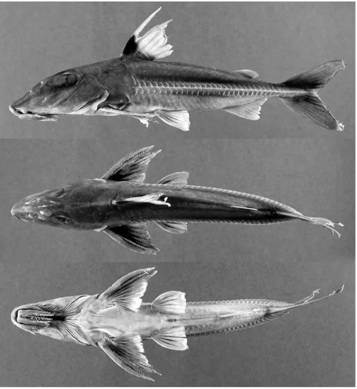
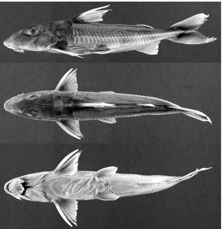
Fig. 12.Leptodoras hasemani, ANSP 175885 (SL 156.4 mm), Essequibo river (Atlantic dr.), Guyana.
PROOFS
Nov 1985, V85-54. L. cf. copei: Brazil: Amazonas: ANSP 180895
(1), rio Solimões (Amazonas dr.), 62.8 km downstream from Bela Vista, 25 km upstream from Manaus (3°12’08"S, 59°53’42"W), 24 Oct 1993, JPF-93-011. Para: ANSP 178540 (1, 49.5 mm), rio Amazonas (Atlantic dr.), 13.6 km downstream from Óbidos, 26.6 km below mouth of rio Trombetas (1°59’58.6"S, 55°25’46.3"W), 27 Oct 1994, JGL-94-081. Peru: Loreto: ANSP 149948 (1, 49.08 mm), ANSP 150185 (1, 42.08 mm), río Amazonas (Atlantic dr.), vicinity of Iquitos, between Isla Iquitos and Isla Lapuna, near Lapuna shore, 9 Oct 1955, P55-9.
Leptodoras hasemani (Steindachner, 1915)
Figs. 4d and 12
Hemidoras hasemani Steindachner 1915: 218 (type locality: rio Branco, Negro dr., Boa Vista, Roraima, Brazil).
Diagnosis. Uniquely distinguished among Leptodoras by having a large black blotch in the distal half of the anterior dorsal-fin rays and membranes. Also in L. hasemani the basal portion of the lower labial extension is uniformly widened and flat compared to other Leptodoras.
Description. Body elongate, weakly compressed; head
com-paratively short, deep, weakly compressed with bluntly pointed snout; ventrally flattened from snout to vent; caudal peduncle moderately elongate, depressed. Eye large with well-developed adipose eyelid.
Mouth subterminal, jaws edentulous in adults and juve-niles as small as 39 mm SL. Oral hood comparatively large and somewhat unique in that origins of first secondary maxillary barbels and upper labial extensions occur well beyond distal margin of dentary. Maxillary barbel long, usually reaching to or slightly beyond ventromedial extent of gill opening. Sec-ondary maxillary barbels 6-9 (modally 7 or 8), flattened, largely overlapping; proximal secondary maxillary barbels with fim-briate anterior margin and smooth posterior margin; distal ones smooth. Upper labial extension lanceolate (tapered dis-tally), moderately elongate, usually straight with smooth ven-tral surface; lateral margin with small distal fimbriae. Lower labial extension finishing more or less even with upper labial extension, basal portion uniformly broad with conspicuously smooth surface, distal portion narrow, attenuate; margins smooth. Interlabial membrane narrow to moderate width (la-bial extensions parallel or convergent), comparatively thin with few very small papillae. Dorsolabial membrane weakly developed, evenly attenuate posteriorly. Two pairs of jaw barbels with comparatively few elongate papillae and broadly cojoined by basal membrane; outer pair only slightly longer than inner pair and shorter than maxillary barbel, cojoined with lower labial extension via narrow membrane. Branchiostegal membrane with greatly expanded fleshy mar-gin that strongly overlaps ventral gill opening; fleshy inner flap along cleithrum well developed, reaching cleithral notch for pectoral spine insertion. Morphology of first gill arch simi-lar to L. praelongus and L. copei.
Sum of midlateral plates 75-80 (modally 78, Table 3). Ante-rior midlateral plates deep, covering more than half of corre-sponding body depth; posterior margins of dorsal and ven-tral wings rounded with many small serrae. Tympanum usu-ally with 3 small ossifications (plates or weak spines) along postotic laterosensory canal; small fixed spine usually vis-ible along posterior margin of neurocranium where postotic laterosensory canal exits supracleithrum. Postcleithral pro-cess short, nearly uniform width or slightly expanded poste-riorly. Middorsal groove on nuchal shield weakly defined. Nuchal foramina absent. Skin comparatively smooth, some-times with minute papillae on head in region between eyes and posterior nares.
Dorsal-fin rays I,6; pectoral-fin rays I,10-11 (modally 10); pelvic-fin rays i,6; total anal-fin rays 14-16 (first 4 to 6 un-branched); fin rays i,8/9,i; dorsal procurrent caudal-fin rays 14-17, ventral procurrent caudal-caudal-fin rays 13-17. Dor-sal-fin spine of moderate length, rather sturdy, weakly curved for much of length, becoming straight near sharp tip; anterior denticulations antrorse, small, crowded basally and largely absent from distal third; posterior denticulations retrorse, moderately sized and regularly spaced to tip. Pectoral-fin spine sturdy, long, distinctly bowed with sharp tip; anterior den-ticulations antrorse, rather small, largely absent from distal third; posterior denticulations strong, retrorse, size rather uniform from midlength to tip; last denticulation subterminal. Pelvic fin of moderate length, very weakly pointed. Anal fin triangular, tip of longest branched ray more or less even with base of last ray, distal margin nearly straight. Caudal fin forked with rounded to bluntly pointed lobes.
Coloration in alcohol. Head and body weakly countershaded,
upper sides relatively uniform tan or light gray, lower sides and ventral surfaces white to cream. Distal half of soft dorsal fin with large black blotch; melanophores concentrated on first two rays and three inter-radial membranes; remaining fin hyaline. Melanophores concentrated on skin covering dor-sal-locking spine and posterior margin of nuchal shield. Dor-sal spine depigmented or with few melanophores along ante-rior margin. Pectoral fin with few melanophores scattered along base and inter-radial membranes, remaining portions cream. Pelvic fins and anal fin relatively depigmented, cream, hya-line. Caudal fin with melanophores forming parallel pair of faint dusky stripes, one on lower rays and membranes of upper lobe and other on upper rays and membranes of lower lobe (portions of stripe overlying membranes appear darker); remaining portions of caudal fin light by comparison with sparsely scattered melanophores, particularly on rays.
PROOFS
Type-material examined.Hemidoras hasemani: lectotype by present designation, NMW 46381 (82 mm, largest), rio Branco (Negro dr.), Boa Vista, Brazil, J. D. Haseman. Paralectotypes: NMW ex. 46381 (3, 77.2-80.1 mm), NMW 46382 (4, 73.1-80.5 mm), same data as lectotype; NMW 46383 (5, 66.0-76.6 mm), rio Branco (Negro dr.), Serra Grande, Brazil, J. D. Haseman; NMW 46384 (1, 46.9 mm), rio Negro (Amazonas dr.), delta of río Negro, Brazil, J. D. Haseman (not this species and reiden-tified here as Hemidoras stenopeltis [Kner 1855]); NMW 46385 (1, 68.6), Conceição, below Boa Vista, Brazil, J. D. Haseman; NMW 46470 (3, 58.8-64.5 mm), same data as NMW 46383.
Non-type material.Brazil: Amazonas: INPA 17705 (12), rio Demini
(Negro dr.), near mouth of rio Aracá, 21 Jun 1993. Roraima: ANSP 178533 (1), rio Branco, 9.3 km upstream from confluence with rio Negro, between Atauba and Caruna (1°19’34"S, 61°52’13"W), 8 Dec 1993, JGL-93-171. Guyana: ANSP 175884 (4, 89.0-167 mm), Essequibo River (Atlantic Dr.), sandbars in vicinity of Maipuri campsite (4°34’17"N, 58°35’17"W), 31 Jan 1997, WGS97-28; ANSP 175885 (1, 157 mm), Essequibo river (Atlantic dr.), 180 yards upstream from Essequibo campsite (Maipuri) (4°45’43"N, 58°45’52"W), 27 Jan 1997, WGS97-23; ANSP 175886 (5), Essequibo river (Atlantic dr.), Essequibo campsite (4°45’41"N, 58°45’53"W), 26 Jan 1997, WGS97-19; ANSP 175887 (3), Essequibo river (Atlantic dr.), sandbars in vicinity of Maipuri campsite (4°34’17"N, 58°35’17"W), 2 Feb 1997, WGS97-31; ANSP 175888 (4), same data as ANSP 175887; ANSP 175889 (1, 86.7 mm), Essequibo river (Atlantic dr.), sand-bar ca. 800 m downstream from Essequibo campsite (Maipuri) (4°45’43"N, 58°45’52"W), 29 Jan 1997, WGS 97-25; ROM 62633 (43), Essequibo river (Atlantic dr.), at southern tip of Indian House Island just north of Kurupukari (4°40’23"N, 58°40’50"W), 15 Oct 1990, H90-78; ROM 62643 (2), Essequibo river (Atlantic dr.), inlet and beach downstream from Kurupukari (4°42’57"N, 58°42’40"W), 10 Oct 1990, H90-43. Venezuela: Amazonas: ANSP 165788 (1, 57.1 mm), río Orinoco (Atlantic dr.), shores of Isla de Raton (5°05’N, 67°48’W), 14 Nov 1985, V85-18; ANSP 180897 (2), río Orinoco (Atlantic dr.), island west of Puerto Venado, 4.5 km south of Samariapo, 56.5 km SW of Puerto Ayacucho (5°12’25"N, 67°48’32"W), 28 Feb 2005, VEN 05-04. Bolivar:
FMNH 109976 (2, 76.8, 152.5 mm), río Caura (Orinoco dr.), sand island 1 km upstream from mouth of caño Mato (7°11’49"N, 65°8’53"W), 9 Dec 2000; LACM 43011 (1), río Orinoco (Atlan-tic dr.), river channel between Palua and San Felix, 180 nau(Atlan-tical miles upstream from sea buoy, 15 Feb 1978; MCNG 19239 (2, 39.2, 43.6 mm), puente del río Orocopiche (Orinoco Dr.), near Ciudad Bolivar, 23 Sep 1988, ABD87-33.
Leptodoras linnelli Eigenmann, 1912
Figs. 4e, 5a, 14 and 15a,b
Leptodoras linnelli Eigenmann 1912:191, pl. 17 (fig. 1), pl. 18 (fig. 1) (type locality: Potaro river (Essequibo dr.) at Tumatumari, Mazaruni-Potaro, Guyana).
Diagnosis. Uniquely distinguished among Leptodoras by shape of upper labial extension: very elongate, straight to weakly curved medially, and nearly uniform in width with a bluntly rounded tip vs. moderately elongate and attenuate (in L. praelongus, copei, hasemani) or moderately to strongly curved and distal portion distinctly expanded with wide lat-eral flap (in L. acipenserinus, nelsoni, rogersae, cataniai, juruensis, myersi).
Leptodoras linnelli is further distinguished from all Leptodoras except L. acipenserinus by having an adipose fin that extends anteriorly as a low thin ridge to a point midway between the anterior insertion of the adipose fin and the pos-terior insertion of the dorsal fin. Leptodoras linnelli is distin-guished from L. acipenserinus by a number of characters including a relatively longer predorsal distance (35.5-38.7% SLvs. 31.6-35.6% in acipenserinus), inner flap of gill opening incomplete (vs. usually complete, nearly reaching cleithral notch in acipenserinus), lower sum of midlateral plates (74-81vs. 77-86 in acipenserinus), anal-fin shape (tip of longest branched ray falls short of vertical through tip of last branched ray in extended anal fin and is more or less even with vertical through base of last ray; line defined by tips angled anteri-orly, forming a 45-90° angle with long axis of body vs. tip of longest branched ray falls well short of base of last branched ray, line defined by tips sharply angled anteriorly, forming a 30-45° angle with long axis of body in acipenserinus), pecto-ral-fin spine length (19.9-26.2% of SL vs. 18.6-21.3% in acipenserinus) and pectoral-fin spine dentation (teeth strongly retrorse along majority of posterior margin, becom-ing less retrorse distally, size relatively uniform along distal half, last denticulation usually subterminal vs. teeth becom-ing gradually larger and less retrorse towards tip of spine, last and/or penultimate denticulation nearly perpendicular to long axis of spine, last denticulation terminal, its lateral mar-gin continuous with tip of pectoral spine in acipenserinus).
Description. Morphometrics summarized in Table 1. Similar
in shape to L. hasemani except head very weakly depressed, snout comparatively longer and more acute; caudal peduncle moderately elongate, depressed. Eye large but with weakly developed adipose eyelid.
PROOFS
Mouth subterminal, jaws edentulous in adults andjuve-niles as small as 50 mm SL. Maxillary barbel long, usually reaching ventromedial extent of gill opening. Secondary max-illary barbels 7-11 (modally 10), flattened, largely overlapping; proximal secondary maxillary barbels with fimbriate anterior margin and smooth posterior margin; distal ones smooth.
Upper labial extension rather elongate, straight to weakly curved medially, width nearly uniform, tip bluntly rounded, ventral surface smooth, distal lateral margin weakly expanded and with small fimbriae. Lower labial extension narrow, at-tenuate, usually finishing even with or slightly beyond upper labial extension; margins smooth, distal tip often with few
PROOFS
small papillae or fimbriae. Interlabial membrane of narrow tomoderate width (labial extensions parallel or convergent),
comparatively thin with few small papillae. Dorsolabial mem-brane weakly developed, evenly attenuate posteriorly. Two
PROOFS
pairs of jaw barbels with scattered elongate papillae; cojoinedby basal membrane; outer pair slightly longer than inner pair and shorter than maxillary barbel, cojoined with lower labial extension via narrow membrane. Branchiostegal membrane with moderately expanded fleshy margin overlapping ventral gill opening; fleshy inner flap along cleithrum incomplete, not reaching cleithral notch for pectoral spine insertion. First gill arch with 15-20 weakly developed gill rakers (length about 2-3 times width); accessory lamellae on medial face of arch and continue well onto medial face of gill filaments (present on every second or third filament); each accessory lamella appears as a column of lappets with first one (opposite rak-ers) enlarged and deflected medially; accessory lamellae ab-sent or only very weakly developed on lateral faces of gill arch and filaments (may appear as one or few small lappets near base of filaments).
Sum of midlateral plates 74-81 (modally 76, Table 3). Ante-rior midlateral plates moderately deep, covering about half of corresponding body depth; posterior margins of dorsal and ventral wings rounded with many small serrae. Tympanum usually with 3 small ossifications (weak spines) along postotic laterosensory canal; small fixed spine usually visible along posterior margin of neurocranium where postotic laterosensory canal exits supracleithrum. Postcleithral pro-cess short, deep and slightly expanded posteriorly. Middor-sal groove on nuchal shield usually well-defined. Nuchal fo-ramina present as small lenticular opening partially or wholly replacing suture between anterior nuchal plate and epioccipital. Skin relatively smooth in adults; juveniles with few, very small papillae on snout and upper head not forming distinct ridges.
Dorsal-fin rays I,6; pectoral-fin rays I,9-10 (modally 10); pelvic-fin rays i,6; total anal-fin rays 12-16 (first 4 to 6 un-branched); fin rays i,8/9,i; dorsal procurrent caudal-fin rays 14-19, ventral procurrent caudal-caudal-fin rays 13-17. Dor-sal-fin spine of moderate length, sturdy, nearly straight (rarely weakly angled) and evenly attenuate with sharp tip; anterior denticulations antrorse, moderately sized and evenly spaced basally, largely absent from distal third; posterior denticula-tions small, retrorse basally, becoming straight and more spaced distally, present nearly to tip. Pectoral-fin spine sturdy, long, weakly bowed with blunt tip; anterior denticulations antrorse, moderately sized nearly to tip; posterior denticula-tions strongly retrorse along most of posterior margin, be-coming less retrorse distally, moderately sized and relatively uniform along distal half; last denticulation usually subtermi-nal. Pelvic fin of moderate length, rounded. Anal fin triangu-lar, tip of longest branched ray more or less even with vertical through base of last anal fin ray, distal margin nearly straight or very shallowly concave. Caudal fin forked with rounded or weakly pointed lobes.
Coloration in alcohol. Head and body coloration similar to L. hasemani; weakly countershaded, upper sides somewhat uniform tan or light gray, occasionally darker brown (Brazil-ian specimens). Wide dusky middorsal stripe evident in some
specimens, flanked by lighter areas along upper sides. Mel-anophores sometimes weakly concentrated in skin between and above dorsal wings of midlateral plates, effecting ap-pearance of faint dusky stripe. Lower sides and ventral sur-faces white to cream.
Dorsal, anal and paired fins without distinct markings. Dorsal fin with faint stippling on rays and skin covering in-sertion; darker stippling concentrated along anterior margin of dorsal spine and around perimeter of locking spine. Pecto-ral and pelvic fins cream to white with faint stippling on dor-sal surfaces. Anal fin cream, hyaline. Caudal fin with parallel pair of dusky stripes as in L. hasemani.
Distribution and habitat.Leptodoras linnelli is known from the upper río Orinoco (ríos Ventuari and Mavaca) and a tribu-tary of the Casiquiare (río Siapa), Venezuela; Atlantic Coast drainages of the Guianas and northern Brazil (e.g., Essequibo, Demerara, Araguari); and the rios Uatumã (Amazonas dr.), Tacutu (Branco dr.) and lower Demini (Negro dr.), Brazil (Fig. 16). The distribution of L. linnelli appears to be restricted to river systems draining the Guiana Shield. Most collections are from whitewater or turbid rivers, often in places with sandy beaches and swift currents (e.g., cataracts).
Type-material examined. Leptodoras linnelli: holotype, FMNH 53561 [ex. CM 1626a] (153.5 mm), Potaro river (Essequibo dr.), Tumatumari, Guyana, 1908, C.H. Eigenmann et al. Paratypes (33 of 36, 3 missing): Guyana: BMNH 1911.10.31.73-74 [ex. CM 1627a-e, ex. IU 12022] (1 + 1*, 162.2 mm), same data as holotype; BMNH 1911.10.31.75 [ex. CM 1627a-e, ex. IU 12022, Tumatumari or ex. CM 1629a, ex. IU 12023, Crab Falls] (1, 70.9 mm), Potaro river (Essequibo dr.), Tumatumari or Essequibo river (Atlantic dr.), Crab Falls, 1908, C.H. Eigenmann et al.; CAS 59775 [ex. CM 1627a-e, ex. IU 12022] (5, 52.1-173.5 mm), same data as holotype; CAS 59776