Mycobacterium bovis
BCG but not
Mycobacterium leprae
Induces TNF-
α
Secretion in Human Monocytic THP-1 Cells
Martha M Oliveira, Rosane Charlab*, Maria Cristina V Pessolani/
+Laboratório de Hanseníase, Instituto Oswaldo Cruz-Fiocruz, Av. Brasil 4365, 21045-900 Rio de Janeiro, RJ, Brasil *Centro Brasileiro de Pesquisas Físicas, CNPq, Rio de Janeiro, RJ, Brasil
In this study, we compared the level of TNF-α secretion induced in monocytic THP-1 cells after phagocytosis of Mycobacterium leprae, the causative agent of leprosy, and M. bovis BCG, an attenu-ated strain used as a vaccine against leprosy and tuberculosis. The presence of M. leprae and BCG was observed in more than 80% of the cells after 24 h of exposure. However, BCG but not M. leprae was able to induce TNF-α secretion in these cells. Moreover, THP-1 cells treated simultaneously with BCG and M. leprae secreted lower levels of TNF-α compared to cells incubated with BCG alone. M. leprae
was able, however, to induce TNF-α secretion both in blood-derived monocytes as well as in THP-1 cells pretreated with phorbol myristate acetate. The inclusion of streptomycin in our cultures, together with the fact that the use of both gamma-irradiated M. leprae and heat-killed BCG gave similar results, indicate that the differences observed were not due to differences in viability but in intrinsic properties between M. leprae and BCG. These data suggest that the capacity of M. leprae to induce TNF-α is dependent on the stage of cell maturation and emphasize the potential of this model to explore differ-ences in the effects triggered by vaccine strain versus pathogenic species of mycobacteria on the host cell physiology and metabolism.
Key words: Mycobacterium leprae - BCG - THP-1 - TNF-α
Mononuclear phagocytes are target cells for pathogenic mycobacteria that generally require an intracellular environment in which they survive and replicate. Although these cells can offer a quite hostile environment to the entering pathogen, they can also provide unique advantages to the infec-tious organism. They are long-living cells and thus provide a potential long-term habitat for the bac-terial invader. In addition, macrophages play a pivotal role in host defense against infection, pri-marily due to the vast array of mediators that these cells can secrete, offering in this way to the in-vader organism an opportunity to manipulate the immune system to its own advantage (Russel 1995). Immunity to intracellular pathogens de-pends mainly upon the activation of IFN-γ produc-ing CD4+ T cells, the so called Th1 response, which
increases the microbicidal capacity of macroph-ages (Kaufmann 1995). In this context, microor-ganisms that supress or avoid, early during
infec-This work was supported by a Papes 2 grant from the Oswaldo Cruz Foundation and CNPq (Brazil).
+Corresponding author. Fax: +55-21-2270.9997. E-mail:
[email protected] Received 14 November 2000 Accepted 3 May 2001
tion, infected macrophages to produce cytokines such as TNF-α and IL-12, cricial for the emergency of a Th1-like response (Trinchieri 1993, Flesch et al. 1995), will favor the development of a suscep-tible phenotype.
emphasized in a report that describes the deleteri-ous effects of deletion in TNF-α gene on myco-bacterial infections (Flynn et al. 1995). Our re-sults demonstrate that BCG but not M. leprae in-duces TNF-α secretion in THP-1 cells, emphasiz-ing the potential of this model to explore differ-ences in signaling pathways triggered by vaccine versus pathogenic species of mycobacteria.
MATERIALS AND METHODS
Source of reagents - RPMI 1640 medium, peni-cillin/streptomycin, L-glutamine were obtained from Gibco BRL (Gaithersburg, MD, USA); phorbol-12-myristate-13-acetate (PMA) was pur-chased from Sigma Chemical Co. (St. Louis, Mis-souri, USA).
Mycobacteria - Frozen aliquots of M. bovis
BCG (Pasteur strain) had been produced by the Pasteur Institute (Paris, France), and were kindly donated by Dr Sylvio CG Costa (Oswaldo Cruz Foundation, Brazil). The mycobacteria was stored in vials at a concentration of about 5x10⁸ CFU/ml
in PBS at -20°C until use. Armadillo-derived M. leprae was kindly donated by Dr PJ Brennan (De-partment of Microbiology, Colorado State Univer-sity, Fort Collins, CO) through NIAID contract NO155262. For each experiment, a sample was thawed and diluted in a small volume of PBS supplemented with 0.005% Tween 80. Before addition to blood-derived macrophages or THP-1 cells, clumps of bacteria were removed by repeated passages through a 27 gauge needle and mild soni-cation for 10 min. Total M. leprae and BCG counts were estimated as described by Shepard and MacRae (1968).
Cell culture and infection
THP-1 cells - Cells from a human myelomono-cytic cell line, THP-1 (American Type Culture Collection, Rockville, MD), were grown as sus-pension cultures in RPMI medium supplemented with 10% heat-inactivated fetal calf serum, 100 U/ ml penicillin, 100 µg/ml streptomycin and 2 mmol/ l glutamine (RPMI complete medium) at 37°C in a humidified atmosphere of 5% CO2 and used be-tween 3-14 passages. For the experiments, 2x10⁵
cells/ml/well were dispensed in 24-well microtiter plates in the same medium and further incubated with or without mycobacteria (BCG and/or M. leprae) or PMA (0.16-160 nM) up to 48 h at 37°C in a humidified atmosphere of 5% CO2. A frac-tion of the PMA- or BCG-treated THP-1 cells hered to culture wells and for analysis of the ad-herent population, glass cover slips were placed in culture wells before cell addition. The number of adherent cells was estimated by subtracting the number of suspended cells from that of seeded cells. In some experiments, THP-1 cells were first
culti-vated in the presence of PMA for 4 h and then washed and incubated in fresh RPMI complete medium for an additional 24 h in the presence of
M. leprae.
Human blood-derived monocytes - Normal human peripheral blood mononuclear cells (PBMC) were separated by Ficoll-Hypaque den-sity gradients from sterile heparinized blood. The cells from the interface were washed three times in PBS (without calcium and magnesium) supple-mented with 1% AB human serum, seeded at a density of 1x10⁶ per well per ml in 24-well
microtiter plates in RPMI-1640 medium supple-mented with 5% AB human serum, 100 U/ml peni-cillin, 100 µg/ml streptomycin and 2 mmol/l glutamine, and allowed to settle at 37°C in a hu-midified atmosphere of 5% CO2. After 24 h, the non-adherent cells were removed with three changes of warm PBS and to the adherent cells (about 2x10⁵ per well), 1 ml of complete RPMI
containing BCG, M. leprae or no mycobacteria was added and the cells were cultured up to 48 h.
Enough mycobacteria were added to the wells to achieve bacteria/cell ratios of approximately 1:1, 10:1 and 100:1. Since most armadillo-derived M. leprae are likely dead, experiments were always performed in the presence of streptomycin, an an-tibiotic known to be effective against M. leprae
and BCG (Winder 1982, Gelber 1987). By doing this, the differences observed in TNF-α secretion in this study would be essentially due to intrinsic differences between M. leprae and BCG, and not to differences in their viability. After 2, 24 and 48 h of incubation, the culture supernatants of PBMC or THP-1 cells were collected and recentrifuged at 14,000 xg to remove extracellular mycobacteria and stored at -70°C until determination of TNF-α levels by ELISA. Viability of the blood-derived monocytes or THP-1 cells was assessed by stain-ing the cells with PBS containstain-ing etidium bromide (4 µg/ml) and fluorescein diacetate (20 µg/ml) for 10 min at room temperature. Glass slides were analyzed and viewed using a Zeiss fluorescent microscope.
Evaluation of mycobacteria phagocytosis
-Duplicate wells of THP-1 cells were infected with BCG or M. leprae as described above and after variable times of incubation, cells were harvested by centrifugation and washed three times to remove extracellular mycobacteria. The percentage of cells containing acid-fast organisms was determined by cytospin and Ziehl-Neelsen staining, and a minimun of 300 cells in each of duplicate slides were counted.
(Pharmingen, San Diego, CA) using specific pairs of anti-TNF-α monoclonal antibodies and human recombinant TNF-α as standard. The detection limit of TNF-α was 250 pg/ml.
RESULTS
BCG but not M. leprae induced TNF-α secre-tion in THP-1 cells - The capacity of M. leprae
and BCG of eliciting TNF-α secretion in THP-1 was evaluated using PMA as a positive control. After 2, 4, 24 and 48 h of continuous exposure, the TNF-α levels in culture supernatants were deter-mined by ELISA. Light microscopic examination of acid-fast stained cultures treated with a bacte-ria/cell ratio of 10:1 showed that at least 80% of THP-1 cells contained M.leprae or BCG after 24 h treatment. Fig. 1 shows that the different stimuli evoked distinct kinetics and levels of TNF-α se-cretion in THP-1 cells. While PMA-induced
TNF-α secretion could be observed only after 48 h of incubation in a dose-dependent manner (Fig. 1a), BCG-induced TNF-α peaked after 2 h of culture, and was continuously detected up to 48 h (Fig. 1b). In contrast, M. leprae induced no detectable
TNF-α in the conditions examined (Fig. 1b). While BCG was able to induce TNF-α production even at bac-teria/cell ratio of 1:1, no detectable levels of this cytokine were observed even when cultures were stimulated with M. leprae at bacteria/cell ratio of 100:1 (data not shown). Experiments performed with gamma-irradiated M. leprae and heat-killed BCG gave identical results (data not shown).
To ensure that the lack of detectable levels of TNF-α in M. leprae-stimulated THP-1 cultures were not related to the M. leprae sample used throughout this work, blood-derived monocytes of two healthy individuals were stimulated for up to 48 h with the same M. leprae sample used through
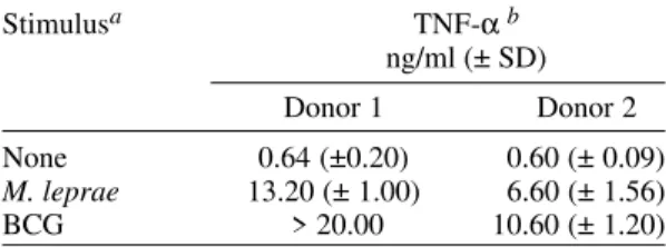
TABLE
TNF-α production of human monocytes infected with BCG and Mycobacterium leprae
Stimulusa TNF-αb
ng/ml (± SD)
Donor 1 Donor 2
None 0.64 (±0.20) 0.60 (± 0.09)
M. leprae 13.20 (± 1.00) 6.60 (± 1.56)
BCG > 20.00 10.60 (± 1.20)
a: mycobacteria was added to cultures at bacteria/cell ratio of 1:1; b: monocytes of two healthy subjects were stimulated with BCG or M. leprae. Culture supernatants were harvested at 24 h and TNF-α concentration was determined by ELISA. Results are expressed as mean ± SD of duplicates.
Fig. 1: kinetics of TNF-α production by THP-1 cells exposed to PMA (a), BCG (b), or Mycobacterium leprae (b). THP-1 cells
(2x10⁵/ml) were treated with PMA (0.16-160 nM, M. bovis BCG or M. leprae (ratio bacteria/cell of 10:1). Supernatants were
harvested at the indicated times and TNF-α levels were determined by ELISA. Data are expressed as mean ± S.D. of ELISA
duplicates and are representative of three independent assays.
out this study and with BCG (bacteria/cell ratio of 1:1 and 10:1). As can be seen in the Table, both M. leprae and BCG were able to induce TNF-α in blood-derived monocytes. TNF-α was detected in culture supernatants as early as 2 h of incubation and maximum values were generally observed at 24 h.
M. leprae inhibited TNF-α induced by BCG
-To determine whether M. leprae suppressed
TNF-α production by THP-1 cells or simply acted as a poor stimulus, mixtures of M. leprae and BCG were used to treat THP-1 cells and the TNF-α produc-tion was measured by ELISA. Mixtures contain-ing equal amounts of BCG and M. leprae (both mycobacteria at bacteria/cell ratio of 10:1) elicited the production of lower levels of TNF-α when com-pared with BCG alone at ratios of 10:1 or 20:1 (Fig. 2), suggesting that M. leprae affects the ability of THP-1 cells to respond to BCG.
0.16 1.6 16 40 80 160 0
2 4 6 8 10
PMA, nM
a
M. leprae BCG
Control 0
1 2 3 4
5
b
TN
F-α
(n
g
/m
l)
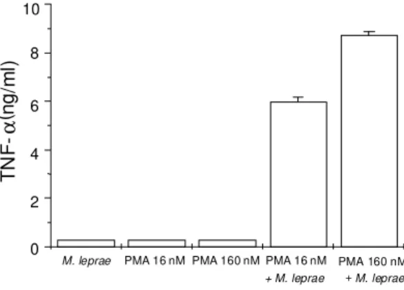
2h4hTHP-1 cells pretreated with PMA were able to produce TNF-α in response to M. leprae - Since THP-1 cells represent a relative immature mono-cytic cell line (Tsuchiya et al. 1980, Friedland et al. 1992), it was of interest to investigate whether more differentiated THP-1 cells could secrete
TNF-α in response to M. leprae. Differentiation of THP-1 cells was induced by exposing cells (2 x THP-105 cells/
ml) to 16 or 160 nM PMA, described as a potent inducer of differentiation in THP-1 cells (Tsuchiya et al. 1982). After an additional 4 h incubation, cells were washed and treated with M. leprae (bac-teria/cell ratio of 10:1) or incubated with medium alone (control) for 24 h more. Supernatants were then collected for TNF-α measurement by ELISA. Some cultures were continuously exposed to PMA throughout the experiment as an additional con-trol. Fig. 3 shows that while cultures continuously exposed to PMA or to M.leprae or exposed to PMA for 4 h and then incubated with medium alone se-creted either undetectable or low levels of TNF-α, cultures pretreated with PMA and then infected with M. leprae secreted up to 9 ng/ml of TNF-α.
DISCUSSION
Interaction of bacterial pathogens with their host cells triggers signal transduction pathways which lead to a variety of cellular responses and ultimately favor their perpetuation in the host. Among these responses there is an extensive reor-ganization of the cytoskeleton resulting in morpho-logical changes, and the secretion of cytokines into the medium. Although this interference in host
Fig. 2: effect of Mycobacterium leprae on TNF-α production
of BCG-treated THP-1 cells. THP-1 cells (2x10⁵/ml) were
either exposed to M. bovis BCG (ratio of 10:1) plus M. leprae
(ratio 10:1), to M. leprae alone (ratios of 10:1 and 20:1) or to
BCG alone (ratios of 10:1 and 20:1). Supernatants were
har-vested after 2 h of incubation and TNF-α levels determined
by ELISA. Data are expressed as mean ± S.D. of three inde-pendent experiments carried out in duplicates.
cell metabolism could represent a central feature of their pathogenesis, these events are poorly un-derstood in mononuclear phagocytes infected with mycobacteria and have only recently received at-tention by a few investigators (Kusner et al. 1996, Mendez-Samperio et al. 1996, Olivier et al. 1998, Malik et al. 2000). In the present study, we have employed an in vitro model to investigate the early molecular events evoked by mycobacteria after their phagocytosis by human monocytes. THP-1 cells, a human myelomonocytic cell line that lately has been extensively employed in studies of mecha-nisms of maturation/differentiation from mono-cytes to macrophages (for review see Hass 1992), were used as host cells. To our knowledge, this is the first report describing the challenge of THP-1 cells with M. leprae, and its effects were compared with those provoked by BCG.
We investigated TNF-α production by THP-1 cells challenged with M. leprae, BCG or PMA. Besides representing a good parameter for mono-cyte activation/differentiation, TNF-α is known to play a critical role in mycobacterial infection, be-ing involved both in protective as well as in patho-logical response (Kindler et al. 1989, Lima et al. 2000). Our data indicate that phagocytosis of ar-madillo-derived M. leprae by THP-1 cells is ac-companied by undetectable levels of TNF-α se-cretion under the experimental conditions em-ployed in this study. Conversely, entry of BCG in these cells, even at a very low bacteria-to-cell ra-tio, resulted in appreciable secretion of TNF-α. The
Fig. 3: TNF-α production by PMA-differentiated THP-1 cells
in response to Mycobacterium leprae. THP-1 cells (2x10⁵/
ml) were plated and incubated in RPMI complete medium for 4 h. After this time, THP-1 cells were stimulated with PMA
(16 or 160 nM) for more 4 h, then washed and exposed to M.
leprae (MOI=10) or incubated in medium alone, as in
Meth-ods. Some cultures were continuously exposed to PMA throughout the experiment as an additional control.
Superna-tants were harvested after 24 h of incubation and TNF-α
lev-els were determined by ELISA. Data are expressed as mean ± S.D. of ELISA duplicates and are representative of two inde-pendent assays.
0 2 4 6 8 10 12 14
BCG 10:1 + M. leprae 10:1 BCG
20:1 BCG 10:1 M. leprae 20:1 M. leprae
10:1 Control
TN
F
-
α
(n
g
/m
l)
0 2 6 8 10
PMA 160 nM + M. leprae PMA 16 nM
+ M. leprae PMA 160 nM PMA 16 nM
M. leprae
TN
α
(ng
/m
l)
induction of TNF-α by BCG was not due to LPS contamination, since the same levels of the cytokine were assayed in the presence of polymyxin B (data not shown). The different behavior of THP-1 cells after challenge with BCG or M. leprae is also not attributed to differences in the extent of mycobac-teria uptake: examination of cells by light micros-copy after 24 h of exposure to mycobacteria at a ratio bacteria/cell of 10:1 revealed the presence of acid fast bacilli in more than 80% of the cells. Moreover, the inclusion of streptomycin in our cultures, together with the fact that the use of both gamma-irradiated M. leprae and heat-killed BCG gave similar results (data not shown), indicate that the differences observed were not due to differ-ences in viability but to intrinsic properties between
M. leprae and BCG.
Next, we examined the effect of M. leprae on TNF-α secretion using primary human monocytes instead of THP-1 as host cells. In accordance with other findings (Santos et al. 1993, Suzuki et al. 1993, Lima et al. 2000), exposure of these cells to
M. leprae induced TNF-α secretion. Moreover, pretreatment of THP-1 cells with PMA, a well known inducer of differentiation, led these cells to produce TNF-α in response to M. leprae. There-fore, the incapacity of M. leprae to induce detect-able levels of TNF-α was inherent to THP-1 cells and can perhaps related to the fact that they can be considered immature monocytes.
The employment of THP-1 as host cells is in agreement with earlier observed differences be-tween BCG and M. leprae in modulating host cell metabolism previously reported by other groups. In agreement with its well known immunostimu-latory effect, it has been shown that BCG is a bet-ter inducer of TNF-α, IL-1 and IL-6 from human and animal macrophages, when compared to M. leprae (Suzuki et al. 1993). It has also been shown that phagocytic cells (monocytes/neutrophils) pro-duce high levels of oxygen free radicals when chal-lenged with BCG, but not with M. leprae (Holzer et al. 1988). Indeed, several reports suggest that
M. leprae has immunosupressive rather than
immunostimulant effects on macrophages. In this regard, it has been shown that M. leprae infected macrophages became deactivated by inducing the secretion by these cells of suppressive mediators such as prostaglandins, IL-1 receptor agonist and TGF-β (Sibley & Krahenbuhl 1988, Suzuki et al. 1993, Goulart et al. 1996). Along these lines, in this study we also found that when mixtures of M. leprae and BCG were used to challenge THP-1, significantly lower levels of TNF-α were secreted, indicating a suppressive effect by M. leprae.
The formation of granuloma can be considered, the principle host defense to mycobacterial
infec-tion in mice infected with BCG. It has been dem-onstrated that local TNF-α production is crucial to granuloma formation (Kindler et al. 1989). In this regard, THP-1 is a relative immature human mono-cytic cell line, representative of cells involved in granuloma formation. The findings of the present study may then suggest that the capacity of young monocytes to respond to BCG by TNF-α secre-tion would tend to markedly amplify the initial in-flammation and creates conditions favoring pro-gression of granuloma.
In conclusion, the in vitro model herein em-ployed allows the detection of differences in cell metabolism, including cytokine production, trig-gered by host-derived M. leprae and BCG. These results are in agreement with previous data that indicate THP-1 as being a useful model to investi-gate the nature of mycobacteria/monocyte inter-actions (Stokes & Doxsee 1999). The use of THP-1 as host cells have two advantages over the use of blood-derived monocytes in that (1) the variabil-ity between donors commonly encountered with primary cells is avoided; and (2) large numbers of cells can be readily cultivated. Our data suggest that THP-1 cells can be useful for a better under-standing of early signal transduction pathways in-volved in the host response against vaccine versus pathogenic species of mycobacteria.
REFERENCES
Flesch IEA, Hess JH, Huang S, Aguet M, Rothe J, Bluethmann H, Kaufmann SHE 1995. Early interleukin 12 production by macrophages in re-sponse to mycobacterial infection depends on inter-feron γ and tumor necrosis factor α. J Exp Med181: 1615-1621.
Flynn JL, Goldstein MM, Chan J, Triebold KJ, Pfeffer K, Lowenstein CJ, Schreiber R, Mak TW, Bloom BR 1995. Tumor necrosis factor-alpha is required in the protective immune response against Mycobac-terium tuberculosis in mice. Immunity2: 561-572. Friedland RJ, Shattock JD, Remick DG, Holliman RE,
Griffin GE 1992. Secretion of Interleukin-8 follow-ing phagocytosis of Mycobacterium tuberculosis by human monocyte cell lines. Eur J Immunol22: 1373-1378.
Gelber RH 1987. Further studies of the killing of M. leprae by aminoglycosides: reduced dosage and fre-quency of administration. Int J Lepr Other Mycobact Dis 55: 78-82.
Goulart IM, Figueiredo F, Coimbra T, Foss NT 1996. Detection of transforming growth factor-beta 1 in dermal lesions of different clinical forms of leprosy. Am J Pathol148: 911-917.
Hass R 1992. Retrodifferentiation - An alternative bio-logical pathway in human leukemia cell. Eur J Cell Biol58: 1-11.
re-sponses to Mycobacterium leprae and Mycobacte-rium bovis Bacillus Calmette-Guérin. J Immunol 141: 1701-1708.
Kaufmann SHE 1995. Immunity to intracellular micro-bial pathogens. Immunol Today16: 338-342. Kindler V, Sappino AP, Grau GE, Piguet PF, Vassalli P
1989. The inducing role of tumor necrosis factor in the development of bactericidal granulomas during BCG infection. Cell56: 731-740.
Kusner DJ, Hall CF, Schlesinger LS 1996. Activation of phospholipase D is tightly coupled to the phago-cytosis of Mycobacterium tuberculosis or opsonized zymosan by human macrophages. J Exp Med184: 585-595.
Lima MCBSL, Pereira GMB, Rumjanek FD, Gomes HM, Duppre N, Sarno EN, Pessolani MCV 2000. Immunologycal cytokine correlates of protective immunity and pathogenesis in leprosy. Scand J Immunol51: 419-428.
Malik ZA, Denning GM, Kusner DJ 2000. Inhibition of Ca2+ signaling by Mycobacterium tuberculosis is associated with reduced phagosome-lysosome fusion and increased survival within human macrophages. J Exp Med 191: 287-302.
Mendez-Samperio P, Hernandez-Garay M, Vazquez NA 1996. Cellular activation induced by BCG is a PTK-dependent event. Cell Immunol 171: 147-152. Olivier M, Cook P, Desanctis J, Hel Z, Wojciechowski
W, Reiner NE, Skamane E, Radzioch D 1998. Phe-notypic difference between Bcgr and Bcgs macroph-ages is related to differences in protein-kinase-C-dependent signalling. Eur J Biochem 251: 734-743. Russell DG 1995. Of microbes and macrophages: en-try, survival and persistence. Curr Opin Immunol7: 479-484.
Santos DO, Suffys PN, Bonifácio K, Marques MA, Sarno
EN 1993. In vitro tumor necrosis factor production by mononuclear cells from lepromatous leprosy pa-tients and from papa-tients with erythema nodosum leprosum. Clin Immunol Immunopathol67: 199-203. Shepard CC, MacRae DH 1968. A method for counting
acid-fast bacteria. Int J Lepr36: 78-82.
Sibley LD, Krahenbuhl JL 1988. Induction of unre-sponsiveness to gamma interferon in macrophages infected with Mycobacterium leprae. Infect Immun 56: 1912-1919.
Stokes RW, Doxsee D 1999. The receptor-mediated up-take, survival, replication, and drug sensitivity of Mycobacterium tuberculosis within the macrophage-like cell line THP-1: a comparison with human mono-cyte-derived macrophages. Cell Immunol197: 1-9. Suzuki K, Fukutomi Y, Matsuoka M, Toni K, Hayashi
H, Takii T, Oomoto Y, Onozaki K 1993. Differen-tial production of interleukin 1 (IL-1), IL-6, tumor necrosis factor, and IL-1 receptor antagonist by hu-man monocytes stimulated with Mycobacterium leprae and M. bovis BCG. Int J Lepr61: 609-618. Trinchieri G 1993. Interleukin-12 and its role in the
gen-eration of Th1 cells. Immunol Today 14: 335-337. Tsuchiya S, Kobayashi Y, Goto Y, Okumura H, Nakae
S, Konno T, Tada K 1982. Induction of maturation in cultured human monocytic leukemia cells by a phorbol diester. Cancer Res42: 1530-1536. Tsuchiya S, Yamabe M, Yamaguchi Y, Kobayashi Y,
Konno T, Tada K 1980. Establishment and charac-terization of a human acute monocytic cell line (THP-1). Int J Cancer26: 171-176.