Behavioral and synaptic circuit
analysis in models of
neuropsychiatric disorders
Dissecting the
in vivo
role of the
postsynaptic density proteins
nArgBP2 and Shank3 using genetic
engineered mice
Cátia Gisela Rebordelo Marques Feliciano
Dissertation presented to obtain the Ph.D degree in Biology
Instituto de Tecnologia Química e Biológica | Universidade Nova de LisboaOeiras,
September, 2011
Behavioral and synaptic circuit
analysis in models of
neuropsychiatric disorders
Dissecting the
in vivo
role of the
postsynaptic density proteins
nArgBP2 and Shank3 using genetic
engineered mice
Cátia Gisela Rebordelo Marques Feliciano
Dissertation presented to obtain the Ph.D degree in Biology
Instituto de Tecnologia Química e Biológica | Universidade Nova de Lisboa
Research work coordinated by:
Oeiras,
Behavioral and synaptic circuit analysis in models of
neuropsychiatric disorders
Dissecting the in vivo role of the postsynaptic density proteins nArgBP2 and Shank3 using genetic engineered mice
Cátia Gisela Rebordelo Marques Feliciano
Dissertação apresentada para obtenção do grau de doutor em Biologia pelo Instituto de Tecnologia Química e Biológica da Universidade de Nova de Lisboa
Orientação do Professor Doutor Guoping Feng
Duke University Medical Center and Massachusetts Institute of Technology Co-orientação do Professor Doutor Sukalyan Chatterjee
Director do Programa Gulbenkian de Doutoramento em Biomedicina (PGDB) Investigador Principal no Centro de Neurociências e Biologia Celular (CNC)
Esta tese foi desenvolvida com apoio financeiro da FCT e do FSE no âmbito do Quadro Comunitário de Apoio (SFRH/BD/15855-2005)
Oeiras
!
!
!
!
!
!
Table of Contents
T
ABLE OFC
ONTENTS... iii
A
CKNOWLEDGMENTS... v
S
UMÁRIO... vii
S
UMMARY... xi
A
BBREVIATIONS... xiii
CHAPTER 1 ... 1
G
ENERALI
NTRODUCTION... 1
Neuropsychiatric Disorders ... 3
Overview of Neuropsychiatric Disorders ... 3
Mood Disorders ... 5
Heritability of Mood Disorders ... 8
Bipolar Disorder ... 10
Candidate Genes for Bipolar Disorder ... 10
Sleep and Bipolar Disorder ... 13
Circadian Rhythms ... 14
Modeling Bipolar Disorder ... 19
Circuit Dysfunctions in Bipolar Disorder ... 25
Synaptic Dysfunctions in Bipolar Disorder ... 28
Shank/SAPAP/nArgBP2 protein network at the PSD ... 31
Autism Spectrum Disorders ... 33
Overview of Autism Spectrum Disorders ... 33
Neuroligins and Neurexins in Autism ... 34
Phelan-McDermid Syndrome and Shank3 ... 36
O
BJECTIVES ANDT
HESISO
UTLINE... 39
CHAPTER 2 ... 41
S
HANK3
MUTANT MICE AS A MODEL FORASD
S... 41
CHAPTER 3 ... 69
C
HARACTERIZATION OF NA
RGBP2
EXPRESSION IN THE MOUSE BRAIN... 69
CHAPTER 4 ... 103
N
A
RGBP2
MUTANT MICE AS A MODEL FORBPD ... 103
CHAPTER 5 ... 141
F
INALD
ISCUSSION... 143
R
EFERENCES... 147
!
Acknowledgments
I am sure I will never be able to completely thank everyone who supported me throughout this journey. Nonetheless, I will take this opportunity to express my gratitude to those whose friendship, mentoring and support were critical at the personal and academic level.!
First and foremost I would first like to thank my Ph.D. supervisor Dr. Guoping Feng for taking me as a student and integrating me in his team. I thank him for being an enthusiastic leader and mentor, for the stimulating discussions, for his patience, his tireless energy, encouragement and insightful advices. I also thank my Ph.D. supervisor Dr. Sukalyan Chatterjee for making me push myself and enriching my scientific horizon since my first day in graduate school. I am very thankful for his support and ideas concerning my future.
I would like to gratefully acknowledge my professors at Universidade da Beira Interior; especially Dr. João Queiroz, Dr. Ignacio Verde, Dra. Graça Baltazar and Dr. José Francisco Cascalheira, for their teaching excellence and for providing a first contact with outstanding research.
I thank all the members of the Feng lab, for the stimulating and fun work environment and for all their help and advices. I am particularly thankful to Jonathan, Zhonghua, Shengli, Molly and Dongqing. I also would like to acknowledge Li Qiu, Jimmy Gross and Qun Liu for technical support and assistance with the injection of ES cells and Dr. Ramona Rodriguiz and Dr. William Wetzel for advice in behavioral experiments. Finally, I am particularly grateful to João Peça for his endless patience, encouragement and support during every step of graduate school.
I am also indebted to the Portuguese community at Duke for making Durham a familiar and enjoyable environment in which to grow as a person and as a scientist. I am especially grateful to Albino Jorge Oliveira-Maia, Teresa Carona Maia, Susana Silva, Rui Peixoto, Ana Oliveira and Catarina Cunha. Finally, Dr. Rui Costa was a very good friend, first welcoming in me to North Carolina and always providing outstanding scientific advices and encouragement.
place to go back to. Molly Heyer, Marek Łaska, Michael Wells, Katie King and Thais Vinholo for making my stay in North Carolina happier and full of good memories.
Agradeço ao IGC e ao PGDB pela oportunidade conferida e pela excepcional formação científica e pedagógica durante os cursos leccionados e o apoio financeiro. Agradeço também à Fundação para a Ciência e a Tecnologia pelo financiamento através de uma bolsa individual de doutoramento (SFRH/BD/15855-2005). I would also like to thank the National Institute of Mental Health and the Simons Foundation Autism Research Initiative for the financial support to the projects I was involved.
!
Sumário
Um ambicioso objectivo em neurociências consiste em compreender como os genes afectam a biologia neuronal, a função sináptica e o comportamento. Os genes que se pensam estar envolvidos em doenças psiquiátricas atraem maior atenção devido à alta probabilidade da sua perturbação originar alterações salientes em processos neurobiológicos. Desta forma, estes genes permitem também uma melhor compreensão de questões biomédicas relevantes. Usando o ratinho (murganho) como animal modelo pode acelerar este processo de descoberta pois trata-se de uma espécie capaz de padecer de manipulações ao nível genético, permitindo assim a recriação ortóloga de mutações humanas. Ao mesmo tempo, o conhecimento actual da neurobiologia comportamental em ratinhos pode conectar certos endofenótipos como sendo reminiscentes de perturbações humanas.
No meio pós-sináptico de neurónios glutamatérgicos encontra-se um aglomerado de proteínas essenciais para o funcionamento sináptico. Esta estrutura conhecida como densidade pós-sináptica, contém um vasto número de receptores de neurotransmissores, proteínas adaptadoras, proteínas de sinalização e proteínas de adesão, que desempenham um papel crucial na regulação da comunicação neuronal. Mais ainda, nos recentes anos, muitos dos genes que codificam estas proteínas da densidade pós-sináptica têm sido associados a um vasto leque de doenças psiquiátricas. No entanto, os mecanismos celulares envolvidos, os circuitos afectados
in vivo e como estas mutações se traduzem em comportamentos anormais, permanece um fenómeno desconhecido para a vasta maioria destes mesmos genes.
genes no cérebro do ratinho e, através de engenharia genética, descreve as linhagens de ratinhos mutantes que foram produzidas para avaliar in vivo as consequências da disrupção de Shank3 e nArgBP2.
A caracterização detalhada do RNAm do gene nArgBP2 no cérebro do ratinho mostra que este é expresso na parte frontal do cérebro. Um anticorpo anti-nArgBP2 foi gerado para caracterizar a expressão proteica ao longo do desenvolvimento e isto levou à observação de que existe um forte enriquecimento da proteína nArgBP2 na camada externa do giro dentado do hipocampo. Animais mutantes com uma disrupção da nArgBP2 apresentam comportamentos hiperactivos em ambientes novos e familiares, aumento de estereotipias, redução de comportamentos-tipo depressão e uma disfunção acentuada nos ritmos circadianos, todos estes endofenótipos característicos de comportamento-tipo maníaco. Para testar se estes comportamentos-tipo maníacos poderiam ser revertidos foi administrado lítio, que se mostrou efectivo em melhorar vários dos comportamentos anormais, incluindo as disfunções nos ritmos circadianos e a redução de comportamentos-tipo depressão. Usando técnicas de electrofisiologia e bioquímica, descobriu-se um fortalecimento na sinalização glutamatérgica e também um aumento no nível de subunidades de receptores do glutamato. No conjunto, estes dados sugerem que a nArgBP2 pode desempenhar um papel importante na regulação do comportamento e das sinapses glutamatérgicas.
!
!
Summary
Understanding how discrete genes affect neuronal biology, synaptic function and, ultimately, behavior is a major goal in neuroscience. Not surprisingly, genes believed to be involved in human psychiatric and developmental brain disorders garner the most attention due to the likelihood that their disruption will promote salient changes in neurobiological functions. They also promise to nurture further understanding of relevant biomedical questions. Using the mouse as a model organism accelerates this discovery process because the species is amenable to manipulation at the genetic level, allowing for the orthologous recreation of human mutations. Simultaneously, our understanding of murine behavioral outputs can now be linked to particular endophenotypes reminiscent of human disorders.
One large cluster of proteins important for synaptic function is situated at the postsynaptic site of glutamatergic neurons. This structure, known as the postsynaptic density, contains large numbers of neurotransmitter receptors, scaffolding, signaling and adhesion proteins that play a critical role in the regulation of neuronal communication. Moreover, several of the genes which code for proteins of the postsynaptic density have in recent years been implicated in a wide spectrum of neuropsychiatric disorders. Nevertheless, the cellular mechanism involved, the neuronal circuits affected in vivo and the manner in which discrete mutations translate into behavioral abnormalities remain unknown for a large number of these genes.
This project aimed at increasing our understanding of the function of two postsynaptic proteins, nArgBP2 (neural Arg-binding protein 2) and Shank3 (SH3 and multiple ankyrin repeat domains protein 3). These proteins were selected because both can form dyadic interaction with SAPAP3 (SAP90/PSD-95-associated protein 3), a key protein at the postsynaptic density. In humans, Shank3, in particular, has been strongly linked with the development of autism, while nArgBP2 sits in a chromosomal region possibly linked to bipolar disorder. Using a multi-level approach, this provides a detailed characterization of the localization and expression of these genes in the mouse brain. By means of genetic engineering, mutant mouse lines were also generated to probe the
in vivo consequences of their disruption.
antibody generated to characterize protein expression across development led to the observation of a striking enrichment of nArgBP2 in the outer molecular layer of the dentate gyrus. Furthermore, nArgBP2 disruption is shown to generate animals that display hyperactivity in novel environments and in the home cage, increased stereotypies, reduced depressive-like behaviors and a pronounced disruption in circadian rhythms, all endophenotypes of manic-like behavior. To test if the manic-like behaviors might be reversed, lithium was administered and shown to ameliorate several behavioral abnormalities including the circadian rhythm dysfunction and the reduced depressive-like behaviors. Probing the mutant animals at the electrophysiological and biochemical level uncovered an increase in glutamatergic signaling and in levels of glutamate receptor subunits at the synapse. Together, these results suggest that nArgBP2 may play an important role in the regulation of glutamatergic synapses and behavior.
!
Abbreviations
AMPA α-amino-3-hydroxy-5-methyl-4-isoxazolepropionic acid AOB Accessory olfactory bulb
Arc Arcuate hypothalamic nucleus
ARNTL, or BMAL1 Aryl hydrocarbon receptor nuclear translocator-like protein ASDs Autism spectrum disorders
BAC Bacterial artificial chromosome BDNF Brain-derived neurotrophic factor BLA Basolateral anterior amygdaloid nucleus BLP Basolateral posterior amygdaloid nucleus BMP Basomedial amygdaloid nucleus
BPD Bipolar Disorder
Clock Circadian Locomotor Output Cycles Kaput CREB cAMP response element-binding
Cry Cryptochrome CT Circadian time
D1R Dopamine D1 Receptor D2R Dopamine D2 Receptor
DAOA D-amino acid oxidase activator DISC1 Disrupted-in-Schizophrenia 1
fMRI functional magnetic resonance imaging gcl Granule cell layer
Gl Glomerular layer
GSK-3β Glycogen synthase kinase-3 beta Hb Habenula
KO Knockout
LSD Lateral septal nucleus, dorsal
MAGUK Membrane-associated guanylate kinase
NRC/MASC NMDA receptor complex/MAGUK-associated signaling complex MDD Major Depressive Disorder
MH Medial hypothalamus ML Mitral cell layer NAc Nucleus Accumbens
nArgBP2 neural Arg-binding protein 2 NMDA N-Methyl-D-aspartic acid NR1 NMDA receptor subunit 1 NR2A NMDA receptor subunit 2A NR2B NMDA receptor subunit 2B PCR Polymerase chain reaction Per gene Period gene
PFC Prefrontal cortex Pir Piriform cortex
PKCI Protein kinase C interacting protein
PMS Phelan-McDermid syndrome (22q13.3 deletion syndrome) PSD Postsynaptic Density
PSD-93 Postsynaptic density protein 93 PSD-95 Postsynaptic density protein 95 SAP102 Synapse-associated protein 102 SAPAP 1-3 SAP90/PSD95-associated protein 1-3 SCN Suprachiasmatic Nucleus
SPON Superior paraolivary nucleus
SHANK 1-3 SH3 and multiple ankyrin repeat domains protein 1-3 SORBS2 Sorbin and SH3 domain-containing protein 2
SPM Synaptosomal plasma membrane St Striosomes
TPH Tryptophan hydroxylase protein UPD Unipolar Depression
!
Chapter 1
!
Neuropsychiatric Disorders
Overview of Neuropsychiatric Disorders
The World Health Organization has estimated that 20 to 25% of all individuals will experience some form of mental or behavioral disorder during their lifetime. Assessments from 2001 show that neuropsychiatric disorders have a point prevalence of 10% in the general adult population, which in that year corresponded to approximately 450 million afflicted individuals (WHO, 2001).
In terms of “years lost to a disability”, neuropsychiatric disorders are at the top of disability-causing diseases or disorders together and represent over a third of time lost while disabled. In 2004, the leading cause for disability was Unipolar Depressive Disorder which corresponded to 8.3% of the total disabilities for males and 13,4% for females. Schizophrenia and bipolar disorder, each representing 2 to 3% of years lost to a disability also rank consistently among the top leading global causes of medical burden for non-fatal disabling conditions (WHO, 2004b).
These disorders also play an important role in economic cost to society. The aggregate yearly cost in the United States was pointed to be as high as 2.5% of the gross domestic product (Rice, 1990), while in the European Union it is situated between 3% and 4%. Furthermore, the expenditure on health related mental disorders accounts for approximately 10-20% of the total health care costs in most European countries (Meerding et al., 1998; OECD, 2008; Patel and Knapp, 1998).
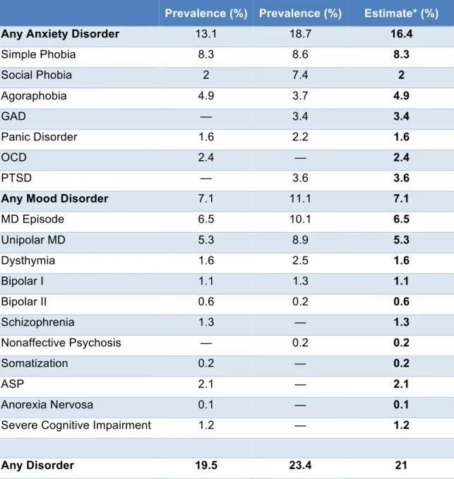
Table 1. – 1-year prevalence of neuropsychiatric disorders in 18-54 year olds. *ECA study, Epidemiologic Catchment Area from 1980-1985 (NIMH, 1992). **NCS study, National Comorbidity Survey 1990-1992 (Kessler et al., 1994). ***Adapted from the “Report of the Surgeon General on mental health” (Satcher, 2000). GAD, generalized anxiety disorder; OCD, obsessive-compulsive disorder; PTSD, post-traumatic stress disorder; MD, major depression; ASP, antisocial personality disorder.
!
ECA* Prevalence (%) NCS** Prevalence (%) Best*** Estimate* (%)Any Anxiety Disorder 13.1 18.7 16.4 Simple Phobia 8.3 8.6 8.3 Social Phobia 2 7.4 2 Agoraphobia 4.9 3.7 4.9
GAD — 3.4 3.4
Panic Disorder 1.6 2.2 1.6
OCD 2.4 — 2.4
PTSD — 3.6 3.6
Any Mood Disorder 7.1 11.1 7.1 MD Episode 6.5 10.1 6.5 Unipolar MD 5.3 8.9 5.3 Dysthymia 1.6 2.5 1.6 Bipolar I 1.1 1.3 1.1 Bipolar II 0.6 0.2 0.6 Schizophrenia 1.3 — 1.3 Nonaffective Psychosis — 0.2 0.2 Somatization 0.2 — 0.2
ASP 2.1 — 2.1
Anorexia Nervosa 0.1 — 0.1 Severe Cognitive Impairment 1.2 — 1.2
!
Mood Disorders
Mood disorders encompass a number of psychiatric diagnoses which describe “disorders in which the fundamental disturbance is a change in affect or mood to depression (with or without associated anxiety) or to elation” (WHO, 2004a). Changes in the underlying mood are hypothesized to be the main feature in these disorders. These diagnosis broadly encompass both the manifestation of single episodes of depressive or manic diagnoses to a combination or oscillations between altered mood states (American Psychiatric Association, 2000).
One end of the mood spectrum is occupied by Unipolar Depression (UPD or Major Depressive Disorder, MDD), a mental disorder characterized by a generalized low mood accompanied by low self-esteem and anhedonia. Patients in a depressive state experience strong emotions of sadness and grief which in contrast to a healthy individual do not remit when the external cause of these emotions is ameliorated. Additionally, these emotions can also be disproportionate to their underlying cause or occur without any triggering external effects (Belmaker and Agam, 2008; Wakefield et al., 2007). Clinically, the diagnosis for UPD requires the observation of distinct altered emotional state and the presence of several psychophysiological changes. These primarily include a depressed mood (subjective or reported by others) and a markedly diminished interest in pleasurable activities (anhedonia). Physiological signs can include significant weight loss or weight gain, disturbed sleep patterns with episodes of insomnia or hypersomnia, extreme fatigue, psychomotor agitation and slowing of speech and action. Other common symptoms described by patients include: feelings of worthlessness, guilt, indecisiveness, diminished ability to think and concentrate and recurrent suicidal thoughts. Diagnostic for UPD is usually attained when several of these symptoms persist for a minimum of 2 weeks and interfere considerably with work and family relations (American Psychiatric Association, 2000). Depressive episodes in UPD are also highly recurrent. Of the individuals who suffer a single episode, 60% of them go on to develop a second, and of those, 70% recur in a third episode (American Psychiatric Association., 2000). Depression also tends to emerge early in adolescence and progress into adulthood suggesting a developmental course in neuropathogenesis (Kim-Cohen et al., 2003; Pine et al., 1998).
Dysthymia originates from the ancient Greek word for “bad mood”. This disorder is defined by milder, non-disabling symptoms that persist for two years or longer (Akiskal, 1983; American Psychiatric Association, 2000). Dysthymia is an independent diagnosis but can also be present in the course of a major depressive state. Specifically, a long term study has shown that patients with major depressive disorder spent on average 44% of their course in a low-grade depressive state (dysthymic) and only 15% of time in major depressive episodes (Judd et al., 1998).
Psychotic depression is characterized by the presence of delusions or hallucinations and specific biological alterations, including a chronic over-activation in the production of glucocorticoids (Schatzberg et al., 1985). This latter observation has led to the successful treatment of Psychotic Major Depression with glucocorticoid receptor antagonists (Belanoff et al., 2001; Schatzberg et al., 1983). Furthermore, Psychotic Major Depression is more resistant to classic pharmacological treatments than non-psychotic MDD while on the other hand, electroconvulsive therapy was reported as more effective for Psychotic Major Depression (Buchan et al., 1992; Petrides et al., 2001).
Postpartum dysphoric mood states comprise a range of severity, from the commonly denominated “baby blues” (a highly prevalent disorder of a mild and transient nature) to the more severe and persistent Postpartum Affective Psychosis and Postpartum Depression. In Western countries the incidence of Postpartum Depression, in particular, has been reported to be as high as 10-20% (Grace et al., 2003; Hopkins et al., 1984).
Lastly, and originally described by Rosenthal and colleagues at the NIH, Seasonal Affective Disorder is also described as change in mood. Most commonly associated with depression affect occurring during the winter (or less frequently in the summer) (Rosenthal et al., 1984), Seasonal Affective Disorder is also positively correlated to latitude and diminished duration of daytime light (Rosen et al., 1990; Vera, 1998).
!
Psychiatric Association, 2000). Bipolar and unipolar disorders share overlapping symptomatology; namely in the presence of episodes of depression in certain forms of bipolar illness. However, the distinction in nosological diagnosis between these two types of mood disorders is supported by distinct differences in disease outcome, response to pharmacological treatment and genetic studies (Farmer and McGuffin, 1989; Kendell, 1987).
Bipolar Spectrum Disorders are also subdivided into several categories: for Bipolar I Disorder (BPDI), a positive diagnostic includes at least one manic or mixed episode (i.e. dysphoric mania or agitated depression) with or without concurrent episodes of hypomania or major depression. Bipolar II Disorder (BPDII) is diagnosed with the occurrence of at least one hypomanic episode and one major depressive episode. Additionally, in BPDII, depressive episodes are more frequent and more intense than manic episodes. Furthermore, some individuals can undergo what is known as “rapid cycling”, a condition in which depressive (dysthymic) and manic (euphoric) states rapidly alternate (Dunner et al., 1977). Interestingly, tricyclic antidepressants can induce rapid cycling in bipolar patients (Wehr and Goodwin, 1979). Finally, another bipolar-like state includes Cyclothymia, a condition considered to be the most milder form of the bipolar spectrum, where affected patients exhibit both hypomanic and mild depressive states (Akiskal et al., 1977).
Historically, depression and mania are some of the oldest known forms of mental disorder. Predating psychiatry by several centuries, Hippocrates was the first to describe these illnesses as disconnected from mysticism and supernatural causes, but instead linked them to a biological cause originating in the brain (Zarate and Manji, 2009). Recently, with the contemporary advent of human genetics, epidemiological studies and heightened clinical awareness, the potential genetic influences that precipitate these disorders have garnered substantial interest.
!
Heritability of Mood Disorders
The heritability of UPD and BPD was originally addressed from quantitative data obtained from interviews of affected patients and family members (James and Chapman, 1975; McGuffin et al., 1988; Reich et al., 1987; Smeraldi et al., 1977; Trzebiatowska-Trzeciak, 1977). The prevalence of similar disorders in first degree relatives was found to be consistently higher than the expected value for the general population. For UPD, in particular, incidences as high as 15% and 20% have been reported for first degree relatives (Shih et al., 2004; Sullivan et al., 2000). A meta-analysis study on familial transmission, twin studies and environmental influences led Sullivan and colleagues to propose that: “Major depression is a familial disorder, and its familiality mostly or entirely results from genetic influences.” (Sullivan et al., 2000).
In terms of gender, although there is strong evidence that UPD affects more women than men (Kessler et al., 1993; Weissman et al., 1993), most studies do not find significant differences in the genetic heritability between males and females (Kendler and Prescott, 1999; Lyons et al., 1998; McGuffin et al., 1996), suggesting that both genders share the majority, but not totality of genetic influences for UPD (Kendler and Prescott, 1999; Sullivan et al., 2000).
Genetic heritability also plays a major role in the development of Bipolar Disorder (Andreasen et al., 1987; Goldin et al., 1983; Reich et al., 1969; Weissman et al., 1984). Interestingly, the lifetime risk of mood disorders for relatives of bipolar patients was reported to be higher than that for unipolar probands (24% versus 20%) (Ciaranello and Ciaranello, 1991; Meltzer, 1987). Furthermore, the lifetime risk of bipolar disorder in relatives of bipolar probands is 40-70% for monozygotic twins and 5-10% for first degree relatives, whereas an overall risk in the general population of 0.5-1% suggests a 10-20 fold increase in risk for first degree relatives of afflicted individuals (Craddock and Jones, 1999).
!
Eley, 2010; Zarate and Manji, 2009). Additional factors play a role confounding the interpretation of genetic inheritance, these include: assortative pairing (defined by the tendency for individuals with related phenotypes to mate more commonly than what
would be expected by random chance) (Baron et al., 1981; Mathews and Reus, 2001; Merikangas et al., 1988), genetic anticipation (surfacing of symptoms at an earlier age in familial transmission as it is passed on to the next generation) (Lange and McInnis, 2002; McInnis et al., 1993; Nylander et al., 1994), genomic imprinting (McMahon et al., 1995; Ohara et al., 1998) or potential effects from mitochondrial genes (Clay et al., 2010; Kato and Kato, 2000; McMahon et al., 1995).
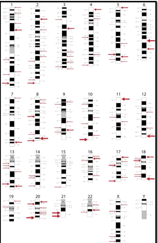
It is therefore clear that bipolar disorder is a complex genetic disorder. However, the high heritability of this condition has attracted intensive research activity aiming at identifying disease causing mutations or mutations associated with increased susceptibility to BPD. Surprisingly, large quantities of data from human genetics, in the form of linkage and association studies have identified a multitude of putative regions of interest for BPD susceptibility in virtually every human chromosome except the Y chromosome (Figure 1). These results have muddled a strong conclusion as far as precise genetic influences on the etiology of this disorder but have, nonetheless, put forward a few “hot spot” regions, which have been reproducibly identified by independent groups of researchers (Serretti and Mandelli, 2008).
specific attention to genes that are known to be involved in biological pathways relating to the disease. Importantly, the synergy between unbiased genomic approaches and target based approach has put forward several promising targets that may be involved in the neuropathology of BPD.
Bipolar Disorder
Candidate Genes for Bipolar Disorder
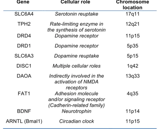
Mutations in the promoter region of the serotonin transporter -SLC6A4- in the 17q11 region have been implicated in several studies into BPD. Multiple groups have identifying a 44 base pair insertion/deletion and another polymorphism in the second intron of the gene as potentially being involved in BPD (Anguelova et al., 2003; Heils et al., 1996; Lasky-Su et al., 2005; Lesch et al., 1996; Rotondo et al., 2002). The tryptophan hydroxylase (TPH) protein is the rate-limiting enzyme in the synthesis of serotonin. In humans, TPH1 and TPH2 proteins are coded by two separate genes with only TPH2 (in chromosome 12q21) being strongly expressed in the brain (Walther et al., 2003). TPH2 polymorphisms have also been implicated in susceptibility to BPD (Cichon et al., 2008; Grigoroiu-Serbanescu et al., 2008; Harvey et al., 2007; Lopez et al., 2007; Van Den Bogaert et al., 2006).
!
The Disrupted-in-Schizophrenia-1 (DISC1) gene (1q42) is another candidate gene for which there is strong evidence implicating its dysfunction in BPD (Blackwood et al., 2001; Hodgkinson et al., 2004; Palo et al., 2007). This clear overlap in a gene potentially involved in schizophrenia as well as BPD is perhaps not surprising given the endophenotypical similarities between the two disorders (Zarate and Manji, 2009). Other strongly implicated genes include the D-amino acid oxidase activator (DAOA) (13q33) (Detera-Wadleigh and McMahon, 2006), FAT1 (4q35) (Abou Jamra et al., 2008; Blair et al., 2006), the Brain-derived neurotrophic factor (BDNF) (11p14) (Kanazawa et al., 2007; Sklar et al., 2002) and interestingly, the aryl hydrocarbon receptor nuclear translocator-like protein (ARNTL, also known as Bmal1) (11p15) which heterodimerizes with the CLOCK gene as part of the molecular regulation of circadian rhythms (discussed below) (Mansour et al., 2006; Nievergelt et al., 2006).
Gene Cellular role Chromosome location SLC6A4 Serotonin reuptake 17q11
TPH2 Rate-limiting enzyme in the synthesis of serotonin
12q21 DRD4 Dopamine receptor 11p15 DRD1 Dopamine receptor 5p35 SLC6A3 Dopamine reuptake 5p15 DISC1 Multiple cellular roles 1q42 DAOA Indirectly involved in the
activation of NMDA receptors
13q33
FAT1 Adhesion molecule and/or signaling receptor (Cadherin-related family)
4q35
BDNF Neurotrophin 11p14 ARNTL (Bmal1) Circadian clock 11p15
!
Sleep and Bipolar Disorder
Sleeping disorders have long been associated with mood disorders. Disturbances in sleep are included as part of the symptomatology for the diagnoses of both MDD (when present as hypersomnia or insomnia) and BPD, where reduced need for sleep can be part of a positive diagnostic for manic episodes (American Psychiatric Association, 2000). Classically, sleep deregulation has often been considered an epiphenomena of mood disorders and not necessarily a causal or primary driving force for either UPD or BPD, but more so, a consequence of the individuals’ altered mood. Recently, however, a shift in paradigm is emerging whereby a deeper understanding of the biology of circadian rhythms and their relationship to mood disorders is seen as an important mechanism contributing to the genesis and/or maintenance of mood disorders (Harvey, 2001; Harvey, 2011; McCrae and Lichstein, 2001; NIH, 2005).
Both insomnia or hypersomnia are highly prevalent in UPD, with 60% to 84% of patients reporting sleep related symptoms (Ford and Kamerow, 1989). Insomnia is also hypothesized to precede and constitute a risk factor for depression (Riemann and Voderholzer, 2003). Interestingly, in a 20 year study from a representative sample of the Swiss population (Vollrath et al., 1989), researchers uncovered that long lasting insomnia predicted major depressive episodes. Specifically, 17% to 50% of subjects with insomnia lasting more than 2 weeks developed a major depressive episode at a later time (Riemann and Voderholzer, 2003).
In BPD, 69 to 99% of patients experiencing manic episodes report a reduction in time spent sleeping. Furthermore, in the course of a depressive episode 23-78% experience hypersomnia (Harvey, 2008). Finally, in the inter-episode periods, BPD patients report sleep disturbances with a higher degree of fragmentation in the sleep/wake rhythm when comparing to healthy patients (Jones et al., 2005).
depressive episodes (Jackson et al., 2003; Sierra et al., 2007). Additionally, sleep disturbances not only predate recurrence into manic episodes, but they can also predict the first onset into BPD. A study performed in high-risk offspring (i.e. children coming from families in which one parent was diagnosed with BPD) concluded that sleep disturbances and anxiety disorders were antecedent conditions to develop BPD (Duffy et al., 2007).
Interestingly, sleep deprivation (partial or total), or circadian phase advances in the sleep-wake cycle have long been known to produce an antidepressant effect in bipolar patients undergoing depressive episodes (Barbini et al., 1998; Wehr et al., 1979; Wu et al., 2009). This effect has been hypothesized to be linked to different mechanism, including the serotonergic system (Benedetti et al., 2008), the dopaminergic system (Ebert and Berger, 1998) or as a consequence of a rebound or reset in the Process S (slow wave sleep) sleep stage (Endo et al., 1998; Endo et al., 1997).
Sleep is one of the biological processes controlled by circadian rhythms. The understanding of the molecular underpinnings of mammalian circadian rhythms has seen a surge in recent years. The circadian control of sleep and wakefulness, the genes and molecules involved and the understanding of the neural circuits that play a role in these processes are paving new discoveries into the neurobiology of mood disorders. Therefore, it is acknowledged that a deeper comprehension in the molecular biology of circadian rhythms can provide new clues into the etiology of Bipolar Disorder as well as new avenues for treatment (Jones, 2001).
!
Circadian Rhythms
!
subjective period lengths that usually deviates from a 24h duration (Schwartz and Zimmerman, 1990) and although light is the predominant Zeitgeber (time giver), entrainment to a 24h cycle can also occur with other stimuli, such as food availability (Lax et al., 1999) or social cues (Goel and Lee, 1997).
The “master clock” for the mammalian brain is present in the Suprachiasmatic Nucleus (SCN). Synchronization through daily dark-light cycle originates in photosensitive retinal ganglion cells which function independently of photoreceptor cones and rods and project to the SCN via the retinohypothalamic tract and thus initiate photonic entrainment (Berson, 2007; Berson et al., 2002). However, humans that are completely blind and display “free-running” circadian rhythms, melatonin can be administered to promote circadian entrainment (Sack et al., 2000).
In contrast, homozygous mice for a deletion of Bmal1, a critical binding partner for Clock and an essential molecule in the molecular circadian regulation, display a complete loss of circadian rhythmicity in free-running conditions at both the behavioral and molecular level (Bunger et al., 2000). CLOCK and BMAL1 heterodimerize in the cytoplasm before entry into the nucleus, this is followed by phosphorylation events which promote their function as a transcriptional complex. With the above observations from mouse molecular genetics, this suggests that BMAL1 is essential for the transcriptional control and circadian oscillations, whereas, Clock can be functionally substituted by another protein (Debruyne et al., 2006). One possible candidate is Npas2, which has been suggested as having an overlapping role with Clock in the mouse brain (DeBruyne et al., 2007). At the molecular level, CLOCK interacts with BMAL1 to activate Per (period homologue) and Cry (cryptochrome) genes. PER and CRY can then heterodimerize, translocate to the nucleus, interact with the CLOCK:BMAL1 complex to inhibit self-expression (i.e. PER and CRY). During subjective evening, the PER:CRY repressor complex is degraded and the CLOCK:BMAL1 complex initiates a new cycle of transcription (Takahashi et al., 2008). Although these molecular pathways are central to circadian biology there are other known pathways that intersect this protein expression and degradation cycles which are summarized in Figure 2.
!
Symptoms of mania Clock mutant mice
Disrupted circadian rhythms Disrupted circadian rhythms Hyperactivity Hyperactivity
Decreased sleep Decreased sleep
Feelings of extreme euphoria Hyperhedonia/less helplessness Increased risk-taking Reduced anxiety
Propensity toward drug abuse Increased preference for cocaine
!
Table 3. – Clock display behavioral abnormalities consistent with symptoms from bipolar patients in the manic state.
Moreover, lithium salts, which are administered as a mood stabilizer for BPD patients, ameliorates several of the behavioral abnormalities in the ClockΔ19 mice in measures of anxiety- and depression-related behavioral tasks (Roybal et al., 2007).
Therefore, it is now acknowledged that circadian biology is well positioned to play a major role in BPD. Moreover, human genetic data and mouse modeling have implicated several of the genes crucial for normal circadian to susceptibility to BPD.
!
!
Modeling Bipolar Disorder
Although mouse models of BPD have been proposed, the critical feature of bipolar cycling between mania and depression has not been successfully observed for genetic mouse models. It is unknown if this limitation is inherent to the murine model or if the manipulations performed have yet to tap on a truly cyclic pathological pendulum capable of pushing mice from depressive-like into manic-like behavior. Nevertheless, useful models of depression and of mania have been reported which replicate several of the features of both types of episodes.
Several pharmacological models of BPD have been proposed in rodents. Amphetamines and other psychostimulants, such as D-amphetamine, 3,4-methylenedioxymethamphetamine (MDMA) and cocaine can induce psychomotor hyperactivity in mammals (Koob and Bloom, 1988). Administration of psychostimulants, amphetamines or methamphetamines, have been used to simulate manic episodes and a concomitant administration of therapies, such as lithium, valproic acid or transcranial magnetic stimulation can successfully rescue pathological behaviors (Frey et al., 2006a; Frey et al., 2006b; Gould et al., 2001; Shaldivin et al., 2001). Repeated administration of methamphetamines, cocaine or morphine can produce behavioral sensitization and be used as a model of bipolar disorder which produces some cyclical manic-depressive activity. Again, the behavioral effect produced by these drugs are reversed or prevented by administration of lithium (Antelman et al., 1998; Kucinski et al., 1998; Namima et al., 1999). Of particular interest, in one study, a model of repeated high-dose cocaine administration produces behavioral hyperactivity response on some days and behavioral hypoactivity on alternating days (Antelman et al., 1998). Another pharmacological model for BPD includes the combination of amphetamine and chlordiazepoxide to induce hyperactivity, which can be rescued by lithium treatment (Vale and Ratcliffe, 1987). The drug ouabain, an inhibitor of the sodium-potassium pump, has also been used to model mania. This drug is known to induce hyperactivity in rodents, again an effect which can be countered by administration of lithium (El-Mallakh et al., 2003; Li et al., 1997).
The acute locomotor hyperactivity due to amphetamines and metamphetamine can be suppressed by Dopamine D1 and D2 receptor antagonists or Dopamine D2 Receptor (D2) agonist –quinpirole- (Kuribara and Uchihashi, 1993; Steketee and Kalivas, 1992). Amphetamines and cocaine tap into the dopamine neurotransmission by promoting an increase of presynaptic dopamine release (Jones et al., 1998; Kokoshka et al., 1998) and cocaine by blocking the reuptake of dopamine (Kitayama et al., 1992; Ritz et al., 1987). Therefore, strong evidences suggest this neurotransmitter may be involved in the development of mania (Diehl and Gershon, 1992).
One interesting behaviorally-induced model for mania involves inducing sleep-deprivation. Originally developed to study sleep deprivation in cats (Jouvet et al., 1964), a common version of this test in rodents prevents sleep by stationing the animal in a platform covered in water. At the onset of REM sleep, muscle relaxation puts the animal in contact with water and induces awakening. This test, compounds sleep deprivation with other stressors such as social isolation, immobilization and contact with water, which impart several behavioral responses similar to idiopathic mania. Interestingly, haloperidol and lithium treatment reduced the sleep deprivation induced manic behavioral manifestations (Gessa et al., 1995).
Another behavioral model is the dominant–submissive test to study mania and depression. Animals screened in this test display mania-like or depression-like behavior depending on their social position. Interestingly, this effect was determined to be reversed by anti-manic or anti-depressive drugs (Malatynska and Knapp, 2005; Malatynska et al., 2007).
With the above described ClockΔ19 mice, other genetically manipulated animals have also been proposed to model aspects of BPD. At the most basic level, these include the analysis of the inbred strains of laboratory mice (Einat, 2007). Others include the Disrupted-in-Schizophrenia 1 gene (DISC1) originally identified in a Scottish family strongly afflicted by schizophrenia and mood disorders. Although not thoroughly explored, mutant mice for DISC1 remains an interesting putative model for bipolar disorder (Ishizuka et al., 2006; Lipina et al., 2010; O'Tuathaigh et al., 2007).
!
the glucocorticoid receptor displays obesity and increased activity in the HPA (Hypothalamic–pituitary–adrenal) axis, consistent with severe depression (Pepin et al., 1992). Treatment of these animals with moclobemide, a monoamine oxidase type A inhibitor, reverted both behavioral and hormonal alterations (Montkowski et al., 1995). Another mouse model includes a forebrain specific over-expression of the glucocorticoid receptor. These animals displayed increased depression-like behaviors, anxiety-like behaviors, hypersensitivity to cocaine administration and a high sensitivity to anti-depressants (Wei et al., 2004). In addition to the above, there are other manipulations on this same gene which promote relevant phenotypes to depression in the mouse. Interestingly, one group simultaneously examined two lines of mutant mice, one with a heterozygous reduction in glucocorticoid receptor expression, and the other with an overexpression of the receptor. Similarly to the other loss of function mutants, the reduction in expression generated an animal prone to depression, while the overexpression produced an animal resistant to depression (Ridder et al., 2005); however, this is different from what was seen in an earlier model of glucocorticoid receptor overexpression (Wei et al., 2004).
The 22q11 deletion in humans is associated with a conserved deletion of approximately 40 genes which impart a high risk for mental disorders, most notably schizophrenia and early onset BPD (Papolos et al., 1996). The prevalence of 22q11 deletion syndrome is estimated to be approximately 1:6000 in the live births, making this syndrome the second most common chromosomal disorder (after Down’s syndrome) and the most common microdeletion syndrome (Botto et al., 2003; Devriendt et al., 1998; Paylor and Lindsay, 2006). One of the most robust findings in a mouse model of 22q11 microdeletion are deficiencies in sensory motor gating (Kimber et al., 1999). This endophenotype is mostly linked to schizophrenia in humans (Braff and Geyer, 1990; Braff et al., 2001), but nevertheless may also be strongly linked to bipolar disorder since BPD patients also display sensory motor gating deficits (Perry et al., 2001).
stabilization (Gould et al., 2004). GSK-3β has also been linked to regulation of the circadian rhythm (Kaladchibachi et al., 2007). Pharmacologically, GSK-3β is efficiently inhibited by lithium and valproate, both of which are used to treat BPD (Chen et al., 1999; Klein and Melton, 1996). Interestingly, transgenic mice overexpressing GSK-3β
express several features reminiscent of mania when tested behaviorally, these included, hypophagia, increased locomotor activity and decreased habituations in the open field test and increased acoustic startle response with decreased habituation (Prickaerts et al., 2006).
The Protein kinase C interacting protein (PKCI) is a small protein belonging to the histidine triad family proteins. In a meta-analysis of 12 microarray studies of bipolar disorder, this gene was found to be decreased in the dorsolateral PFC (Elashoff et al., 2007). Relevant for mood disorders, the PKCI/HINT1 mutant mice display less immobility in the forced swim and the tail suspension tests, both of which are indicative of lower levels of depression. These animals are also less anxious than their wildtype littermates, at the same time as the HPA axis activity was reported to be potentiated in the knockouts (Barbier and Wang, 2009).
Electrophysiological kindling has also been used to model aspects of BPD (Post et al., 2001). This procedure has been used to indirectly address illness progression and development of new BPD episodes (Machado-Vieira et al., 2004). A typical kindling paradigm involves a daily subthreshold stimulation of the amygdala. With the progression of stimulations, same intensity depolarization leads to motor seizures and, subsequently, the occurrence of spontaneous seizures in the absence of stimulation (Goddard et al., 1969; Racine, 1972). Phenotypically, kindled rats display an increase in “emotionality” and increased resistance to capture in an open field test (Kalynchuk et al., 1998). On a similar paradigm hippocampal kindling induces increased locomotor activity, which can be suppressed by the injection of a D2 receptor antagonist in the NAc (Leung et al., 2000). Nevertheless, the validity of the kindled model directly relating to the pathophysiological mechanisms of BPD is controversial (Post and Weiss, 1998).
!
attained when phenomenological similarities are seen between the model and the studied phenomena (i.e. the animal displays behavioral responses reminiscent of the human condition). Construct validity references to a strong theoretical support for the model (i.e. targeting a gene which is implicated in a human disorder). Predictive validity pertains at correcting abnormalities in the model after treatments or manipulations which replicate what is seen in affected individuals after homologous treatments (e.g. correction of behavioral defects in a model of BPD following a treatment with lithium) (Boulton et al., 1991).
In BPD research, most models only approximate neuropathogenesis to a certain degree. Most pharmacological models display a good level of face validity but are somewhat lacking in construct validity (i.e. most bipolar onsets are not brought about by amphetamine or cocaine administration). On the other hand, genetic models display in some cases robust construct validity but only moderately approximate face validity (e.g. none of the genetic models display cyclic manic-depressive episodes). Predictive validity however is somewhat robustly attained across most modalities when measuring responsiveness to conventional treatment in reverting phenotypical manifestations (e.g. lithium or other pharmacological treatments successfully correct several abnormal phenotypes in the majority of models).
Model Main effects Relevant Reference Amphetamine
Hyperactivity/ Reversibility and prevention of symptoms by Li, valproic acid or transcranial magnetic stimulation
(Frey et al., 2006a; Frey et al., 2006b; Gould et al., 2001; Shaldivin et al., 2001)
Metamphetamines Hyperactivity/ reversibility of symptoms by Li (Namima et al., 1999)
Cocaine Potential cycling model of BPD (Antelman et al., 1998)
Morphine Oscillatory effects on shock induced
hypoalgesia/ Li attenuates phenotype (Kucinski et al., 1998) Amphetamine &
chlordiazepoxide Hyperactivity/ reversibility of symptoms by Li
(Vale and Ratcliffe, 1987)
Ouabain Hyperactivity/ reversibility of symptoms by Li 2003; Li et al., 1997) (El-Mallakh et al.,
Sleep deprivation
Insomnia, hyperactivity, irritability, aggressiveness, hypersexuality and increased stereotypies/ Behavioral effects reverted by haloperidol or lithium
(Gessa et al., 1995)
Dominance-submission
Drugs used to treat mania inhibit the dominant behavior| Antidepressants
counteract the behavioral consequences of encounter defeats
(Malatynska et al., 2007)
ClockΔ19
Hyperactivity, decreased sleep, lowered depression-like behavior, lower anxiety, increase in the reward value for cocaine and sucrose/lithium rescues phenotypes
(Roybal et al., 2007)
DISC1 Hyperactivity, deficits in PPI, deficits in sociability
(Ishizuka et al., 2006; Lipina et al., 2010; O'Tuathaigh et al., 2007)
22q11 deletion Deficits in sensory motor gating
(Paylor and Lindsay, 2006; Paylor et al., 2001)
GSK-3β
overexpression
Hypophagia, increased locomotor activity with decreased habituations, increased acoustic startle response with decreased habituation
(Prickaerts et al., 2006)
PKCI mutant mice Less anxious, less prone to depression, increased HPA axis activity
(Barbier and Wang, 2009)
Amygdala and hippocampal kindling
Increase in emotionality response/increased activity in the open field reverted by D2 antagonist
(Kalynchuk et al., 1998; Leung et al., 2000)
!
Circuit Dysfunctions in Bipolar Disorder
The neural networks most strongly implicated as dysfunctional in BPD patients are comprised of the prefrontal cortical-striatal-limbic circuits. These regions form a key system in the control of complex socio-emotional behaviors (Cerullo et al., 2009; Strakowski et al., 2005). Broadly, prefrontal cortical areas are involved in executive function, decision-making and attention. Most notably, damage to orbitofrontal cortex is known to affect patients and produce impairments in everyday decisions-making, whilst simultaneously not affecting most other cognitive abilities (Wallis, 2007). Perhaps the two most well studied examples of damage to frontal cortical regions are the case of Phineas Gage and patient EVR. Both men were reported to display intact functioning in higher cognition (memory, reading and writing, verbal communication, sensorial processing, facial recognition and fine motor function) but an extremely damaged decision-making process profoundly altered their personalities and behaviors (Damasio, 2005; Eslinger and Damasio, 1985). In terms of circuitry, the PFC receives afferent connections from all sensorial modalities (Carmichael and Price, 1995b; Cavada et al., 2000). Conversely, projections from the PFC extend to synapse onto premotor cortical neurons as well as somatosensory association cortical structures. Additionally, the PFC is known to heavily converge onto limbic regions (amygdala, cingulate gyrus, hippocampus and thalamus) (Carmichael and Price, 1995a) and the striatum (Haber et al., 1995; Kemp and Powell, 1970).
Lastly, the limbic system is also hypothesized to be involved in the broad neuronal circuitry potentially dysfunctional in BPD. Even though this structure is comprised of several well defined brain sub-regions, there is no precise neuroanatomical consensus as to which of these compose the limbic system (LeDoux, 2000). The amygdala, cingulate gyrus, hippocampus, thalamus and hypothalamus are some of the more “consensual limbic areas”. Functionally, the limbic system is classically described as an important hub for memory formation, arousal and autonomic nervous system (Stephen et al., 1997).
!
on a lithium treatment, BPD patients under a different drug treatment (the anticonvulsantes valproic acid or lamotrigine) and healthy controls. In this work, there was a trend for a smaller hippocampal region in untreated BPD patients when compared to controls, but conversely, an increased hippocampal volume in lithium treated patients when compared to healthy controls (Yucel et al., 2008). This finding highlights the potential neuronal effects of this drug in humans and addresses some of the conflicting evidences in the field pertaining to anatomical brain volume.
Nevertheless, even though structural abnormalities provide some clues into affected neural circuits, functional MRI (fMRI) has also been used to probe how brain regions are affected in terms of neuronal activity. One work found an increase in amygdala activation and a reduction in dorsolateral prefrontal cortex activation in response to fearful facial stimuli (Yurgelun-Todd et al., 2000). The same group however, found an increase in dorsolateral prefrontal cortex and decrease anterior cingulate cortex (limbic system) activation during a cognitive test (Gruber et al., 2004). BPD patients also display increased activation in the amygdala, thalamus, hypothalamus (limbic system) and medial globus pallidus (basal ganglia) (Malhi et al., 2004). Qualitatively similar results were found by a separate group where patients observing hostile faces had greater activation in the limbic regions (left amygdala) and the basal ganglia region (nucleus accumbens and putamen) (Rich et al., 2006).
Interestingly, some researchers have proposed that UPD and BPD can perhaps be distinguished based on fMRI activation patterns. Specifically, responses to positive and negative emotional expressions in BPD patients revealed increased subcortical and ventral PFC responses, when comparing to healthy controls or UPD patients (Lawrence et al., 2004). It is worth noting however, that even though these studies are informative, they carry some limitations in interpretation. Confounding factors include not only the effect of medication on neuronal activation patterns, but also the contaminating effect that comorbid conditions may have on neuronal activity. Also, while some studies look at whole brain during imaging, many only analyze discrete regions of interest. Finally, experimental differences in the tasks performed by patients during fMRI analysis make a combined analysis of data across multiple research groups difficult to attain (Yurgelun-Todd and Ross, 2006).
(Yurgelun-Todd and Ross, 2006). At the same time, this information provides important and complementary information when attempting to model BPD with genetic mouse model and broadly point to an over-activation of neuronal circuits in BPD.
Synaptic Dysfunctions in Bipolar Disorder
At the most fundamental level, the behavioral manifestations in neuropsychiatric disorders are a direct consequence of dysfunction in neuronal communication. Specifically, schizophrenia, autism and mood disorders are an example of disorders which have accrued significant converging evidence implicating synaptic dysfunction to their respective etiological basis (Bourgeron, 2009; Sanacora et al., 2008; Sodhi et al., 2008). For BPD, key findings pertaining to molecular dysfunctions are postulated to affect either the neurons as a whole or specifically the signaling networks at the postsynaptic site. Therefore, the molecular complexity of the postsynaptic site may be of key interest in understanding BPD and how synaptic dysfunction may contribute to the development of this disorder.
!
In the mammalian brain, glutamate is the most abundant excitatory neurotransmitter (Purves, 2008). Glutamatergic synapses form the prototypical junctions and the ones that best represent the asymmetric nature of information flow between neurons in the central nervous system. In these types of synapses, glutamate is released from the axonal presynaptic bouton onto the synaptic cleft. The neurotransmitter then transverses the synaptic cleft to bind and activating glutamate receptors on the postsynaptic neuron and generate excitatory postsynaptic currents (EPSCs) or changes in intracellular biochemical processes (Kandel et al., 2000). The glutamate receptors which effect this communication are subdivided into two major subtypes, ionotropic and metabotropic glutamate receptors. Metabotropic glutamate receptors (mGluRs) are G-protein-coupled receptors further subdivided into three groups, group I (mGluR1a–d, mGluR5a,b), group II (mGluR2 and mGluR3) and group III (mGluR4, mGluR6–8). These transmembrane receptors transduce neurotransmitter binding to intracellular activation of phospholipase C, modulation of adenylyl cyclase and through other second messengers (Conn and Pin, 1997). Glutamate ionotropic receptors are also subdivided according to their physiological and pharmacological properties into three groups, the N-Methyl-D-aspartic acid (NMDA) glutamate receptors; the α-amino-3-hydroxy-5-methyl-4-isoxazolepropionic acid (AMPA) glutamate receptors and kainate receptors (Dingledine et al., 1999). Ionotropic glutamate receptors are ligand gated channels which depending on physiological conditions and selective permeability, allow the passage of ions across the cell membrane.
PSDs and proteomic analysis, over 1000 different types of proteins at varying stoichiometric ratios have been identified at the PSD (Sheng and Hoogenraad, 2007). Interestingly, many of these proteins are not only present at the PSD, but are also biochemically enriched at this structure, when comparing the relative abundance between the PSD and the synaptic cytoplasm (Collins et al., 2006). By function, some of the best characterized types of PSD protein include: glutamate neurotransmitter receptors; scaffolding and adaptor proteins involved in the anchoring, trafficking and regulation of postsynaptic receptors; cell-adhesion molecules, which form contact across the synaptic cleft; kinases involved in downstream signaling and proteins involved in interaction and remodeling of the actin cytoskeleton (Sheng and Hoogenraad, 2007).
The molecular function of many of these proteins revolves around the formation of defined macro-molecular complexes which orchestrate biochemical function in discrete sub-networks. The NMDA glutamate receptors, known to play an important role for synaptic plasticity and cellular information processing (Daw et al., 1993; Malenka and Nicoll, 1993), form one of the largest complexes within the PSD (Husi and Grant, 2001; Husi et al., 2000). One example is the NMDA receptor/MAGUK-associated signaling complex (MASC), named after the receptors and their primary interaction proteins, the “membrane associated guanylate kinase” (MAGUK) family of proteins. The MASC complex is composed of at least 186 different proteins which form a 2000 KDa structure within the larger PSD (Collins et al., 2006; Husi and Grant, 2001). Other large complex is formed by mGluRs which can associate with at least 64 other types of proteins (Farr et al., 2004) and AMPA receptors complex which directly or indirectly associate with at least 9 other proteins in the PSD (Collins et al., 2006).
!
disorder therapeutics can themselves be informative on the underlying mechanisms surrounding BPD.
Mood stabilizer drugs, such as lithium and valproic acid, are generally prescribed to BPD patients to alleviate manic episodes and also to prevent recurrent manic-depressive oscillations. In cultured neurons, lithium displays a protective effect on NMDA mediated excitotoxicity due to calcium influx (Hashimoto et al., 2002; Nonaka et al., 1998). Indirectly, this suggests that abnormality in glutamatergic neurotransmission may underlay certain cellular aspects of BPD. Also, AMPA receptor function is known to be modulated by both lithium and valproic acid. It is interesting that although these two drugs are structurally dissimilar, both are able to affect AMPA receptor synaptic expression (Du et al., 2004). Specifically, both drugs lead to a downregulation of AMPA receptor GluR1 and GluR2 subunits, while conversely, the antidepressant drug imipramine, which can trigger manic episodes, increases synaptic expression of GluR1 in hippocampus in vivo (Du et al., 2008; Du et al., 2004).
It is therefore not surprising that synaptic and postsynaptic proteins directly or indirectly involved in the anchoring, regulation and trafficking of glutamate receptors may play a role in psychiatric disorders in general or in BPD in particular. In fact, several canonical PSD scaffolding proteins are strongly implicated in a multitude of disorders, such as mental retardation (SAP102 – synapse associated protein 102 a MAGUK), autism (Shank3 – SH3 and multiple ankyrin repeat domains protein 3) or obsessive-compulsive disorder (SAPAP3 - SAP90/PSD95-associated protein 3) (Bienvenu et al., 2009; Durand et al., 2007; Tarpey et al., 2004; Welch et al., 2007).
!
Shank/SAPAP/nArgBP2 protein network at the PSD
dendritic spines (Elias and Nicoll, 2007; Hung et al., 2008; Roussignol et al., 2005; Welch et al., 2007). Importantly, genetic disruption of Shank3 is thought to be a primary cause for the deficits in the 22q13.3 deletion syndrome (Phelan-McDermid syndrome), a syndrome characterized by the presence of autistic symptoms in a high percentage of affected individuals (Discussed in next section) (Abrahams and Geschwind, 2008).
The SAPAP family of proteins is another group of proteins involved in the regulation of the PSD and glutamate receptors (Takeuchi et al., 1997; Welch et al., 2007; Welch et al., 2004). SAPAP3, in particular, binds to Shank3 and has also been demonstrated to affect NMDA receptors at the PSD (Welch et al., 2007). In vivo, mutant mice for the SAPAP3 gene display OCD-like behaviors which are ameliorated by the administration of fluoxetine (Welch et al., 2007). In humans, mutations in the SAPAP3 gene have also been identified in OCD patients (Bienvenu et al., 2009; Boardman et al., 2010; Zuchner et al., 2009). Interestingly, both SAPAP3 and Shank3 proteins are enriched in the striatum and both mutant animals display cortico-striatal defects (Peca et al., 2011; Welch et al., 2007).
nArgBP2 (neural Arg-binding protein 2) is another binding partner to SAPAP3 identified using a Yeast Two-hybrid screening (Kawabe et al., 1999). This protein is the neuronal specific form of ArgBP2, a protein which associates and is a phosphorylation target for Abl and Arg protein-tyrosine kinases (Wang et al., 1997). ArgBP2 and nArgBP2 are reported to closely regulate multiple mechanisms converging on the cytoskeleton (Cestra et al., 2005; Ronty et al., 2005). At the same time, ArgBP2 was also identified to be present at the nucleus (Kioka et al., 2002; Wang et al., 1997). nArgBP2 however, is specifically expressed in the brain, much like SAPAP3 and Shank3, and is also highly enriched in the PSD. Therefore, nArgBP2 is well positioned to bridge the interaction between the PSD and the actin cytoskeleton (Cestra et al., 2005; Kawabe et al., 1999). In terms of structure, nArgBP2 is composed of a conserved Sorbin homology domain (which was demonstrated to target proteins to lipid rafts) (Kimura et al., 2001), a zinc finger domain in the brain specific section of the protein and three SH3 domains near the C-terminus (Kawabe et al., 1999).
!
role for this gene in related neuropsychiatric disorders. Importantly, the concept of a human phenome supports the functional analysis of genes and their association with similar disorders in a landscape of interrelated diseases which may originate from an overlapping molecular causation (Barabasi et al., 2011; Goh et al., 2007; Linghu et al., 2009; Oti and Brunner, 2007; Oti et al., 2008; van Driel et al., 2006). This is particularly relevant to diseases which share common endophenotypes that may be brought about by functionally related genes which produce proteins that, at the cellular level, associate in a multiprotein complex (e.g. the complexes in the PSD) in the same pathway, or as part of the same subcellular organelle (Linghu et al., 2009; Oti and Brunner, 2007). This is particularly relevant for the study of nArgBP2 since the interaction of this protein with SAPAP3 and its enrichment at the PSD promotes an association of nArgBP2 with a cluster of proteins that are strongly implicated in the pathogenesis of several neuropsychiatric disorders.
Finally, nArgBP2 is coded from the SORB2 gene which is present in chromosome 4q35, a region identified in BPD susceptibility. Therefore, the in vivo
characterization and the exploration of the cellular roles of nArgBP2 are underexplored, at the same time as the potential involvement of this gene in neuropsychiatric disorders remains an interesting hypothesis.