Insert here a
Streptococcus pneumoniae
streptococci in the era of co
Dissertation presented to obtain a Ph.D
Instituto de Tecnologia Química e Biológica | U
Oeiras,
Insert here a
with rounded
e an image
ae and closely-related
conjugate vaccines
Alexandra Sofia Simões
degree in Biology/Molecular Biology
| Universidade Nova de Lisboa
e an image
Alexandra Sofia Simões
Dissertation presented to obtain a Ph.D
I
nstituto de Tecnologia Química e Biológica | Un
resistant
Streptococcus
closely-related streptoc
conjugate vaccines
I
nstituto de Tecnologia Química e Biológica | Un
Oeiras, December 2011
h.D degree in Biology/Molecular Biology
ca | Universidade Nova de Lisboa
cus pneumoniae
and
eptococci in the era of
Portugal through grant SFRH/BD/27325/2006 awarded to Alexandra S. Simões.
First Edition, October 2011 Second Edition, December 2011 © Alexandra S. Simões
Raquel Sá-Leão Hermínia de Lencastre
Examiners:
Josefina Liñares Annalisa Pantosti José Melo-Cristino Adriano Henriques
I am deeply grateful to Dr. Raquel Sá-Leão, my supervisor. Thanks for all the confidence, guidance, critical sense and friendship. I’ve learned almost everything I know about science with you. Your support has made me grow, not only as a researcher but also, and more importantly, as a person.
I would like to thank Professor Hermínia de Lencastre, also my supervisor, for the opportunity of doing this PhD in the Laboratory of Molecular Genetics. Thank you for your critical sense, helpful commentaries and for your conception of perfection that led me to be here today.
I would like to thank my PhD Thesis Committee, Professor Alexander Tomasz and Dr. Sérgio Filipe for their critical sense, interesting discussions and for sharing their knowledge with me.
All my colleagues at the Laboratory of Molecular Genetics at the Instituto de Tecnologia Química e Biológica for their encouragement and warm work environment. I would like to show my appreciation, in particular to Sónia Nunes, Carina Valente, Débora Tavares, Nelson Frazão, Sónia Almeida and Sofia Félix. Thank you all for your support in all experimental work, for being always available to discuss the work and the results, and also for the good moments that we spent together.
of Fundação para a Ciência e Tecnologia (SFRH/BD/27325/2006).
This PhD thesis has become possible only because of the support of all my family and friends. You all make me happy and without you all it would have been much harder than it was. In particular, Hugo, Joana, Patricia, and Mário: you made it seem easy. Thank you for being always so present and for all the “Let´s go, you can do it!”
In Portugal, the introduction of the seven-valent pneumococcal conjugate vaccine (PCV7) has led to significant changes in the population structure of Streptococcus pneumoniae. However, the levels of antimicrobial resistance have not decreased and have been a matter of concern.
In this thesis, two studies were performed aiming to understand the reasons for maintenance of antimicrobial resistance.
among colonizing pneumococci.
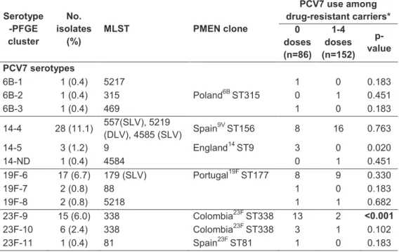
In the second study, it was found that, despite the extensive pneumococcal serotype replacement, observed among children in DCC due to PCV7 introduction, no significant changes were observed in the rates of non-susceptibility to penicillin resistance, to macrolides, or multidrug resistance in general. In order to investigate the mechanisms leading to the maintenance of antimicrobial resistance rates, we compared through molecular typing antibiotic resistant pneumococci recovered from young carriers in 2006 and 2007 (era of high-PCV7 uptake) with collections of isolates from 2002-2003 (low-PCV7 uptake era) and 1996-2001 (pre-(low-PCV7 era). We observed that the group of clones that accounted for antimicrobial resistance since 1996 was essentially the same as the one identified in the PCV7 era. However, the relative proportions of such clones had evolved substantially overtime. The expansion of clones expressing non-PCV7 capsular variants of the original strains was the major mechanism leading to the maintenance of antimicrobial resistance rates. Emergence of novel clones and de novo acquisition of resistance contributed little to the observed scenario. No evidence of capsular switch events occurring after PCV7 introduction was found.
samples.
We developed a low cost and easy assay to detect and quantify NTPn in primary samples obtained from nasopharyngeal swabs. The strategy was based on a multiplex PCR targeting lytA (a virulence factor ubiquitous in pneumococci that is often used as an identification marker of this species), cpsA (a conserved pneumococcal capsular gene), aliB-like ORF2 (a gene described as present in the capsular region of non-typeable pneumococci) and 16S rDNA (used as a positive internal control) genes, plus a restriction fragment length polymorphism (RFLP) assay to differentiate typical from atypical lytA. The application of this new methodology found that the prevalence of NTPn in colonization was three-fold higher than estimated by routine methods.
Em Portugal, a introdução da vacina pneumocócica conjugada sete valente (PCV7) levou a mudanças significativas na estrutura populacional de Streptococcus pneumoniae. Contudo, os níveis de resistência aos antimicrobianos em isolados de colonização não diminuíram o que é motivo de grande preocupação a nível nacional.
Realizámos dois estudos com o objectivo de perceber quais as razões que levaram à manutenção dos níveis elevados de resistência aos antimicrobianos.
mantido estável.
No segundo estudo observou-se que após a introdução da PCV7, ocorreu uma substituição de serótipos em crianças portadoras de pneumococos. Contudo, não foram observadas alterações significativas nas taxas de não-susceptibilidade à penicilina, resistência aos macrólidos ou multirresistência em geral. A fim de investigar os mecanismos que conduziram à manutenção da resistência aos antimicrobianos, foram comparados, pneumococos resistentes aos antimicrobianos isolados de colonização em 2006-2007 (período de alto consumo de PCV7) com colecções de isolados de 2002-2003 (período de baixo consumo de PCV7) e isolados de 1996-2001 (pré-PCV7). A caracterização por tipagem molecular permitiu-nos observar que o grupo de clones que apresentam resistência antimicrobiana, é essencialmente o mesmo na era pré-PCV7 e na era da pré-PCV7. No entanto, as proporções relativas dos respectivos clones, sofreram alterações substanciais ao longo dos anos. A expansão de clones a expressar variantes capsulares (não incluídos na PCV7) das estirpes originais, foi o principal mecanismo que levou à manutenção da resistência. O aparecimento de novos clones resistentes e a aquisição de resistência em clones já existentes pouco contribuiu para o cenário observado. Não se encontraram exemplos de transformação capsular devido à introdução da PCV7.
Os PnNT são difíceis de identificar e a diferenciação de espécies estritamente relacionadas, tais como Streptococcus pseudopneumoniae e SMG nem sempre é clara.
O nosso objectivo foi desenvolver um método que facilmente identifique estes isolados e calcular a sua prevalência real em amostras de colonização. Neste estudo, desenvolveu-se um método fácil e de baixo custo que permite detectar e quantificar PnNT em amostras primárias obtidas a partir de zaragatoas da nasofaringe. A estratégia foi baseada num PCR para detecção simultânea do lytA (um factor de virulência ubíquo em pneumococos e que é frequentemente usado para identificação da espécie), cpsA (gene capsular conservado em pneumococos), aliB-like ORF2 (gene presente na região capsular nos PnNT) e 16S rDNA (usado como controlo interno positivo), seguido de um ensaio de RFLP (“Restriction Fragment Length Polymorphisms” - Análise do polimorfismo dos fragmentos de restrição do DNA) para diferenciar lytA típicos de lytA atípicos. A aplicação desta nova metodologia permitiu determinar uma prevalência de PnNT em colonização três vezes maior do que o estimado pelos métodos de rotina.
The purpose of the work presented in this thesis was to gain insights into the epidemiology of Streptococcus pneumoniae and closely-related streptococci in the era of pneumococcal conjugate vaccines.
Chapter I is a general introduction where important aspects of S. pneumoniae epidemiology are reviewed in the context of the results described in the thesis. Topics such as epidemiology, identification and typing methods, resistance to antimicrobial agents, and vaccines are discussed. The major recent findings about non-typeable S. pneumoniae and pneumococcus-like streptococci are also addressed.
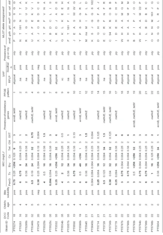
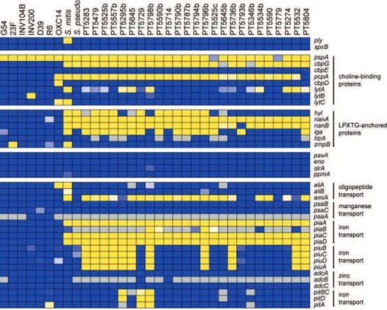
Chapter II describes a detailed analysis of a collection of penicillin-resistant multidrug-penicillin-resistant pneumococcus-like strains colonizing children in the era of pneumococcal conjugate vaccines. The analyses include several phenotypic and genotypic assays complemented by multilocus sequence analysis and comparative genomic hybridization.
Chapter III describes the mechanisms that led to the maintenance of antibiotic resistance rates among colonizing pneumococcus in Portugal, besides a significant shift in serotypes following the introduction of the seven-valent pneumococcal conjugate vaccine.
pneumococci.
In Chapter V, the results obtained in chapters II to IV are discussed as a whole and future perspectives are presented.
Chapters II to IV are reproductions of the publications indicated below. They can be read independently.
Chapter II - Simões, A. S., R. Sá-Leão, M. J. Eleveld, D. A.
Tavares, J. A. Carriço, H. J. Bootsma, and P. W. Hermans. 2010. Highly penicillin-resistant multidrug-resistant pneumococcus-like strains colonizing children in Oeiras, Portugal: genomic characteristics and implications for surveillance. J Clin Microbiol
48:238-246.
Chapter III - Simões, A. S., L. Pereira, S. Nunes, A. Brito-Avô, H.
de Lencastre, and R. Sá-Leão. 2011. Clonal evolution leading to maintenance of antibiotic resistance rates among colonizing pneumococci in the PCV7 era in Portugal. J Clin Microbiol 49: 2810-2817.
Chapter IV - Simões, A. S., C. Valente, H. de Lencastre, and R.
Acknowledgments V
Abstract VII
Resumo XI
Thesis outline XV
Table of contents XVII
Chapter I 1
Streptococcus pneumoniae – Historical Background 3 Pneumococcus and closely related species 4
S. pneumoniae 6
S. pseudopneumoniae 7
S. mitis and S. oralis 7 Pneumococcus-like streptococci 8 Pneumococcal identification methods and differentiation from other streptococci 9 Epidemiology of S. pneumoniae 13 Pneumococcal resistance to antimicrobial agents 16 Pneumococcal vaccines 18 Vaccine impact on pneumococcal population structure 19 Pneumococcal typing methods 22 Non-typeable S. pneumoniae 24 Population structure: clones and antimicrobial resistance 27 Identification of non-typeable pneumococci 29
Aim of the work 30
Highly penicillin-resistant multidrug-resistant pneumococcus-like strains colonizing children in Oeiras, Portugal: genomic characteristics and implications for surveillance
Summary 51
Introduction 51
Materials and Methods 54
Results 61
Discussion 70
Acknowledgments 73
References 74
Supplementary Material 80
Chapter III 83
Clonal evolution leading to maintenance of antibiotic resistance rates among colonizing pneumococci in the PCV7 era in Portugal
Summary 85
Introduction 86
Materials and Methods 87
Results 90
Discussion 101
Acknowledgments 106
References 106
Chapter IV 113
Introduction 115 Materials and Methods 118
Results 124
Discussion 130
Acknowledgments 133
References 133
Chapter V 139
Concluding Remarks 141
References 145
STREPTOCOCCUS PNEUMONIAE - HISTORICAL BACKGROUND
Streptococcus pneumoniae or pneumococcus was one of the first bacterial pathogens to be isolated and characterized. It was first described in 1881 by Louis Pasteur and Georg Sternberg while working independently, although the first to recognize pneumococci was Edwin Klebs in 1875 when he observed infected sputum and lung tissue (reviewed in (50)). Since then, S. pneumoniae has played an important role in the development of microbiology, molecular biology, bacteriology, genetics and vaccines. The use of polysaccharide antigens as vaccines (13), the ability of polysaccharides to induce antibodies (66), the mechanism of bacterial gene transfer (51), the identification of DNA as the genetic material (12), the therapeutic efficacy of penicillin (126), the role of bacterial capsule in resistance to phagocytosis (43), and the first bacterial quorum sensing factor (127), were firstly described in pneumococci.
Following its first description, its name has changed several times. “Microbe septicémique du saliva” and Micrococcus pasteuri were the first names given by Pasteur and Sternberg, respectively. In 1883, Mátray used the term “pneumoniekokken” and in 1886 Albert Fraenkel used the word “pneumokokkus”. In the same year, the name Diplococcus pneumoniae became officially recognized due to its appearance when observed under the microscope, and, in 1974, it was reclassified as S. pneumoniae based on its growth in chains in liquid media (50).
PNEUMOCOCCUS AND CLOSELY RELATED SPECIES
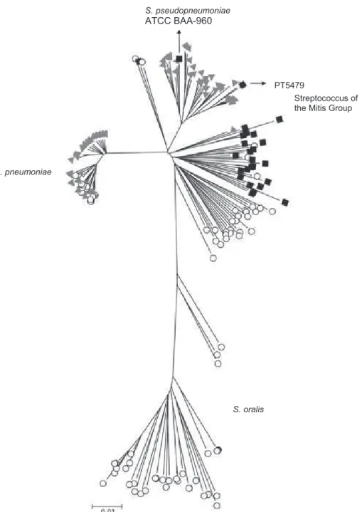
S. pneumoniae is part of the bacterial streptococcus group. This is a heterogeneous group of gram positive, catalase-negative, spherical or ovoid bacteria comprising species that colonize and infect humans and animals (139). Streptococci can be classified according to the hemolytic pattern (partial, complete or no blood hemolysis when grown in blood agar plates); Lancefield group (serological reaction against the major cell-wall carbohydrate antigen); or species (according to their metabolic reaction in various culture media) (139). Figure 1 shows the phylogenetic relationships among 34 species of the genus Streptococcus.
S. pneumoniae is part of the mitis group. Streptococci of the mitis group are part of the commensal flora of the upper respiratory tract of humans. This group includes not only S. pneumoniae but also S. pseudopneumoniae, S. mitis, S. oralis, S. gordoni, S. sanguis and S. parasanguis (Figure 1) (10, 16).
G e n e ra l I n tr o d u c tio n 5 re 1 . P h y lo g e n e tic r e la tio n s h ip s a m o n g 3 4 S tr e p to c o c c u s s p e c ie s ( 7 3 ). te ria a re p ro m is c u o u s ( 4 6 ) a n d , i n th e n a s o p h a ry n x , p n e u m o c o c c i a b it w ith s e v e ra l o th e r b a c te ria l s p e c ie s , in c lu d in g its c lo s e s t tiv e s : S . p s e u d o p n e u m o n ia e , S . m itis , a n d S . o ra lis . G e n e tic S. pneumoniae S. mitis S. gordonii S. sanguis S. oralis S. parasanguis mitis group salivarius group bovis group mutans group pyogenic group anginosus group S. vestibularis S. salivarius S. thermophilus S. anginosus S. constellatus S. intermedius S. bovis
S. equinus S. alactoluticus S. equi S. pyogenes S. porcinus S. canis S. uberis S. suis S. agalactiae S. dysgalactiae S. parauberis S. hyointestinalis S. iniae S. downeii S. sobrinus S. macacae S. acidominimus S. pleomorphus S. rattus
exchange with related species sharing the same ecological niche is one of the mechanisms of S. pneumoniae evolution. Indeed, S. mitis has been described as the main reservoir of genetic diversity of S. pneumoniae (37).
The exchange of genetic elements between species can lead to difficulties in species identification. The mitis group has caused considerable confusion for both clinical microbiologists and taxonomists (42). The correct differentiation between S. pneumoniae and closely related species is essential, not only for clinical diagnostics, but also for colonization studies. For example, the incorrect identification of these isolates could falsely increase the rates of pneumococci non-susceptible to antimicrobials (136).
Since the correct identification of pneumococci is one of the major subjects of this thesis, the following sections will describe S. pneumoniae and closely related species and the methods used to distinguish them.
S. pneumoniae
S. pneumoniae is a gram-positive bacterium, lancet-shaped, typically less than 2µm in diameter, which can grow in pairs or short chains. It is catalase negative, facultative anaerobic, usually susceptible to optochin, soluble in bile (deoxycholate) salts and shows -hemolysis in blood agar plates. It is a fastidious organism that requires the presence of blood or serum in the medium for growth, and also the presence of 5% of CO2 (121). Most of the isolates gave a positive
More detailed information about S. pneumoniae will be described later in this chapter.
S. pseudopneumoniae
S. pseudopneumoniae, first described in 2004 (10), is a gram-positive bacterium, similar to pneumococcus when grown in agar media, is not soluble in bile, and is resistant to optochin when incubated under an atmosphere of increased CO2, although it is susceptible to optochin
when incubated in ambient atmosphere. It has a negative result in the Quellung reaction (10).
S. pseudopneumoniae has low incidence in clinical samples but it has been associated with chronic obstructive pulmonary disease (75) and it has been demonstrated to have disease potential. In a study by Harf-Monteil et al, it was demonstrated that were able to kill 100% of infected animals (immunocompetent 6-week-old Swiss mice) in 36 hours in a peritonitis/sepsis model (60).
In two studies, in New Zealand, that described S. pseudopneumoniae isolates recovered from respiratory samples between 2000 and 2007, high rates of resistance to erythromycin (c.a. 60%) and tetracycline (c.a. 70%) have been observed. No resistance to any -lactam agent has been observed, although about one-third of the isolates showed reduced susceptibility to penicillin and ampicillin (74, 75).
S. mitis and S. oralis
in bile, and are resistant to optochin. Similarly to S. pseudopneumoniae they give a negative result in the Quellung reaction (reviewed in (35)).
Although commensals, these streptococci are isolated from a variety of infections but most significantly from patients with subacute bacterial endocarditis and from neutropenic patients with cancer (17, 38).
Regarding antimicrobial resistance, several studies have indicated that S. mitis and S. oralis strains are commonly found in blood cultures of cancer patients and are commonly resistant to -lactam antimicrobials (reviewed in (42)). In fact, high rates of resistance to penicillin and high MICs (minimum inhibitory concentration) are frequent among S. mitis isolates (69).
Pneumococcus-like streptococci
Atypical pneumococcal isolates that are putatively identified as pneumococci but that give negative results in one or more of the classical assays have been described (87, 88, 138). These isolates were considered to be pneumococcus-like streptococci and the denomination “streptococci of the mitis group” (SMG) has been proposed (87).
S. pneumoniae would have been acquired more recently, presumably by individual S. mitis lineages (77). In addition, most genes associated with pathogenicity which are shared by S. pneumoniae strains, can also be present in S. mitis, S. oralis, and S. infantis (37).
Like pneumococci, most members of the mitis group are naturally competent for genetic transformation, i.e., they are able to take up DNA fragments and incorporate them into their genome (65, 93). Another theory for the origin of these bacteria is that horizontal gene transfer between naturally competent streptococci that share the same ecological niche contributes to the attenuation of putative barriers between these species (10, 55, 56). In fact, the group of Regine Hakenbeck described the genome of S. mitis B6 (a high level penicillin and multiple antibiotic resistant isolate) and suggested that the genome of this strain is an example of genome modification by the acquisition of genes and gene clusters from other sources such as S. pneumoniae, since both species share a core genome of over 900 genes (43% of total S. mitis genome) (34). The same group described an identical mosaic structure of pbp2x gene in S. pneumoniae, S. mitis and S. oralis, suggesting inter- and intraspecies recombination events (28).
Still, several attempts to correctly distinguish pneumococci from closely relatives have been made, as detailed in the following section.
PNEUMOCOCCAL IDENTIFICATION METHODS AND
DIFFERENTIATION FROM OTHER STREPTOCOCCI
(colonies with a depression in the center showing alpha-hemolysis), susceptibility to optochin when incubated in CO2 atmosphere,
solubility in 1% of sodium deoxycholate, and assignment of a capsular type through serotyping (76). For most pneumococci, which are capsulated, its identification is straightforward. However some isolates give an atypical result in one or more of these assays (80, 87, 138).
Susceptibility to optochin has been described as a method with 90% to 100% of sensitivity and 99% to 100% of specificity (76). However, optochin-resistant pneumococcal isolates have been described (4, 94, 97, 104) and optochin-susceptible non-pneumococcal isolates have been observed (76).
Solubility in 1% of sodium deoxycholate (or bile salts) is considered by some authors the standard method to identify pneumococci since it has a sensibility of 98% and a specificity of 100% (112). However, some strains can harbor a mutation in the lytA gene (a deletion of two amino acids responsible for cell wall anchoring in the carboxy-terminal domain) that confers a deoxycholate-insoluble phenotype (100). Additionally, some streptococcus of the mitis group were described as bile soluble (87).
non-pneumococcal streptococci with positive cross-reactions with antipneumococcal polysaccharide capsular antibodies (67).
In addition, in recent years several molecular methods have been proposed to differentiate pneumococcus from closely related species. For example, amplification of ubiquitous pneumococcal genes such as autolysin (lytA), surface antigen A (psaA) and pneumolysin (ply) have been proposed to identify pneumococci (92).
In 2004, Messmer et al compared the specificity of these three genes for the correct identification of S. pneumoniae and concluded that genes lytA and psaA had a specificity of 100% and 99%, respectively (92). However, for the ply gene, the specificity was 50%, since it was found in eight of the 16 atypical isolates tested (92). In addition, in 2007, Jefferies et al described a homologue of the ply gene named mitilysin (mly) that could be found in some S. mitis isolates (71). Furthermore, homologues of lytA, psaA and ply have been detected in strains of closely related streptococcal species (70, 95, 111, 138).
More recently, detection of the pneumococcal species specific gene piaA (which encodes for a lipoprotein component of two iron ABC transporters) has been proposed as a diagnostic tool (137). However, this gene is not ubiquitous in pneumococci (137).
The DNA fragment Spn9802 has also been described as a specific target for S. pneumoniae (1, 125) but some authors have described its presence in S. pseudopneumoniae and S. mitis (40, 101).
A 181-bp S. pneumoniae-specific fragment, located between the gene Spn23F20600 (from nucleotide 2,002,250 to nucleotide 2,000,793), and gene Spn23F20590 (from nucleotide 2,000,770 to nucleotide 2,000,489), in S. pneumoniae ATCC700669, was described as being able to discriminate S. pneumoniae from other streptococci (106).
The sequence of the intergenic spacer (ITS) region and part of gdh gene was also proposed but ITS region cannot be used for the species-level identification of the mitis group (only at the group level) and sequence analysis of the partial gdh gene is apparently able to differentiate S. mitis, S. oralis, and S. pneumoniae, but is not able to differentiate S. mitis from S. pseudopneumoniae (96). In addition, this method cannot be easily applied in a clinical microbiology laboratory that has not sequencing facilities (96).
A recA PCR assay was described as useful to differentiate pneumococcus from other viridans streptococci (142) but do not correctly differentiate S. pneumoniae from S. mitis and S. pseudopneumoniae (142).
In addition, multilocus sequence typing (MLST) has been described as an important tool to define pneumococcus (55) and the construction of phylogenetic trees using concatenated sequences of all MLST loci except ddl (an approach denominated multilocus sequence analysis (MLSA)), has been proposed as a good alternative molecular technique to differentiate pneumococci from other closely related streptococci (55). However, the average sequence divergence between S. mitis and S. pneumoniae clusters is 5.8%, only slightly greater than within the S. mitis cluster itself (54).
Overall, methods based on lytA gene appear to be the most reliable (24, 87). In particular, in 2006, Llull and co-workers described a RFLP method to differentiate pneumococcus from closely related species based on signatures characteristic of typical pneumococcal lytA or atypical lytA (87). More recently, a lytA based asymmetric PCR for the specific detection of pneumococcus was developed and it has been claimed that it has no cross reactions with S. mitis or S. pseudopneumoniae (24).
A simple and low cost method that can be easily applied in a microbiology laboratory is essential to correctly identify pneumococci and differentiate them from closely related species.
In summary, differentiation between atypical S. pneumoniae and closely related species remains a challenge since they share several phenotypic and genotypic characteristics.
EPIDEMIOLOGY OF S. PNEUMONIAE
asymptomatic and it is common in children under five years old. The colonization occurs soon after birth (9) and it peaks around the age of three (18). After that, there is a steady decline until the age of ten and remains low in adulthood (18).
The duration of carriage is variable (typically ranging between one to seventeen months (49)) and depends on several factors such as the host’s age and the serotype of the colonizing strain (30). The transmission is either person-to-person by direct contact, or by aerosols (134). Recently, it was described that pneumococci are able to survive long-term desiccation, suggesting that environmental surfaces may also serve as sources of pneumococcal infection (132).
The risk factors associated with pneumococcal carriage include age (children less than two years old, and adults older than 65 years old), frequent contact with children, crowded environments (day care center attendance, military training camps, prisons, nursing homes), prior respiratory infection, cigarette smoking, asthma and other chronic respiratory diseases (82).
Colonization precedes pneumococcal disease (18). As a pathogen, S. pneumoniae can cause a wide range of diseases from otitis media, to pneumonia, bacteremia, and meningitis. Worldwide, the morbidity and mortality associated with pneumococcal infections remain very high (98).
Most S. pneumoniae strains are covered by a polysaccharide capsule that constitutes the main virulence factor of this pathogen. The capsule is 200 to 400nm thick (120) and plays a major role in colonization and disease by promoting bacterial interaction with the epithelium and provide protection against complement-mediated opsonophagocytosis (141).
There are more than 90 different capsules described, however, only a restricted number of serotypes are responsible for disease. Differences in virulence between pneumococcal serotypes have been observed and the association serotype disease depends on several factors, such as, the age of the host (different serotypes between children and adults), geographic location, and disease manifestation (62).
and 33F) were associated with more than 80% of the pneumococcal infections (19).
Regarding disease manifestation some studies described serogroups 6, 14, 19 and 23 associated with otitis media (11, 78, 131), serotypes 1, 4 as 14 isolated from blood (63) and serotypes 1 and 3 associated with pneumonia (62). In addition, some serotypes appear to have higher invasive disease potential than others. In a Finnish study, serotypes 6B, 14, 18C and 19A were more related with invasive disease and serotypes 6A, 35F and 11A were associated with carriage (57). In Portugal, in a study to estimate the invasive disease potential of serotypes and clones circulating in Portugal before extensive use of the seven-valent pneumococcal conjugate vaccine it was found that serotypes 1, 3, 4, 5, 7F, 8, 9N, 9L, 12B, 14, 18C, and 20 had an enhanced propensity to cause invasive disease, while serotypes 6A, 6B, 11A, 15B/C, 16F, 19F, 23F, 34, 35F, and 37 were associated with carriage (115).
The susceptibility to antimicrobial agents appears to be associated with some serotypes. For instance, penicillin resistance and multiple resistance appears to be restricted to few serotypes such as 6A, 6B, 9V, 14, 19A, 19F and 23F (32). In the same way, strains isolated from the nasopharynx were frequently more resistant to antimicrobial agents than those isolated from infections (62).
PNEUMOCOCCAL RESISTANCE TO ANTIMICROBIAL AGENTS
of patients was documented in 1917 (reviewed in (79)). However, the first S. pneumoniae clinical strain resistant to an antimicrobial agent (penicillin) was described in 1967 from a 25-year-old woman (reviewed in (79)). Since then, emergence of resistant strains to several classes of antimicrobials such as -lactam and macrolides, has been observed (79).
Nowadays, the overall rates of antimicrobial resistant isolates vary from country to country being high in several geographical areas. In Europe, the prevalence of penicillin-non susceptible S. pneumoniae varies between 1-5% in the Netherlands, Norway, Germany, the United Kingdom, Sweden, and Denmark; and 20% in Ireland, Spain, and France. Resistance levels to macrolides also vary significantly: 4-10% in Sweden, the United Kingdom, Denmark, the Netherlands, Norway and Germany; and 20-30% in Spain, Belgium, France, and Finland (39). In the United States the rates of non-susceptible S. pneumoniae are of approximately 10% for penicillin and 25% for macrolides (25).
In Portugal, in 2006 the antimicrobial resistance rates were comparable between carriage and invasive isolates from children up to six years old. Non-susceptibility to penicillin was 22% for carriage and 23% for invasive disease isolates, macrolide resistance was observed in 23% of colonizing isolates and 27% of invasive isolates, and resistance to tetracycline was found for 18% of carriage and 22% of invasive disease isolates (3, 114).
moderate to high, such as southern and eastern European countries (48) and remain low in northern European countries where antibiotic consumption is low (109).
PNEUMOCOCCAL VACCINES
As in other diseases, prevention of pneumococcal disease is the first step to control it. A 23-valent pneumococcal polysaccharide vaccine (PPV23) has been available since 1983, and contains the 23 most common serotypes causing disease in adults from the United States of America (USA) at the time it was launched. This vaccine is not effective in children less than two years old as it elicits a T-cell-independent immune response, which is underdeveloped in young children (19, 123). PPV23 is used nowadays to vaccinate high risk adults and older children, and the elderly.
In 2000, a seven-valent pneumococcal conjugate vaccine (PCV7) was introduced in the USA universal immunization program. This vaccine targets the seven most common serotypes (4, 6B, 9V, 14, 18C, 19F and 23F) that caused invasive pneumococcal disease (IPD) in children in the USA (27). More recently, two other pneumococcal conjugate vaccines were licensed - a ten-valent pneumococcal conjugate vaccine (PCV10) which includes the PCV7 serotypes plus serotypes 1, 5 and 7F, as well as, an antigen that stimulates antibody production against non-typeable Haemophilus influenzae (www.gsk.com/products/vaccines/synflorix); and a 13-valent pneumococcal conjugate vaccine (PCV13) that includes the PCV10 serotypes plus serotypes 3, 6A and 19A (www.prevnar13.com).
available. Despite widespread vaccination, none of these vaccines is part of the National Vaccination Program nor is reimbursed by the state. However, available data indicates that around 60% of children up to 5 years old were vaccinated in 2006 (107, 114,Pfizer Portugal data).
Vaccine impact on pneumococcal population structure
In areas where there has been a massive use of PCV7, the pneumococcal population has changed substantially: a significant reduction in the incidence of pneumococcal disease and a replacement of serotypes in circulation has been observed; non-vaccine types have increased among asymptomatic carriers and as causes of invasive pneumococcal disease - for a review see (133).
In colonization, in studies conducted in Portugal and Norway (five and two years after PCV7 was commercially available, respectively), carriage by vaccine serotypes decreased; however, the overall prevalence of pneumococcal carriage among vaccinated and unvaccinated children remained the same (114, 130). The maintenance of rates of carriage was due to serotype replacement. In Norway, an increase of serotypes 9N, 16F, 24F, 35F and 35B (130) was observed. In Portugal, rates of non-vaccine types 1, 6C, 7F, 15A, 16F, 21, 23A, 29 and non-typeable strains increased significantly (114).
vaccination. In fact, several studies described changes in the pneumococcal population prior to the introduction of PCV7 (8, 45, 59, 72).
In contrast with what had been anticipated by some authors (83) the proportion of antimicrobial resistant pneumococci did not decrease in all countries that introduced PCV7 (6, 108, 114). In Portugal, in colonization isolates, rates of intermediate resistance to penicillin and resistance to macrolides, tetracycline and multidrug resistance remained unchanged due to a balance between reduction of antimicrobial resistant vaccine types and an increase among non– vaccine types (114). Regarding invasive isolates, there was not an overall reduction in the proportion of infections caused by resistant pneumococci in children and adults (6). In the USA, in invasive and non invasive infections isolates from all age groups, the prevalence of isolates with intermediate penicillin resistance and resistant to erythromycin has decreased, but the levels of multidrug resistance have not changed (108).
dominant clones in the USA (108). In Norway, the expansion of Portugal19FST177 and the emergence of Taiwan19FST236 were observed (130). Regarding capsular switch events, isolates with genotypes that previously were only associated with vaccine serotypes, but now express non-vaccine serotypes due to recombination at the capsular locus, have been well documented (22). In Portugal, no information about the mechanisms that led to the maintenance of antimicrobial resistance levels in colonization, despite serotype replacement among carriage isolates had been unavailable until recently (see Chapter III).
The introduction of PCV10 and PCV13 will also have an impact in pneumococcal invasive disease (IPD) and carriage. A study conducted in 2010 in the USA predicted that vaccination with PCV13 would reduce the incidence of IPD and acute otitis media by approximately 106 thousand and 16.3 million cases over a ten-year period (113). In a study from the United Kingdom, the serotype coverage of IPD in children under five years of age for PCV10 and PCV13 was less in 2007/2008 than in 2005/2006 which suggests that the potential coverage of PCV10 and PCV13 will be less than expected due to the routine use of PCV7 and the associated serotype replacement (47).
PNEUMOCOCCAL TYPING METHODS
of the world, or for protection against different kinds of pneumococcal infections. Methods for bacterial typing have also had a high importance in studies of outbreaks cases (129).
Pneumococcus can be typed by phenotypic and genotypic methods. Genotypic methods have generally higher discriminatory power than phenotypic methods.
The most common phenotypic methods are serotyping and antimicrobial susceptibility testing. Serotyping was already described here, and consists on the determination of capsular type using antipneumococcal polysaccharide capsule antibodies (81). In the antimicrobial susceptibility testing, the isolates are tested by diffusion or dilution methods against a panel of antimicrobial agents and an antibiotype is obtained.
The most common molecular typing methods are pulsed-field gel electrophoresis (PFGE) and multilocus sequence typing (MLST). PFGE is based on the digestion of the total chromosomal DNA with a restriction endonuclease that cleaves the DNA infrequently. The macro-restriction fragments are separated by gel electrophoresis according to molecular weight in an apparatus that switches the direction of current according to a predetermined pattern. The different orientation of the electric pulses during electrophoresis allows the separation of large DNA fragments. In S. pneumoniae, typically, 10 to 19 bands of 20 to 300 kb are obtained when de DNA is digested with SmaI (85).
consists on the amplification of approximately 450bp of the internal fragments of seven housekeeping genes – aroE, gdh, gki, recP, spi, xpt, and ddl (41). The sequences are submitted to an international database available online (available at www.mlst.net) and a number is attributed to each allele. The seven alleles defined the sequence type (ST).
As described early in this chapter, multilocus sequence analysis (MLSA) use concatenated sequences from the multiple house-keeping loci to construct a dendrogram and observe the patterns of clustering of genotypes (54) and has also been proposed as a good alternative molecular technique to differentiate pneumococci from other closely related streptococci (55).
Comparative genomic hybridization (CGH) is based on the hybridization of DNA samples, labeled with fluorescent dyes, to microchips containing multiple oligonucleotides spotted on the chip surface. The fluorescence pattern is recorded by a scanner, quantified and analyzed. In pneumococcus, CGH has been used to compare the genomic content between clinical isolates (99, 119) and differentiate/compare pneumococcus from closely related species (84).
NON-TYPEABLE S. PNEUMONIAE
colonies are generally rough (12). These atypical isolates are called non-typeable S. pneumoniae (NTPn).
The mechanisms leading to non-typeability by the Quellung reaction may be at least four: (i) the expression of capsule may be severely down-regulated; (ii) the cps region, which contains the genes encoding the enzymes responsible for capsule biosynthesis, may have been disrupted; (iii) a novel capsular type not described yet for which there is no specific antisera may be expressed; and (iv) the capsular operon may be absent.
In 2003, Hathaway analyzed 27 non-capsulated pneumococci (seven from invasive disease and 20 from colonization) and divided them into two groups (61). Group I strains were closely related to capsulated strains since they had typical capsule genes and their genetic backgrounds, as determined by MLST, were characteristic of typeable strains. Group II strains were less closely related to capsulated pneumococci by MLST but had a clonal population structure among themselves and a genetic background associated with two geographically widespread clones able to cause epidemic conjunctivitis (89). Group II strains lacked typical capsule genes, but instead possessed two ORFs, which exhibited 61% to 64.5% homology to the ATP-binding cassette (ABC) transporter gene aliB and were designated aliB-like ORF 1 and aliB-like ORF 2 (61).
Hanage and colleagues used MLST to characterize a set of 121 non-typeable presumptive pneumococci from Finland and identified three classes: (i) NTPn isolates with STs identical to capsulated pneumococci (suggesting pneumococci in which capsular expression had been down regulated or lost), (ii) isolates that clustered with capsulated pneumococci in a phylogenetic tree based on MLSA but had STs that differed from those of capsulated pneumococci in the MLST database (these isolates seemed to lack the capsular operon); and (iii) isolates that did not have typical pneumococci MLST alleles and did not cluster with capsulated pneumococci (all seven MLST alleles were 5% to 10% divergent from those of pneumococci) (56).
Based in these and other studies, non-capsulated strains are often referred to as non-typeable strains (in this thesis the two names are often used as synonyms).
Non-capsulated strains are less virulent than capsulated strains but have been associated to conjunctivitis outbreaks in rates that range from 23% to 51% (14, 15, 91). They have also been described as otopathogens (140). Regarding invasive disease, these isolates represented 2% of the cases in some studies (21, 44, 52) and in carriage, these rates were up to 10% (7, 116, 117, 130). In a study performed in Israel, NTPn was the most common pneumococcal type in acute conjunctivitis cases (14.1%), it was the seventh among carriage isolates (5.1%), was rarely found in acute otitis media (0.7%), and was not found in the invasive pneumococcal disease group (105).
demonstrated to increase the adherence to the epithelial tissue (2, 31, 135), biofilm formation (36), and facilitates transformation events (102, 134).
Furthermore, it has been suggested that NTPn may act as privileged vectors to horizontal gene transfer: non-capsulated pneumococci may acquire DNA fragments from commensal streptococci and act as a shuttle vector of resistance genes for capsulated pneumococci.
Specifically, it has been shown that a serotype 19F ST444 has evolved from low level to high level penicillin resistance by uptaking penicillin binding proteins gene fragments from a NTPn isolate (64).
In addition non-typeable pneumococci were detected often in co-colonized samples (i.e., samples that contain more than one pneumococcal isolate) (23, 116) which may lead to increased opportunities for horizontal gene transfer between capsulated and non-capsulated pneumococci.
Population structure: clones and antimicrobial resistance
internationally spread and have been described in acute otitis media, conjunctivitis and carriage (56, 116, 140).
Figure 2. Go-eburst of the NTPn isolates available in the MLST database.
NTPn are often multidrug resistant (116). The international clone NorwayNTST344 has a characteristic pattern of multiresistance to penicillin, erythromycin, clindamycin, tetracycline and sulfamethoxazole-trimethoprim (122). However, it seems that NTPn isolates recovered from nasopharynx are more resistant than those recovered from other sites. In a study performed in Israel, 82% of NTPn isolates recovered from carriage were non-susceptibility to penicillin in contrast with rates of 65% and 53% found for acute conjunctivitis and acute otitis media respectively (105). In a Spanish collection ST941, ST942, and ST943 (isolated from the conjunctiva) were fully antibiotic susceptible (15) and the same happened with ST448 described as othopathogen (140) and associated with conjunctivitis outbreaks (89, 105).
These high rates of antibiotic resistance among NTPn (especially the ones from colonization) could possibly be explained by a combination of two factors: (i) increased exposure to transforming DNA due to high transformability rates (NTPn have transformability rates up to 1,075-fold higher than capsulated strains (102)) and, (ii) greater selective antibiotic pressure during nasopharyngeal colonization (33).
Identification of non-typeable pneumococci
is of clinical importance, since a misidentification of the causative agent can influence the diagnosis and treatment of disease (138).
Non-agglutination with any available capsular anti-serum is the method that per se defines NTPn. However, this may be tricky. For example, in a study by Sá-Leão et al, the presumptive identification of NTPn based on colony morphology, optochin susceptibility, and a negative capsular serotype reaction led to the misclassification of 19% NTPn isolates (116). Molecular methods are useful for NTPn identification and have been largely used (55, 116, 128). Sá-Leão and colleagues suggested the simultaneous detection of lytA and psaA, since the odds of having a non-pneumococcal isolate carrying these both pneumococcal genes is smaller (116). Hanage and co-workers described MLSA as an important tool to identify NTPn isolates (55). A serotype microarray that can detect NTPn in co-colonization has also been proposed (128).
AIM OF THE WORK
Since the introduction of pneumococcal conjugate vaccines, changes have been observed in the ecology of nasopharynx. In Portugal, the maintenance of antimicrobial resistance rates among disease and carriage isolates (5, 114) and an increase of rates of non-typeable strains among carriage isolates (114) was observed.
REFERENCES
1. Abdeldaim, G. M., K. Stralin, P. Olcen, J. Blomberg, and B.
Herrmann. 2008. Toward a quantitative DNA-based definition of
pneumococcal pneumonia: a comparison of Streptococcus
pneumoniae target genes, with special reference to the Spn9802
fragment. Diagn Microbiol Infect Dis 60:143-150.
2. Adamou, J. E., T. M. Wizemann, P. Barren, and S. Langermann.
1998. Adherence of Streptococcus pneumoniae to human bronchial
epithelial cells (BEAS-2B). Infect Immun 66:820-822.
3. Aguiar, S. I., M. J. Brito, J. Goncalo-Marques, J. Melo-Cristino,
and M. Ramirez. 2010. Serotypes 1, 7F and 19A became the
leading causes of pediatric invasive pneumococcal infections in
Portugal after 7 years of heptavalent conjugate vaccine use. Vaccine
28:5167-5173.
4. Aguiar, S. I., M. J. Frias, L. Santos, J. Melo-Cristino, and M.
Ramirez. 2006. Emergence of optochin resistance among
Streptococcus pneumoniae in Portugal. Microb Drug Resist 12:
239-245.
5. Aguiar, S. I., F. R. Pinto, S. Nunes, I. Serrano, J. Melo-Cristino,
R. Sá-Leão, M. Ramirez, and H. de Lencastre. 2010.
Denmark14-230 clone as an increasing cause of pneumococcal infection in
Portugal within a background of diverse serotype 19A lineages. J
Clin Microbiol 48:101-108.
6. Aguiar, S. I., I. Serrano, F. R. Pinto, J. Melo-Cristino, and M.
Ramirez. 2008. Changes in Streptococcus pneumoniae serotypes
causing invasive disease with non-universal vaccination coverage of
the seven-valent conjugate vaccine. Clin Microbiol Infect 14:
835-843.
7. Allen, U. D., S. Thomas, J. Carapetis, S. Henry, S. Wasfy, M.
Lovgren, S. Richardson, and D. E. Low. 2003. Serotypes of
respiratory tract isolates of Streptococcus pneumoniae from
8. Amrine-Madsen, H., J. Van Eldere, R. M. Mera, L. A. Miller, J. A.
Poupard, E. S. Thomas, W. S. Halsey, J. A. Becker, and F. P.
O'Hara. 2008. Temporal and spatial distribution of clonal complexes
of Streptococcus pneumoniae isolates resistant to multiple classes
of antibiotics in Belgium, 1997 to 2004. Antimicrob Agents
Chemother 52:3216-3220.
9. Aniansson, G., B. Alm, B. Andersson, P. Larsson, O. Nylen, H.
Peterson, P. Rigner, M. Svanborg, and C. Svanborg. 1992.
Nasopharyngeal colonization during the first year of life. J Infect Dis
165 Suppl 1:S38-42.
10. Arbique, J. C., C. Poyart, P. Trieu-Cuot, G. Quesne, G. Carvalho
Mda, A. G. Steigerwalt, R. E. Morey, D. Jackson, R. J. Davidson,
and R. R. Facklam. 2004. Accuracy of phenotypic and genotypic
testing for identification of Streptococcus pneumoniae and
description of Streptococcus pseudopneumoniae sp. nov. J Clin
Microbiol 42:4686-4696.
11. Arguedas, A., R. Dagan, S. Guevara, N. Porat, C. Soley, A.
Perez, and R. Brilla. 2005. Middle ear fluid Streptococcus
pneumoniae serotype distribution in Costa Rican children with otitis
media. Pediatr Infect Dis J 24:631-634.
12. Avery, O. T., C. M. MacLeod, and M. McCarty. 1944. Studies on
the chemical nature of the substance inducing transformation in
pneumococcal types. J Exp Med 79:137-159.
13. Avery, O. T., H. T. Chickering, R. Cole, and A. R. Dochez. 1917.
Acute lobar pneumonia, prevention and serum treatment. Monogr
Rockefeller Inst 7:1-110.
14. Barker, J. H., Musher, D. M., Silberman, R., Phan, H. M., Watson,
D. A. 1999. Genetic Relatedness among Nontypeable Pneumococci
Implicated in Sporadic Cases of Conjunctivitis. J Clin Microbiol
37:4039-4041.
15. Berron, S., A. Fenoll, M. Ortega, N. Arellano, and J. Casal. 2005.
Analysis of the genetic structure of nontypeable pneumococcal
16. Bishop, C. J., D. M. Aanensen, G. E. Jordan, M. Kilian, W. P.
Hanage, and B. G. Spratt. 2009. Assigning strains to bacterial
species via the internet. BMC Biol 7:3.
17. Bochud, P. Y., T. Calandra, and P. Francioli. 1994. Bacteremia
due to viridans streptococci in neutropenic patients: a review. Am J
Med 97:256-264.
18. Bogaert, D., R. De Groot, and P. W. Hermans. 2004.
Streptococcus pneumoniae colonisation: the key to pneumococcal
disease. Lancet Infect Dis 4:144-154.
19. Bogaert, D., P. W. Hermans, P. V. Adrian, H. C. Rumke, and R.
de Groot. 2004. Pneumococcal vaccines: an update on current
strategies. Vaccine 22:2209-2220.
20. Bogaert, D., M. N. Engelen, A. J. M. Timmers-Reker, K. P.
Elzenaar, P. G. H. Peerbooms, and R. A. Coutinho. 2001.
Pneumococcal carriage in children in the Netherlands. A molecular
epidemiological study. J Clin Microbiol 39:3316-3320.
21. Broome, C. V., and R. R. Facklam. 1981. Epidemiology of clinical
significant isolates of Streptococcus pneumoniae in the United
States. Rev Infect Dis 3:277-280.
22. Brueggemann, A. B., R. Pai, D. W. Crook, and B. Beall. 2007.
Vaccine escape recombinants emerge after pneumococcal
vaccination in the United States. PLoS Pathog 3:e168.
23. Brugger, S. D., P. Frey, S. Aebi, J. Hinds, and K. Muhlemann.
2010. Multiple colonization with S. pneumoniae before and after
introduction of the seven-valent conjugated pneumococcal
polysaccharide vaccine. PLoS One 5:e11638.
24. Campuzano, S., M. Pedrero, J. L. Garcia, E. Garcia, P. Garcia,
and J. M. Pingarron. 2011. Development of amperometric
magnetogenosensors coupled to asymmetric PCR for the specific
detection of Streptococcus pneumoniae. Anal Bioanal Chem
25. CDC 2009, posting date. Active Bacterial Core Surveillance Report.
Emerging Infections Program Network for Streptococcus
pneumoniae. [Online.]
26. CDC. 2005. Direct and indirect effects of routine vaccination of
children with 7-valent pneumococcal conjugate vaccine on incidence
of invasive pneumococcal disease--United States, 1998-2003.
MMWR Morb Mortal Wkly Rep 54:893-897.
27. CDC. 2000. Preventing pneumococcal disease among infants and
young children Recommendations of the Advisory Committee on
Immunization Practices (ACIP). Morb Mortal Wkly Rep 49:1–35.
28. Chi, F., O. Nolte, C. Bergmann, M. Ip, and R. Hakenbeck. 2007.
Crossing the barrier: evolution and spread of a major class of
mosaic pbp2x in Streptococcus pneumoniae, S. mitis and S. oralis.
Int J Med Microbiol 297:503-512.
29. Chistenson, B., S. P. Sylvan, and B. Noreen. 1997. Carriage of
multiresistant Streptococcus pneumoniae among children attending
day-care centers in the Stockholm area. Scand J Infect Disease
29:555-558.
30. Crook, D. W., A. B. Brueggemann, K. L. Sleeman, and T. E. A.
Peto. 2004. Pneumococcal Carriage, p. 136-168. In E. I. Tuomanen,
T. J. Mitchell, D. A. Morrison, and B. G. Spratt (ed.), The
Pneumococcus. ASM Press, Washington, DC.
31. Cundell, D. R., J. N. Weiser, J. Shen, A. Young, and E. I.
Tuomanen. 1995. Relationship between colonial morphology and
adherence of Streptococcus pneumoniae. Infect Immun 63:757-761.
32. Dagan, R., and K. P. Klugman. 2008. Impact of conjugate
pneumococcal vaccines on antibiotic resistance. Lancet Infect Dis
8:785-795.
33. Dagan R, M. L. 2004. Changing the ecology of pneumococci with
antibiotics and vaccines, p. 283–331. In M. T. Tuomanen EI,
Morrison DA, Spratt BG, (ed.), The Pneumococcus. ASM Press,,
34. Denapaite, D., R. Bruckner, M. Nuhn, P. Reichmann, B. Henrich,
P. Maurer, Y. Schahle, P. Selbmann, W. Zimmermann, R.
Wambutt, and R. Hakenbeck. 2010. The genome of Streptococcus
mitis B6--what is a commensal? PLoS One 5:e9426.
35. Doern, C. D., and C. A. Burnham. 2010. It's not easy being green:
the viridans group streptococci, with a focus on pediatric clinical
manifestations. J Clin Microbiol 48:3829-3835.
36. Domenech, M., E. Garcia, and M. Moscoso. 2009. Versatility of
the capsular genes during biofilm formation by Streptococcus
pneumoniae. Environ Microbiol 11:2542-2555.
37. Donati, C., N. L. Hiller, H. Tettelin, A. Muzzi, N. J. Croucher, S. V.
Angiuoli, M. Oggioni, J. C. Dunning Hotopp, F. Z. Hu, D. R.
Riley, A. Covacci, T. J. Mitchell, S. D. Bentley, M. Kilian, G. D.
Ehrlich, R. Rappuoli, E. R. Moxon, and V. Masignani. 2010.
Structure and dynamics of the pan-genome of Streptococcus
pneumoniae and closely related species. Genome Biol 11:R107.
38. Douglas, C. W., J. Heath, K. K. Hampton, and F. E. Preston.
1993. Identity of viridans streptococci isolated from cases of infective
endocarditis. J Med Microbiol 39:179-182.
39. EARS. 2009. Annual report of the European Antimicrobial
Resistance Surveillance Network.
40. El Aila, N. A., S. Emler, T. Kaijalainen, T. De Baere, B. Saerens,
E. Alkan, P. Deschaght, R. Verhelst, and M. Vaneechoutte. 2010.
The development of a 16S rRNA gene based PCR for the
identification of Streptococcus pneumoniae and comparison with
four other species specific PCR assays. BMC Infect Dis 10:104.
41. Enright, M. C., and B. G. Spratt. 1998. A multilocus sequence
typing scheme for Streptococcus pneumoniae: identification of
clones associated with serious invasive disease. Microbiology
144:3049-3060.
42. Facklam, R. 2002. What happened to the streptococci: overview of
taxonomic and nomenclature changes. Clin Microbiol Rev 15:
43. Felton, L. D., G. Kauffmann, B. Prescott, and B. Ottinger. 1955.
Studies on the mechanism of the immunological paralysis induced in
mice by pneumococcal polysaccharides. J Immunol 74:17-26.
44. Fenoll, A., G. Asensio, I. Jado, S. Berron, M. T. Camacho, M.
Ortega, and J. Casal. 2002. Antimicrobial susceptibility and
pneumococcal serotypes. J Antimicrob Chemother 50 Suppl S2:
13-19.
45. Fenoll, A., I. Jado, D. Vicioso, A. Perez, and J. Casal. 1998.
Evolution of Streptococcus pneumoniae serotypes and antibiotic
resistance in Spain: update (1990 to 1996). J Clin Microbiol
36:3447-3454.
46. Fraser, C., W. P. Hanage, and B. G. Spratt. 2007. Recombination
and the nature of bacterial speciation. Science 315:476-480.
47. Gladstone, R. A., J. M. Jefferies, S. N. Faust, and S. C. Clarke.
2011. Continued control of pneumococcal disease in the UK - the
impact of vaccination. J Med Microbiol 60:1-8.
48. Goossens, H., M. Ferech, R. Vander Stichele, and M. Elseviers.
2005. Outpatient antibiotic use in Europe and association with
resistance: a cross-national database study. Lancet 365:579-587.
49. Gray, B. M., G. M. Converse, 3rd, and H. C. Dillon, Jr. 1980.
Epidemiologic studies of Streptococcus pneumoniae in infants:
acquisition, carriage, and infection during the first 24 months of life. J
Infect Dis 142:923-933.
50. Gray, B. M., and D. M. Musher. 2008. The history of pneumococcal
disease, p. 3-18. In G. R. Siber, K. K. P., and P. H. Makela (ed.),
Pneumococcal vaccines. Amercican Society of Microbiology,
Washington.
51. Griffith, F. 1928. The significance of pneumococcal types. J Hyg
27:113-159.
52. Gross, J., and O. Fulco. 1985. Pneumococcal pneumonia and
septicemia resulting from a nontypable strain of pneumococcus. J
53. Hanage, W. P., C. J. Bishop, S. S. Huang, A. E. Stevenson, S. I.
Pelton, M. Lipsitch, and J. A. Finkelstein. 2011. Carried
pneumococci in Massachusetts children: the contribution of clonal
expansion and serotype switching. Pediatr Infect Dis J 30:302-308.
54. Hanage, W. P., C. Fraser, and B. G. Spratt. 2006. Sequences,
sequence clusters and bacterial species. Philos Trans R Soc Lond B
Biol Sci 361:1917-1927.
55. Hanage, W. P., T. Kaijalainen, E. Herva, A. Saukkoriipi, R.
Syrjanen, and B. G. Spratt. 2005. Using multilocus sequence data
to define the pneumococcus. J Bacteriol 187:6223-6230.
56. Hanage, W. P., T. Kaijalainen, A. Saukkoriipi, J. L. Rickcord, and
B. G. Spratt. 2006. A successful, diverse disease-associated
lineage of nontypeable pneumococci that has lost the capsular
biosynthesis locus. J Clin Microbiol 44:743-749.
57. Hanage, W. P., T. H. Kaijalainen, R. K. Syrjanen, K. Auranen, M.
Leinonen, P. H. Makela, and B. G. Spratt. 2005. Invasiveness of
serotypes and clones of Streptococcus pneumoniae among children
in Finland. Infect Immun 73:431-435.
58. Hanquet, G., E. Kissling, A. Fenoll, R. George, A. Lepoutre, T.
Lernout, D. Tarrago, E. Varon, and J. Verhaegen. 2010.
Pneumococcal serotypes in children in 4 European countries. Emerg
Infect Dis 16:1428-1439.
59. Harboe, Z. B., T. L. Benfield, P. Valentiner-Branth, T. Hjuler, L.
Lambertsen, M. Kaltoft, K. Krogfelt, H. C. Slotved, J. J.
Christensen, and H. B. Konradsen. 2010. Temporal trends in
invasive pneumococcal disease and pneumococcal serotypes over 7
decades. Clin Infect Dis 50:329-337.
60. Harf-Monteil, C., C. Granello, C. Le Brun, H. Monteil, and P.
Riegel. 2006. Incidence and pathogenic effect of Streptococcus
pseudopneumoniae. J Clin Microbiol 44:2240-2241.
61. Hathaway, L. J., P. Stutzmann Meier, P. Battig, S. Aebi, and K.
region of nonencapsulated Streptococcus pneumoniae. J Bacteriol
186:3721-3729.
62. Hausdorff, W. P., D. R. Feikin, and K. P. Klugman. 2005.
Epidemiological differences among pneumococcal serotypes. Lancet
Infect Dis 5:83-93.
63. Hausdorff, W. P., J. Bryant, C. Kloek, P. R. Paradiso, and G. R.
Siber. 2000. The contribution of specific pneumococcal serogroups
to different disease manifestations: implications for conjugate
vaccine formulation and use, part II. Clin Infect Dis 30:122-140.
64. Hauser, C., S. Aebi, and K. Muhlemann. 2004. An internationally
spread clone of Streptococcus pneumoniae evolves from low-level
to higher-level penicillin resistance by uptake of penicillin-binding
protein gene fragments from nonencapsulated pneumococci.
Antimicrob Agents Chemother 48:3563-3566.
65. Havarstein, L. S., P. Gaustad, I. F. Nes, and D. A. Morrison.
1996. Identification of the streptococcal competence-pheromone
receptor. Mol Microbiol 21:863-869.
66. Heidelberger, M., and O. T. Avery. 1923. The Soluble Specific
Substance of Pneumococcus. J Exp Med 38:73-79.
67. Holmberg, H., D. Danielsson, J. Hardie, A. Krook, and R. Whiley.
1985. Cross-reactions between alpha-streptococci and Omniserum,
a polyvalent pneumococcal serum, demonstrated by direct
immunofluorescence, immunoelectroosmophoresis, and latex
agglutination. J Clin Microbiol 21:745-748.
68. Hoshino, T., T. Fujiwara, and M. Kilian. 2005. Use of phylogenetic
and phenotypic analyses to identify nonhemolytic streptococci
isolated from bacteremic patients. J Clin Microbiol 43:6073-6085.
69. Ioannidou, S., P. T. Tassios, A. Kotsovili-Tseleni, M.
Foustoukou, N. J. Legakis, and A. Vatopoulos. 2001. Antibiotic
resistance rates and macrolide resistance phenotypes of viridans
group streptococci from the oropharynx of healthy Greek children.