Liliana Sofia Gomes Raimundo
Drugging p53 in cancer: from yeast to human cells
Dissertação de Candidatura ao grau de Mestre em
Oncologia Molecular submetida ao Instituto de Ciências
Biomédicas Abel Salazar da Universidade do Porto.
2014
Orientador: Professora Doutora Lucília Saraiva
Categoria: Professora auxiliar
Afiliação: Faculdade de Farmácia da Universidade do
Porto
Coorientador: MSc Joana Soares
Categoria: Estudante de doutoramento
Afiliação: Faculdade de Farmácia da Universidade do
Porto
Acknowledgements
First of all I would like to thank Professor Lucília Saraiva, my supervisor, for giving me this wonderful opportunity to work in her research group, for all the trust and for believing in my abilities. Thanks for all the guidance, constant monitoring, availability and generosity revealed, as well as all the criticisms, corrections and relevant suggestions made during the course of this work.
To the PhD student Joana Soares, my co-supervisor, for all the valuable lessons, for all conversations, reprimands, patience, support, enthusiasm and dedication throughout the past year. I wold never be able to thank you enough for everything you did and I feel grateful to have worked and learned so much with you.
To all the other members of the group, Mariana Leão, Claudia Bessa and Sara Gomes, for making me feel welcome, for the constant support, for always being available and especially for all the knowledge and motivation. Each one of you was important in distinct ways along the past year.
To Professor Berta Martins, MSc of Molecular Oncology director, for the guidance and support during the past two years.
To my parents which always believed in me and always supported me to follow my dreams. For everything I own you, for all the life and love you both gave me, my sincere thanks. To José Pedro Tavares, thank you for all your patience, love, complicity and encouragement. Tanks for believing in me even when I don´t.
To my little sisters, for all the amazing things you gave me. Thanks for the support during this year and for our long weekends studding together along with the “small moments” of distraction.
To my dearest friends, for all the companionship and for always being present, even when we are far away from each other. Thanks for always understand me.
This work was supported by FCT (Fundação para a Ciência e a Tecnologia) through REQUIMTE (PEst-C/EQB/LA0006/2014), and FEDER funds through the COMPETE program under the project FCOMP-01-0124-FEDER-015752 (PTDC/SAU-FAR/110848/2009).
Abstract
The tumour suppressor p53 is a major transcription factor activated in response to cellular stresses to induce cell cycle arrest, senescence, and apoptosis. TP53 is the most frequently mutated gene in human cancers. Actually, about half of all human tumours express inactive mutant forms of the p53 tumour suppressor, which often correlates with high resistance to conventional chemotherapy and poor prognosis. Additionally, in tumours retaining a wild-type p53 status, the activity of this protein is suppressed due to the overexpression of two structurally related p53-negative regulators, murine double minute (MDM)2 and MDMX. The simultaneous inhibition of the interactions of MDM2 and MDMX with p53, resulting in a full p53 reactivation represents a promising anticancer strategy, due to the critical and non-redundant role of these negative modulators. In this work, from the screening of a small library of tryptophanol-derived oxazolopiperidone lactams, using a yeast target-directed approach, a potential dual inhibitor of the p53 interaction with MDM2 and MDMX, the N-tosylindole OXAZ-1, was discovered. OXAZ-1 exhibited a p53-dependent in vitro antitumour activity against tumours retaining wild-type p53 and distinct levels of MDM2 and MDMX. The promising antitumour activity of OXAZ-1 was reinforced by its ability to trigger a mitochondria-mediated apoptotic cell death and to sensitize tumour cells to the effects of conventional chemotherapeutic drugs. The N-tosylindole OXAZ-1 opens the way to the development of a new class of dual MDM2/MDMX inhibitors, with promising applications in anticancer therapy.
An additional outcome of this work was the development of new yeast target-directed screening assays for reactivators of some of the most prevalent human mutant p53 forms, namely R280K, R175H, R273H and Y220C. The pharmacological restoration of the wild-type activity to mutant p53 is an appealing and selective anticancer therapeutic strategy, since besides their high prevalence, expression of p53 mutants is restricted to tumour cells. The developed yeast target-directed screening assays may also represent a relevant tool for the further elucidation of the mutant p53 network, since the biology of p53 mutants remains largely unknown.
Keywords: anticancer agents; p53-MDM2 interaction; p53-MDMX interaction; mutant p53; tumour suppressor proteins; yeast target-directed assays
Resumo
A proteína supressora tumoral p53 representa um dos principais fatores de transcrição ativados em resposta a stresses celulares levando à paragem do ciclo celular, senescência e apoptose. O gene TP53 é o mais comumente mutado em cancros humanos. De facto, cerca de metade dos cancros humanos expressa uma forma mutada da proteina oncossupressora p53, a qual está frequentemente associada a resistências à quimioterapia e a um mau prognóstico. Para além disso, em tumores que mantêm a forma nativa da p53, a atividade da proteína é suprimida devido à sobrexpressão de dois reguladores negativos estruturalmente relacionados, murine double minute (MDM)2 e MDMX.
A inibição simultânea da interação das proteínas MDM2 e MDMX com a p53 nativa representa uma estratégia promissora no tratamento do cancro, tendo em conta as funções distintas e cruciais destes moduladores negativos da p53. Neste trabalho, a partir da análise de uma biblioteca de compostos derivados da família química triptofanol oxazolopiperidona lactamas, e usando o modelo de pesquisa direcionada com células de levedura, foi descoberto um potencial inhibidor dual da interação da p53 com MDM2 e MDMX, o composto N-tosilindole OXAZ-1. O OXAZ-1 exibe atividade antitumoral in vitro dependente da p53 em células tumorais que possuem a p53 nativa e diferentes níveis de MDM2 e MDMX. A promissora atividade antitumoral do composto OXAZ-1 foi reforçada pela sua capacidade de ativar a via apoptótica mitocondrial e de sensibilizar as células tumorais para os efeitos de quimioterápicos convencionais. O composto N-tosilindole OXAZ-1 abre assim caminho para o desenvolvimento de uma nova classe de inibidores duais de MDM2/MDMX, com promissora aplicabilidade em terapias antitumorais.
Para além disso, neste trabalho, foi desenvolvido um novo modelo de levedura para a pesquisa de reativadores de algumas das formas mutadas mais prevalentes da p53 humana, nomeadamente R280K, R175H, R273H e Y220C. A restituição farmacólogica da função nativa da p53 é uma estratégia apelativa e seletiva no tratamento de tumores que expressam mutantes da p53, uma vez que, para além de serem muito prevalentes, a expressão de formas mutadas da p53 é restrita a células tumorais. O modelo de levedura aqui desenvolvido pode também representar uma valiosa estratégia para estudos funcionais das mutantes da p53, uma vez que muitos aspetos biológicos de formas mutadas da p53 continuam amplamente desconhecidos.
Palavras-chave: agentes anticancerígenos; interação p53-MDM2; interação p53-MDMX; p53 mutante; proteínas supressoras tumorais; modelo de levedura para pesquisa direcionada
Table of contends
Acknowledgements ... iii
Abstract ... v
Resumo ... vii
Table of contends ... ix
List of figures ... xiii
List of tables ... xv
Abbreviations ... xvii
Chapter 1: General Introduction ………..……….... 1
1 General introduction ... 3
1.1 Cancer epidemiology and general concepts about cancer ... 3
1.2 The p53 tumour suppressor protein: A major therapeutic target in cancer ... 4
1.3 Structural organization of p53 protein ... 7
1.3.1 The p53 role in cell cycle regulation ... 9
1.3.2 Apoptosis regulation by p53 protein ...12
1.4 Endogenous negative regulators of p53: MDM2 and MDMX ...16
1.4.1 Inhibitors of MDM2 and MDMX in anticancer therapy ...20
1.5 p53 mutant forms ...23
1.5.1 Reactivation of wild-type function to mutant p53 ...27
1.6 Yeast model in the study of p53 related proteins ...29
1.7 Aims of research ...33
Chapter 2: OXAZ-1: a new small molecule with in vitro antitumour activity through selective activation of a mitochondrial-mediated p53 pathway and potential MDM2/MDMX inhibition ………..… 35
2 Introduction ...37
2.1 Material and methods ...37
2.1.1 Compounds ...37
2.1.2 Synthesis of OXAZ-1 ...38
2.1.4 Yeast target-directed screening assay ...38
2.1.5 Human tumour cell lines and growth conditions ...39
2.1.6 Sulforhodamine B (SRB) assay ...39
2.1.7 Analysis of cell cycle and apoptosis in human tumour cell lines ...39
2.1.8 Western blot analysis ...40
2.1.9 Co-immunoprecipitation assay ... 41
2.1.10 Analysis of mitochondrial transmembrane potential (∆ψm) ...41
2.1.11 Analysis of reactive oxygen species (ROS) production ...41
2.1.12 Flow cytometric data acquisition and analysis ...41
2.1.13 Computational chemistry ...42
2.1.14 Statistical analysis ...42
2.2 Results ...42
2.2.1 Identification of OXAZ-1 as potential dual inhibitor of the p53 interaction with MDM2 and MDMX in yeast ...42
2.2.2 OXAZ-1 selectively activates the p53 pathway in human tumour cells expressing wt p53 ...46
2.2.3 MDMX-overexpression tumour cells are highly sensitive to OXAZ-1 ...49
2.2.4 OXAZ-1 sensitizes tumour cells to chemotherapeutic drugs ...50
2.2.5 Analysis of the predicted binding model of OXAZ-1 to MDM2 and MDMX supports that OXAZ-1 binds to both MDM proteins ...51
2.3 Discussion ...53
Chapter 3: Development of a yeast target-directed screening assay for biological and pharmacological studies of mutant p53 ……….……… 57
3 Introduction ...59
3.1 Material and methods ...59
3.1.1 Plasmids and compounds. ...59
3.1.2 Yeast strain, transformation, and growth conditions ...59
3.1.3 Yeast protein extraction and Western Blot analysis ...60
3.1.4 Yeast growth screening assay ...61
3.2 Results ...61
3.3 Discussion ...63
Chapter 4:General conclusions and final remarks ...65
List of figures
Figure 1. The hallmarks of cancer ... 4 Figure 2. Schematic representation of p53 functions in tumour prevention, suppression and development ... 6 Figure 3. Schematic representation of the domain structure of p53 ... 7
Figure 4. Regulation of cell cycle by p53, which transcriptional activation may lead to growth arrest at both G1 and G2 cell cycle phases ...10 Figure 5. Schematic representation of the regulation of intrinsic and extrinsic apoptotic pathways by p53 ...15 Figure 6. Schematic representation of the primary structure of MDM2 and MDMX proteins ...18 Figure 7. Regulation of p53 by its negative regulators MDM2 and MDMX ...19 Figure 8. Schematic representation of small molecule inhibitors of MDM proteins ...22
Figure 9. Schematic representation of the prevalence of TP53 missense mutations in human cancers ...24 Figure 10. Functional impact of TP53 mutations ………. 26 Figure 11. Schematic representation of three yeast models to study p53. A) Yeast-based p53 dual-luciferase transactivation assay……….. 32 Figure 12. Chemical structure of tryptophanol-derived oxazolopiperidone lactams evaluated in yeast ………. 43 Figure 13. Identification of OXAZ-1 as potential dual inhibitor of the p53 interaction with MDM2 and MDMX, using a yeast screening assay ……… 45 Figure 14. OXAZ-1 induces a p53-dependent growth inhibition associated with cell cycle arrest and apoptosis in human colon adenocarcinoma HCT116 tumor cells ……….. 47 Figure 15. OXAZ-1 leads to p53 stabilization and to the up-regulation of p53 target genes by blocking the p53 interaction with MDM2 and MDMX in HCT116 p53+/+ tumor cells … 48 Figure 16. OXAZ-1 induces a mitochondrial-dependent apoptotic pathway in HCT116 p53+/+ tumour cells ……….. 49 Figure 17. OXAZ-1 exhibits antitumour activity against MDMX-overexpressing human breast adenocarcinoma MCF-7 tumour cells through activation of the p53 pathway ……. 50 Figure 18. OXAZ-1 sensitizes tumour cells to the effects of doxorubicin and etoposide .. 51
Figure 19. Docking pose of OXAZ-1 within the MDM2 and MDMX hydrophobic clefts limits depicted with a surface ……… 52 Figure 20. Proposed molecular mechanism underlying OXAZ-1 antitumour activity …... 55 Figure 21. Effect of wt and mut p53 forms in yeast cell growth ………. 61 Figure 22. Development of a new yeast target-directed screening assay for the identification of reactivators of mut p53 forms ……….. 62
List of tables
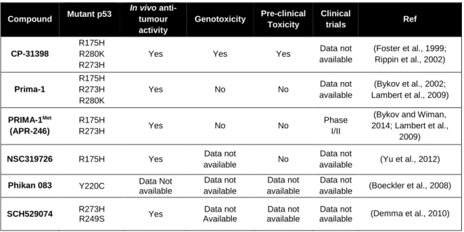
Table 1. Small molecule reactivators of mut p53 through restoration of wt p53 conformation ...29 Table 2. EC50 values obtained for the compounds tested in yeast ...44
Abbreviations
Bax Bcl-2 associated X protein
Bid BH3 interacting-domain death agonist CAK Cdk-activating kinase
Cdc25C Cell division cycle 25C Cdk Cyclin-dependent kinase CFU Colony-forming unit COX Cytochrome c oxidase cyt c Cytochrome c
DBD DNA-binding domain
DISC Death-inducing signaling complex DiOC6(3) 3,3'-Dihexyloxacarbocyanine Iodide
DMSO Dimethyl sulfoxide
FADD Fas-associated protein with death domain
FCCP Carbonyl cyanide p-(trifluoromethoxy)phenylhydrazone GADD45 Growth arrest and DNA damage protein 45
GOF Gain-of-function
MDM2 Murine double minute 2 MDMX Murine double minute x M-phase Mitotic phase
MOMP Mitochondrial outer membrane permeabilization MMP Mitochondrial membrane permeabilization
mut Mutant
OD Optical density
PUMA p53 up-regulated modulator of apoptosis p21 Cdk inhibitor
RE Responsive element
ROS Reactive oxygen species SRB Sulforhodamine B
S. cerevisiae Saccharomyces cerevisiae
TAD Transactivation domain Y2H Yeast-two hybrid
wt Wild-type
Chapter 1
.
1 General introduction
1.1
Cancer epidemiology and general concepts about cancer
Cancer is the leading cause of death in economically developed countries and the second in developing countries. Despite the improvement in the relative survival rates for many cancer types, cancer prevalence is increasing in economically developed countries as a result of population aging and growth, as well as an increased adoption of cancer-associated lifestyle choices including smoking, physical inactivity, and ‘‘westernized’’ diets. According to the World Health Organization, in 2012, there were 14.1 million new cancer cases, 8.2 million cancer deaths and 32.6 million people living with cancer (within 5 years of diagnosis). In the same year, in Europe, 3.7 million new cases of cancer were diagnosed and 1.9 million people died with the disease. In Portugal, there were 49.2 thousand new cancer cases, 24.1 thousand cancer deaths and 134.3 thousand people living with the disease (Bray et al., 2012; Ferlay et al., 2013; Soussi and Béroud, 2012). These statistics data may justify why cancer has been the focus of an intense worldwide scientific research. During development and cellular proliferation there is an intrinsic control system that regulates the balance between cell division and death in response to growth signals. The loss of this balance is the basis of carcinogenesis, a multistage process, involving oncogene activation and tumour suppressor gene inactivation that promotes cell proliferation and/or impairs apoptosis. This process also involves complex interactions between tumour and host tissues, ultimately leading to an aggressive metastatic phenotype [reviewed in (Wang and Sun, 2010)].
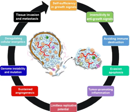
A normal cell needs to acquire several characteristics, called hallmarks of cancer, in order to progress to a cancer cell (Figure 1). In 2000, six hallmark capabilities were originally proposed, namely self-sufficiency in growth signals, insensitivity to anti-growth signals, evasion of apoptosis, limitless replicative potential, sustained angiogenesis and tissue invasion and metastasis (Figure 1). However, recent data support that there are two additional hallmarks of cancer, named emerging hallmarks, involved in the pathogenesis of perhaps all cancers. One involves the capability to modify, or reprogram, cellular metabolism in order to most effectively support neoplasic proliferation, and the second allows cancer cells to evade immunological destruction. Additionally, two consequential characteristics of neoplasia facilitate acquisition of both core and emerging hallmarks. Genomic instability and thus mutability endow cancer cells with genetic alterations that drive tumour progression. Inflammation by innate immune cells designed to fight infections and heal wounds can instead result in their inadvertent support of multiple hallmark capabilities, thereby manifesting the now widely appreciated tumour-promoting consequences of
inflammatory responses (Figure 1). Each of these new abilities acquired during cancer development represents the successful breaching of an anticancer defence mechanism hardwired into cells and tissues [reviewed in (Hanahan and Weinberg, 2000; Hanahan and Weinberg, 2011)].
1.2
The p53 tumour suppressor protein: A major therapeutic target in cancer
Despite the huge diversity of tumour suppressor genes implicated in carcinogenesis, the TP53 gene, which encodes the transcription factor p53, has been shown to play a pivotal role protecting cells against cancer development [reviewed in (Suzuki and Matsubara, 2011)].
Figure 1. The hallmarks of cancer. This illustration encompasses the ten hallmark capabilities currently
proposed. Some of these critical features are: self-sufficiency in growth signals, insensitivity to anti-growth signals, evasion of apoptosis, limitless replicative potential, genome instability and mutation, sustained angiogenesis, tissue invasion and metastasis [adapted from (Hanahan and Weinberg, 2011)]
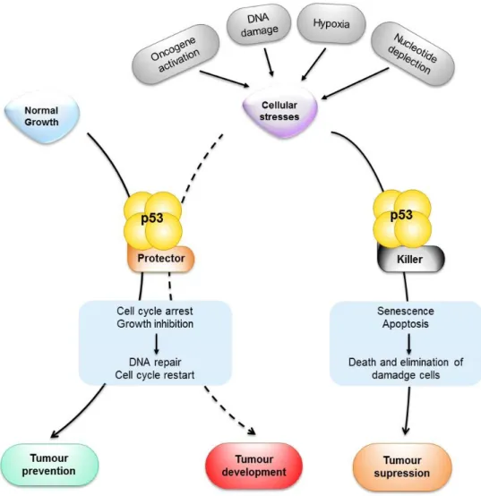
Figure 2. Schematic representation of p53 functions in tumour prevention, suppression and development. When activated by stress signals, p53 can halt the cell cycle and initiate the DNA repair.
Under a successful repair, the cell cycle is restarted, and the cellular and genomic stability are re-established. When the damage is too extensive and the repair is not an option, cells induce the killer function of p53 to activate apoptosis or senescence. This activation results in the permanent elimination of damage cells. Both apoptosis and senescence have been linked to tumour suppression, whereas the cell cycle arrest has been linked to tumour prevention. Notably, the protective functions of p53 may contribute to tumour development if not properly regulated (dashed arrow).Figure 3. The hallmarks of
cancer. This illustration encompasses the ten hallmark capabilities currently proposed. Some of these
critical features are: self-sufficiency in growth signals, insensitivity to anti-growth signals, evasion of apoptosis, limitless replicative potential, genome instability and mutation, sustained angiogenesis, tissue invasion and metastasis [adapted from (Hanahan and Weinberg, 2011)]
The TP53 gene was the first tumour suppressor gene identified. First described in 1979, it was initially reported as a protein interacting with the oncogenic T antigen from SV40 virus (Lane and Crawford, 1979; Linzer and Levine, 1979). Based on these observations, p53 began to be classified as an oncogene, with many studies describing proliferative and transforming activities of the protein. However, this classical point of view has been challenged by the findings that the initially discovered p53 was a mutant (mut) and not the wild-type (wt) form, as was originally considered. Only ten years later, subsequent studies revealed that the wt p53 is capable to suppress the malignant growth of transformed cells as well as tumours, supporting the fact that p53 is a potent tumour suppressor instead of an oncogene [reviewed in (Bourdon, 2007; Freed-Pastor and Prives, 2012)].
The human TP53 gene, located at position 17p13.1 of the small arm of chromosome 17, has been called the “guardian of the genome”, due to its pivotal role in maintaining genomic stability and tumour prevention [reviewed in (Bourdon, 2007; Freed-Pastor and Prives, 2012)]. Indeed, the p53 tumour suppressor acts as a major regulator in a complex signalling pathway that involves to sense a broad range of cellular stresses such as DNA damage, hypoxia, nutritional deprivation, nucleotide depletion or oncogene activation (Figure 2) [reviewed in (Amaral et al., 2010; Liu et al., 2014; Suzuki and Matsubara, 2011)].
In the absence of cellular stresses, the p53 protein is kept at low steady-levels, through the interaction with murine double minute (MDM) proteins, and exerts little effect on cell fate. However, upon exposure to these countless cellular stress signals, and depending on the tissue-type and the extension of the damage, p53 undergoes post-translational modifications that leads to its stabilization and activation, namely phosphorylation, acetylation and methylation, among others. Once activated, p53 is able to inhibit cell cycle progression, promote senescence, or induce apoptotic cell death, in order to prevent the replication of damaged DNA or the proliferation of genetically altered cells that could lead to tumour formation and development (Figure 2) [reviewed in (Bourdon, 2007; Brosh and Rotter, 2009; Freed-Pastor and Prives, 2012)]. Actually, when the DNA damage is too profound to be successfully repaired by cell cycle arrest, p53 triggers apoptosis. However, when p53 is mutated or absent, damaged cells become resistant to apoptosis and survive (Figure 2) [reviewed in (Amaral et al., 2010)].
The TP53 gene is inactivated by mutation or deletion in nearly 50% of human cancers. In the remaining cases, p53 retains its wt status but its function is compromised by distinct mechanisms, as the overexpression of its two major negative regulators, the homologs MDM2 and MDMX [reviewed in (Shadfan et al., 2012)].
Furthermore, the TP53 gene belongs to a highly conserved gene family containing at least two related genes, TP63 and TP73 (encoding p63 and p73, respectively) [reviewed in (Bourdon, 2007; Freed-Pastor and Prives, 2012; Millau et al., 2009)]. Actually, the exogenous expression of p63 or p73 causes growth arrest, apoptosis, and differentiation, which are often due to the transcriptional activation of p53 target genes (Strano et al., 2002). To date, both p63 and p73 have been found rarely mutated in human tumours (Gaiddon et al., 2001; Strano et al., 2002). However, several forms of mut p53 were reported to interact with p63 and p73 through their DNA-binding domain, inhibiting their transcriptional activities (Gaiddon et al., 2001; Strano et al., 2002).
Figure 2. Schematic representation of p53 functions in tumour prevention, suppression and development. When activated by stress signals, p53 can halt the cell cycle and initiate the DNA repair.
Under a successful repair, the cell cycle is restarted, and the cellular and genomic stability are re-established. When the damage is too extensive and the repair is not an option, cells induce the killer function of p53 to activate apoptosis or senescence. This activation results in the permanent elimination of damage cells. Both apoptosis and senescence have been linked to tumour suppression, whereas the cell cycle arrest has been linked to tumour prevention. Notably, the protective functions of p53 may contribute to tumour development if not properly regulated (dashed arrow).
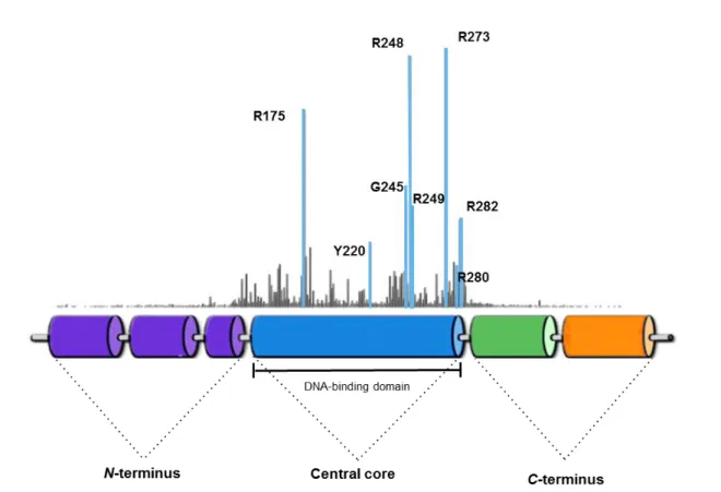
Figure 4. Schematic representation of the domain structure of p53. The human p53 protein
comprises several well-defined domains, including an N-terminal transactivation domain (TAD), followed by a central core DNA-binding domain, an oligomerization domain and a basic domain at the extreme C-terminus. The p53 TAD is able to mediate p53 transcriptional activity and to regulate its stability through interaction with components of the transcription initiation complex, with co-activators, such as p300 and CBP, and with several regulatory proteins, such as MDM2 and MDMX.Figure 5. Schematic
representation of p53 functions in tumour prevention, suppression and development. When
activated by stress signals, p53 can halt the cell cycle and initiate the DNA repair. Under a successful repair, the cell cycle is restarted, and the cellular and genomic stability are re-established. When the damage is too extensive and the repair is not an option, cells induce the killer function of p53 to activate apoptosis or senescence. This activation results in the permanent elimination of damage cells. Both apoptosis and senescence have been linked to tumour suppression, whereas the cell cycle arrest has been linked to tumour prevention. Notably, the protective functions of p53 may contribute to tumour development if not properly regulated (dashed arrow).
Therefore, the dynamic and multiple functions of p53, highlight the importance in understanding its activity, since the loss of p53 function can lead to more aggressive cancer forms or to chemotherapy resistance making of p53 one of the most appealing therapeutic targets in cancer therapy [reviewed in (Amaral et al., 2010)].
1.3
Structural organization of p53 protein
The 393-amino acid p53 protein has a complex domain structure, which can be schematically divided into three main domains (Figure 3): the N-terminal transactivation domain (TAD), the core DNA-binding domain (DBD), the most conserved region of the p53 protein, and the C-terminal domain [reviewed in (Millau et al., 2009)]. Each of these domains plays an important role in p53 function.
The N-terminal domain consists of two continuous transactivation subdomains (TADI and TADII) followed by an adjacent proline-rich domain (Figure 3). In fact, it has been shown that the transactivation function of wt p53 is dependent on four critical hydrophobic amino Figure 3. Schematic representation of the domain structure of p53. The human p53 protein
comprises several well-defined domains, including an N-terminal transactivation domain (TAD), followed by a central core DNA-binding domain, an oligomerization domain and a basic domain at the extreme C-terminus. The p53 TAD is able to mediate p53 transcriptional activity and to regulate its stability through interaction with components of the transcription initiation complex, with co-activators, such as p300 and CBP, and with several regulatory proteins, such as MDM2 and MDMX.
Figure 6. Regulation of cell cycle by p53, which transcriptional activation may lead to growth arrest at both G1 and G2 cell cycle phases. A) Under normal conditions the cyclin/Cdk complexes are
functional and Rb family members are found hyperphosphorylated (yellow) leading to the release and activation of the E2F transcriptional factor and, consequently, to G1 progression. Under stress conditions (red arrows) p53 mediates the transcriptional induction of p21, which plays a pivotal role in mediating the arrest at G1-phase. p21 binds to, and inhibits, cyclin/Cdk complexes resulting in the accumulation of hypophosphorylated Rb (green) that interact with and inhibit E2F transcription factors activity. These results in an arrest at the G1-phase. B) p53 has also been implicated in the control of the G2/M checkpoint. Transcriptional down-regulation of cyclin B1 by p53 may be involved in p53-mediated G2 arrest, once that cyclin B1/Cdc2 complex is the major regulatory factor required for entry into mitosis. p53 is able to control G2 arrest by inhibition of Cdc25C and CAK, both required for activation of cyclin B1/Cdc2 complex. CAK
acids in its N-terminus: Leu22, Trp23, Trp53, and Phe54. When these four residues are mutated, the transactivation capability of p53 is completely abolished. The p53 TAD is involved in the p53 transcriptional activity and in the regulation of its stability. Both of these capabilities of the TAD are mediated through interaction with components of the transcription initiation complex, with acetyltransferases p300 and CBP (which act as co-activators of p53), and with several regulatory proteins, such as MDM2 and MDMX. Actually, in unstressed cells, p53 would be kept at low levels essentially due to the action of MDMs, which inhibit its transcriptional activity (Figure 3). Upon stress, the N-terminus of p53 may suffer modifications that lead to the disruption of its contacts with MDMs. Therefore, p53 can interact with transcriptional co-activators, that leads to modifications, such as acetylation in the C-terminus, and allows transcriptional activation and induction of different cellular responses (Figure 3) [reviewed in (Freed-Pastor and Prives, 2012; Joerger and Fersht, 2007)]. Although its precise function has not yet been clearly defined, the proline-rich domain seems to be involved in growth suppression and apoptosis and its deletion results in a complete loss of this function (Sakamuro et al., 1997; Walker and Levine, 1996). Besides that, it is involved in p53 stabilization regulated by MDM2, since p53 becomes more susceptible to degradation if this region is deleted. It has also been reported that this domain comprises a negative auto-regulatory domain reducing the DNA-binding ability (Muller-Tiemann et al., 1998).
The DBD is responsible for the binding to sequence-specific DNA elements, located close to promoters of the p53 target genes. Indeed, this ability of p53 to bind to sequence-specific DNA elements is tightly linked to its pro-apoptotic activity [reviewed in (Ozaki et al., 2013)]. The C-terminal domain contains an oligomerization domain, involved in the ability of p53 to form active tetramers, followed by a basic domain, that interacts directly with single strands DNA (Figure 3) [reviewed in (Millau et al., 2009)].The basic domain of p53, acts as a flexible linker region that connects the DBD and the C-terminus of p53 (Cho et al., 1994). Besides that, the basic region of the C-terminus, which contains acetylation sites that binds DNA non-specifically, acts as a negative regulatory domain, and has been implicated in the induction of cell death [reviewed in (Joerger and Fersht, 2007)].
Interestingly, p63 and p73 share a significant homology with each other and with p53, having the same modular organization, which comprises an N-terminal TAD, a DBD and a C-terminal oligomerization domain and, consequently, similar functions (Strano et al., 2002).
1.3.1 The p53 role in cell cycle regulation
One of the most important cellular processes regulated by p53 is the cell cycle progression. This process consists on a series of steps at which the cell checks for the accuracy of the process and instructs itself to proceed to the next step. Cell cycle consists in a G1-phase, in which cells increase in size, in a S-phase, where the DNA is duplicated, in a G2-phase, in which cells continue to grow, and in a mitotic phase (M-phase), where chromosome segregation and cells division occurs. The G1 and G2 phases also provide the cell more time to grow and to ensure that the conditions are suitable for entry into the S-phase or mitosis [reviewed in (Nurse, 2000)].
The cell cycle is tightly regulated by stimulators and inhibitors containing intrinsic checkpoint controls, preventing in this way the transmission of damaged genetic material to the daughter cells and the consequent replication of abnormal material. Thus, there are three cell cycle checkpoints that regulate cells progression through each stage: the G1/S, the intra-S and the G2/M checkpoints. In case of DNA damage, the G1 checkpoint will lead to cell cycle arrest, ensuring that the DNA is not replicated during the S-phase. In response to damaged and/or un-replicated DNA the G2 checkpoint leads to cell cycle arrest safeguarding the proper completion of S-phase. The M-checkpoint leads to the arrest of chromosomal segregation in response to misalignment on the Mitotic spindle [reviewed in (Morgan, 1995)].
The cell cycle checkpoints are regulated by cyclin-dependent kinase (Cdk) complexes formed through association of a Cdk and a regulatory protein, a cyclin, that controls and activates the Cdk and whose levels change in a cyclical manner. So, since Cdk levels are usually constant throughout the cell cycle, its regulation and progression depends on variations of the levels of each cyclin (Sheaff et al., 1997). These cyclin/Cdk complexes regulate the phosphorylation of proteins involved in cell cycle progression, leading to DNA replication and mitosis. There are two types of cyclin/Cdks that regulate the transit of mammalian cells from quiescence into S-phase: the cyclin D, which activate Cdk4/6, and the cyclin E, which activates Cdk2 (Figure 4A) [reviewed in (Sherr and Roberts, 1999)]. Depending on the type of the cellular stress, the p53-mediated cell cycle arrest may be considered the primordial response to DNA damage. Actually, in response to a variety of stress conditions, such as chemotherapeutic treatments, p53 can trigger protective, pro-survival responses, such as a temporary cell cycle arrest at the G1-phase, regulation of the G2/M transition, DNA repair and antioxidant protein production to maintain the genome integrity and the viability of cells that sustain limited reparable damage. All this is possible due to the ability of p53 to up-regulate the expression of several cell cycle proteins, such as GADD45 (Growth arrest and DNA damage protein 45), Cdc25C (Cell division cycle 25C)
and p21 (cdk inhibitor also known as CIP1/WAF1) (Figure 4A and B) [(Amundson et al., 1998; El-Deiry, 1998); reviewed in (Nag et al., 2013)].
Figure 4. Regulation of cell cycle by p53, which transcriptional activation may lead to growth arrest at both G1 and G2 cell cycle phases. A) Under normal conditions the cyclin/Cdk complexes are
functional and Rb family members are found hyperphosphorylated (yellow) leading to the release and activation of the E2F transcriptional factor and, consequently, to G1 progression. Under stress conditions (red arrows) p53 mediates the transcriptional induction of p21, which plays a pivotal role in mediating the arrest at G1-phase. p21 binds to, and inhibits, cyclin/Cdk complexes resulting in the accumulation of hypophosphorylated Rb (green) that interact with and inhibit E2F transcription factors activity. These results in an arrest at the G1-phase. B) p53 has also been implicated in the control of the G2/M checkpoint. Transcriptional down-regulation of cyclin B1 by p53 may be involved in p53-mediated G2 arrest, once that cyclin B1/Cdc2 complex is the major regulatory factor required for entry into mitosis. p53 is able to control G2 arrest by inhibition of Cdc25C and CAK, both required for activation of cyclin B1/Cdc2 complex. CAK can be inhibited by p21 and Cdc25C by phosphorylation after activation of the Chk1 and Chk2 kinases, establishing a bind site to 14-3-3σ (green) proteins and leading to inactivation of Cdc2 activity. Besides that, the progression into the M-phase requires Cdc2 which can be inhibited by overexpression of p21, GADD45 or 14.3.3σ, being the expression of these inhibitory proteins regulated by p53 in order to induce growth arrest. p53 has also other targets that do not affect cyclin B1/Cdc2 complex but contribute to G2 arrest, namely Reprimo, B99 andpoly(rC) binding protein 4 (PCBP4); P – phosphorylation; All the red arrows and crosses correspond to the p53 effect on cell cycle progression.
Figure 8. Regulation of cell cycle by p53, which transcriptional activation may lead to growth arrest at both G1 and G2 cell cycle phases. A) Under normal conditions the cyclin/Cdk complexes are
functional and Rb family members are found hyperphosphorylated (yellow) leading to the release and activation of the E2F transcriptional factor and, consequently, to G1 progression. Under stress conditions (red arrows) p53 mediates the transcriptional induction of p21, which plays a pivotal role in mediating the arrest at G1-phase. p21 binds to, and inhibits, cyclin/Cdk complexes resulting in the accumulation of hypophosphorylated Rb (green) that interact with and inhibit E2F transcription factors activity. These results in an arrest at the G1-phase. B) p53 has also been implicated in the control of the G2/M
During the G1-phase, the expression of cyclin D is stimulated, forming cyclin D/Cdk4/6 complexes that phosphorylate the retinoblastoma (Rb) protein. Under normal conditions, the exit from G1 to S-phase requires the phosphorylation of Rb protein by cyclin/Cdk complexes. This results in the activation of the S-phase-promoting E2F transcription factor (required for entry into the S-phase), which binds to hypophosphorylated Rb protein (Figure 4A) (Amundson et al., 1998; Chellappan et al., 1991). One of the target genes of E2F is cyclin E, which forms a complex with Cdk2 that phosphorylates and consequently inhibits the Rb protein, leading to DNA replication and progression though cell cycle (Koff et al., 1992). Under stress conditions, p53 induces the transcriptional activation of p21 (major effector of G1 arrest), which in turn inactivates the cyclin/Cdk complexes. This enables the activation of Rb proteins and leads to the inhibition of E2F transcription factors (Figure 4A) (El-Deiry, 1998).
The p53 protein is able to regulate the G2/M transition, blocking cell entry into mitosis by inhibition of Cdc2 (Cdk1), which needs to bind to cyclin B1 in order to be functional. Thus, the repression of cyclin B1/Cdc2 complex by p53 leads to an arrest in G2/M transition (Figure 4B) [reviewed in (Suzuki and Matsubara, 2011)]. Additionally, the biochemical pathways involved in the DNA damage-induced G2 arrest involve signalling cascades that converge to inhibit the activation of Cdc2. Therefore, p53 impacts the G2/M checkpoint by transcriptional modulation of the expression of multiple, physically and functionally intertwined targets, including Cdc25C, 14-3-3σ, p21, and GADD45 (Figure 4B) (El-Deiry, 1998; Giono and Manfredi, 2006; Zhan et al., 1998). Mechanistically, p53 leads to G2/M arrest by repressing Cdc25C, a mitosis promoting phosphatase, which dephosphorylates and activates the cyclin B1/Cdc2 complex. In addition, the Cdc25C activity can be inhibited via phosphorylation by checkpoint kinases, Chk1 and Chk2, which, in turn, generate a consensus binding site for 14-3-3σ proteins. Binding of 14-3-3σ protein to Cdc25C results in the nuclear export of Cdc25C, sequestration of the phosphatase in the cytoplasm and, consequent inhibition of Cdc25C (Figure 4B) [reviewed in (Taylor and Stark, 2001)]. Besides the activity of Cdc25C, the Cdk-activating kinase (CAK) phosphorylates specific amino acid residues required for the activation of the cyclin B1/Cdc2 complex [reviewed in (Kishimoto and Okumura, 1997; Sherr and Roberts, 1999)]. Interestingly, some studies suggest that p21, involved in G1 arrest, is also implicated in G2 arrest by disruption of cyclin B1/Cdc2 complex through CAK (Figure 4B) (Smits et al., 2000; Zhan et al., 1999).
The p53 protein has been reported to transcriptionally activate GADD45, which dissociates cyclin B1/Cdc2 complex by binding to Cdc2 [reviewed in (Taylor and Stark, 2001). This prevents the formation of the protein complex necessary for entry into the M-phase, resulting in G2 arrest (Zhan et al., 1999). p53 can also induce the expression of other
proteins involved in G2 arrest, namely Reprimo (a glycosylated, cytoplasmic protein that plays a role in cyclin B1/Cdk1 localization), B99 (a protein that may cause G2 arrest independently of Cdc2) [reviewed in (Taylor and Stark, 2001)] and poly(rC) binding protein 4, a RNA-binding protein (Zhu and Chen, 2000) (Figure 4B).
1.3.2 Apoptosis regulation by p53 protein
Apoptosis is an evolutionary conserved and genetically controlled process of cell death that is essential, not only for development and maintenance of tissue homeostasis, but also for the elimination of unwanted cells during normal development and as a component of multistep carcinogenesis and therapy treatment resistance [reviewed in (Amaral et al., 2010)]. This cellular mechanism is induced by a tightly regulated suicide programme characterized by several hallmarks as cell shrinkage, condensation and fragmentation of nuclear chromatin, DNA fragmentation, mitochondrial swelling and membrane blebbing as well as by loss of mitochondrial membrane integrity. This mechanism of cell death is considered very “clean” occurring without stimulation of an inflammatory response [reviewed in (MacFarlane, 2003; Sharp et al., 2010)].
Alteration of many proteins involved in the apoptotic signalling pathways have been described and it is clear that alterations of upstream regulators of these pathways are the most common alterations found in cancer cells. As an example p53, that can induce cell death in response to a number of different stress stimuli, regulates the expression of a wide variety of genes involved in apoptosis [reviewed in (Vaseva and Moll, 2009)]. The apoptotic function of p53 has been first reported in mouse thymocytes, in response to irradiation (Clarke et al., 1993; Lowe et al., 1993). Since then, the p53-dependent apoptosis has been reported in a wide range of cells in response to many different stress signals [reviewed in (Amaral et al., 2010)].
p53 stimulates a wide network of signals that can act through two major apoptotic pathways (Figure 5): the extrinsic (or death receptor) and the intrinsic (or mitochondrial) [reviewed in (Amaral et al., 2010; MacFarlane, 2003)]. The apoptotic extrinsic pathway is triggered by extracellular signals that result in the binding of ligands to specific trans-membrane death receptors located at the plasma membrane, such as KILLER/DR5 and Fas (also called APO-1). Fas is a member of the tumour necrosis factor receptor (TNFR) superfamily and, in turn, KILLER/DR5 is a member of the TNF-related apoptosis-inducing ligand (TRAIL) family of death receptors [reviewed in (Ashkenazi and Dixit, 1998; Haupt et al., 2003; Sharp et al., 2010)]. The activation of these receptors, after binding to their ligands, leads to their trimerization and consequent clustering of the intracellular death domain, which
subsequently recruits the protein adaptor FADD (Fas-associated protein with death domain) through homotypic death domain interactions. The death effector domain of FADD then recruits procaspase-8 forming the death-inducing signalling complex (DISC) (Yu et al., 2001). The formation of the DISC complex results in caspase-8 activation and consequently in executioner caspases activation (Figure 5A), resulting in DNA fragmentation as an hallmark of apoptosis [reviewed in (Schuler and Green, 2001)].The p53 overexpression enhances cell surface levels of Fas by promoting its trafficking from Golgi complex. Thus, p53 status may influence the chemosensitivity via Fas signalling [reviewed in (Suzuki and Matsubara, 2011)] and activate KILLER/DR5, which is induced in response to DNA damage and, consequently, promote cell death through caspase-8 activation (Liedtke et al., 2003) (Figure 5A).
On the other hand, the intrinsic apoptotic pathway can be activated in response to a variety of cellular insults or other damaged signals such as radiation. This apoptotic pathway is regulated by members of the Bcl-2 family and involves the disruption of mitochondrial membrane integrity and subsequent release of apoptogenic proteins from the mitochondria into the cytosol (Voortman et al., 2007). The disruption of mitochondrial membrane integrity results in biochemical and structural changes of mitochondria including mitochondrial swelling, changes at the mitochondrial outer membrane permeabilization (MOMP), loss of mitochondrial membrane potential (∆ψm), and cytochrome c (cyt c) release (Figure 5B).
Actually, it has been proposed that mitochondrial depolarization defines an early and irreversible stage of apoptosis preceding other manifestations in this process, such as DNA fragmentation and reactive oxygen species (ROS) production. Additionally, the increase of ∆ψm is often observed in human cancer cells (Li et al., 1998; Vrablic et al., 2001; Zamzami
et al., 1995).
Therefore, MOMP is essential for the regulation of the apoptotic process and the proteins of the Bcl-2 family are the main regulators of this deadly switch (Renault et al., 2013). The members of the Bcl-2 family include anti-apoptotic, such as Bcl-2 and Bcl-xL, and pro-apoptotic proteins, such as Bax, Bak, Bid, NOXA and PUMA (p53 Up-regulated Modulator of Apoptosis), that can be classified on basis of structural similarity to the BH (Bcl-2 Homology) domains (BH1, BH2, BH3 and BH4) and a transmembrane domain, being the BH3 domain crucial for the pro-apoptotic function [reviewed in (Amaral et al., 2010; Igney and Krammer, 2002; Manfredi, 2003)]. Bax and Bak are the direct pro-apoptotic effectors of MOMP as they can translocate and/or insert into the outer mitochondrial membrane, oligomerize and form pores, leading consequently to cyt c release. Despite Bax and Bak are functionally redundant proteins, Bak has a constitutive mitochondrial localization in healthy cells, whereas Bax is mostly cytosolic, awaiting for activation signals that will trigger
its translocation to mitochondria and further activation (Figure 5B) (Renault et al., 2013). Bax activation is favoured by the pro-apoptotic BH3-only proteins, who can act both as direct activators of Bax (tBid, Bim, PUMA), by interacting directly with him, or as derepressors (Bad and Noxa), by interacting with anti-apoptotic members of the Bcl-2 family. The anti-apoptotic family members bind the BH3 domains of pro-apoptotic family proteins, preventing Bak/Bax homo-oligomerization and BH3-only proteins activation. The overexpression of anti-apoptotic members has been reported in most human cancers, favouring survival of neoplastic cells and consequently the resistance to chemotherapeutic agents [(Gallenne et al., 2009; Renault et al., 2013); reviewed in (Vaseva and Moll, 2009)]. Curiously, the binding region for Bcl-xL and Bcl-2 on p53 is located in the DBD, the same region that harbours the vast majority of “hotspot” mutations found in human cancers. Indeed, it was shown that such mut p53 proteins are defective in their ability to interact with Bcl-xL and Bcl-2 family of proteins (Mihara et al., 2003).
After several studies, it is clearly established that in response to a stress signal, cytoplasmic p53 rapidly translocates to mitochondria, where it can promote apoptosis in a transcriptional-independent manner, preceding the loss of ∆ψm (Mihara et al., 2003). At the
mitochondria, p53 is able to interact, either to inhibit or activate, with several anti- and pro-apoptotic genes of the Bcl-2 family, such as Bcl-2, BAX, NOXA, PUMA and BID (BH3-only proteins). Indeed, by directly interact with the anti-apoptotic proteins Bcl-xL and Bcl-2, p53 is able to inhibit these proteins inducing cyt c release [reviewed in (Amaral et al., 2010; Igney and Krammer, 2002; Manfredi, 2003; Vaseva and Moll, 2009)]. These interplay between pro- and anti-apoptotic members of the Bcl-2 family, after p53 activation, has been found to control the MOMP, and consequently the cyt c release from the mitochondria intermembrane space into the cytosol, unleashing the activation of the apoptotic machinery of caspases, chromatin degradation and apoptosis (Figure 5B) (Xiao et al., 2005). Once in the cytosol, cyt c participates in the formation of the apoptosome complex together with its adaptor molecule, Apaf-1 (Apoptotic Protease-Activating Factor-1), which activates procaspase-9 and promotes activation and cleavage of executioner caspases (procaspase-3, -6 and -7) (Figure 5B). This event leads to the cleavage of other specific death substrates, cellular and nuclear morphological changes, and ultimately, to cell death (Li et al., 1998). Thus, it is logical to assume that cyt c release is the rate-limiting step in initiating the caspase activation cascade and the main consequence of p53-mediated signals leading to apoptosis (Shinoura et al., 2001). Additionally, PUMA may play a pivotal role in determining cell fate (programmed cell death versus cell cycle arrest) in response to p53 activation, being an essential mediator for p53-dependent apoptosis in vivo (He et al., 2013).
Interestingly, there are evidences that the two apoptotic pathways are not exclusive. Instead, they can be linked and molecules in one pathway can influence the other. In fact, both pathways activate caspases, proteolytic enzymes functionally divided into two groups (initiator and executioner) that carry out apoptotic cell death. Once initiator caspases (such Figure 5. Schematic representation of the regulation of intrinsic and extrinsic apoptotic pathways by p53. The p53 protein is able to induce transcription of proteins involved in both apoptotic pathways. A) In the extrinsic pathway, the binding of ligands to specific trans-membrane death receptors (Fas and
KILLER), leads to the recruitment of the adaptor molecule, FADD. Then, the procaspase-8 binds to FADD leading to DISC formation and resulting in caspase-8 activation. Activated caspase-8 directly activates executioner caspases (caspase-3, -6, and -7) or cleaves the pro-apoptotic protein Bid. B) In the intrinsic/mitochondrial pathway the stress-induced mitochondrial translocation of p53 results in interactions between several anti- and pro-apoptotic members of the Bcl-2 family in order to induce MOMP and cyt c release. Indeed, p53 interacts with Bcl-xL/Bcl-2 blocking their inhibitory effect on pro-apoptotic Bax. Besides that it is able to activate genes such as PUMA and NOXA, which binds to and inhibit the activity of the anti-apoptotic proteins Bcl-2/Bcl-XL, unleashing the translocation of cytosolic Bax to the mitochondria. This leads to mitochondrial permeabilization and activation. The release of cyt c from the mitochondria results in the formation of the apoptosome and caspase-9 activation. Then, both caspase-8 and -9 activate executioner caspases, namely caspases-3, -6 and -7, resulting in cell death. Additionally, the MOMP can lead to a loss of the ∆ψm and to increase ROS production [adapted from
(Haupt et al., 2003)].
Figure 10. Schematic representation of the primary structure of MDM2 and MDMX proteins. Both
MDM2 and MDMX proteins comprises a p53-binding domain, an acidic and a zinc-finger domain in their central portion followed by a C-terminal Ring-finger domain. Additionally, the MDM2 protein possess a nuclear localization signal (NLS) and a nuclear export signal (NES) sequence.Figure 11. Schematic
representation of the regulation of intrinsic and extrinsic apoptotic pathways by p53. The p53
protein is able to induce transcription of proteins involved in both apoptotic pathways. A) In the extrinsic pathway, the binding of ligands to specific trans-membrane death receptors (Fas and KILLER), leads to the recruitment of the adaptor molecule, FADD. Then, the procaspase-8 binds to FADD leading to DISC formation and resulting in caspase-8 activation. Activated caspase-8 directly activates executioner caspases (caspase-3, -6, and -7) or cleaves the pro-apoptotic protein Bid. B) In the intrinsic/mitochondrial pathway the stress-induced mitochondrial translocation of p53 results in interactions between several anti- and pro-apoptotic members of the Bcl-2 family in order to induce MOMP and cyt c release. Indeed,
Extrinsic Pathway (A)
as caspases-8, -9 and -10) are activated, they cleave and activate (directly or indirectly) executioner caspases (such as caspase-3, -6 and -7) that, in turn, cleave intracellular substrates, such as poly(ADP-ribose)polymerase (PARP) (Figure 5A and B), leading to biochemical and morphological changes that are characteristics of apoptosis. This makes the activation of initiator caspases a key step to cell death [reviewed in (Igney and Krammer, 2002; MacFarlane, 2003)]. One of the major links between the two pathways is provided by the pro-apoptotic Bcl-2 family member Bid. Activation of Bid can promote MOMP and involves cleavage of cytoplasmic Bid by caspase-8 to expose a new N-terminal glycine residue that is subsequently myristoylated. Myristoylated Bid translocates to the mitochondria, inserts into the membrane and activates Bax and Bak to initiate mitochondrial events leading to apoptosome formation. The BID gene is transcriptionally regulated by p53 in response to -irradiation. Therefore, p53 appears to promote the convergence of the intrinsic and extrinsic pathways through Bid regulation (Li et al., 1998; Zha et al., 2000). Finally, increased mitochondrial production of ROS, has been related to the loss of ∆ψm, in
cancer cells, but not in normal cells (Gogvadze et al., 2008). While normal levels of ROS can be pro-proliferative, excess of ROS production can damage DNA and proteins, contributing to the development of various diseases (Maddocks and Vousden, 2011). Interestingly, it was showed that p53 protects cells from oxidation by reducing the production of intracellular ROS (Bensaad et al., 2006). Although ROS are by-products of normal mitochondrial function, high levels of ROS have been associated with p53-induced apoptosis (Figure 5B). The p53 deficiency in cells and mouse tissues results in the elevation of intracellular ROS levels, which in turn leads to the increased DNA oxidation and mutation rates in cells (Budanov et al., 2004; Sablina et al., 2005).
1.4
Endogenous negative regulators of p53: MDM2 and MDMX
Under normal conditions, p53 remains at low levels due to its short half-life allowing normal cell proliferation and viability. However, in response to cellular stresses, p53 becomes stabilized and modulates the transcription of target genes. As referred above, in tumours in which p53 is not mutated, its function is inhibited by overexpression of its two major negative regulators MDM2 and MDMX. Actually, both of them regulate the p53 activity by inhibiting its transcriptional activity (Jones et al., 1995; Parant et al., 2001).
The MDM2 gene encodes a protein that was first described in the late 1980s as an amplified gene responsible for the spontaneous transformation of an immortalized murine cell line, BALB/c 3T3 (Cahilly-Snyder et al., 1987). Within 5 years, the oncogenic potential of MDM2
gene was demonstrated, with several studies showing that MDM2 interacts directly with p53 and inhibits its ability to function as a transcriptional activator [reviewed in (Manfredi, 2010)]. In the middle 90s, a screen for p53-binding proteins led to the identification of a protein sharing a structural homology with MDM2. This protein was first called MDMX and later given the official name of MDM4. The high level of structural similarity makes us thought that these two proteins derived from the duplication of a single ancestral gene [reviewed in (Toledo and Wahl, 2006; Toledo and Wahl, 2007)].
The MDM2 gene is overexpressed in a wide range of human cancers expressing wt p53 including breast carcinomas, glioblastomas and sarcomas, whereas overexpression of MDMX can be detected in melanomas, retinoblastomas and breast carcinomas, being both associated with resistance to treatment, poor prognosis, metastasis and advanced forms of the disease [reviewed in (Nag et al., 2013)].
The full-length transcripts of MDM2 and MDMX genes encode structural related proteins of 491 and 400 amino acids, respectively (Figure 6). Thus, both MDMs bind to a short α-helical stretch on the p53 TAD domain through their N-terminal hydrophobic region, which is the best conserved region between the two proteins [reviewed in (Toledo and Wahl, 2007; Wang and Jiang, 2012)]. By blocking the p53 TAD domain and occupying the binding site of co-activators, such as p300 [reviewed in (Hock and Vousden, 2014)], these proteins lead to the inhibition of the p53 transcriptional function (Kussie et al., 1996; Momand et al., 1992). Both MDM2 and MDMX possess a zinc-finger domain together with a central acidic domain, followed by a C-terminal RING-finger domain, with this last domain receiving special attention for its multiple functions (Fang et al., 2000). Additionally, only MDM2 possesses a nuclear localization signal (NLS) and a nuclear export signal (NES), responsible for shuttling MDM2 between the nucleus and the cytoplasm (Figure 6). This suggests that MDM2 binds to p53 in the nucleus and transports it to the cytoplasm for ubiquitin-mediated degradation by the proteasome [reviewed in (Freedman et al., 1999)] (Figure 7).
Several studies have reported the distinct but complementary roles of MDM2 and MDMX in p53 regulation. Indeed, although both proteins inhibit the p53 transcriptional activity, they have significant differences in their p53 regulation mechanisms. Previous in vivo studies confirmed the separated importance of this two proteins indicating that while MDM2 mainly regulates p53 stability, MDMX has a major role in regulating its activity [reviewed in (Toledo and Wahl, 2006; Toledo and Wahl, 2007; Vassilev, 2007)]. Interestingly, although the RING-finger domains of MDM2 and MDMX share a high level of homology, only MDM2 possess the ability to act as an E3 ubiquitin ligase, leading p53 and itself for degradation by ubiquitination (Marine and Jochemsen, 2005), whereas MDMX controls the levels of p53 by modulating the levels of MDM2 (Linke et al., 2008). Several studies, have shown that MDM2 and MDMX function in a non-overlapping manner, creating a negative feedback loop, which precisely controls the level and activity of p53 [reviewed in (Lenos and Jochemsen, 2011; Popowicz et al., 2011; Shadfan et al., 2012; Vassilev, 2007)]. Additionally, if in one way the endogenous negative regulators of p53 are able to inhibit and regulate its expression levels, p53 has shown to be capable to induce the transcription of both MDM2 [reviewed in Figure 6. Schematic representation of the primary structure of MDM2 and MDMX proteins. Both
MDM2 and MDMX proteins comprises a p53-binding domain, an acidic and a zinc-finger domain in their central portion followed by a C-terminal Ring-finger domain. Additionally, the MDM2 protein possess a nuclear localization signal (NLS) and a nuclear export signal (NES) sequence.
Figure 12. Regulation of p53 by its negative regulators MDM2 and MDMX. In normal cells, p53
increases the MDM2 and MDMX transcription over basal levels (red arrows). MDM2 and MDMX cooperate to inhibit the p53 function by modulating its transcriptional activity and by preventing its interaction with the general transcription machinery. As a homodimer, MDM2 is able to promote p53 nuclear export to the cytoplasm for ubiquitin-mediated degradation by the proteasome. Additionally, MDM2 can also form a heterodimeric complex with MDMX promoting the nuclear degradation of p53 (polyubiquitination) and MDMX ubiquitination by MDM2. These cellular mechanisms result in subtle control of p53 levels; (Ub: ubiquitin)Figure 13. Schematic representation of the primary structure of MDM2 and MDMX proteins. Both MDM2 and MDMX proteins comprises a p53-binding domain, an acidic and a zinc-finger domain in their central portion followed by a C-terminal Ring-finger domain. Additionally, the MDM2 protein possess a nuclear localization signal (NLS) and a nuclear export signal (NES) sequence.
(Manfredi, 2010)] and MDMX proteins (Li et al., 2010), thus establishing a negative feedback loop (Figure 7).
It has been described that, under normal conditions, MDM2 is able to inhibit the p53 activity through ubiquitination, either in the form of a homodimer or of a heterodimer, being the RING-finger domains integrity essential for these hetero or homodimerization (Lenos and Jochemsen, 2011; Linke et al., 2008). This complex results in the stabilization of MDM2 and, consequently, the ability of MDM2 to monoubiquitinate or polyubiquitinate p53 depends on its expression levels (Li et al., 2003). In fact, when the levels of MDM2 are low, monoubiquitination of p53 promotes the export of p53 from the nucleus to the cytoplasm Figure 7. Regulation of p53 by its negative regulators MDM2 and MDMX. In normal cells, p53
increases the MDM2 and MDMX transcription over basal levels (red arrows). MDM2 and MDMX cooperate to inhibit the p53 function by modulating its transcriptional activity and by preventing its interaction with the general transcription machinery. As a homodimer, MDM2 is able to promote p53 nuclear export to the cytoplasm for ubiquitin-mediated degradation by the proteasome. Additionally, MDM2 can also form a heterodimeric complex with MDMX promoting the nuclear degradation of p53 (polyubiquitination) and MDMX ubiquitination by MDM2. These cellular mechanisms result in subtle control of p53 levels; (Ub: ubiquitin)
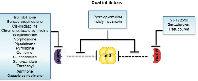
Figure 14. Schematic representation of small molecule inhibitors of MDM proteins. Isoindolinones,
cis-imidazolines, xanthones, oxazoloisoindolinones, among others, act as inhibitors of the p53-MDM2 interaction. SJ-172550, benzofuroxan and pseudorea act as inhibitors of p53-MDMX interaction. The pyrrolopyrimidine-based small molecules and the compound RO-5963 from the Indolyl hydantoin chemical family, as shown to inhibit both p53-MDM2 and p53-MDMX interactions.Figure 15. Regulation of p53 by
its negative regulators MDM2 and MDMX. In normal cells, p53 increases the MDM2 and MDMX
transcription over basal levels (red arrows). MDM2 and MDMX cooperate to inhibit the p53 function by modulating its transcriptional activity and by preventing its interaction with the general transcription machinery. As a homodimer, MDM2 is able to promote p53 nuclear export to the cytoplasm for ubiquitin-mediated degradation by the proteasome. Additionally, MDM2 can also form a heterodimeric complex with MDMX promoting the nuclear degradation of p53 (polyubiquitination) and MDMX ubiquitination by MDM2. These cellular mechanisms result in subtle control of p53 levels; (Ub: ubiquitin)
and consequent inhibition of p53 transcriptional activity (Figure 7). Once in the cytoplasm, p53 can be further ubiquitinated and degraded by p300. However, processes such as apoptosis has been described to be positively regulated by a number of different functions of cytoplasmic p53 [reviewed in (Hock and Vousden, 2014)]. Instead, high levels of MDM2 favour the p53 polyubiquitination, which results in the nuclear degradation of p53 (Figure 7). This degradation mechanism is responsible for the maintenance of low p53 levels in unstressed cells (Li et al., 2003).
Interestingly, the MDM2/X heterodimer complex induces polyubiquitination of p53, whereas MDM2 alone is primarily responsible for its monoubiquitination with further degradation of p53. Another consequence of MDMs heterodimerization is the MDMX ubiquitination (Figure 7) (Linke et al., 2008). The MDM2/MDMX E3 ligase as well as p53 can be regulated by deubiquitinases. Indeed, the deubiquitination and stabilization of both MDM2 and MDMX by the ubiquitin-specific protease 7(USP7) plays a pivotal role in the control of p53 stability [reviewed in (Hock and Vousden, 2014; Wang and Jiang, 2012)].
Many currently used cancer therapies, such as chemotherapy and radiation, have been primarily focused on p53 reactivation to trigger an apoptotic response. Unfortunately, high doses of these genotoxic treatments can also induce p53-independent pathways and thus may cause severe toxicities in normal tissues, which could eventually lead to secondary malignancies. In this context, efforts have been made in order to develop new selective and nongenotoxic inhibitors of the p53-MDMs interactions as an alternative to conventional cytotoxic chemotherapy [reviewed in (Shangary and Wang, 2009; Tisato et al., 2013)]. In fact, over the past years, the concept of selective chemotherapy has dominated the field of drug discovery and development. In this regard, the pharmacological restoration of the impaired function of p53, by disrupting its interaction with MDMs proteins has been shown to be an alternative therapeutic strategy against a broad spectrum of cancers with wt p53. Besides, the combination of various drugs that target multiple p53 pathways may be a useful strategy to achieve synergistic drug efficacy by reducing the genotoxic burden with the same or better anti-tumour effect [reviewed in (Popowicz et al., 2011; Zhao et al., 2013a)].
1.4.1 Inhibitors of MDM2 and MDMX in anticancer therapy
Over the past years, it has been difficult to develop small molecule inhibitors that disrupt large protein-protein interactions. Despite all the difficulties, and few progresses made in the first years, small molecules have been developed that compete for the p53 binding site of MDM2, inhibiting the binding and degradation of p53 by MDM2. The disruption of the p53-MDM2 interaction has shown to restore the wt p53 activity and to drive cancer cells
selectively into apoptosis [reviewed in (Shangary and Wang, 2009)]. Several studies of small molecule inhibitors of p53-MDM2 interaction, carried out in different cancer cell lines and animal models, support their usefulness as potential anticancer agents in tumours with overexpression of MDM2 [reviewed in (Vassilev, 2007)].
Several classes of chemical families have been reported as potent inhibitors of the p53-MDM2 interaction like the cis-imidazoline compounds, most commonly known by nutlins (Vassilev et al., 2004), the spiro-oxindoles (Zhao et al., 2013b), the isoindolinones (Hardcastle et al., 2011), the piperidinones (Rew et al., 2012), the xanthones (Leão et al., 2013a; Leao et al., 2013) and the oxazoloisoindolinones (Soares et al., 2014b), among others (Figure 8). A common way of action of several compounds, like nutlins and spiro-oxindoles, is mimicking the three pivotal amino acids that mediate the interaction of p53 with MDM2: Phe19, Trp23 and Leu26, blocking the p53-MDM2 interaction (Zhao et al., 2013a). However, only few inhibitors of the p53-MDM2 interaction have advanced into clinical trials [reviewed in (Hoe et al., 2014)] namely RG7112, an oral formulation of nutlin series (Ray-Coquard et al., 2012; Vassilev et al., 2004), and MI-773 (spiro-oxindole) (Hoe et al., 2014; Zhao et al., 2013b).
Encouraged by the success in the design of small molecule inhibitors of the p53-MDM2 interaction, efforts have been made towards the development of small molecule inhibitors of the p53-MDMX interaction, such as SJ-172550 (Reed et al., 2010). Interestingly, high levels of MDMX protein are known to confer resistance to MDM2 inhibitors, which have shown very low binding affinity to MDMX. In fact, the apoptotic response of some p53-MDM2 interaction inhibitors, such as nutlin-3a, is markedly attenuated in MDMX-overexpressing tumours, such as in retinoblastomas and melanomas [reviewed in (Hoe et al., 2014)]. Since the simultaneous inhibition of the p53-MDM2 and p53-MDMX interaction is required for a full reactivation of p53, efforts have been made for the development of dual inhibitors of MDM2 and MDMX. Actually, very few compounds with dual specificity against both MDM2 and MDMX, namely pyrrolopyrimidine-based small molecules (Lee et al., 2011) and RO-5963 from the Indolyl hydantoin chemical family (Graves et al., 2012), were reported. Being these two compounds the only dual inhibitors found to date, the search for new ones is highly required.