UNIVERSIDADE DE TRÁS-OS-MONTES E ALTO DOURO
NECROPSY PROCEDURES IN VETERINARY FORENSIC:
IDENTIFICATION OF CRITICAL POINTS
Dissertação de Mestrado Integrado em Medicina Veterinária
VANESSA CARINA MENDONÇA DELGADO
ORIENTADOR
Professora Doutora Isabel Cristina Ribeiro Pires
COORIENTADOR
Professora Doutora Justina Maria Prada Oliveira
iii
UNIVERSIDADE DE TRÁS-OS-MONTES E ALTO DOURO
NECROPSY PROCEDURES IN VETERINARY FORENSIC:
IDENTIFICATION OF CRITICAL POINTS
Dissertação de Mestrado Integrado em Medicina Veterinária
VANESSA CARINA MENDONÇA DELGADO
ORIENTADOR
Professora Doutora Isabel Cristina Ribeiro Pires
COORIENTADOR
Professora Doutora Justina Maria Prada Oliveira
COMPOSIÇÃO DO JÚRI
Professora Doutora Maria da Conceição Fontes Professora Doutora Anabela Gouveia Antunes Alves Professora Doutora Paula Avelar Rodrigues
Professora Doutora Isabel Cristina Ribeiro Pires
v
DECLARAÇÃO
NOME: VANESSA CARINA MENDONÇA DELGADO
C.C.:13605803 CORREIO ELECTRÓNICO: [email protected]
DESIGNAÇÃO DO MESTRADO: MESTRADO INTEGRADO EM MEDICINA
VETERINÁRIA
TÍTULO DA DISSERTAÇÃO DE MESTRADO EM MEDICINA VETERINÁRIA:
NECROPSY PROCEDURES IN VETERINARY FORENSIC: IDENTIFICATION OF CRITICAL POINTS
ORIENTADOR:
PROFESSORA DOUTORA ISABEL CRISTINA RIBEIRO PIRES
CO-ORIENTADOR:
PROFESSORA DOUTORA JUSTINA MARIA PRADA OLIVEIRA
ANO CONCLUSÃO: 2018
DECLARO QUE ESTA DISSERTAÇÃO DE MESTRADO É RESULTADO DA MINHA PESQUISA E TRABALHO PESSOAL E DAS ORIENTAÇÕES DOS MEUS SUPERVISORES. O SEU CONTEÚDO É ORIGINAL E TODAS AS FONTES CONSULTADAS ESTÃO DEVIDAMENTE MENCIONADAS NO TEXTO, E NA BIBLIOGRAFIA FINAL. DECLARO AINDA QUE ESTE TRABALHO NÃO FOI APRESENTADO EM NENHUMA OUTRA INSTITUIÇÃO PARA OBTENÇÃO DE QUALQUER GRAU ACADÉMICO.
VILA REAL, 25 DE OUTUBRO DE 2018
vii
ACKNOWLEDGMENTS
I start to thank the Universidade de Trás-os-Montes e Alto Douro, personified by its Rector, Professor António Fontainhas Fernandes, for providing the necessary conditions for this graduation to happen. I also would like to thank all the personnel of the Department of Veterinary Sciences of this University for the global support during all the graduation period.
First and foremost, I want to give large part of the credits to my advisor Doctor Isabel Cristina Ribeiro Pires, and co-advisor, Doctor Justina Maria Prada Oliveira, on the guidance of this work. I would like to reinforce my sincere appreciation to Doctor Isabel Cristina Ribeiro Pires for all the dedication and enthusiasm conveyed, for her invaluable advices, and for being always present and available in any way during my internship and in the writing of this dissertation.
A special thank you to Doctor Leonor dos Santos Diniz Orge and Doctor Madalena Monteiro, for welcoming me at the laboratory of pathology of the Instituto Nacional de Investigação Agrária e Veterinária (INIAV) at Oeiras, and for supervise me throughout my internship; another appreciation to Doctor Paula Mendonça and Doctor Paulo Carvalho for guide me and shared their knowledge; finally but not least, my thankfulness to Doctor Miguel Fevereiro, the director of the department of animal health production of INIAV, for receiving me in the premise, and also to all the personnel of the institute that I came across with for all the sympathy.
To the Centro di Referenza Nazionale per la Medicina Forense Veterinaria dell’Istituto Zooprofilattico Sperimentale Di Lazio e Toscana, I could not forget to show my gratitude to Doctor Alberigo Nardi, director of the Grosseto section, and to Doctor Alessia Mariacher and Doctor Rosario Fico, for the willingness to receive me on their department, and in special way to Doctor Antonella Bozzano and Doctor Valeria Mariano for all the patience and the time expended on helping me in the foreign country, even though it was not gone has planned, it was a short but good experience thanks to your kindness.
To my parents, who always been supportive of my academic choices.
To my closest friends, who were my anchor along this path, and became my big family.
ix
RESUMO
A medicina veterinária forense é definida como o uso do conhecimento dos ramos da medicina veterinária para resolver casos criminais e/ou de responsabilidade civil, pela aplicação da lei. Os métodos forenses são aplicáveis a situações fora dos tribunais, como reclamações de seguro, comissões de serviço público, para defender ou apresentar alegações de má conduta profissional ou outras medidas disciplinares, entre outros. Apesar da definição de medicina veterinária forense parecer limitada, a verdade é que a maioria dos casos forenses carecem do conhecimento de outras ciências para além da área da veterinária; uma equipa forense deve ser multidisciplinar e ter um contacto próximo com outras ciências, como a balística, entomologia, genética, e muitas outras. Esta é uma área em fase de crescimento rápido, o que provavelmente se deve ao aumento das leis relacionadas com o bem-estar animal, aumentando assim a tendência para que as pessoas exijam compensações em questões relacionadas com morte e agressões dos seus animais. A patologia forense é um ramo da medicina veterinária forense, em que os seus principais objetivos são documentar, analisar e explicar os achados patológicos de um caso, de forma acessível para aqueles que irão ler o relatório, que presumivelmente não serão da área da medicina, mas sim da lei (juristas, advogados, etc.). A necrópsia forense é, por sua vez, uma subdivisão da patologia forense e é um dos principais componentes em investigações de mortes.
Este trabalho compreende uma revisão bibliográfica dos procedimentos de necrópsia forense e a descrição de três casos de morte violenta não acidental (causa legal da morte), em anonimato, com as seguintes causas de morte: Provável afogamento de dois canídeos, com tentativa de suicídio do seu detentor; (2) Estrangulamento de um canídeo; (3) Disparo de projétil a dois bovinos. Através do seguimento dos diversos casos forenses durante o período de estágio, em conjunto com a revisão bibliográfica, foi elaborada a identificação de pontos críticos nos procedimentos de necrópsia em veterinária forense, que se centram na especialização académica do veterinário patologista; no seu conhecimento jurídico; no exame do local do crime; na manipulação das provas; nos próprios procedimentos de necrópsia; na redação do relatório; na biossegurança; e na cadeia de custódia.
Palavras-chave: Pontos críticos; necrópsia forense; patologia forense; medicina veterinária
xi
ABSTRACT
Forensic veterinary medicine is defined as the use of the knowledge of veterinary medicine branches to solve cases of civil and/or criminal liability, supported by the application of the law. Forensic methods are largely applicable to situations outside the courts, such as insurance claims, public service commissions, to defend or propound allegations of professional misconduct or other disciplinary measures, and others. Despite the definition of forensic veterinary medicine seems limited, the true is that most of the forensic veterinary cases require recourse to others sciences knowledge beyond the veterinary area; a forensic veterinary team should be multidisciplinary as well as it should sustain an intimate contact with other departments/sciences such as ballistics, entomology, genetics, and many others. It is a rapidly growing field probably due to the increasing of legislation relating to animal welfare, accruing the tendency for animal owners to seek compensation in matters regarding death and injuries. The forensic pathology is a branch of the forensic veterinary medicine, and its main objectives are to document, analyse, and elucidate the pathological findings of a case in a comprehensible way for those who will read the report, which probably are non-medical people, but people related to the law (jurists, lawyers, etc.). The forensic necropsy is, in its turn, a subdivision of forensic pathology and it is a main component in deaths investigations.
This work comprehends a literature review of the forensic necropsy procedures and the description of three cases of non-accidental violent death (legal cause of death), in anonymity, with the following causes of death: (1) Probable drowning of two canids, with attempted suicide of their caretaker; (2) Strangulation of a canid; (3) Forearm shooting on two cattle. Through following-up the forensic cases presented on the internship, along with the literature review, it was elaborated the identification of critical points regarding to necropsy procedures in veterinary forensic, which focus on the academic specialization of the veterinary pathologist; his/her legal knowledge; the crime scene examination; the evidence handling; the necropsy procedures itself; the report writing; the biosafety; and the chain of evidence.
Keywords: Critical points; forensic necropsy; forensic pathology; forensic veterinary
xiii
LIST OF CONTENTS
ACKNOWLEDGMENTS ... vii
RESUMO ... ix
ABSTRACT ... xi
LIST OF CONTENTS ... xiii
LIST OF FIGURES ... xvii
LIST OF TABLES ... xxiii
ABBREVIATIONS, SYMBOLS AND ACRONYMS ... xxv
CHAPTER I - LITERATURE REVIEW ... 1
Forensic Veterinary Medicine ... 1
1.1. Definition ... 1
1.2. State of the art ... 2
1.3. The role of the forensic veterinary pathologist ... 5
1.4. Other forensic sciences ... 8
1.5. The legal system ... 12
1.5.1. Governing laws ... 13
1.5.2. Legal description of death ... 14
1.5.2.1. Cause of death ... 14 1.5.2.2. Mechanism of death ... 15 1.5.2.3. Manner of death ... 15 1.6. Chain of Custody ... 17 1.6.1. Case folder ... 17 1.6.1. Evidence identification ... 18 1.6.2. Secure storage ... 20
Necropsy Procedures in Veterinary Forensic ... 21
2.1. Introduction to forensic necropsy ... 21
2.2. Reception and labels ... 22
2.3. Background information ... 23
2.4. Records and case notes ... 25
2.5. Forensic photography ... 26
2.6. Forensic radiography ... 33
xiv
2.7.1. External examination ... 35
2.7.2. Skinning ... 36
2.7.3. Internal examination ... 36
2.7.4. Opening of the skull ... 37
2.8. Estimation the time of death ... 38
2.9. Necropsy approach to animal abuse and neglect (AAN) ... 39
2.9.1. Blunt force trauma ... 40
2.9.1.1. Bruising/contusions... 41
2.9.1.2. Abrasions ... 42
2.9.1.3. Lacerations ... 44
2.9.1.4. Avulsion injuries ... 45
2.9.1.5. Fractures of the skeletal system ... 46
2.9.1.6. Specific types of cruelty associated with blunt force injuries... 46
2.9.1.6.1. Fall injuries ... 46
2.9.1.6.2. Swinging/ dragging injuries... 47
2.9.2. Sharp force injuries ... 47
2.9.2.1. Incised wounds... 48
2.9.2.2. Stab wounds ... 50
2.9.2.3. Incised-stab wounds ... 51
2.9.2.4. Chop wounds ... 51
2.9.2.5. Specific types of cruelty associated with sharp force injuries ... 52
2.9.2.5.1. Mutilations, predator attacks and dog attacks ... 52
2.9.3. Thermal injuries ... 54
2.9.4. Firearms injuries ... 55
2.9.5. Asphyxia and drowning ... 57
2.9.6. Poisoning ... 60
2.9.7. Neglect ... 61
2.9.8. Sexual abuse ... 62
2.10. Sample collection and ancillary tests ... 63
2.11. Forensic necropsy report ... 64
2.11.1. External exam ... 69
xv
2.11.3. Radiographic interpretation ... 69
2.11.4. Internal exam ... 69
2.11.5. Evidence of injury ... 70
2.11.6. Photographs and diagrams ... 70
2.11.7. Time of death ... 70
CHAPTER II: AIMS ... 73
CHAPTER III: PRESENTATION OF CASES ... 75
Case 1 ... 75
Case 2 ... 89
Case 3 ... 98
CHAPTER IV: CRITICAL POINTS OF NECROPSY PROCEDURES IN VETERINARY FORENSIC ... 111
Academic specialization ... 111
Legal knowledge ... 112
Crime scene examination ... 112
Evidence handling ... 113
4.1. Conservancy of the cadaver ... 113
4.2. Evidence reception ... 114
4.3. Evidence labelling and identification ... 114
Necropsy procedures ... 115
5.1. Systems and protocols ... 115
5.2. Records and case notes ... 116
5.3. Sample collection ... 116
5.4. Expertise of the forensic veterinary pathologist ... 117
Report writing ... 118 Biosafety ... 118 Chain of evidence ... 119 CHAPTER V: CONCLUSIONS ... 121 REFERENCES ... 123 ANNEX I ... xxv
xvii
LIST OF FIGURES
Figure 1: Tamper evident braided metal cable seal. Adapted from https://seals.com/ ... 18
Figure 2: Tamper evident braided plastic cable seal. Adapted from https://seals.com/ ... 18
Figure 3: Tamper evident container of plastic envelope. Adapted from https://www.henryschein.com/us-en/Global.aspx ... 19
Figure 4: Tamper evident container of paper envelope. Adapted from https://www.beaglelegal.com/tamper-evident-tyvek-envelopes_10x13 ... 19
Figure 5: Tamper evident tape. Adapted from https://www.aeharris.com/home/76-tamper-evident-tape.html ... 19
Figure 6: Example of a possible Portuguese laboratory labels that are unaffected by freezing, with the laboratory reference number printed permanently in black letters on a white background. Authored by the own. ... 23
Figure 7: Example of a basic pre-made dog outline drawings. Adapted from http://wiringdiagramblog.today/pet-wound-diagram.html ... 26
Figure 8: Photographic documentation of the cadaver– Lateral right ... 28
Figure 9: Photographic documentation of the cadaver – Lateral left ... 28
Figure 10: Photographic documentation of the cadaver– Front/facial ... 28
Figure 11: Photographic documentation of the cadaver – hind/rear ... 28
Figure 12: Photographic documentation of the cadaver – dorsal ... 28
Figure 13: Photographic documentation of the cadaver - ventral ... 28
Figure 14: Example of an overall orientation view, in a distant plan, putting the lesion in the context of its surrounding anatomic landmarks ... 29
Figure 15: Example of a closeup view of the finding before, granting it is the central piece in the view-finder and that the label is also included. ... 29
Figure 16: L-shaped forensic scale. Adapted from http://www.bvda.com/en/rulers-5-cm-2-inch ... 30
Figure 17: Example of the use of a blunt instrument – surgical stylet - as pointer, to identifying the track of a gunshot injury at the scalp of a dog. ... 30
xviii
Figure 18: Example of a reference scale ruler, in a L-shape, prepared for serve as well as identification card. Authored by the own, based on http://www.bvda.com/en/rulers-5-cm-2-inch ... 31 Figure 19: Several sizes of reference scale rulers. All of them can be used in one case, depending on the size and type of the lesion. Adapted from http://www.bvda.com/en/rulers-5-cm-2-inch ... 31 Figure 20: An ante-mortem abrasion on a cat that died in a clothes dryer. Note the reddish-brown appearance - impact abrasions - on the bony prominences. Adapted from Merck et al. (2012g). ... 43 Figure 21: An ante-mortem laceration of the skin in the internal left thigh of a female dog due to road trauma. Denote that the skin lesion is deeper than an abrasion, extended into the subcutis, and with adjacent bruising; it is also possible to observe the bridging tissue in the depth of the laceration, as multiple white fascia (arrows). ... 45 Figure 22: Examples of ante-mortem incised wounds. Denote that the defect is generally longer than it is deep, and as all sharp injuries. 22a. A superficial incised wound (cut); 22b. A slash wound caused by a sharp blade in a dog. Adapted from Merck et al. (2012f) and H. Munro and R. Munro (2008). ... 49 Figure 23: A characteristic stab wound, from a single-edged blade knife. Notice the depth of the wound exceeds its length in the skin, and its edges in the skin are sharp, without abrasion or contusion. Adapted from Merck et al. (2012f). ... 50 Figure 24: Multiple chop wounds in the head of a dog made from a meat cleaver. Notice the sharp force injury through the edge of the object along with abrasions, lacerations, contusions and/or fractures. Adapted from Merck et al. (2012f). ... 52 Figure 25: Chin abrasions on a cat from dragging in a predator attack. Adapted from Merck et al. (2012f). ... 53
Figure 26: A red, circular mark, with a diameter of 0,5 to 1,0 cm, cratered, appearing to be a
non-accidental burn lesion, in a dog. Adapted from
xix
Figure 27: A gunshot injury. 27a. The entry wound in the right flank of a cat. 27b. The matching exit wound in the left flank, observe that his hole is marginally larger than the entrance hole
and shows a halo of bruising. Adapted from H. Munro and R. Munro (2008). ... 56
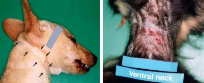
Figure 28: A situation of a dog strangulation, being possible to see the indentation of hair in the neck. Adapted from (H. Munro & R. Munro, 2008). ... 58
Figure 29: A situation of a dog strangulation, being possible to see a mild bruising and reddening of the skin on the ventral surface and both sides of the neck. Adapted from (H. Munro & R. Munro, 2008). ... 58
Figure 30: A frozen pink frothy fluid in the lumen of the trachea of a drowned dog. Adapted from (H. Munro & R. Munro, 2008). ... 59
Figure 31: View of the cadaver 1A in a left lateral decubitus. ... 83
Figure 32: Packaging of the cadaver 1A. ... 83
Figure 33: A closer view of the Figure 31; denote the wet fur around the right pinna (black arrow) and the bloody soiling fur perioral (white arrow). ... 83
Figure 34: Petechiae and haemorrhagic suffusions in the ventral region of the cadaver 1A. .. 83
Figure 35: Circular chop-wound in the right temporo-parietal area of the cadaver 1A. ... 83
Figure 36: Closer view of the previous s figure. ... 83
Figure 37: Pulmonary edema - presence of foam in the trachea and bronchi – in the lungs of the cadaver 1A. ... 84
Figure 38: Linear fracture in the parietal/temporal bone (black arrows) of the cadaver 1A. ... 84
Figure 39: Contusion in the temporal muscles under the external wound observed and described above. ... 84
Figure 40: In situ view of the haemorrhage in the right hemisphere of the brain (frontal, parietal and occipital lobes) under the external injuries described above. ... 84
Figure 41: Ex situ view of the previous injury in the cadaver 1A. ... 84
Figure 42: View of the cadaver 1B in a left lateral decubitus. ... 85
Figure 43: Packaging of the cadaver 1B. ... 85
xx
Figure 45: Bilateral subconjunctival haemorrhage of the cadaver 1B. ... 85
Figure 46: Ring bruise in the neck muscles, found after the skinning on the cadaver 1B. ... 85
Figure 47: Presence of foam in the trachea lumen and haemorrhage in the ventral trachea wall of cadaver 1B. ... 85
Figure 48: Closer view of the previous figure. ... 86
Figure 49: Elliptical shape fracture in the nasal bones corresponding to the external chop wound of the cadaver 1B. ... 86
Figure 50: Generalized congestion and haemorrhage of the brain of the cadaver 1B. ... 86
Figure 51: View of the weight (5,830 Kg) of the cadaver. ... 94
Figure 52: View of the cadaver along with the black plastic bag in which was conditioned and with the transparent bag of the suspected bait. ... 94
Figure 53: Both tamper evident bags, one containing the suspected bait and the other unidentified. ... 94
Figure 54: Radiograph showing the microchip (blue arrow) and the subcutaneous emphysema (red arrows). ... 94
Figure 55: Ring shape indentation of the fur around the neck. ... 94
Figure 56: Closer view of the previous photo, being possible to see a bruise of the corresponding tissues (black arrow). ... 94
Figure 57: Mouth defiled with soil and congestion of the oral mucosa. ... 95
Figure 58: Subconjunctival haemorrhage of the right eye. ... 95
Figure 59: Subconjunctival haemorrhage of the left eye. ... 95
Figure 60: Extensive contusion on the ventral neck region. ... 95
Figure 61: Extensive contusion on the dorsal neck region, extended to the frontal-parietal region of the head. ... 95
Figure 62: Extensive laceration of the ventral cervical muscles, exposing the trachea. ... 95
Figure 63: Ring shape contusion on the cervical region of the trachea, with 5 cm length (white arrows). ... 96
xxi
Figure 64: Cadaver 3A in lateral view. ... 105 Figure 65: Right scapular region with a perforating circular wound, of 1 cm diameter, with no contour halo (red arrow). ... 105 Figure 66: Left scapular region with a cutaneous perforation and contusion associated, measuring the larger diameter 2,5 cm, with everted and irregular edges (red arrow). ... 105 Figure 67: Contusion and laceration of the right scapular muscles, with extrusion of pulmonary tissue outside the thoracic cavity (white arrow). ... 105 Figure 68: Fracture of the scapula and laceration of the underlying muscles and extrusion of the pulmonary tissue. ... 105 Figure 69: Presence of a blood clot in the thoracic cavity – haemothorax - rupture of the pleura serosa and an injury of the left lung. ... 105 Figure 70: Fracture on the 6th right rib. ... 106 Figure 71: Laceration of the intercostal muscles between the left 4th and 5th ribs and comminuted fractures of the same ribs. ... 106 Figure 72: Blood in the pericardial sac due to a traumatic rupture of the left atrium with irregular borders. ... 106 Figure 73: Frontal region of the head with the hair stained with blood. ... 107 Figure 74: Circular perforated wound, without contusion halo, adjacent to the cornual process of the frontal bone. ... 107 Figure 75: Circular perforated wound, without contusion halo, adjacent to the cornual process of the frontal bone, seen after better dissection. ... 107 Figure 76: Female foetus on the uterus of about six months, meaning that the animal was pregnant when was killed. ... 107 Figure 77: Female foetus from the previous figure, exteriorized from the uterus. ... 107 Figure 78: Laceration and contusion of the neck muscles. ... 107 Figure 79: Metal fragments (white arrows), compatible with projectile, detected during the dissection of the injured muscles. ... 108
xxii
Figure 80: The bigger metal fragment (white arrow), detected on deeper dissection, in the fracture of the right wing of the atlas bone. ... 108 Figure 81: Blood clot in the sinuses. ... 108 Figure 82: Metal fragments collected from the Cadaver 3B. ... 108 Figure 83: Trajectory of the projectile, using a surgical instrument. ... 108
xxiii
LIST OF TABLES
xxv
ABBREVIATIONS, SYMBOLS AND ACRONYMS 3D: Three-dimension
AAN: Animal abuse and neglect
ACVP: American College of Veterinary
Pathology
ALS: alternate light source BCS: Body condition score BPA: Blood stain pattern analysis C.L.: Charles Louis
COD: Cause of death CoE: Council of Europe CT: Computed tomography DL: Decree-Law
DNA: Deoxyribonucleic acid EU: European Union
FVP: Forensic veterinary pathologist GNR: Guarda Nacional Republicana INIAV: Instituto Nacional de Investigação
Agrária e Veterinária
LCOD: Legal cause of death
MOD: Mechanism of death
MSCT: Multislice computed tomography NUIPC: Unique process identification
number crime / Número único de identificação do processo crime
PMCT: Post-mortem computed
tomography
PMCTA: Post-mortem computed
tomography angiography
PMMRI: Post-mortem magnetic
resonance
SIRCA: Sistema de Recolha de
Cadáveres de Animais Mortos na Exploração / Portuguese system of death farm animals collection
SPPA: Sociedade Portuguesa de patologia
animal / Portuguese society of animal pathology
WSAVA: World small animal veterinary
1
CHAPTER I - LITERATURE REVIEW
Publications related with the subject “Forensic veterinary medicine” were searched in “PubMed” and “ScienceDirect”, with the keywords “Forensic veterinary”, “Forensic necropsy”, “Chain of custody” and “Forensic veterinary pathology”.
Forensic Veterinary Medicine
1.1. Definition
Forensic veterinary medicine can be summarily defined as “the application of veterinary knowledge to the purpose of the law” (J. Cooper, 1998, p. 161); this means, using the knowledge of the several veterinary medicine branches to solve cases of civil and/or criminal liability, supported by application of the law (Peleteiro et al., 2016). Since this definition is very general, and it can be applied to civil cases per se, some authors prefer to name it as legal veterinary medicine rather than forensic veterinary medicine, as the last usually has a direct connection with crime. The studying of the origin of the word forensic is very important to comprehend its generality. It has derived from the Latin word ‘forensic’ which means ‘public’, that in turn was derived from ‘forum’ - a place of debate usually in the public arena of court or other legal proceedings that happens in open doors (J. Cooper & M. Cooper, 2007); and it is defined by the concise Oxford dictionary as “relating to, used in, or connected with a court of law” (J. Cooper & M. Cooper, 2007, p. 3).
Furthermore, forensic - originally meaning ‘relating to the law’- has broadened even more the generality of the word; it is nowadays implied to be “a detailed investigation and collection of evidence regardless of whether or not there is a specific legal case or enquiry pending” (p. x), so the methods used in conventional forensic work can now be largely applied in situations outside the courts, such as insurance claims, inquiries, environmental impact assessments, public service commissions or when defending or propounding allegations of professional misconduct or other disciplinary measures (J. Cooper & M. Cooper, 2007). In addition, there is an increasing demand by the animal owners to seek compensation in matters regarding death and injuries, especially about deaths, which often triggers an investigation that may lead to a
2
legal case, disagreements over an insurance claim or allegations of professional malpractice (J. Cooper & M. Cooper, 2007).
Veterinary forensic medicine is not only restricted to animal deaths, as the growth of this science may have an increased acknowledgment in some human forensic cases, as it is already described by Tsokos, Byard, and Puschel (2007), which an human victim succumbed to a dog attack, and veterinary studies assisted on the human investigation, as a necropsy of the offending animal provided information that helped to establish the identity and ownership of the animal, as well as trace evidence linked the dog to the victim; other-like case has been described by Aquila et al., 2014, (p. e1) about "a case study concerning a car accident where both humans and pets were involved; where investigation and reconstruction of the crime scene were conducted by a team consisting of forensic pathologists and forensic veterinarians”. Both an autopsy and a necropsy were conducted on the man and the dog, respectively, and the results were compared, and the information was used to reconstruct the collision. “This unusual case was solved through the collaboration between forensic pathology and veterinary forensic medicine, emphasising the importance of this kind of co-operation to solve a crime scene concerning both humans and animals”. Therefore, we should be aware that forensic veterinary deals with cases of animal abuse and smuggling, but also with the role of animals in court cases involving people (Ottinger et al., 2014).
After all, “the main objectives of forensic medicine are to document, analyse, and elucidate scientific medical findings in a comprehensible way for courtroom presentation”, regardless the type of case (Dirnhofer, Jackowski, Vock, Potter, & Thali, 2006, p. 1306). For instance, we should have in mind that an animal can be witness, victim or perpetrator in a forensic case (J. Cooper & M. Cooper, 2008). When being the perpetrator, the animal can injury the human in a variety of ways such as bites, stings, electrocution, transmission of infectious agents or presentation of allergens - and veterinarians are often called to help understand the case (J. Cooper & M. Cooper, 2008).
1.2. State of the art
Forensic veterinary medicine is not yet a recognized discipline but is rapidly evolving (J. Cooper & M. Cooper, 1998); after all, according to McEwen (2012) as cited in Gerdin and
3
McDonough (2013): “cases involving insured animals, possible medical malpractice, and alleged animal abuse and neglect are not new; new is the increasing amount of media coverage and degree of public awareness of these cases” (p. 994); mainly due to the changes in laws and their enforcements and because of the public concern about the health, welfare, and conservation of animals, both domesticated and wild, more legal cases relating to such issues are being taken to the courts and a higher standard of expert evidence is expected, becoming widely recognized the need for veterinarians to be more actively involved in forensic work (J. Cooper & M. Cooper, 2008; McEwen, 2012).
Listos, Gryzinska, and Kowalczyk (2015) in their review ‘Analysis of cases of forensic veterinary opinions produced in a research and teaching unit’, observed an increase in the demand for the services of veterinary forensic experts at the Department of Pathological Anatomy, Faculty of Veterinary Medicine, University of Life Sciences in Lublin, beginning in 2006 and persisting through 2014 (H. Munro & R. Munro, 2013). Also, recently, Gerdin and McDonough (2013) affirmed the frequent increasing of cases of suspected animal abuse and neglect submitted to veterinary pathologists, as well as Ottinger et al. (2014) quoted McEwen (2012) and Salvagni et al. (2012), whose reports showed that the number of medico-legal veterinary pathology cases had increased. For instance, Portugal has been witnessing, since 2014, the creation of legal norms designed to protect the animals, either by criminalizing the abuse and neglect of companion animals or by officially acknowledging their status as sentient beings (Moreira, 2017). Harris (1998), declared: “we live in a time in which society is very prone to litigation” (p. 1), and therein lies exciting possibilities for the expansion and refinement of the forensic veterinary pathology knowledge base (H. Munro & R. Munro, 2013).
J. Cooper & M. Cooper at ‘Future trends in forensic veterinary medicine’ (1998), early suggested that “one of the main elements that hampers the development of veterinary forensic science and its acceptance as a specialty is the absence of standard systems and protocols” (p. 213), and they recommended the establishment of protocols that would serve the veterinarian well when dealing with forensic cases.
The non-recognized path to forensic veterinary specialization diverges on the growing demand of the forensic veterinary services, and those veterinarians who become involved in this field are usually self-taught or, in a few cases, have attained a qualification based on training for
4
human medicine (J. Cooper, 1998). Also, the increasing of legislation relating to animal welfare and conservation, makes the demand for specialists higher, thus the continued absence of such a specialty is surprising (J. Cooper, 1998). In Portugal, however, some Universities have already in the Veterinary Medicine course, some subjects of the forensic field. This situation is in stark contrast to human forensic medicine, which is a recognised independent discipline with training opportunities and certified specialists with full-time employment (Ottinger et al., 2014). And while human forensic medicine is a well-recognized and highly developed speciality, the forensic veterinary medicine is a relatively young science (J. Cooper, 1998; Listos et al., 2015). So, much can be gained studying the procedures, methods, and standards used in human forensic cases, especially establishing and using the systems and protocols in a similar way to those used in human forensic medicine, but wisely, because extrapolation from human work of research in the field of forensic science, although necessary, is not always satisfactory (J. Cooper & M. Cooper, 1998; R. Munro, 1998). However, more than grounding the veterinary forensics on the human forensic knowledges, it shall be, instead, linked both areas of practice, as the opportunity to foster collaboration may be seized upon (Byard & Boardman, 2011).
As mentioned before, the growth of the veterinary forensic sciences creates a need to educate and teach and to ensure that the training offered is of a satisfactory standard and objectively assessed and accredited by an independent body (J. Cooper & M. Cooper, 2007); to overcome this, forensic pathologists can contribute in lecturing to veterinary science students on basic injury assessment and evaluation (Byard & Boardman, 2011). The raising number of textbooks and journal articles covering domestic animal and wildlife forensic investigations as well as forensic veterinary lectures are a modern strategic – academic education like the international veterinary forensic science association in 2008, the several presentations in the field of veterinary forensic pathology at national and international meetings at 2011 (such as the C. L. Davis/ACVP symposium on forensic veterinary pathology; the American academy of forensic sciences conference, or the European veterinary conference Voorjaarsdagen (Holland)); and the increasing submissions of forensic cases to veterinary pathology facilities as result of investigations of animal-related crime, are happening world-wide (Brownlie & R. Munro, 2016; Gerdin & McDonough, 2013; H. Munro & R. Munro, 2013; Ottinger et al., 2014). Along with that is Portugal, where in 2017 only, had one theoretical-practical course on veterinary forensic pathology by the Portuguese society of animal pathology (SPPA) and one forensic formation of two days by a Portuguese veterinary university.
5
Essentially, it can be assumed that more and more people care about animal welfare, leading to a greater public concern about a fair and accurate prosecution of the animal cruelty perpetrators (McDonough, Gerdin , Wuenschmann , McEwen , & Brooks, 2015). However this demand is not always linked with society empathy towards animals, but with other factors and various public concerns that are helping to mould a new approach to forensic medicine, such as the recognition of the link between cruelty to animals and violence toward humans, and that animal abuse is often one of the indicators of family violence and child abuse (Balkin, Janssen, & Merck, 2012; J. Cooper & M. Cooper, 2007). McDonough et al. (2015) alerted that “more than 70% of battered women who own pets report that their batterer threatened, injured, or killed family pets as a form of revenge or to psychologically control the victim” (p. 5). It is also alarming that multiple studies showed that more than 60% of violent adult offenders have a history of childhood animal abuse (Ascione, 2001). Therefore, the law enforcement community has becoming to recognize that early intervention in animal cruelty cases has a positive and proactive impact on public safety and human welfare (Balkin et al., 2012).
Allen, Gallagher, and Jones (2006) were vigilant about an underestimate collateral fact from domestic violence, that was an important factor to lead the women victim to stay longer under the condition of domestic violence, that was the lack of protection of victim’s companion animals at refuges in Ireland, where neither were provided facilities for animal care nor was there an animal foster service, for instance, similar as those operated in the UK by Paws for Kids and First Strike. These types of facilities are starting to appear in certain developed countries as a recognition of the importance of safeguarding the victim’s companion animals in the decision of refugeeing; however, in Portugal, there is not yet such services.
1.3. The role of the forensic veterinary pathologist
Forensic veterinary pathology is a diverse discipline, included under the term ‘forensic science’, that is on an early phase of its development. It walks by side with other forensics sciences, such as crime scene examination, ballistics tests, genetics analysis, toxicology and others that will be described later (H. Munro & R. Munro, 2013). However, in contrast with some forensic sciences, forensic pathology still utilises the time-old, evidence-based methods introduced centuries ago, like the dissection of the cadaver (necropsy), cytology and histopathology and the oral/written description (Bolliger et al., 2008); nevertheless, forensic cases requires
6
modification of post-mortem procedures and written reports compared to those of routine diagnostic cases, as the questions imposed are lightly different, specially since the questions come mostly from the court and are basically to determine the cause, mechanism, manner and time of death and injuries (Gerdin & McDonough, 2013); thus, the duties of the forensic pathologist are to “collect evidence from the body, document injuries or lack of them, deduce how the injuries occurred, (…), determine or exclude other contributory or causative factors to the death, and provide expert testimony if the case goes to trial” (D. DiMaio & V. DiMaio, 2001). The veterinary pathologists play a crucial role in such cases as the identification of lesions and their accurate description and photographic documentation are the key to criminal death investigations (J. Cooper & M. Cooper, 2008; de Siqueira, Cuevas, Salvagni, & Maiorka, 2016; Gerdin & McDonough, 2013; McEwen, 2012).
The veterinary forensic pathologist is a neutral observer, making careful notes of the events leading up to death, the location and position of the animal, environmental considerations, and overall preservation and condition of the animal - specially about the internal normal and abnormal tissues – and collecting samples to perform relevant tests for the case, because, after all, and quoting Brownlie and R. Munro (2016), “the veterinary pathologist is required to act in an independent, objective, and unbiased manner in forensic investigations” (p. 919); and his/her role is “not to pass judgment but to document, interpret, and explain the pathological findings to the investigators and ultimately to the court, thereby assisting the court to reach a decision on the case” (p. 919).
As a matter of fact, veterinarians have for long played a part in legal cases, especially those relating to such matters as the purchase and sale of horses and other livestock, animal welfare, and food hygiene, but nowadays is expected to have a forensic veterinary pathologist, this is, a denote trained and certified veterinary pathologist who have additional forensic qualifications or who have documented, relevant experience in forensic practice to handle forensic cases, since he/she must have a systematic and meticulous approach and should be prepared to present and defend evidence in court (Brownlie & R. Munro, 2016; J. Cooper & M. Cooper, 2008). Although formal training is a part of the necessary specialization, sometimes the courts may place most weight on the experience that the veterinarian has in the field directly related to the case (R. Munro, 1998). Nevertheless, any veterinary practitioner may become involved in a
7
legal case in countless ways, whether being a "witness of fact", a "professional witness" or an "expert witness" (J. Cooper, 1998).
It is important to note that the veterinary pathologist do not have to accept a forensic case; the forensic necropsy is an intensive labour and requires meticulous documentation and strict maintenance of the chain of custody, thus if the forensic veterinary pathologist (FVP) are not sure if he/she has the time or if it is convenient for him/her, must do not accept the case (McDonough et al., 2015). Another reason for declining a forensic case is the mistaken belief that the veterinary pathologist must determine if a crime was committed; but it should be comprehend that cannot be asked to the FVP if a crime was committed, since animal cruelty is a legal, not medical, determination, therefore, stablishing if acts are criminal is the duty of the members of the court (Benetato, Reisman, & McCobb, 2011; Gerdin & McDonough, 2013; McDonough et al., 2015). The common type of questions made to the forensic veterinary pathologist are to confirm whether suspected inflicted injuries can be confirmed as such, and how the injury may have been caused, or if an injury is consistent with an alleged incident or with the account of the accused, or to confirm if the injuries were sustained ante-mortem, and for how long might the animal have survived after the injury and, often the crux of a case, if it was suffering involved. On more specific situations, the questions may be related to if the animal was already dead when thrown into water, or if there was evidence to suggest burial when still alive, or if the animal die in the fire, or if it is possible to estimate the duration of deprivation of food or water (Brownlie & R. Munro, 2016).
As seen before, forensic veterinary pathology is guided in many ways on the knowledge of human forensic pathology, since the pathologic changes in animals are closely parallel to those in humans, whether are electrothermal burns, toxicities or gunshot wounds, the effects on the body of both Homo sapiens and Canis lupus are similar (Viner, 2016). One of the major differences in human and veterinary forensic pathology is the sheer number of species of animals and birds that the veterinarian might be asked to examine, unlike in human pathology; but is an impossible expectation to be knowledgeable about all of them, thus is important and humbled to establish contacts with specialists, who can provide advice on species identification, feeding habits, anatomy, diseases, etc. (H. Munro & R. Munro, 2008).
8
1.4. Other forensic sciences
The increasingly widespread use of the term ‘forensic science’ is a reminder that “it is an interdisciplinary subject, par excellence”, where numerous fields such as toxicology, ballistics, entomology (a ramification of parasitology), genetics, anatomopathology, bacteriology, virology, as well as anthropology, biology, botanic, palynology, blood stain pattern analysis (BPA), alternative light source (ALS), as well as justice, bioethics, and many others, find themselves (J. Cooper, 1998; Lima, Ochôa, & Orge, 2016; Peleteiro et al., 2016). There is an excellent citation from J. Cooper and M. Cooper (2007) about this subject showing the immensity of the forensic sciences: “We speak now of ‘forensic scientists’ who, in turn, describe themselves as ‘forensic chemists’, ‘forensic botanists’ or ‘forensic biologists’, and so on” (p. 5).
It is important to recognize when there is a need to resort to other forensic professionals, because all the veterinary forensic cases involve a multidisciplinary approach, that may lead to take assistance from other specialists, and perhaps specialists from human forensic (Touroo & Fitch, 2016). J. Cooper and M. Cooper (2007) wrote that “working with people from other backgrounds is often the key to fruitful investigation and production of sound evidence” (p. 36).
Would be unrealistic extensive describing all the forensic sciences possible to exist. There are some more commonly used nowadays at veterinary forensic, and others, on the other side, that barely take place at the daily veterinary forensic, so the intent here is to summarily describe the ones more common, as well as, in contrast, present the ones innovators in the field.
For instance, in cases of death by killing, blood is the most common body fluid found at crime scenes, and blood stain pattern analysis (BPA) may indicate the angle and distance of the blood fall, helping to solve some cases (de Siqueira et al., 2016).
One science regularly used by the forensic pathologists is toxicology. Occasionally, the cause and manner of death suspected at the necropsy table is completely changed by the toxicology data (Gill, 2005). The toxic panel asked by the pathologist should be synthetize to the most likely suspicions. And in the end, even though pathologists need the toxicology results for the death investigation, they are the expert ones in interpreting the results in the setting on an entire
9
case, meaning that the toxicologists give to the forensic pathologists information but not conclusions (Gill, 2005).
The determination of the time of the death or post-mortem interval is a major topic in forensic pathology as well it is in forensic entomology. The entomologic method is based on the correlation between the developmental stages of arthropods (especially of blowfly larvae) and the time of the death; the major advantage of entomology against the pathological standard methods for the determination of the post-mortem interval (body temperature, post-mortem lividity and rigidity, and chemical investigations) is that arthropods can represent an accurate measure even in later stages of the post-mortem interval when the classical forensic pathological methods fail, due to the advance of putrefaction (Benecke, 2005). But, entomology is much more; it can also give information about the geographic localization of a cadaver - insects that live in restricted areas but are found on a cadaver in a different area can prove that the body had been moved after death – or give toxicological information – drugs that cannot be detected in severely decomposed tissue of a corpse may still be found in the insects that fed on the corpse – or, for instances, about the site of injuries - the location of a stab wound can be determined by unusual feeding sites of beetles and maggots (Benecke, 2005). So, it is easy to perceive that a forensic science team should have a consulting entomologist (a biologist who studies insects) with forensic specialization as a member, and such a specialist should be consulted for the accurate identification and interpretation of insect evidence (Castner, 2009). Although identification of the types and stages of maggots and beetles is outside the competence of most veterinary pathologists, the correct procedures for the collection of entomological evidence must be an inherent skill of those (Byrd, Lord, Wallace, & Tomberlin, 2010; H. Munro & R. Munro, 2013). Forensic entomology can be very valuable for numerous veterinary forensic cases and in Portugal there are some forensic pathology facilities where it is already a common practice.
In the field of genetics, DNA research for forensic identification prospers at human forensic medicine (Kondo, 2007). Animal DNA results have also been used successfully in cases that come to court. Animals live in close contact with humans and animal-derived trace and DNA evidence is often found at a crime scene or on a suspect (Merck & LeCouteur, 2012a). The application of DNA analysis has a huge potential in cases involving companion animals, and when possible has been used to tie suspects to crime scenes or specific dogs to specific bites, or to identify stolen dogs, among others. Animal DNA also can link a suspect with a crime
10
scene or victim, being the animal a witness. Transfer of DNA from hair, saliva, blood, urine or faeces will occur during a crime, either from the victim's pet to the suspect or crime scene, or from the suspect's pet to the victim or crime scene, as dictated by Locard’s exchange principle, and can be found on wounds, clothing, or property (Gerdin & McDonough, 2013). Likewise, the DNA test take a major importance in animal attacks, at identifying the correct attacker, and preventing innocent animals from being euthanised for aggressive behaviour (Merck & LeCouteur, 2012a). However, we must be realistic and be aware of the different economic power of the countries; in Portugal this field is economic unavailable for veterinary forensic pathology.
An uncommon, or not so known, forensic sciences are the forensic botany and palynology.
Forensic botany refers to the forensic analysis of the plant anatomy, plant growth and
behaviour, plant reproductive cycles and population dynamics, and plant classification schemes to species identification (Merck & LeCouteur, 2012a). Plant matter may be found in stomach contents or faeces, and on fur or clothing, and can or cannot be associated with poisoning, and also may be found around a body or area from a weapon (Merck & LeCouteur, 2012a). Plant-derived evidence can be linked to specific locations and certain seasons, which can be useful to verify an alibi, track movements of the suspect or victim, aid in determination of time of death or determine the primary crime scene in cases in which there is a secondary body dump site (Merck & LeCouteur, 2012a).
Forensic palynology is the study of pollen, spores, and other acid-resistant microscopic plant
bodies (palynomorphs), and it is valuable because of the microscopic size of the organisms, their large production and high resistance to decay and also because they can be identified in a plant taxon (Milne, Bryant Jr., & Mildenhall, 2005). Pollen is often present on a body or object of a crime scene due to their ubiquitous nature - they can be found on shoes, clothing, fur, rope, or carpet, and inside the nasal cavity or upper airways when inhaled (Merck & LeCouteur, 2012a). Submerged water plants also rely on pollination to reproduce, leading to be a valued evidence in suspicious cases of drowning (Merck & LeCouteur, 2012a). “The recognition and identification of pollen, seeds, diatoms, flowers and plant fibres sometimes play a unique part in providing ‘trace’ evidence in veterinary and comparative studies” (J. Cooper & M. Cooper, 2007, p. 28).
11
Imaging techniques take a special importance at forensic work, mainly concerning to post-mortem radiography, which is getting more and more common its performance prior to the necropsy examination. However, there are other radiological areas - ones recent on the forensic field and others quite recent as a science itself. A modern imaging technique, started as a project founded at the Institute of Legal Medicine of the University of Berne in Switzerland at the turn of the millennium is the virtopsy (Flach et al., 2014). The term ‘virtopsy’ was created from the terms ‘virtual’ and ‘autopsy’, and it is based on three groundworks: “3D body surface documentation using photogrammetry-based optical surface scanning, and both multislice computed tomography (MSCT) and magnetic resonance imaging (MRI) to visualise the internal body”, giving a resulting data set that contains high-resolution 3D colour-encoded documentation of the body surface and 3D volume documentation of the interior of the body (Bolliger et al., 2008; Dirnhofer et al., 2006).
X-rays are already routinely used in forensic cases, mainly on the detection of the foreign bodies in the cadavers, such as projectiles; but a multislice computed tomography (MSCT) has the advantage of locate with precision the topography of the foreign bodies within the body in a three-dimension (3D) manner, thus facilitating their extraction at necropsy, redirecting pathologists to dissect body parts or areas that are not routinely dissected during a standard necropsy, such as the face bones, shoulder articulation, extremities, outer pelvis, larynx and soft tissue of the back (Flach et al., 2014). “Another advantage of MSCT over conventional X-rays is that MSCT can measure the radiological density” of these objects, as well as having greater precision and definition in detection and demonstration of fractures and, essentially of gas - pneumothorax and gas embolism are difficult to determine and impossible measure the amount of gas at necropsy (Bolliger et al., 2008; Jackowski et al., 2004). According to Jackowski et al. (2004), “MSCT sectional images depict the presence of gas immediately, whilst 3D reconstructions display the gas distribution in the blood vessels and the cardiac chambers, and even the precise amount may be determined” (as cited in Bolliger et al., 2008, p. 276).
Although conventional X-rays are often used in forensic practice, and although PMCT and PMCTA are still not considered a quotidian essential in veterinary pathology, nowadays these techniques have gain some supporters, introducing it to improve the post-mortem diagnosis, by not destroying the relevant forensic findings on the necropsy (Bolliger et al., 2008; Dirnhofer et al., 2006; Lee et al., 2011; Martinez, Hetzel, Thali, & Schweitzer, 2015; Tahli et al., 2007). Even more, “the 3D reconstructions from computed tomography (CT) are especially clear and
12
understandable to lay observers and can be rotated on the computer screen to give a graphic view of areas of interest and angles of an attack” at court (Brownlie & R. Munro, 2016, p. 924). Both techniques - PMCT and necropsy - demonstrated injuries one complementing the other, and the inclusion of the PMCT gives important information to the final diagnosis, playing an important role to the veterinary pathology (Pinto et al., 2017). As Franckenberg, Kern, Vogt, Thali and Flach (2015) acclaimed:
Virtopsy of animals provides not only spectacular insight into the anatomy of the mammals but also aids in the evaluation of hunting-related and veterinary issues and the determination of the cause and manner of death without the need for any manual dissection. (p. 75)
Other imaging techniques, such as endoscopic, although not common in forensic necropsy, are already described in human forensic autopsy, as the inspection of the cavities and sinuses, like the eye ground, the external auditory meatus up to tympanic membrane and the naso- and laryngopharynx, can be done without their morphological damage is a massive advantage (Amberg, Lemmen, Pollak, Strutz, & Unsöld, 1992, cited in Ohshima, 2000, p. 154).
1.5. The legal system
Since the notion that cruelty to animals is a symptom of depravity and that the crimes against animals can go beyond the act itself and be a reflection of other types of violence, like the interpersonal, domestic violence and children or elderly abuse, “there has been a recent global increase in the enforcement of laws regarding animal care and the enactment of tougher laws regarding animal abuse and neglect”, once it started to be seen by the authorities as a risk factor or a possible indicator of another type of violence, leading to collaboration between several entities with the aim of crossing information between them (Gerdin & McDonough, 2013; Moreira, 2017). On the other side, there is the emotional connection between humans and companion animals, leading to a not surprising growth on the demand of a consistent legal board to protect the last ones, approximating those to the norms applicable to humans (Moreira, 2017).
13
1.5.1. Governing laws
Any FVP undertaking a forensic case must be familiar with the applicable laws in his/her country, as well as any person involved in animal forensic work should have a sound background in knowledge of the law and be competent in identifying, collecting and preserving evidence, because a thorough knowledge of the relevant legislation is invaluable in the full preparation for a court appearance (J. Cooper & M. Cooper, 1998; J. Cooper & M. Cooper, 2007; Touroo & Fitch, 2016).
As known, the approach to animal forensic work differs between the countries, even within Europe, due to legal variations and different public opinions about the importance of animal welfare (Ottinger et al., 2014); however the European convention for the protection of animals produced by the European Union (EU) and the Council of Europe (CoE), in 1987, comprehends a large body of regulations and directive relating to animal health, farm animal welfare, research animals, wildlife conservation and the international trade in endangered species (J. Cooper & M. Cooper, 2007). This became a law to the Member States countries, being thereby subjected to penalties if there were some failures on the implementation of the directives (J. Cooper & M. Cooper, 2007). In Portugal, this European legislation was approved and ratified in 1993 by the Decree No. 13/93 of 13 April 1993; however, only at 2001 it was become regulated, by the Decree-Law No. 276/2001 of 17 October, and its successive amendments, being the last one the Decree-Law No. 260/2012 of 12 December 2012, which, summarily, declares the prohibition of all violence against animals with the purpose of inflicting death, suffering or injury, being the offenses punishable by a fine (Moreira, 2017). Since this date, Portugal has been having successive law adjustments, for instance, the embracing of the Law No. 69/2014 of 29 August 2014, which criminalizes the abuse and abandon of companion animals, introduced on the Penal Code’s Title VI “Crimes against companion animals” (Moreira, 2017). At 2015 emerge the Law No. 110/2015 of 26 August 2015 establishing the framework of accessory penalties applicable to crimes against pet animals; recently, at 2017, the amendment to the Civil Code by the Law No. 8/2017 of 3 March 2017 was introduced, which established a legal status of animals, recognizing their nature as sentient beings (Moreira, 2017).
Regardless all legislation, it should be remembered that any laws, including those of animal protection, can be effortless if not correctly applied. Each legislation is supported by powers of
14
implementation and enforcement, and this depends on the will of the authority but mainly on the powers provided in the legislation (J. Cooper & M. Cooper, 2007).
1.5.2. Legal description of death
The concepts of cause, mechanism and manner of death are vital to the comprehension of the death of any animal, however these concepts are usually confused between each other (Peleteiro et al., 2016). In forensic pathology the accurate knowledge of each individual ones of these concepts is required since they must be clearly expounded on the necropsy report to be correctly perceived by the law related personnel who will read it (Brownlie & R. Munro, 2016).
1.5.2.1. Cause of death
The cause of death (COD) is the event that produces the physiological alteration that leads to death, this mean “any injury or disease that produces a physiological derangement in the body that results in the death of the individual” (D. DiMaio & V. DiMaio, 2001, p.; Peleteiro et al., 2016); in other words is “the injury or disease that began a sequence of events that ultimately led to the death of the animal” (Merck, Miller, & Maiorka, 2012e, p. 65). Thus, a gunshot wound to the head or to the chest, a run over by a car, a stab wound to the chest, an adenocarcinoma of the lung, a coronary atherosclerosis, or a canine parvovirus are some examples of COD (D. DiMaio & V. DiMaio, 2001); denote that in most cases, euthanasia is not considered as a COD, but indeed is the injury or illness that lead to the euthanasia (Merck et al., 2012e). Determining the COD is often the primary goal of the post-mortem investigations, however, sometimes the FVP are not entirely certain about the COD, but are not completely uncertain either, not justifying the use of the term “undetermined”; in such cases it is rather to use “probable” before the cause of death opinion (Gerdin & McDonough, 2013); Merck et al. (2012e) claim that this is a safeguard measure to the FVP, who is saying that has some degree of certainty, although his/her opinion may change based on any future information provided.
When stablishing the COD, a concept to keep in mind is the contributory cause; it refers to “any condition the animal had that could have contributed to death or injury (Merck et al., 2012e, p. 65). An example is a case of multiple stab wounds in the chest, causing extensions haemorrhages leading to a hypovolemic shock, which the COD is the multiple stab, but maybe a clotting disorder would be a contributory caused for the death due to a massive
15
exsanguination; other example given by Merck et al. (2012e) is a starvation case which an emaciated animal died from hypothermia, the cause of death is the hypothermia but the contributory cause would be starvation, the cause of an severe emaciated state, that limit the ability to regulate the body temperature.
1.5.2.2. Mechanism of death
The mechanism of death (MOD) is defined by Gerdin and McDonough, (2013) as “the pathophysiologic events and pathways that precede termination of brain, heart, and/or lungs function” (p. 996); in other words, is “the physiological derangement produced by the cause of death, and that is responsible by the exitus” (Peleteiro et al., 2016). Haemorrhages, septicaemia, and cardiac arrhythmia are some examples of different MOD (D. DiMaio & V. DiMaio, 2001). Must be noticed that a mechanism of death can be produced by multiple causes of death and vice versa. For instance, if an animal dies of a massive haemorrhage (MOD), this haemorrhage could have been produced by a cause of death like a gunshot wound, or a stab wound, or a malignant tumour of the lung eroding into a blood vessel, and so forth. The reverse is also true, for example, a gunshot wound of the abdomen (COD) can result in many possible mechanisms of death as haemorrhage or peritonitis (D. DiMaio & V. DiMaio, 2001); a blunt-force injury to the skull (COD) can be deathful by either due a cerebral edema or due penetration of bone fragments on the brain (Brownlie & R. Munro, 2016).
Determine the MOD can be challenging, and “in some cases, the COD may be known with certainty but the mechanism unknown” (Gerdin & McDonough, 2013, p. 996). Thus, the FVP must be kept in mind that the mechanism of death, as well, can be stated as undetermined when the necropsy findings are inconclusive (de Siqueira et al., 2016).
1.5.2.3. Manner of death
The manner of death, synonymous of legal cause of death (LCOD) and medical-legal differential diagnosis, is defined as “the set of circumstances that led to the occurrence of the cause of death”, giving a legal explanation of how the cause of death came about (D. DiMaio & V. DiMaio, 2001; Peleteiro et al., 2016). In human forensic, the LCOD is classified in five categories but is grouped in two main ones - natural death (from natural causes) and violent
16
suicidal or undetermined deaths (Gerdin & McDonough, 2013). In veterinary forensic, violent
deaths only comprises three subtypes: accidental, non-accidental and undetermined death, especially since suicidal is not considered to occur in veterinary (Merck et al., 2012e).
A natural death only applies to deaths caused exclusively by a disease or senescence. An
accidental death is caused by violent means, but not due to an intentional or criminal act, whilst
a non-accidental death refers to deaths caused by a person which may be a criminal offense under the animal cruelty laws; and an undetermined death means that a reasonable classification could not be made because the pathologist could not define what occurred (Merck et al., 2012e).
Just as a mechanism of death can have many causes and a cause can have many mechanisms, a cause of death can also have multiple manners. An animal can die of a massive haemorrhage (MOD) due to a gunshot wound to the heart (COD), with the manner of death being homicide (someone shot the animal), accident (the weapon fell and discharged throw the animal), or undetermined (D. DiMaio & V. DiMaio, 2001).
“The necropsy findings could be directly linked or not to the understanding of how the death occurred” or sometimes the LCOD simply may remain unknow (D. DiMaio & V. DiMaio, 2001). Other times, despite unknowing with certain the COD and the MOD, the circumstances strongly suggest a criminal manner of death, e.g. if a cadaver is found within a container submerged in water, and at necropsy there’s no significant lesions and no evidence of euthanasia, and in this situations, the FVP can describe an animal death to have a presumptive abuse or neglect as LCOD, however he/she should be caution of the legal implications this can have on his/her country jurisdiction (Gerdin & McDonough, 2013). Therefore, the FVP should not feel pressure about requests to provide a manner of death merely because the courts may expect one (Brownlie & R. Munro, 2016; D. DiMaio & V. DiMaio, 2001; Gerdin & McDonough, 2013).
17
1.6. Chain of Custody
In forensic cases, the chronological history of all the evidence has to be exhaustively documented in order to ensure the reliability and tracking of evidence used in court proceedings (Lima et al., 2016); this process is the definition of chain of custody or, by other term, chain of evidence, which its maintenance is emphasized by all forensic books (R. Munro, 1998). It serves as a guarantee and prove of the integrity of the whole process to which the samples were submitted, requiring that “all persons handling or having custody of an evidence can testify that the sample that passed to the next stage of the investigation was the same sample as the one received” (Lima et al., 2016; R. Munro, 1998). Therefore, the information from the field, from the laboratory, and all people who manipulated the sample, should be registered, what requires a close teamwork that involves all parts, both internal and external to the laboratory, including those responsible for sample collection or removal from the body (Lima et al., 2016).
Overall, the chain of custody guarantees the control of all evidences of a case through a nominal identification of the people involved in each phase of the forensic process, discerning their responsibilities (Lima et al., 2016); the first person involved in the case usually is the enforcement officer who went on the field; it is his/her responsibility to maintain custody over the animal or its carcass until it reaches to the laboratory, when it becomes the pathologist’s responsibility, and the continuation of possession registration is required to allow an evidence to be acceptable, meaning that whether the live animal, the carcass, the sample or other must be physically accounted for at all times (Green, 1979; Wobeser, 1996).
1.6.1. Case folder
To maintain the chain of evidence, records from the chronological history of the sample, starting on its transportation, to the reception, storage, necropsy, collection of samples and its transfer to other services when needed, must be kept with written and photographic documentation (through logs or diagrams) of the various stages of the process and evidence (Lima et al., 2016). It must be created a securely filed folder for each case, named the case log, where it should be hard copies of significant paperwork such as the cadaver receipt (signed and dated by the person delivering the body and by the recipient), case number and the original labelling of the specimen