Raquel Nadine
Cadete Vieira
Nanopartículas de magnetite revestidas com quitosana
para hipertermia magnética
Coating of magnetite nanoparticles with chitosan for
magnetic hyperthermia
Raquel Nadine
Cadete Vieira
Nanopartículas de magnetite revestidas com quitosana
para hipertermia magnética
Coating of magnetite nanoparticles with chitosan for
magnetic hyperthermia
Tese apresentada à Universidade de Aveiro para cumprimento dos requisitos necessários à obtenção do grau de Mestre em Materiais e Dispositivos Biomédicos, realizada sob a orientação científica da Doutora Paula Celeste da Silva Ferreira, Investigadora Principal do Centro de Investigação em Materiais Cerâmicos e Compósitos da Universidade de Aveiro e do Doutor Manuel António Martins da Silva, Bolseiro de pós-Doutoramento do Centro de Investigação em Materiais Cerâmicos e Compósitos da Universidade de Aveiro
Trabalho realizado com o apoio financeiro do projeto CICECO-Instituto de Materiais de Aveiro, POCI-01-0145-FEDER-007679 (FCT UID/CTM/50011/2013), financiado por fundos nacionais através da FCT/MEC e quando aplicável cofinanciado pelo FEDER, no âmbito do Acordo de Parceria PT2020.
o júri
Presidente Professor Doutor Manuel Almeida Valente
Professor Associado da Universidade de Aveiro
Vogal - Arguente principal Doutor Ricardo João Borges Pinto
Bolseiro de Pós-Doutoramento da Universidade de Aveiro
Vogal - Orientadora Doutora Paula Celeste da Silva Ferreira
Acknowledgements I would like to thank my supervisor Dr. Paula Ferreira for all the support during this work, especially in the last days.
To my supervisor, Dr. Manuel Silva, I thank for the introduction to the materials world and the initial support.
I would like to thank Prof. Liliana Ferreira and Prof. Maria de Deus Carvalho from the Faculty of Science of the University of Lisbon for the magnetic and hyperthermia measurements.
I would like to thank Dr. Rosário Soares from the Laboratório Central de Análises of University of Aveiro for her support in XRD analyses and to technical services from Department of Materials and Ceramics Engineering, namely Ana Ribeiro, Artur Sarabando and Marta Ferro. Thank you, Celeste Azevedo for all support during the analysis of FTIR and TGA.
To my laboratory colleagues thank you, especially you Ana.
And most important, I want to thank my family, specially my Mum and Dad for all the support.
palavras-chave Nanopartículas magnéticas, óxido de ferro, hipertermia, dopamina, quitosana, genipina, ácido cafeico.
resumo O cancro é uma das doenças com maior ocorrência na população mundial e com uma elevada taxa de mortalidade. Os principais problemas na luta contra o cancro prendem-se com a dificuldade de diagnóstico precoce, a citotoxicidade associada aos fármacos anticancerígenos usados em quimioterapia convencional e a falta de tratamentos mais eficazes. Com o advento da nanotecnologia, tem havido um crescente interesse na aplicação de nanopartículas e nanoestruturas, nas mais diversas áreas da ciência, nomeadamente em aplicações biomédicas. Neste contexto em particular, as nanopartículas magnéticas apresentam propriedades interessantes, por exemplo, em sistemas de libertação controlada de fármaco e em hipertermia. A sua aplicação em áreas relacionadas com a saúde, como o tratamento de cancro por hipertermia magnética, passa necessariamente por uma boa caracterização das suas propriedades e pela correta avaliação das suas capacidades de libertação de energia sob a forma de calor por indução magnética.
Nesse sentido, este trabalho teve como objetivo a síntese de nanopartículas de magnetite devido a sua compatibilidade com o organismo humano e propriedades magnéticas. No entanto, devido ao seu elevado grau de agregação assim como facilidade de oxidação em meios aquosos existe uma necessidade de revestir estas partículas. Para tal, foi utilizado um biopolímero: a quitosana.
A ligação do revestimento da quitosana ao núcleo do óxido de ferro foi realizada através de dois tipos de ancoragem: através da dopamina, conhecida pela sua grande afinidade aos grupos aminas e através do ácido cafeico, por apresentar uma similaridade estrutural à dopamina.
Para a caracterização estrutural e morfológica das partículas recorreu-se à difração de raios-X (DRX), à espetroscopia de infravermelhos com transformada de Fourier (FTIR), à dispersão dinâmica da luz (DLS), ao Potencial Zeta e à microscopia eletrónica de transmissão (TEM). As propriedades magnéticas foram medidas por magnetometria de SQUID (Superconducting Quantum Interferance Device). Por fim foi avaliada a capacidade das partículas sintetizadas para aplicação em hipertermia magnética.
keywords Magnetic nanoparticles, iron oxide, hyperthermia, dopamine, chitosan, genipin, caffeic acid
abstract Cancer is a disease with high incidence in the world population and equally with a high mortality rate. The main problems in the fight against cancer are linked to the difficulty of early diagnosis, the cytotoxicity associated with anticancer drugs used in conventional chemotherapy and the lack of more effective treatments. With the advent of nanotechnology, there has been increasing interest in the application of nanoparticles and nanostructures, in several areas of science, such as biomedicine. In this context, the magnetic nanoparticles have interesting properties in controlled drug release systems and hyperthermia. Its application in areas related to health, such as the treatment of cancer by magnetic hyperthermia, necessarily requires a good characterization of their properties and the correct assessment of their ability to release energy in the form of heat by magnetic induction.
Therefore, this study aimed the synthesis of nanoparticles of magnetite due to their biocompatibility and magnetic properties. However, due to their high degree of aggregation as well as facile oxidation in aqueous media there is a need to coat these particles. For this purpose, a biopolymer was used: chitosan. The binding of the coat to the core of the iron oxide was accomplishment through two types of anchorages molecules: dopamine, knowing for their great affinity with amine groups and through caffeic acid due to structural similarity to dopamine.
The structural and morphological characterization was performed using X-ray diffraction (DRX), Fourier transformed infrared spectroscopy (FTIR), dynamic light scattering (DLS), Zeta Potential; thermalgravimetric analysis and transmission electron microscopy (TEM). The magnetic properties were studied using a Superconducting Quantum Interference Device (SQUID) magnetometer. Finally, we evaluated the ability of some of the synthesized NPs for use in magnetic hyperthermia.
i 1.1CONTEXT ... 3 1.2LITERATURE REVIEW ... 5 1.2.1. Cancer ... 5 1.2.2. Nanotechnology ... 5 1.2.3. Magnetic materials ... 6 1.2.3.1 The M-H curve ... 6 1.2.3.2 Magnetism at nanoscale ... 7 1.2.4. Hyperthermia ... 9
1.2.4.1 Advantages and disadvantages of hyperthermia ... 10
1.2.5. Magnetic Hyperthermia ... 11
1.2.5.1 Advantages of magnetic hyperthermia treatment ... 12
1.2.5.2 Disadvantages of magnetic hyperthermia treatment ... 13
1.2.6. Magnetic nanoparticles in the treatment of cancer ... 14
1.2.6.1 Why Fe3O4? ... 15
1.2.6.2 Toxicity ... 16
1.2.7. Synthesis and coating of MNPs ... 17
1.2.7.1 Chemical Precipitation ... 20
1.2.7.2 Geometry and Size ... 21
1.2.7.3 Surface modification of iron oxide MNPs ... 21
1.2.7.4 Chitosan ... 24
1.3 MOTIVATION ... 25
1.4 OBJECTIVES ... 25
2.1 SYNTHESIS OF IRON OXIDE MNPS ... 30
2.2 PROCEDURES FOR COATING OF MNPS ... 31
2.2.1 Functionalization of the MNPs with dopamine ... 31
2.2.2 Addition of genipin molecule as “tethering” agent ... 31
2.2.3 Functionalization of the MNPs with caffeic acid ... 32
2.2.4 Coating of the functionalized MNPs with chitosan... 32
2.2.4.1 Coating of F.DOPA.GP with chitosan ... 32
2.2.4.2 Coating of F.CA NPs with chitosan ... 33
2.2.4.3 Coating of F.CA with chitosan using ceric ammonium nitrate ... 33
2.2.4.4 Coating of F.CA with chitosan via 1-Ethyl-3-(3-dimethylaminopropyl)carbodiimide ... 33
2.3 CHARACTERIZATION OF THE NPS ... 34
ii
2.3.1.1 X-ray diffraction ... 34
2.3.1.2 Fourier transformed infrared spectroscopy ... 35
2.3.1.3 Thermogravimetric Analysis (TGA) ... 35
2.3.1.4 Transmission Electron Microscopy (TEM) ... 35
2.3.1.5 Hydrodynamic Size (DLS) and Zeta Potential ... 36
2.3.2 Magnetic Characterization ... 36
2.3.3 Magnetic hyperthermia measurements ... 37
RESULTS AND DISCUSSION ... 41
3.1. SYNTHESIS OF IRON OXIDE MNPS ... 41
3.2LINKAGE AGENTS TO ENABLE THE MNPS COATING... 47
3.2.1 Linking with Dopamine ... 48
3.2.2 Addition of Genipin ... 49
3.2.3 Linking with Caffeic Acid ... 50
3.2.4 Thermogravimetric analyses of the functionalized particles ... 52
3.2.5 Chitosan coating ... 55
3.2.5.1 Coating F.DOPA.GP MNPs with chitosan ... 55
3.2.5.2 Coating of the F.CA MNPs with chitosan ... 56
3.2.6 Thermogravimetric Analyses of chitosan coated MNPs ... 58
3.2.7 Zeta Potential and Hydrodynamic Diameter of funcionalized and coated MNPs ... 60
3.2.8 Magnetic Characterization ... 64
3.2.9 Magnetic hyperthermia measurement ... 66
CONCLUSION ... 71
BIBLIOGRAPHY ... 75 ANNEX ………9 1
iii
INDEX OF FIGURES
Figure 1.1 Scheme of the dipole alignment in zero applied field at room temperature for: (a)
ferromagnetic, antiferromagnetic and ferrimagnetic materials. 6
Figure 1.2 Typical hysteresis loop observed during magnetization/demagnetization of
magnetic nanoparticles. M and H are magnetic flux density and magnetic field strength, respectively. A single magnetization/demagnetization cycle (a→b→c) produces a thermal energy proportional to the shaded area within the hysteresis loop.46
7
Figure 1.3 a) Multidomains and monodomains representation under application and absence
of a magnetic field. (b) Magnetization curves as function of the magnetic field for superparamagnetic particles under temperatures below de blocking temperature – ferromagnetic behaviour (black line) showing hysteresis loop; and above the blocking temperature - superparamagnetic behaviour (blue line) with no remanent magnetization (Mr)
and no coercive field (HC). 8
Figure 1.4 Néel rotation vs. Brownian rotation. (A) Néel rotation: the magnetic moment
(arrow) rotates while the particle remains fixed (inner circle). (B) Brownian rotation: the magnetic moment (arrow) remains fixed with respect to the crystalline axes (lines inside the circle) while the particle rotates.51 9
Figure 2.1 Schematic representation of the main steps during laboratorial work. In top, the
synthesis of Fe3O4 and down, the paths for the functionalization and coating. 29 Figure 2.2 Experimental apparatus used for synthesis of NPs of MNPs. 30 Figure 2.3- Final aspect after coating the MNPs with chitosan via CAN. 33 Figure 3.1 (a) Image showing the response of the produced MNPs in the absence of magnetic
field (B=0) and (b) in the presence of magnetic field (B>0). 43
Figure 3.2 Experimental appearance of iron oxide MNPs: (a) Fresh MNPs; (b) Old MNPs
after 8 months of aging. 43
Figure 3.3 Results of the Rietveld refinement of the X-ray diffraction patterns for
synthetized iron oxide MNPs: (a) Fresh NPs, where the blue line represents the Bragg position; (b) Old NPs where orange line represents the Bragg position of two phases: magnetite (blue) and iron hydroxide (green). 44
iv
Figure 3.4 FTIR spectra of Old (continuous line) and Fresh (dotted line) synthetized iron
oxide MNPs. 46
Figure 3.5 TEM images of fresh MNPs synthetized Fresh MNPs: a) overall view
demonstrating the huge aggregation; b) the lattice fringes of the planes of the iron crystal structure. 47
Figure 3.6 Size histogram showing the average diameter (9.81 nm) of the synthesized
nanoparticles. 47
Figure 3.7 FTIR spectra of pristine Dopamine (DOPA), Fresh Fe3O4 and Fe3O4 conjugated
with dopamine (F.DOPA). 49
Figure 3.8 Chemical structure of genipin. 49 Figure 3.9 FTIR spectra of genipin (GP), F.DOPA and F.DOPA conjugated with GP
(F.DOPA.GP). 50
Figure 3.10 Chemical structure of caffeic acid and dopamine. 51
Figure 3.11 FTIR spectra of pure Caffeic acid CA, Fresh iron oxide MNPs and when
conjugated with caffeic acid (F.CA). 52
Figure 3.12 TGA analysis of a dried samples of Fresh and Old iron oxide; and when both
(Fresh and OLD MNPs) conjugated with dopamine (DOPA) and caffeic acid (CA); and the pure DOPA and pure CA. 54
Figure 3.13 FTIR spectra of chitosan (CS), F.DOPA.GP and F.DOPA.GP coated with CS.
56
Figure 3.14 FTIR spectra of the MNPsfunctionalized with CA, and coated with chitosan
per two different methods: CAN and EDAC. 57
Figure 3.15 TGA analysis of the coated with chitosan of the Fresh MNPs conjugated with
caffeic acid using cerium ammonium nitrate (IV) (CAN) and using EDAC chemistry (EDAC); and the MNPs coated with chitosan conjugated with dopamine and genipin (F.DOPA.GP.CS) and the raw chitosan (CS). 59
Figure 3.16 TEM images of: (a) uncoated MNPs; (b) MNPs functionalized with dopamine
and with genipin and coated with chitosan (F.DOPA.GP.CS); (c) MNPs functionalized with caffeic acid and coated with chitosan via EDAC; d) MNPs functionalized with caffeic acid and coated with chitosan via CAN. The arrows evidence the coat of chitosan. The circle evidence the absence of the coat. 60
v
Figure 3.17 Zeta Potential of the initial MNPs (F) and when functionalized with dopamine
(F.DOPA) and crosslinked with genipin (F.DOPA.GP) and with caffeic acid (F.CA). 62
Figure 3.18 Magnetization curves as a function of temperature (up to 250 K) measured at
50 Oe, and magnetization curves as a function of the applied field of the iron oxide MNPs (Old and Fresh NPs). 64
Figure 3.19 Magnetization curves as a function of temperature (up to 250 K) measured at
50 Oe, and magnetization curves as a function of the applied field of the functionalized and coated iron oxide MNPs (F.CA, F.DOPA, EDAC, CAN, F.DOPA.GP.CS). 65
Figure 3.20 Temperature variation as a function of time for samples synthesized in distilled
vii
INDEX OF TABLES
Table 1.1 -Some examples of different types of NPs used against cancer cells.39 15
Table 1.2 -Summary of some synthetic methods of nanoparticles. 19 Table 1.3 - Some materials reported as coating/crosslinkers and linkers to the iron oxide
(Fe3O4). 23 Table 2.1 Reagent quantities used in the synthesis of MNPs. 30 Table 3.1 Possible oxidations reaction that may occur in aqueous solution when iron (II) and
(III) are present136. 42
Table 3.2 Position of the main diffraction peaks of the synthesized Old and Fresh MNPs and
comparison to the data found in the JCPDF database concerning magnetite (Fe3O4) and
maghemite (γ-Fe2O3).138 45 Table 3.3 Summary of the percentage of mass losses during the TGA analysis observed for
the Fresh and Old NPs and when conjugated with DOPA and CA. 55
Table 3.4 Summary of TGA percentage of weight losses of all samples. 58 Table 3.5 Hydrodynamic diameter of the iron oxide MNPs and with some of MPS with the
ligand and the coat of chitosan. Z- represents the average size; Std - represent the standard deviation; d – represents the diameter in nm. 63
Table 3.6 Values of the specific absorption rate of the synthesized samples, based on the
results obtained. It is assumed that the mass of the dispersion is equal to the volume of water. cwater=4.185 (J / g K) and cglass=0.2 (J / g K). 68
ix
ABBREVIATIONS
AMF Alternating Magnetic Field AC Alternating Magnetic Current CAN Ceric Ammonium Nitrate DLS Dynamic Light Scattering DMSA Dimercaptosuccinic acid
EDAC 1-Ethyl-3-(3-dimethylaminopropyl)carbodiimide FTIR Fourier Transform Infrared Spectroscopy
f Frequency FC Field cooling H Magnetic Field Hc Coercive Field
HEMAGl (2-{[(D-glucosamin-2-N-yl)carbonyl]oxy}ethyl methacrylate HT Hyperthermia
IgG Immunoglobulin ILP Intrinsic Loss Power IS Immune System
JCPDS Joint Committee on Powder Diffraction Standards M Magnetization
MES 2-(N-morpholino)ethanesulfonic acid MHT Magnetic Hyperthermia MNPs Magnetic Nanoparticles Mr Residual Magnetization Ms Saturation Magnetization NPs Nanoparticles PNIPA Poly(N-isopropylacrylamide) ROS Reactive Oxygen Species SAR Specific Absorption Rate SPL Specific Loss Power SPM Superparamagnetic
x TEM Transmission electron microscopy
US FDA United States of Food and Drug Administration VEGF Vascular endothelial growth factor
WHO World Health Organization XRD X-ray Diffraction
3
Introduction
1.1Context
In spite of all progress made in medicine, cancer remains as one of the largest global problem, representing the main cause of illness and mortality worldwide.1 World Cancer
Report 2014 predicted an increase of cancer events from 14, in 2012, to 22 millions within the next two decades.2 Only in 2012 more than 8 millions people died as consequence of a cancer.1 Moreover, in the report “Assessing national capacity for the prevention and control of noncommunicable diseases: global survey” concerning the period 2010–2015, the 160 countries with available data saw an increase from 47 to 63% in number of cases of cancer incidence.3 According to World Health Organization (WHO), the most frequent types of cancers causing deaths are lung, liver, stomach, colorectal, breast and oesophageal.1
Such overwhelming scenario, not only have impact in the huge number of human life losses, but also in the financial costs associated to treatments and palliative cares. According to a 2015 report published in Forbes magazine, the global cancer drug market reached the US $100 billion mark in that year, and it is predicted to achieve to US $147 billion by 2018.4 These appalling numbers reveal the huge need for further research exploring better, safer, and more effective therapeutic and diagnostic strategies than the used so far, enhancing patient’s life quality as well as extending their lifetime. The last decades were characterized by development of novel and advanced strategies to fight cancer illness. The conventional strategies commonly employed include surgery, chemotherapy, radiotherapy, stem cell transplant therapy, immunotherapy, and other varieties of targeted therapies.4 Despite the considerable progress in the developing cancer therapeutics, their harmful side effects are of dominant concern. Osteoporosis, infertility, premature ovarian failure, typhlitis, hair loss, herpesviridae infections, heart diseases, gastrointestinal lesions, gastric pain are few examples of possible side effects of the current therapies.4 One of the most worrying side
effects is the resistance that tumour can acquire from the recurrent use of chemotherapeutic agents in therapies.5,6
In an attempt to improve human’s health, nanotechnology applied to medicine has been proposed in the last decades.7 Nanomedicine, allows to work at molecular and cellular levels,
4 having the potential to achieve important developments in healthcare.8 The nanomaterials have been in the few years a remarkable role in cancer diagnostic and therapeutic, where the magnetic nanoparticles (MNPs) have been in the spotlight due to their unique properties.7–9 MNPs easily match with biomolecules and can be manipulated, for example, by an external magnetic gradient,10 being used for magnetic resonance imaging as contrast agents,11–15 or they can be applied as drug delivery carriers14,16–18 for localized chemotherapy, for bioseparation,19–21 tissue repairing22 and thermal tumour therapy.23–26
5
1.2 Literature Review 1.2.1. Cancer
Cancer is characterized by abnormal cell growth9 where many environment and genetic-related factors are involved.7 When tumour cells first appear, their regular supply of oxygen and nutrients is provided by the normal blood supply of the host organ, in which the tumour ascends. As the tumour expands, it is developed its own functional vascular supply by angiogenesis.27 However, the vasculature that is formed is primitive, chaotic and with a numerous structural and functional abnormalities.27,28 The tumour cells are not able to fulfil their increasing demand for oxygen and nutrients.9,28–30. As a consequence of the hypoxic medium31, the tumour cells tends to be more acidotic,9,27 a feature that make the cells more susceptible to thermal damage.31
The development of the tumour cells mostly happens in localized tissue, but can spread to distant sites within the body by a metastatic growth. In this case, the tumour is referred as “malignant” and is usually associated with a poor prognosis for the patient,7,9
Although the tumour cells are more vulnerable to temperature, the hostile microenvironment where the tumour cells developed make them to exhibit resistance to therapies as radiation or even certain types of chemotherapies.27,31
1.2.2. Nanotechnology
Nanotechnology is the understanding and control of matter at nanometric dimensions.10,32,33 When reducing all three dimensions of a particle of a certain composition to the nanoscale, zero dimensional nanoparticles (NPs)34 and obtained and the original properties are usually
modified. As a consequence of the dimensionality (1-100 nm),7,13,35 they are characterized by high surface area,7,35 which potentiates the interaction with surrounding molecules31 and
size in the range of biomolecule entities. NPs can be very attractive for biomedical applications.35 They may be specifically targeted toward cancer cells, offering a major advantage over other therapeutic agents.7
6 The behaviours in nanoscale are not necessarily predicted from those observed in macroscopic scale. News phenomena may be observed,10,35,36 because properties such as melting point, fluorescence, electrical conductivity, magnetic permeability, and chemical reactivity change as a function of the size. More, the nanoscale materials have larger surface areas than similar masses of larger-scale materials, which allows a greater amount of the material that can interact with surrounding materials, thus affecting reactivity.37
1.2.3. Magnetic materials
Magnetic materials can be classified in one of five types of magnetism: ferromagnetic, diamagnetic, paramagnetic, antiferromagnetic and ferrimagnetic38,39 (Figure 1.1). The type of magnetism is dependent on the magnetic moments response to the application of an external applied magnetic field.38 Ferromagnetic materials display electron spins aligned parallel to each other at absolute zero. When the Curie temperature (Tc) is reached, the ferromagnetism disappears and the material turns paramagnetic. Under cooling, the ferromagnetic domains reform and the materials become ferromagnetic again. An antiferromagnetic behaviour corresponds to the antiparallel alignment of equal moments. If the antiparallel moments have different magnitudes, the materials are called ferrimagnetic.
Figure 1.1 Scheme of the dipole alignment in zero applied field at room temperature for: (a) ferromagnetic, antiferromagnetic and ferrimagnetic materials.
1.2.3.1 The M-H curve
The magnetization (M) and demagnetization of a material under the applied magnetic field (H) is represented as a curve designed the M-H curve.40–43 The characteristic shape of a M-H curve is sigmoidal and depends of the temperature and the magnetic field41. For high
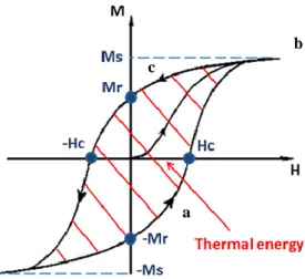
7 values of H, the magnetization (M) approach to the saturation value (Ms) and will not increase no matter how strong H could be,41,44 because in that condition- the saturation, all domains are aligned.45 But not all the magnetic domains return to their original orientation when the applied magnetic field is reduced after reaching the Ms. Thus, when H returns to zero, there is a residual magnetization named remanent magnetization (Mr) which can be removed by applying a reverse magnetic field, called coercive field, Hc.43,45 So, open hysteresis represents an irreversible process of magnetization which is related with both intrinsic (e.g., particle size, shape, and magnetocrystalline anisotropy) and structural characteristics (e.g., grain boundaries, impurities, and vacancies) of the magnetic material.43,44 Typically, big particles (at micrometer range) lead to large hysteresis loops, while small particles lead to narrow hysteresis loops.41 A single magnetization/demagnetization cycle (a→b→c) produces a thermal energy proportional to the shaded area within the hysteresis loop46 (Figure 1.2).
Figure 1.2 Typical hysteresis loop observed during magnetization/demagnetization of magnetic nanoparticles. M and H are magnetic flux density and magnetic field strength, respectively. A single magnetization/demagnetization cycle (a→b→c) produces a thermal energy proportional to the shaded area within the hysteresis loop.46
1.2.3.2 Magnetism at nanoscale
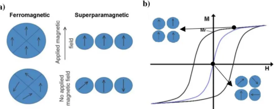
In bulk materials, the magnetic moments are divided in multiple domains (Figure 1.3a). As the particle size decreases, the domains sizes also reduce. Below a certain particle size value (denoted as critical size), the magnetic moments become organized in mono-domains
a c
8 (Figure 1.3a). These mono-domains haves a more favourable energetic configuration,6 displaying different magnetic behaviours according to temperature. Below the blocking temperature, the superparamagnetic (SPM) NPs behave as a ferromagnetic, showing a typical hysteresis loop with net magnetization (black line in Figure 1.3b). For temperatures above the blocking temperature, the remanent magnetization becomes equal to zero as well as the coercive field (blue line in Figure 2.3b).10,13,46 The characteristic M-H curve of SPM NPs at room temperature is an overlap curve of curves with sigmoidal curve. When a magnetic field is applied to the SPM NPs, the magnetic moments tend to be oriented in the direction of the field, which from a certain value, leads all magnetic moments to be aligned parallel to their direction, reaching Ms. As the field is reduced, there is no Mr, and therefore no hysteresis loop47,48 as show in Figure 1.3b.
Figure 1.3 a) Multidomains and monodomains representation under application and absence of a magnetic field. (b) Magnetization curves as function of the magnetic field for superparamagnetic particles under temperatures below de blocking temperature – ferromagnetic behaviour (black line) showing hysteresis loop; and above the blocking temperature - superparamagnetic behaviour (blue line) with no remanent magnetization (Mr) and no coercive field (HC).
The conversion of AMF energy to heat through SPM NPs are explained essentially by two theories: Néel relaxation and Brownian relaxation9,31 (Figure 1.4). The first one causes
energy to be released when the magnetic dipole of a particle flips between two stable orientations within a magnetic field that are separated by an energy barrier.31,46,49 This
physical hindrance is directly proportional to size of the particles.49 The Brownian losses is due to random collision with other particles and the medium (like extracellular or complex components inside the cells that tends to counter the physical rotation of the suspended MNPs) and physical rotation of particles within an AMF.5,9,10,31,46 In a SPM NPs the Brownian fluctuations are sufficiently intense to randomly orient the individual magnetic
b) a)
9 moments of each particle, leading to a zero global magnetization.50 The Brownian loss is not directly related to the magnetism of the SPM MNPs, but can increasingly modulate it as the particle diameter increases within the SPM range.5
Figure 1.4 Néel rotation vs. Brownian rotation. (A) Néel rotation: the magnetic moment (arrow) rotates while the particle remains fixed (inner circle). (B) Brownian rotation: the magnetic moment (arrow) remains fixed with respect to the crystalline axes (lines inside the circle) while the particle rotates.51
1.2.4. Hyperthermia
The concept that heat can be used for therapeutic purposes has been described in the medical literature for centuries.31 The increase of the temperature above normal physiological temperature (ca. 37 ºC in humans) to a mild temperature, which does not cause cell death by itself, may have therapeutic effects and be defined as hyperthermia.28,31,52 Hyperthermia (HT) usually applies temperatures in the range of 41 to 50 ºC to induce apoptosis. Above 50 ºC is considered thermoablation.52 Thermoablation leads to the necrosis of cancer cells, but may also affect healthy cells and therefore it is not desirable.
The success of this approach is based on the differences between healthy cells and cancer cells,29 namely in the difference how the healthy and cancer cells behave when the temperature is increased.52 Cells in the mitosis are sensitive to heat probably because of the injury to the mitotic apparatus.28,52 As cancer cells have a faster cell division to quickly multiply,10,53 making them more sensitive to the increase of temperature. The physiological differences between normal and tumour tissue in the architecture of the vasculature (quite disorganized compared to the normal tissue)28,52,54 and with a low vessel density result in high susceptibility to the heat. In normal body temperature, the cancer cells take advantage comparatively to normal cell as the blood flow in them is faster. When the temperature
10 increase, the opposite happens: the blood flow in normal tissue increases whereas in the cancer tissue decreased. This happens because the cancer tissue cannot dissipate effectively the heat, remaining heated for longer time than the normal tissue. This, make the cancer cells to undergo into apoptosis phenomena, whereas normal tissue manages the high temperatures easily due to the increased blood flow.28 All these aspects make cancer cells more susceptible to heat.28,31 However, the specific mechanism of cell killing by hyperthermia is still unclear.28
The duration, homogeneity of the temperature in tissue, tissue type and context of treatment influence the effects of HT in tumour cells.31,55 Depending on the thermal stimulation, there is a growing amount of experimental evidence showing that 43°C is the breaking point to develop cancer cell death.32,53
A variety of techniques have been employed to achieve HT in the modern clinic practice including electromagnetic radiation, laser, microwave, radiofrequency and high-intensity focused ultrasound.31,43,56
Depending on the extent of the body’s exposure to heat, there are three types of HT: whole body, regional and local5,31,42 HT. Whole body HT is achieved using water blankets and
thermal chambers, whereas regional HT includes the perfusion of a tumour-bearing limb with part of the patient’s blood which is taken out and warmed ex vivo.5,31 Note, that these
methods are not specifics and are technically challenging to perform repeatedly with reproducible heating levels. The last type of HT, the local HT, is more specific,31 and refers to heating only one body area, usually where the tumour is located.55,56
1.2.4.1 Advantages and disadvantages of hyperthermia
HT is independent of the cell cycle and this is a great advantage comparing to chemotherapies, which is cytotoxic to the cells, especially in specific phases of the cell cycle. Furthermore, HT can be used to treat cancer cells localized in many places like brain, thyroid, lungs, breast, prostate, hepatocellular carcinoma among others.53,56 Also, HT activates
immunological responses to tumours for eradication by the host immune response.31
Many clinical trials have documented that mild temperature HT can be safely administered to patients showing clinical benefits in terms of improved local control, palliation of symptoms and even overall survival. However, it is underutilized for a variety of reasons as the invasive methods of generating HT, the difficulty in maintaining temperatures at desires
11 levels, the lack of monitoring and modelling of HT. For example, in local HT, more specifically, luminal and interstitial HT, employ customized probes and applicators placed close to the tumour individually; it can apply a grid to achieve relatively uniform heating of the tumour; or metal antennas that are activated by an external energy source. However the placement of external heating sources usually invasive, challenging to the deep-seated tumour, and sometimes results in a nonuniform heat distribution.31 The external heating techniques such ultrasound and electromagnetic phase arrays are not invasive like the luminal and interstitial implants but the reproducibility and uniformity heating levels are challenging.3
HT treatment is seen a great promise for cancer therapy, however the methods of reaching, maintaining, monitoring and modelling heating are still requiring a huge improvement.3
1.2.5. Magnetic Hyperthermia
Magnetic Hyperthermia (MHT) consists in applying an AMF in a magnetic fluid to produce heating through transformation of magnetic energy in thermal energy, raising the temperature in near the cancer cells.12,15,57–60 Magnetic fluids are class of magnetic materials constituted of colloidal suspension of MNPs.42,61 The first application of ferrofluids for hyperthermia treatment was investigated in the work of Chan et al.59 in 1993.
The MNPs can be used in a specific areas of a human body by a simple direct injection to target tissue14,42 or by tumour specific antibody targeting42, followed by exposing to an external magnetic field.12,36,57,62
As mentioned before, in the location of the malignant tumour, the proliferating cells require extra supplying oxygen and nutrients. Thus, small molecules (e.g., nanoparticles) can circumvent the aberrant endothelial barrier and leave the circulation to concentrate at the site of neoplasia. When a high concentration is reached locally, an alternating magnetic field can be used to target the tumour to raise the temperature of the medium.5,9,63
The capacity of a material to generate heat under the influence of an AMF is characterized by its specific absorption rate (SAR)31,52 or specific loss power (SLP), expressed in Wg-1 (1.1):
12 SAR = SLP = ∆T
∆t C
m𝐹𝑒
where c is the heat capacity of the fluid per unit mass, mFe is the concentration of the colloidal
solution studied (g l-1 of iron) and ΔT/Δt is the measured temperature increase with time.52 High values for SAR offers the advantages of enable the reduction of MNPs dose, and decrease the values of magnetic field strength and frequency that are required for MHT therapy.
The SAR measurement is widely used to characterize the heat generation capability of the system,10 and it is a crucial parameter to modulate the tissue temperature that will be achieved during hyperthermia treatments. Overheating the tumour may result in serious damage to the surrounding healthy cells or in uncontrolled necrosis. On the contrary, the desired therapeutic effect cannot be achieved if the temperature rise is not high enough.5
Although this measure cannot be directly compared between different experimental setup’s once the SAR value depends on the magnetization - strength and frequency of the used magnetic field5,52 and on the chemical, physical and magnetic properties of the material.
Moreover, it also dependent on the dispersion media and agglomeration degree.5 The efficiency of power dissipated from SPM particles is enhanced when the particles’ size distribution is narrow.49
Various factors influence the MHT, the extent of the heating produced and the time during which is maintained the heat as: magnitude of the AMF (the frequency and amplitude); 31,33,42
the size; size distribution and other characteristic of the MNPs,5,31,40,42,46 the concentration
of them in the target place as well the depth of the tumour within the body31 and the viscosity
of surrounding medium.33,42
1.2.5.1 Advantages of magnetic hyperthermia treatment
Magnetic hyperthermia (MHT) have a great potential as a stand-alone treatment or adjacent therapy for cancer.39,52 The first successful clinical trial of interstitial hyperthermia in the treatment of human cancer using magnetic nanoparticles was attempted by Johannsen et.al54
in 2005. Recently, favourable results of the ‘nanothermotherapy’ study in Phase II clinical trials run by MagForce Nanotechnology (Hospital Charité, Berlin) on patients suffering from
13 glioblastoma multiform, show an improvement on patient survival from 6.2 to 13.4 months.52
The use of SPM NPs prevents the agglomeration of particles in the body, avoiding obstruction of blood vessels.47 Furthermore, these particles can reside in the body for long time, allowing several heat treatment of the tumour without re-introduction of the MNPs.15,30 In addition, as SPM NPs tend to be less than 20 nm and resonate with a magnetic fields ranging from 10 KHz-10 MHz, they can easily penetrate soft tissues and bones.31 Besides the easiness in penetrating the cells, small particles (< 20 nm) are also more suited for in vivo applications.12
MHT is advantageous in treating tumours in organs like lungs, which are easy to locate and avoid thermal bystander effect on nearby regions like heart.15 Moreover, MHT reaches deeper tissue than any other activation technology (light or acoustic waves).34 Direct injection to the target tissue allow to put in the target place sufficiently quantities of MNPs to generate the heat needed.31 MHT cal also be used in conjunction with other treatment
modalities of the cancer, such as traditional radiotherapies, like chemotherapy or even surgery, for the purpose of improving the effectiveness of antineoplastic drugs.36
The magnetic fluids have a specifically high absorption rate per mass but the normal tissues have a lower absorption rate, so it is possible to selectively excite areas containing fluids without interference with different neighbouring tissues. Such magnetic-field-induced excitation produces heat absorbed mostly by magnetic fluids and thus increasing the temperature only in regions with accumulated magnetic nanoparticles.14
MHT has fewer side effects than chemotherapy or radiotherapy14,64 or anticancer drugs that have to be administered systemically, and with the lack of specificity towards a pathological site, requires a high dose to reach the local in the needed concentration. Moreover, the cancer cells develop resistance to the therapeutic effects of drugs.15,42 Conventional thermotherapies as photothermal, radiofrequency, infrared or ultrasound exhibit insufficient temperature elevation and non-homogeneity within the tumour15,42,49 and provides a minimally invasive way to deliver a therapeutic dose of heat specifically to cancerous regions.49
1.2.5.2 Disadvantages of magnetic hyperthermia treatment
The inability to precisely control the thermal dose and the dependence of heating efficiency on thermal dose rate is one of the limitations. It is know that large doses of heating can be
14 achieved by applying large fields amplitudes in a short time frame and result in a more sustained rise in temperature before heat is dissipated,31 which can provoke discomfort.15 Non-uniform distribution of nanoparticles throughout the tumour generates cold spots in the regions where no or less MNPs are accumulated, and tumour cells remain untreated in those regions and regrowth occurs subsequently.15,31 Yet, these cold spots eventually get heated up by active thermal bystander effect. This effect can also heat up normal surrounding cells which need to be avoided; so 3D temperature measurement within the tumour region can help to avoid that situations.15
When MNPs are injected intravenously, their biodistribution and achievable temperature is highly dependent on the formulation used, amount of MNPs injected, the target organ and the time point of addition.15,31 So, the desired heat in the local is complicate to achieved by intravenous administration.31 In addition, the interaction of MNPs with cells is complex and depending on the physicochemical conditions or the time of exposure, it may also result in the triggering of cytotoxic processes or the internalization of MNPs.35 Thus, the introduction
of the MNPs relied on intratumourally injection, but it is invasive and is not synonymous with uniformity in the introduction of MNPs in the desired place.31 The optimization process
is not yet well understood, experimental results from MNPs systems vary expressively.52 The hydrophobic surfaces and the large surface area relative to volume48,53,58 and therefore possesses high surface energie,58 MNPs in vivo tend to agglomerate and to be released rapidly by the circulation.17,58,59 Moreover, they tend to have high chemical activity, and are easily oxidized in air (especially magnetite), generally resulting in loss of magnetism and dispersibility.58 To avoid these difficulties, the surface of the NPs must be modified by a biocompatible polymer.60,65
1.2.6. Magnetic nanoparticles in the treatment of cancer
In Table 1.1 are presented some of the nanoparticles used against cancer cells with different types of magnetism.
15 Table 1.1 -Some examples of different types of NPs used against cancer cells.39
Ferrimagnetic Paramagnetic Diamagnetic Antiferromagnetic
iron oxide palladium zinc oxide cobalt oxide
nickel zinc ferrite cerium oxide biosynthesized silver -
magnesium ferrite titanium dioxide gold -
- platinium copper -
- neodymium oxide selenium -
For biomedical applications, MNPs should fulfil a variety of requirements as: (i) SPM behaviour at room temperature, in order to avoid particle aggregation12,40,45,57 (and escape to induce dangerous thrombosis of blood vessels);34 (ii) large Ms, so as to show a large response
under the application of a magnetic field40,45,66 and low coercivity (Hc)49 (iii) limited particle
size (<20 nm)40,66 to escape the phagocytosis of reticuloendothelial system and penetrate into
capillary vessels in body tissues, which ensures an effective distribution in specific tissues;17
(iv) high size unirformity;45,49 (v) biocompatibility40,57,65- nanoparticles are usually coated with either biological or biocompatible molecules;55 (vii) the carrier particles must have a long retention time while circulating in the blood40,53 - preventing agglomeration and minimizing adsorption of plasma proteins that enhances phagocytosis (opsonins) on the MNPs by covering them with a thin layer of biodegradable polymer.40
1.2.6.1 Why Fe3O4?
Fe3O4 or γ-Fe2O3 (magnetite and maghemite respectively) are the MNPs most used in the
research groups and it becomes evident why are attractive candidates for use in human body. Fe3O4 is a ferrimagnetic compound with two different cation sites in a unit cell: 8 tetrahedral
A sites, and 16 octahedral B sites. Magnetite is an inverse spinel, which means, (Fe+3)[Fe+2Fe+3]O4, where the specie in curved parentheses stand for the tetrahedral sites and
the square parentheses stand for the octahedral sites.67
The iron oxide nanoparticles have good biocompatibility, non-toxicity,9,28,31,52 potential to exhibit superparamagnetic properties if the particle sized is below 20 nm,9,28,31 the specific surface is high, biodegradability,52 high chemical stability28,31,68 and are excellent conductor
16 of heating, transmitting it to the adjacent tissues.31 In addition, the heating efficiency in aqueous colloids of these materials is well established.69
Iron is essential in the human body! Besides, it exists in several body entities as in the haemoglobin, which is essential for human survival, it have great stability and unique magnetic properties. Furthermore, iron oxides are not only biocompatible as they can show therapeutic effect, where others nanoparticles as FeSi or FePt fail. Besides, US FDA approve the use of the ultrasmall and/or SPM NPs of iron oxide for MRI (Magnetic resonance imaging)9,17. However, it is important to refer the fact that several agents have been removed from the market in the late 1990s and 2000s because the possible side effects. The toxicity is not always immediately detectable.9
The surface of the iron oxide MNPs has iron atoms on the surface that can coordinate with molecules or get protonated/deprotonated. This capability allows to modifying the surface pre-and post-synthesis with many functional groups whether inorganic and organic as monomeric ligands (dopamine, phosphoric acid), multimeric ligand and zwitterionic ligand31,70; and therefore improve their stability and increase their circulatory halt-time as
preventing aggregation, improving their hydrophilic properties and lower the possible cytotoxicity4,31,70. The possibility of an easy functionalization10,33,53 allow obtain a multimode MNP with potential application in several areas.53
1.2.6.2 Toxicity
The long terms effects of the MNPs still one the major concerns in their use because of the lack of knowledge of the effect of accumulation of particles in body.9,71 For example, in blood transfusion, patients suffering for sickle cell anaemia require the constant administration of iron chelating agents in order to control the excess of Fe. Generally, these patients, have reduced life expectancy due to their condition but also due to the iron overload.9 Landeghem reported in their study that MNPs of iron coated with aminosilanes
applied in gliobastoma on phase II were localized in macrophages in areas of tumour necrosis rather than the cancer cells themselves.72
Spherical NPs with zwitterionic charged surfaces and hydrodynamic diameter of about 5 nm are released by the kidneys and not entrapped within the reticuloendothelial system (RES) while larger particles (20 nm) are captured by reticuloendothelial macrophages.31
Another question, is the time that the iron MNPs can persevere in the body, because they can be uptake by phagocytes cells but also by nonphagocytic cells. For example, the
17 endothelial cells surrounding the blood vessels, are a sub-group of the latter, and they can engulf the MNPs of iron. Due to oxidative stress, in the case of the γ - Fe2O3 uptake, the cells
do not survive frequently more than 24 hours post micropinocytosis, and MNPs are cleared through urine rapidly. However, some organs as the lung, liver or kidney have been identified as the main target organs of bioaccumulation of the MNPs in some studies68,69,71 as well as the macrophages in systemic circulation.68
The possibility of these particles to escape from physiological barriers and contact vital organs as the brains is also of high concern.9
Nowadays some studies report toxicity to healthy skin and lung tissues in vitro tests.9 However others studies report iron oxide as innocuous to normal rat hepatocytes and human fetal lung cells (IMR-90).39 These inconsistent results may be due to factors such as dose and exposure duration;39,68 and MNPs properties and characteristics such as shape, size, charge, biodistribution, functionalizations39,57,66,69,71 or life cycle69,71 which influence the
results. Further, the behaviour of the MNPs in biological media - the interactions with the cells and tissues can alter the physicochemical properties and contribute in the discrepancy of results of in vitro and in vivo.57,69 With a large variety of MNPs and possible behaviours,
it is difficult to draw general conclusions about their possible toxicity.71
As the possibility of toxic effects are not clear, coating is one of the protective measures to be implemented to minimize iatrogenic complications and damage of healthy tissue.9 An appropriate coating diminishes systemic toxicity while improving specificity to cancerous cells. However, complete safety is never guaranteed.9 Also, the MNPs should be tested for microbial contaminations (bacteria, yeast, and mold) eliminating other parameter, which could lead to ambiguous cytotoxic results. The majority of MNPs of iron can be considered sterile due to the synthesis procedures; however, contamination can be introduced in various downstream processes such as surface functionalization.57
The consensus at this moment is in favour of their use in vitro and in vivo experiments involving animals.
1.2.7. Synthesis and coating of MNPs
Various methods of synthesis of magnetic nanoparticles are described in literature.18 The approaches may be chemical,5,42,73 biological42,73 or physical.5,42 The chemical methods are
18 the simplest and the most efficient, having a sensible control of composition, shape and size74 whereas the biological route do not produce homogeneous particles.74 The physical procedures can be used to produce high purity nanomaterials, but the geometry of the synthesized particles is difficult to control.45,74 Both methods are able to prepare magnetite with controllable particle diameters, however, well-dispersed aqueous Fe3O4 nanoparticles
are difficult to achieve.18 In table 1.2, it is presented some possible methods to synthetize iron MNPs.
The synthesis method has an important role in the magnetic properties62 as it the size,33,35,45,62,75 size distribution,35,62 shape,45,75 particle surface characteristics,29,53,62,75 crystalline lattice, oxidation state of iron ions,53, etc..42,45,59,62,74. Various shapes of iron oxides particles such as spheres, cubes, worms, stars, rods, octahedrons, and prism59 are described in literature.
19 Table 1.2 -Summary of some synthetic methods of magnetic nanoparticles.
Methods Ref. Chemical co-precipitation 5,18,35,53,73 thermal decomposition 5,18,29,35,53 microemulsion 18,29,35,53 hydrothermal synthesis 5,53,76
microwave assisted synthesis 73
solvothermal method 45,73 self-assembly 60 combustion synthesis 73 sol–gel 73 polyol method 5 Physical
Gas phase deposition, 5,50
ball milling 5,50
laser-induced pyrolysis 5,45,50
electrodeposition 45
pulsed laser ablation 45,50
aerosol 45,50
Biological
magnetotactic bacteria 73
mediated by proteins 50,53
20
1.2.7.1 Chemical Precipitation
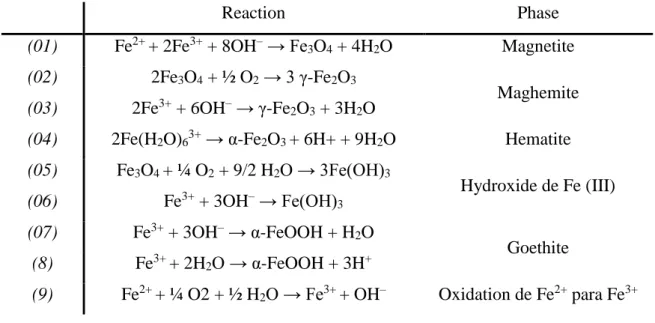
Co-precipitation of ferrous and ferric salts in aqueous solution represents a straightforward, facile and efficient synthesis strategy to synthetize iron oxide NPs on a large scale.70 A wide variety of experimental parameters, such as: i) pH; ii) ionic strength; iii) concentrations ratios of Fe2+/Fe3+; type of precipitation agent; v) strength of the basic agent;5,45,70 vi) reaction temperature;5,18,45,53,70 and vii) stirring rate5,18,45,47,53 were described in the literature that influence size, shape, magnetic and surface properties of the MNP synthetized. For example, if the concentration of the salts is enlarged, there is an increase in the number of particles, with a subsequent loss of uniformity in size.5,45,53 However, the control of the particle size distribution, crystallinity and oxidation is limited once the reactions are thermodynamically driven.13,47,70 This could be overcome by others alternatives as thermal decomposition.
However, the MNPs synthetized by this method generally are non-aqueous, being soluble only in organic solvents. Post-preparative ligand-exchange procedures are require to turn the particle water-soluble and biocompatible.70
The precursors used are ferric (Fe3+) and ferrous (Fe2+) chlorides, sulphates, perchlorates or nitrates, etc..5,45,76 They are, first dissolved in an acidic aqueous solution to prevent the
individual precipitation of hydroxides whose solubility products are very high. Then they are “co-precipitated” (meaning the two valences of iron ions together) under the addition of a strong base (commonly NaOH), according to the reaction 1.2.5,13,45,53 It is not needed high temperatures18,76 and the resulting particles surface possesses hydrophilic properties. Also, it can produce fine, high-purity, stoichiometric particles of single and multicomponent metal oxides.35
2 Fe3+ + Fe2+ + 8 OH- → Fe3O4 + 4 H2O
Controlling the reaction atmosphere is also of crucial importance in this method, since -magnetite (Fe3O4) easily oxides into maghemite (-Fe2O3), a ferrimagnetic iron oxide phase
or hematite (-Fe2O3), a antiferromagnetic iron oxide. Therefore, inert conditions are
required to obtain magnetite, or a controlled oxygen atmosphere.5,45,53 For example, bubbling the solution with an inert gas does not only protect the critical oxidation of magnetite, but also reduces particle size when compared with methods that do not remove oxygen.
( 1.2)
21
1.2.7.2 Geometry and Size
The geometry and size of the MNPs are important to define their lasting time in the bloodstream. Rapid removal of them from circulation systems by opsonins or white cells can substantially reduce their biomedical functionality. The dimensions of the MNPs directly influence their trajectory in the body. MNPs larger than 100 nm are usually opsonized63 and
if lower than 5-6 mm diameter are removed by renal filtration.77 In general, the larger the
particle, the lower their spent time in blood circulation. Thus, it is commonly accepted that MNPs with a diameter of between 1 and 100 nm are the most suitable for in vivo application.63
Spherical MNPs have been the most studied form. They are isotropic whereas the elongated MNPs, exhibit magnetic shape anisotropy. The magnetic shape anisotropy is a property that reflects the existence of a preferential direction magnetization dependent of the particle shape, being an advantage.63 The magnetic moments of spherical nanoparticles are arranged in a random way, and tend to cancel, diminishing the strength of the magnetic signal. In anisotropic MNPs, the magnetic moments tend to orient themselves according to their long axis and they will always be pointed to two possible ways, in the same direction. Thus, the strength of the magnetic signal is enhanced, i.e., elongated particles experience a greater strength when compared to spherical particles of the same volume.63
1.2.7.3 Surface modification of iron oxide MNPs
As MNPs usually tend to aggregate,15,33,35,45 the coating of synthesized MNPs is advised.33,45 Furthermore, the naked MNPs can stimulate ROS generation and non-specific interactions with opsonins5,10,33,53 (albumin, immunoglobulin (IgG), apolipoproteins, etc.), which adsorb onto the MNP surface, thereby completely obstructing their functionality and allowing their uptake by the phagocytes of the immune system (IS)5,33,53 which results in increased cytotoxicity.45
As the microenvironment where the cancer cells are developed are hostiles (acid medium, low oxygen) the surface modifications must take that in account.45,53 For example, if the intend is reduce the adsorption of MNPs by the opsonins, they may be coating with amphiphilic molecules and then increase the time in blood circulation.33
22 The interaction between the coating agent and the MNPs is critical35,45 and the special attention must be taken to insure the stability of the MNPs. For example, physically adsorbed coatings (by electrostatic interactions or hydrogen binding) show limited stability in comparison to chemically adsorbed coatings agents. The steadiness of the coating also depends on the quantity of the chemical interaction that each molecule or macromolecule can establish with the MNPs surface.13
The goal stands by offering a non-toxic35,48 and biocompatible coating5,35,45,47,62 so that the crystalline character of the oxide is retained33,53,62 as well increase the chemical stability of the Fe3O4 NPs5,10,17,18,35,44,45,48,53 in vivo. Further, the route employed should be simplistic
and effortlessly.62
A variety of materials have been reported to coating the Fe3O4 MNPs. Some are presented
in table 2.3.
In this work, it was tried to synthesize magnetic NPs of iron oxide, coated with biocompatible and non-toxic chitosan biopolymer.
23 Table 1.3 - Some materials reported as coating/crosslinkers and linkers to the iron oxide (Fe3O4).
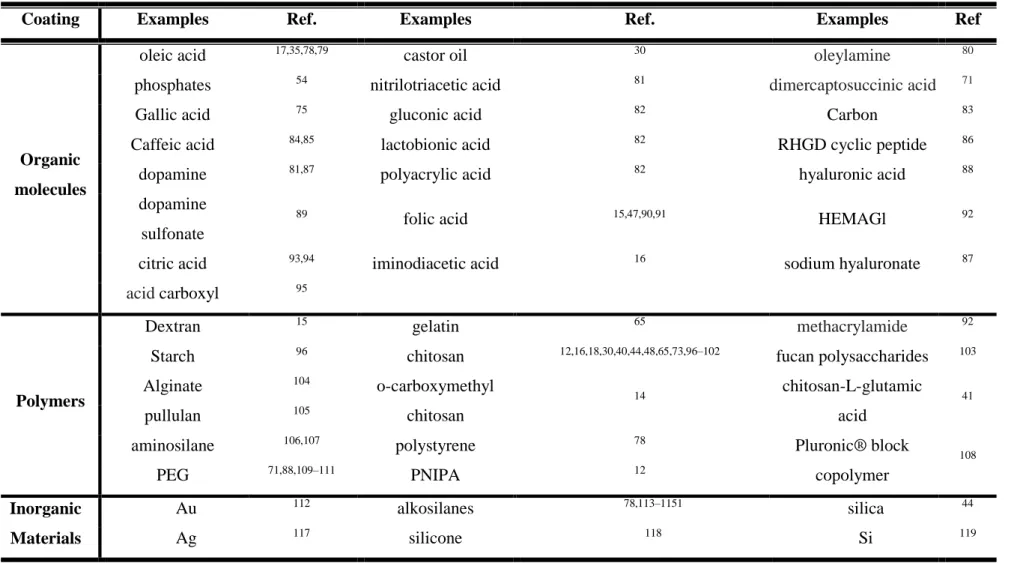
Coating Examples Ref. Examples Ref. Examples Ref
Organic molecules
oleic acid 17,35,78,79 castor oil 30 oleylamine 80
phosphates 54 nitrilotriacetic acid 81 dimercaptosuccinic acid 71
Gallic acid 75 gluconic acid 82 Carbon 83
Caffeic acid 84,85 lactobionic acid 82 RHGD cyclic peptide 86
dopamine 81,87 polyacrylic acid 82 hyaluronic acid 88
dopamine sulfonate
89
folic acid 15,47,90,91 HEMAGl 92
citric acid 93,94 iminodiacetic acid 16 sodium hyaluronate 87
acid carboxyl 95
Polymers
Dextran 15 gelatin 65 methacrylamide 92
Starch 96 chitosan 12,16,18,30,40,44,48,65,73,96–102 fucan polysaccharides 103
Alginate 104 o-carboxymethyl chitosan 14 chitosan-L-glutamic acid 41 pullulan 105
aminosilane 106,107 polystyrene 78 Pluronic® block
copolymer 108 PEG 71,88,109–111 PNIPA 12 Inorganic Materials Au 112 alkosilanes 78,113–1151 silica 44 Ag 117 silicone 118 Si 119
24
1.2.7.4 Chitosan
Chitosan is a biopolymer with a unique chemical structure, high density, free amino and hydroxyl groups120 and easy application.62 Amongst the biocompatible natural materials
employed as coatings, chitosan has been used due to its known properties as: biocompatibility, biodegradability,17,40,44,120–122 bioactivity,18,29,122 hydrophilicity (provides a
barrier to adsorption of protein, reducing the macrophage),30,44,123 bioadhesion,17,73 low
cytotoxicity,15,16,18,30,120 antioxidant, anti-inflammatory120 and antimicrobial properties.16,120 Chitosan have been used for various biomedical applications such as tissue engineering, drug delivery,15,65 cell encapsulation, wound healing along with orthopaedic, periodontal, pharmaceutical and gene therapy65 among others.
Chitosan or poly[β-(1-4)-2-amino-2-deoxy-D-glucopyranose], is obtained by partial alkaline deacetylation of chitin15,29,120 (about >50%), a main component of the exoskeleton of crustacean, mollusc shells, fungal cell walls and insect cuticles.18,65,120 Their structure is very similar to that of cellulose120,124 and is characterized by their molecular weight and degree of acetylation,120 which are determinant on various properties including solubility, biodegradability, toxicity, antimicrobial activity, etc.124 This biopolymer has a positive charge, being soluble in various acids.100
In addition, the free amino groups and hydroxyl groups can be easily used for further functionalization17,33,40,44,73,121 and improve the colloidal stability in vivo in blood circulation.17,122 But chitosan cannot dissolve in a physiological environment (pH = 7.4), only in acidic solution (pH < 6),15,120 where the units of glucosamine (-NH2) can be converted
into their protonated and soluble form (NH3+).120,124 This is the main disadvantage of coating
the MNPs with chitosan because can interact with anionic components (glycosaminoglycans and proteins).125 But, Nunes et al., demonstrated that chitosan can link to genipin and caffeic
acid molecules, becoming less soluble in acid medium between chitosan chains.126 Based on
this result, there is a way to make chitosan more stable in acidic medium. So, the main disadvantage of chitosan can be overcome.
25
1.3 Motivation
As everything it is used inside of human body, biocompatibility is required. More, in the case of the MNPs, to achieve better results in the heat generated, disaggregation is need. So, coating is a possible method to achieve these two requirements, for the use of the MNPs in hyperthermia therapy. Promote a good link between the iron oxide and the coat is fundamental, so adversely effects are avoiding as well to achieve better heat production during the hyperthermia treatment.
In the beginning of this work dopamine never was used to promote this type of bonding (between iron oxide and chitosan) in this type of therapeutic (magnetic hyperthermia). Tumour cells are more acid and chitosan have poor resistance to acid medium. To overcome this issue, crosslink with a known molecule is proposed: genipin. Due to structural similarity with dopamine, caffeic acid was proposed to see if could promote a good link between the iron oxide as well to see of the link to the coat is stronger than using dopamine and genipin.
1.4 Objectives
The general objective of this work was to prepare suitable magnetic nanoparticles to be used in magnetic hyperthermia.
To reach the main purpose of this thesis, it was necessary accomplish minor objectives: To produce magnetic nanoparticles (MNPs) of iron oxide, preferably magnetite
superparamagnetic NPs;
To functionalize the produced nanoparticles through two different ways: one using dopamine (DOPA) and the other using caffeic acid (CA);
To coat the nanoparticles with chitosan;
To characterize structural and morphologically the NPs through different techniques as X-ray diffraction (DRX), Fourier transmission infrared spectroscopy (FTIR), transmission electronic microscopy (TEM), Zeta Potential and hydrodynamic diameter (DLS) and to evaluate the magnetic properties after the coat of chitosan by Superconducting Quantum Interference Device (SQUID);
29
Experimental Procedure
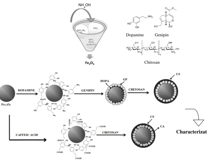
This chapter describes, briefly, the materials and procedures used, as well as the characterization techniques. The reagents used were commercially available and used without pre-treatment. The main characteristics of all reagents used are in Annex A. To avoid the production of impurities was used Type 1 water from Millipore Simplicity UV Water Purification System.Figure 2.1 represent the main steps of this experimental work.
Figure 2.1 Schematic representation of the main steps during laboratorial work. In top, the synthesis of Fe3O4 and down, the paths for the functionalization and coating.
Fe3O4 80ºC, 30min (under N2) FeCl2∙4H2 O FeCl3 NH4OH Characterization Dopamine Genipin Chitosan
30 It is divided in four parts: i) synthesis of iron oxide MNPs; ii) functionalization and coating of the MNPs synthesized; iii) morphological, structural and magnetic characterization and iv) magnetic hyperthermia measurement (based on the release heat by the NPs).
2.1 Synthesis of iron oxide MNPs
The synthesis of the iron oxide MNPs was adapted from the chemical co-precipitation method described on reference (78). The experimental apparatus used in the synthesis is
shown in Figure 2.2. The reagent quantities used in the synthesis of MNPsare presented in Table 2.1.
Figure 2.2 Experimental apparatus used for synthesis of NPs of MNPs.
Table 2.1 Reagent quantities used in the synthesis of MNPs.
FeCl2∙4H2O FeCl3 Fe3+/Fe2+
Mass (g) 7,2021 4,98 -
Molar quantity (mol) 0,0444 0,0250 Ratio=1,76
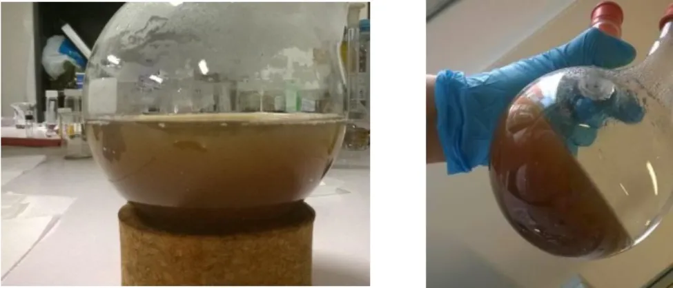
Briefly, the MNPs were produced by co-dissolving FeCl3 and FeCl2∙4H2O in 50 mL of water
under nitrogen gas with vigorous stirring at 80ºC. Then, at this temperature, 25 mL of ammonium hydroxide were rapidly added into the solution. The solution immediately turned black. The solution was kept reacting at 80ºC for 30 minutes and cool down to room
31 temperature. To get particles free from sodium and chloride compounds, the MNPs were magnetically separated and washed with water. This procedure was repeated three times, and then MPNs were dispersed in 72 mL of water. The pH of the suspension of NPs was neutral.
2.2 Procedures for coating of MNPs
2.2.1 Functionalization of the MNPs with dopamine
Dopamine was used as the anchor to present functional molecules on the surface of iron oxide magnetic nanoparticles. Adapting the procedure of (81), dopamine (38.00 mg) was dissolved in 14 mL of water and ethanol (2.5:1). The functionalization was made by mixing 5 mL of iron oxide (dropwise) to dopamine solution under using an ultrasound bath for homogeneizing. After, the pH was adjusted to 4-5 (4.57) with HCl (1 M). The resulting black solution was put in the ultrasound for 20 min. The MNPs were magnetically separated and washed with hexane. This process was repeated three times. The MPNs functionalized with dopamine were dispersed in 22 mL of ethanol and denoted as F.DOPA.
2.2.2 Addition of genipin molecule as “tethering” agent
Genipin molecule was reacted with dopamine to act as bridging ligand to chitosan.
The addition of genipin to the functionalized dopamine’s iron oxide was made in the proportion of 1:2. 30.1 mg of genipin, was dissolved in 20 mL of ethanol followed by 5 min in the ultrasound. 4.8 mL of F.DOPA suspension were diluted in 20 mL of water:ethanol (2:3). The functionalized iron oxide was dropped to the genipin solution under ultrasound and left reacting overnight in the dark. Afterward, the MNPs were centrifuged twice at 6000 rpm during 90 min and then the pellet powders were ressuspended in isopropyl alcohol. Lastly, the MNPs were magnetically separated and suspended in 30 mL of ethanol, designated as F.DOPA.GP.