WENJUAN WU
CARACTERIZAÇÃO DE DUAS NOVAS PROTEÍNAS
REGULADORAS DA PROTEÍNA FOSFATASE TIPO 1:
I2L E NEK2C
CHARACTERIZATION OF TWO NOVEL PROTEIN
PHOSPHATASE 1 REGULATORS: I2L AND NEK2C
WENJUAN WU
CARACTERIZAÇÃO DE DUAS NOVAS PROTEÍNAS
REGULADORAS DA PROTEÍNA FOSFATASE TIPO 1:
I2L E NEK2C
CHARACTERIZATION OF TWO NOVEL PROTEIN
PHOSPHATASE 1 REGULATORS: I2L AND NEK2C
Dissertação apresentada à Universidade de Aveiro para cumprimento dos requisitos necessários à obtenção do grau de Doutor em Biologia, realizada sob a orientação científica do Prof. Doutor Edgar F. da Cruz e Silva, Professor Associado do Departamento de Biologia da Universidade de Aveiro
o júri
presidente Prof. Dr. Joaquim Arnaldo Carvalho Martins
Professor Catedrático da Universidade de Aveiro
Prof. Dr. Andrew M. Fry
Reader, Universidade de Leicester, Leicester, Reino Unido
Prof. Dr. Celso Vladimiro Ferreira de Abreu Cunha
Professor Auxiliar, Universidade Nova de Lisboa
Prof. Dr. Edgar Figueiredo da Cruz e Silva
Professor Associado da Universidade de Aveiro
Prof. Dr. António José de Brito Fonseca Mendes Calado
Professor Auxiliar da Universidade de Aveiro
Dr. Michael Schrader
agradecimentos I want to thank FCT for providing a PhD fellowship (BD/13658/97) and the Centro de Biologia Celular of the University of Aveiro for providing the necessary conditions.
I want to thank my supervisor Prof. Edgar da Cruz e Silva for his instruction during my Ph.D work.
A special thanks is warranted to Dr. Andrew M. Fry, particularly for his help with the Nek2 work. This thesis would not have been possible without the helpful contributions of all the colleagues in the Signal Transduction and Neuroscience Laboratories. My heartfelt thanks go to my colleagues Ana Paula Vintém, Margarida Fardilha and Sandra Vieira for their invaluable help with the writing of my thesis. I also wish to acknowledge Sara Domingues for her help with typesetting and the translation of the abstract.
Finally, I want to thank Prof. Odete A. B. da Cruz e Silva for the great encouragement she provided for me to finish my work.
palavras-chave I2L, Nek2C, ‘splicing’ alternativo, mobilidade dos espermatozóides.
resumo A infertilidade masculina tem vindo a assumir proporções preocupantes na nossa sociedade. Há inúmeros factores que contribuem para esta condição, incluindo problemas associados à motilidade dos espermatozóides. As bases moleculares da motilidade dos espermatozóides ainda não foi completamente desvendada, contudo a incubação de espermatozóides imaturos imóveis com inibidores de proteínas fosfatases induz a sua motilidade. A fosforilação de proteínas é, então, fundamental na regulação da motilidade dos espermatozóides. A fosforilação de proteínas é um dos principais mecanismos reguladores de cascatas de transdução de sinais em organismos eucariotas. Os mecanismos dinâmicos e reversíveis de fosforilação/desfosforilação são catalizados pelas proteínas cinases e pelas proteínas fosfatases, respectivamente. A PP1, uma fosfatase específica para serina/treonina, está envolvida no controlo da motilidade dos espermatozóides, e noutras funções nos testículos. Nas células somáticas, a PP1 está envolvida em diversos mecanismos, que incluem o controlo do ciclo celular, a contracção muscular, a expressão de genes, a actividade neuronal e o metabolismo do glicogénio. A especificidade da função da PP1 depende das proteínas reguladoras que interagem com a sua subunidade catalítica, direccionando-a para um dado substrato ou para uma determinada localização subcelular, e/ou modificando a sua actividade em relação ao substratos. Actualmente conhecem-se mais de cinquenta subunidades reguladoras da PP1, não relacionadas bioquimicamente.
A diversidade da PP1 também se deve à expressão de várias isoformas: existem três genes que codificam a PP1 no genoma humano, denominados PP1α, PP1β e PP1γ. Maior complexidade advem ainda do ‘splicing’ alternativo que é conhecido para a PP1α e PP1γ. O gene da PP1γ origina uma variante ubíqua, a PP1γ1, e uma variante enriquecida em testículo, a PP1γ2, que é também a isoforma da PP1 mais abundante em espermatozóides, e que pode ser alvo de uma terapêutica para a infertilidade masculina ou contracepção. Rastreios de uma biblioteca de cDNA de testículo humano utilizando o sistema dois-híbrido de levedura foram realizados usando como iscos a PP1γ1 ou a PP1γ2. Várias novas proteínas que se ligam à PP1 foram identificadas e as respectivas interacções validadas usando uma diversidade de métodos, quer in vivo quer in vitro (utilizando PP1γ1 e PP1γ2 recombinantes produzidas em sistemas de expressão
bacterianos). Nas condições testadas, ambas as variantes recombinantes da PP1γ apresentaram propriedades enzimáticas semelhantes, o que confirma que a sua especificidade funcional é provavelmente adquirida pela ligação de proteínas reguladoras. Neste trabalho iremos focar em duas proteínas que ligam a PP1 e que foram recentemente identificadas no laboratório, denominadas I2L e Nek2C. A I2L (Inhibitor 2-like) é >90% idêntica ao I2, tanto ao nível nucleotídico como ao nível da sequência de amino ácidos, e constitui uma isoforma nova do I2. Previamente tinha sido identificada como um pseudogene, no entanto apresentamos evidências que apoiam a sua expressão no testículo. A ausência da Thr-73 da I2L é muito significativa e pode ter consequências fisiológicas importantes. Esta ausência resulta numa inibição permanente da actividade da PP1γ2 pela I2L, promovendo potencialmente o desenvolvimento unidireccional da motilidade à medida que os espermtozóides viajam através do epidídimo. A Nek2C é uma nova variante de ‘splicing’ da Nek2A, diferenciando-se apenas pela ausência de 8 aminoácidos N-terminais à sequência consenso de ligação à PP1. Contudo a falta destes 8 aminoácidos tem consequências evidentes, resultando na expressão funcional de um NLS (sinal de localização nuclear). Deste modo, o mecanismo de ‘splice’ alternativo controla a translocação nuclear desta proteína cinase reguladora do ciclo celular, fornecendo um mecanismo pouco usual de modulação da localização da Nek2 e permitindo que a cinase desempenhe funções tanto nucleares como citoplamáticas. A PP1 encontra-se também em ambos os locais, regulando uma enorme variedade de processos celulares. No futuro espera-se que o presente trabalho possa contribuir para uma melhor compreensão dos eventos moleculares envolvidos no controlo da motilidade dos espermatozóides e na fertilização. Estas proteínas que interagem com a PP1 podem constituir alvos para interferir com diferentes funções específicas da PP1. Tanto a I2L como a Nek2C exibem características únicas, podendo constituir candidatos interessantes para futuros desenvolvimento no âmbito do diagnóstico e terapêutica baseadas na transdução de sinais para a contracepção e para o tratamento da infertilidade masculina.
keywords I2L, Nek2C, alternative splicing, sperm motility.
abstract Male infertility is a problem of growing concern in our society. Various factors contribute to this condition including motility defects in sperm. The molecular basis of sperm motility has not been fully elucidated, but non-motile immature sperm acquire motility when incubated with protein phosphatase inhibitors. Thus, protein phosphorylation is a key regulatory mechanism in sperm motility. In eukaryotic cells protein phosphorylation is a major general mechanism regulating signal transduction cascades. Consequently, protein kinases and protein phosphatases are central players in these reversible and dynamic processes. Among them, the serine/threonine-specific protein phosphatase 1 (PP1) appears to play a particularly important role in the control of sperm motility and in the testsis. In somatic cells, PP1 is known to be involved in many diverse processes, including cell cycle control, muscle contraction, gene expression, neuronal activity and glycogen metabolism. Functional specificity is provided by PP1 catalytic subunit interacting proteins, which bind the catalytic subunit and target it to a specific substrate or subcellular location, and/or modify its activity towards those substrates. To date more than 50 unrelated and biochemically diverse regulatory subunits have been described.
PP1 diversity can also arise from the expression of various isoforms: three PP1 genes occur in the human genome, termed PP1α, PP1β and PP1γ. Further complexity derives from alternative splicing events, known to occur at least for PP1α and PP1γ. The PP1γ gene produces a ubiquitously expressed PP1γ1 variant and the alternatively spliced PP1γ2 variant that is highly enriched in testis and is the main PP1 protein found in mammalian sperm. Therefore, it is the latter which has been implicated in the control of sperm motility. Hence, in this work we set out to identify and characterize PP1γ2 interactors expressed in human sperm, which could be targeted for male infertility therapeutics and contraception. Yeast two-hybrid screens of a human testis cDNA library were carried out using either PP1γ1 or PP1γ2 as bait. Several novel PP1 binding proteins were identified and interactions were validated using a variety of methods, both in vivo and in vitro (using purified recombinant PP1γ1 and PP1γ2 expressed in a bacterial expression system). Both recombinant PP1γ variants exhibited similar
Enzymatic profiles under the conditions tested, thus confirming that functional specificity is likely acquired by the binding of regulatory proteins. Here, we will address two recently identified novel PP1 binding proteins, namely I2L and Nek2C.
I2L (Inhibitor 2-Like) is >90% identical to I2, both at the nucleotide and amino acid levels, and constitutes a novel I2 isoform. Previously identified as a pseudogene, we present evidence supporting its testis-specific expression. The lack of Thr-73 from I2L is highly significant and may have important physiological consequences. The missing Thr-73 results in I2L being able to permanently inhibit PP1γ2 activity and potentially promote the unidirectional development of motility as sperm travel through the epidydimis. Nek2C is a novel splice variant of Nek2A, differing solely from the latter in missing 8 amino acids immediately N-terminal to the consensus PP1 binding motif. However, the lack of those 8 amino acids also appears to have marked consequences, resulting in the expression of a functional NLS (nuclear localization signal). Thus, alternative splicing controls nuclear translocation of this cell cycle regulated protein kinase, providing an unusual mechanism for modulating Nek2 localization and enabling it to undertake both nuclear and cytoplasmic functions. PP1 is known to occur in both locations, where it regulates many different cellular processes. In the future, it is hoped that the present work may contribute to a better understanding of the molecular events underlying sperm motility and fertilization. Clearly, such PP1 interacting proteins may provide interesting targets to interfere with specific PP1 functions. Thus, since both I2L and Nek2C exhibit unique characteristics, they may constitute interesting candidates for future developments in signal transduction diagnostics and therapeutics for contraception and the treatment of male infertility.
________________________________________________________________________________________
Centre for Cell Biology 9
University of Aveiro
INDEX
INDEX ... 9 PUBLICATIONS... 13 ABBREVIATIONS ... 15 CHAPTER I: ... 19 INTRODUCTION ... 19 I INTRODUCTION ... 20I.1 PROTEINPHOSPHORYLATIONASADYNAMICPROCESS ... 20
I.1.1 SER/THR-SPECIFIC PROTEIN PHOSPHATASES AND THEIR CLASSIFICATION....21
I.1.2 THE PPP FAMILY ...22
I.1.3 PP1 - PROTEIN PHOSPHATASE 1...23
I.1.4 PHOSPHATASE INHIBITORS ...36
I.1.5 SIGNAL TRANSDUCTION THERAPEUTICS ...38
I.2 PROTEIN PHOSPHATASE INHIBITOR-2 ... 40
I.2.1 I2 AS A PHOSPHOPROTEIN AND MODULATOR OF PP1 ...40
I.2.2 I2 INTERACTION WITH PP1 ...41
I.2.3 PHYSIOLOGICAL FUNCTION OF THE PP1-I2 COMPLEX: FORMATION OF TRIMERIC COMPLEXES WITH OTHER PP1 REGULATORY SUBUNITS ...42
I.3 NIMA-RELATED KINASE 2 (NEK2) ... 45
I.3.1 NEK2 IS THE HUMAN HOMOLOG OF NIMA...46
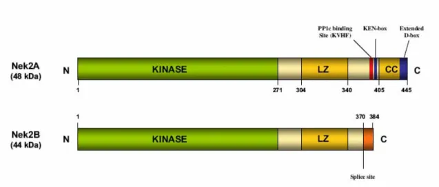
I.3.2 NEK2 STRUCTURE...46
I.3.3 NEK2 IS A CENTROSOME KINASE...47
I.3.4 NEK2 AND CHROMOSOMES...53
I.3.5 ALTERNATIVELY SPLICED VARIANTS OF NEK2...55
I.4 AIMS... 56
CHAPTER II ... 59
EXPRESSION AND PURIFICATION OF PROTEIN PHOSPHATASE 1 GAMMA ISOFORMS... 59
II EXPRESSION AND PURIFICATION OF PROTEIN PHOSPHATASE 1 GAMMA ISOFORMS 60 II.1 INTRODUCTION... 60
II.2 MATERIAL AND METHODS ... 62
II.2.1 PLASMIDS CONSTRUCTION...62
II.2.2 IMMUNOBLOT SCREENING OF RECOMBINANT PROTEINS ...62
II.2.3 MEDIAL AND LARGE SCALE EXPRESSION OF RECOMBINANT PROTEINS ...63
II.2.4 PURIFICATION OF THE TWO ISOFORMS OF HIS-TAGGED PP1 GAMMA UNDER DIFFERENT CONDITIONS...63
II.2.5 THE PROTEIN PHOPHATASE ACTIVITY ASSAY...64
II.2.6 ASSAY OF PROTEIN PHOSPHATASE 1 ACTIVITY INHIBITED BY I2...66
II.3 RESULTS... 67
II.3.1 CONSTRUCTION OF RECOMBINANT PLASMIDS FOR EXPRESSION OF HIS-TAGGED PP1 GAMMA 1 AND PP1 GAMMA 2 ...67
II.3.2 PURIFICATION OF HIS-TAGGED PP1 GAMMA 1 AND PP1 GAMMA 2 USING NI-NTA RESIN 69 II.3.3 LARGE SCALE PURIFICATION OF HIS-PP1 GAMMA 1 AND HIS-PP1 GAMMA 2...72
II.3.4 PHOSPHATASE ACTIVITY OF RECOMBINANT HIS-TAGGED PP1 GAMMA 1 AND PP1 GAMMA 2 ...75
II.4 DISCUSSION ... 78
_______________________________________________________________________________________ 10 Centre for Cell Biology
University of Aveiro
I2-L, A NEW TESTIS-SPECIFIC ISOFORM OF PP1 INHIBITOR-2 ...81
III I2L, A NEW TESTIS-SPECIFIC ISOFORM OF PP1 INHIBITOR-2 ...82
III.1 INTRODUCTION ...82
III.2 MATERIALS AND METHODS...84
III.2.1 LIBRARY SCREENING... 84
III.2.2 I2L CLONING, EXPRESSION AND PURIFICATION... 84
III.2.3 CONSTRUCTION OF AN I2L-GFP FUSION AND EXPRESSION IN MAMMALIAN CELLS 85 III.2.4 YEAST CO-TRANSFORMATION WITH PLASMID DNA ... 85
III.2.5 BLOT OVERLAY ANALYSIS... 86
III.2.6 PHOSPHATASE ACTIVITY ASSAYS... 86
III.2.7 PREPARATION OF HUMAN SPERM HEAT EXTRACT OF I2/I2L ... 86
III.2.8 IMMUNOBLOT ANALYSIS... 87
III.2.9 PHOSPHORYLATION OF I2 AND I2L... 87
III.2.10 DEPHOSPHORYLATION OF HSI2 ... 87
III.2.11 TWO DIMENSIONAL (2D) GEL ELECTROPHORESIS ANALYSIS... 88
III.2.12 IMMUNOCYTOCHEMISTRY OF SPERMATOZOA ... 88
III.2.13 IMMUNOPRECIPITATION OF HUMAN SPERM SAMPLES ... 88
III.3 RESULTS ...90
III.3.1 IDENTIFICATION OF A NEW I2 ISOFORM FROM A YEAST TWO HYBRID SCREEN 90 TABLE III.1. CHARACTERISTIC GENE AND PRIMARY PROTEIN SEQUENCE DIFFERENCES BETWEEN I2 AND I2L ...95
III.3.2 I2L AND I2 INTERACT SIMILARLY WITH THE VARIOUS PP1 ISOFORMs... 96
III.3.3 I2L SUBCELLULAR DISTRIBUTION... 97
III.3.4 COMPARISON OF I2 AND I2L PHOSPHORYLATION BY GSK3 ... 98
III.3.5 PHOSPHORYLATION OF ENDOGENOUS I2/I2L PRESENT IN HUMAN SPERM... 98
III.3.6 INTERACTION OF HUMAN SPERM ENDOGENOUS I2/I2L WITH PP1... 100
III.4 DISCUSSION...102
CHAPTER IV...107
ALTERNATIVE SPLICING CONTROLS NUCLEAR TRANSLOCATION OF THE CELL CYCLE REGULATED NEK2 KINASE ...107
IV ALTERNATIVE SPLICING CONTROLS NUCLEAR TRANSLOCATION OF THE CELL CYCLE REGULATED NEK2 KINASE...108
IV.1 INTRODUCTION ...108
IV.2 MATERIAL AND METHODS ...111
IV.2.1 PLASMID CONSTRUCTION... 111
IV.2.2 CELL CULTURE, TRANSFECTION AND EXTRACTION... 111
IV.2.3 REVERSE TRANSCRIPTASE-POLYMERASE CHAIN REACTION (RT-PCR) ... 111
IV.2.4 IN VITRO TRANSLATION (IVT) ... 112
IV.2.5 IMMUNOPRECIPITATION (IP)-KINASE ASSAYS... 112
IV.2.6 PP1 BINDING, DIMERIZATION AND MICROTUBULE BINDING ASSAYS ... 113
IV.2.7 DEGRADATION ASSAYS ... 113
IV.2.8 IMMUNOFLUORESCENCE MICROSCOPY ... 114
IV.2.9 NUCLEAR-CYTOPLASMIC FRACTIONATION ... 114
IV.2.10 KINASE SUBSTRATE TRACKING AND ELUCIDATION (KESTREL) ANALYSIS.. 115
IV.3 RESULTS ...116
IV.3.1 EXPRESSION OF NEK2C IN HUMAN CELL LINES ... 116
IV.3.2 NEK2C IS AN ACTIVE KINASE THAT BINDS PP1Α AND UNDERGOES DIMERIZATION... 118
IV.3.3 NEK2C IS DEGRADED IN MITOTIC EGG EXTRACTS AND PROMETAPHASE-ARRESTED CELLS ... 119
IV.3.4 NEK2C CAN BIND MICROTUBULES AND LOCALIZE TO CENTROSOMES... 121
________________________________________________________________________________________
Centre for Cell Biology 11
University of Aveiro IV.3.6 NUCLEAR TRANSLOCATION OF NEK2C DEPENDS UPON AN NLS THAT FLANKS THE SPLICE SITE...126
IV.3.7 DETECTION OF A NOVEL NEK2 NUCLEAR SUBSTRATE...128
IV.4 DISCUSSION ... 130
CHAPTER V ... 133
DISCUSSION ... 133
V DISCUSSION... 134
_______________________________________________________________________________________ 12 Centre for Cell Biology
________________________________________________________________________________________ Centre for Cell Biology 13 University of Aveiro
PUBLICATIONS
This thesis contains experimental results included in the publications indicated below already published or under preparation. The author of this thesis declares that she participated in the planning and execution of the experimental work, as well as in data interpretation and in the preparation of the work for publication
Wenjuan Wu, Joanne E. Baxter, Samantha L. Wattam, Daniel G. Hayward, Margarida
Fardilha, Axel Knebel, Eleanor M. Ford, Edgar F. da Cruz e Silva and Andrew M. Fry (2007). Alternative Splicing Controls Nuclear Translocation of the Cell Cycle-regulated Nek2 Kinase. J Biol Chem 282, 26431-26440.
Wenjuan Wu, Ana Paula Vintém, Margarida Fardilha, Odete A. B. da Cruz e Silva and
Edgar F. da Cruz e Silva (2007). I2L, a New Testis-Specific Inhibitor of Protein Phosphatase 1 (in preparation).
Wenjuan Wu, Ana Paula Vintém, Odete A. B. da Cruz e Silva and Edgar F. da Cruz e
Silva (2007). Expression and Purification of Protein Phosphatase 1 gamma Isoforms (in preparation).
Margarida Fardilha, Wenjuan Wu, Rosália Sá, Sara Fidalgo, Cristina Sousa, Catarina Mota, Odete A. B. da Cruz e Silva and Edgar F. da Cruz e Silva (2004). Alternatively spliced protein variants as potential therapeutic targets for male infertility and contraception. Ann N Y Acad Sci 1030, 468-478.
Gareth J. Browne, Margarida Fardilha, Senga K. Oxenham, Wenjuan Wu, Nicholas R. Helps, Odete A. B. da Cruz e Silva, Patricia T. W. Cohen and Edgar F. da Cruz e Silva (2007). SARP, a new alternatively spliced protein phosphatase 1 and DNA interacting protein. Biochem J 402, 187-916.
_______________________________________________________________________________________ 14 Centre for Cell Biology
________________________________________________________________________________________ Centre for Cell Biology 15 University of Aveiro
ABBREVIATIONS
AATYK apoptosis-associated tyrosine kinase AKAP A-kinaseanchoring anchoring protein APC/C anaphase promoting complex/cyclosome Bcl-2 B-cell lymphoma 2
BSA bovine serum albumin
BREK brain-enriched protein kinase CA calyculin A
Cdk cyclin dependent kinase
CFTR cystic transmembrane conductance regulator CIP calf intestinal phosphatase
CKII casein kinase 1 CKII casein kinase 2
C-Nap1 centrosome Nek associated protein 1
CPI-17 C-kinase-dependent phosphatase inhibitor of 17 kDa Cprk cyclin-dependent kinase 5/p35-regulated kinase
DAPI 4,6-diamidino-2-phenylindole
DARPP-32 dopamine and cAMP-regulated protein of 32 kDa D-box destruction box
DMSO dimethylsulfoxide
EDTA ethylenodiaminotetraacetic acid Erk2 extracellular signal-regulated kinase 2 EST expressed sequence tag
FAK focal adhesion kinase.
G subunits glycogen targeting subunits, cGMP-dependent protein kinase substrate Gac1 glycogen accumulation 1
GADDs growth arrest and DNA damage-inducible proteins GIP1 Glc7-interacting protein 1
GIP2 Glc7-interacting protein 2 GL liver-type G subunite Glc7 glycogen-deficient 7
_______________________________________________________________________________________ 16 Centre for Cell Biology
University of Aveiro
GluR glutamate receptor
GM the N-terminal domain of the muscle glycogen-targeting subunit
Grp78 glucose-regulated protein of 78 kDa,member of the HSP-70 family GSK-3 glycogen synthase kinase-3
HCF host cell factor or human factor C1 HCG-V hemochromatosis candidate gene V Hec highly expressed in cancer
His histidine
HMGA2 high mobility group protein A2 Hox11 homeodomain transcription factor
HSI2 human sperm protein phosphatase inhibitor-2 I-1 protein phosphatase inhibitor-1
I-1 PP2A (PHAP-I) inhibitor-1 of PP2A I-2 PP2A (PHAP-I) inhibitor-2 of PP2A I-2 protein phosphatase inhibitor-2 I-3 protein phosphatase inhibitor-3 I-4 protein phosphatase inhibitor-4 IP immunoprecipitation
IVT in vitro transcription
KEN-box protein degradation box, the amino acids sequence is KENIMRSEN KEPI kinase-enhanced protein phosphatase type-1 inhibitor
KESTREL kinase substrate tracking and elucidation KPI-2 kinase/phosphatase/inhibitor-2
LiAc lithium acetate LTD long-term depression LTP long-term potentiation MAD mitotic arrest deficiency
MAPK mitogen-activivated protein kinase MBS myosin binding subunit
MYPT myosin phosphatase targeting subunit MT microtubule
________________________________________________________________________________________ Centre for Cell Biology 17 University of Aveiro
NCLK neuronal cdc2-like protein kinase Nek2 NIMA-related protein kinase NGF neuron growth factor
NIMA never in mitosis A
NKCC1 Na-K-Cl cotransporter 1 Nlp ninein-like protein
NIPP1 nuclear inhibitor of PP1
NMDA N-methyl-D-aspartate receptors OA okadaic acid
ORF open reading frame PBS phosphate-buffer saline PCM pericentriolar matrix PCR polymerase chains reaction PEG polyethylene glycol
PFK Phosphofrutokinase
PHI phosphatase holoenzyme inhibitor PKA protein kinase A
Plk1 pole-like protein kinase 1 PMSF phenyl methylsulfoxide
PRIP-1 phospholipase C-related inactive protein 1 PNUTS phosphatase 1 nuclear targeting subunit PP1 protein phosphatase 1
PP1c catalytic subunit of PP1 PP2A protein phosphatase 2A PP2B protein phosphatase 2B
PSF polypyrimidine tract-binding protein associated splicing factor PTG protein targeting to glycogen
PTP protein tyrosine phosphatase R subunit regulator subunit Rb retinoblatoma protein
RIPP1 ribosomal inhibitor of PP1
_______________________________________________________________________________________ 18 Centre for Cell Biology
University of Aveiro
SARP Several ankyrin repeat protein
Scd5 suppressor of clathrin heavy-chain deficiency 5
Sds22 suppressor of the dis2 mutant; Sla1, synthetically lethal with ABP1 SDS sodium dodecyl sulfate
SDS-PAGE sodium dodecyl sulphate – polyacrylamide gel electrophoresis Ser/Thr-PP serine threonine spicial protein phosphatase
SHP1 Scr homology domain 2 (SH2)-containing protein tyrosine phosphatase 1 (SHP-1) TBS-T tris-buffered saline tween solution
Tris tryptophan
________________________________________________________________________________________ Centre for Cell Biology 19 University of Aveiro
CHAPTER I:
_______________________________________________________________________________________ 20 Centre for Cell Biology
University of Aveiro
I
INTRODUCTION
I.1 PROTEIN PHOSPHORYLATION AS A DYNAMIC PROCESS
The reversible phosphorylation of structural and regulatory proteins is a major intracellular control mechanism in eukaryotes. It is involved in almost all cellular functions, from metabolism to signal transduction, cell division and memory. The phosphorylation state of a protein is a dynamic process controlled by both protein kinases and protein phosphatases.
Protein kinases and protein phosphatases, the key controlling elements, are regulated by a myriad of extracellular and intracellular signals. Unlike the protein kinases that all belong to a single gene family, the protein phosphatases are divided into several distinct and unrelated protein/gene families. The Ser/Thr-specific protein phosphatases (Ser/Thr-PPs) comprise two distinct families, the PP1/PP2A/PP2B gene family and the PP2C gene family. The Tyr-specific phosphatase family, as well as including the Tyr-specific phosphatases, also comprises the so-called dual specificity phosphatases (capable of dephosphorylating Ser, Thr and Tyr residues). Besides these intracellular phosphatases involved in signal transduction, there are also unrelated non-specific alkaline and acid phosphatases that are usually found either in specialized intracellular compartments or in the extracellular milieu.
The sequencing of entire genomes has revealed that approximately 3% of all eukaryotic genes encode protein kinases or protein phosphatases (Plowman et al., 1999). Surprisingly, there appear to be 2-5 times fewer protein phosphatases than protein kinases. This imbalance is even more pronounced when the analysis is limited to Ser/Thr-PPs and kinases, particularly in vertebrates. The human genome, for instance, encodes approximately 20 times fewer Ser/Thr-PP than Ser/Thr-kinases. Thus, whereas the diversity of the Ser/Thr-protein kinases has kept pace with the increasing complexity of evolving organisms, that of Ser/Thr-PP apparently has not. In the past decade it has become apparent that functional diversity of the Ser/Thr-PPs is achieved not only by the evolution of new catalytic subunits, but also by the ability of a single catalytic subunit to interact with multiple regulatory (R) subunits.
________________________________________________________________________________________ Centre for Cell Biology 21 University of Aveiro
I.1.1 SER/THR-SPECIFIC PROTEIN PHOSPHATASES AND THEIR CLASSIFICATION
The Ser/Thr-PPs, based on biochemical parameters, were initially divided into two classes: the type-1 phosphatases (PP1) that were inhibited by two heat-stable proteins, inhibitor-1 (I-1) and inhibitor-2 (I-2), and preferentially dephosphorylated the β-subunit of phosphorylase kinase; and the type-2 phosphatases, insensitive to heat-stable inhibitors and that preferentially dephosphorylated the α-subunit of phosphorylase kinase (Cohen, 1989; Ingebritsen and Cohen, 1983). Type-2 phosphatases were further subdivided into cation independent (PP2A), Ca2+-dependent (PP2B) and Mg2+-dependent (PP2C) types. The use
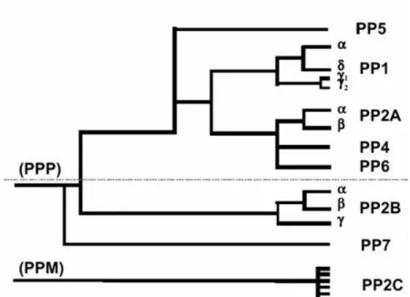
of okadaic acid, a specific phosphatase inhibitor, further facilitated the discrimination between the different classes (Cohen et al., 1989). Although widely in use, this classification does not reflect the currently known phylogenetic relationship between the different Ser/Thr-PPs. Molecular cloning revealed that PP2A was in fact much more related to PP1 than to PP2C (Berndt et al., 1987; Honkanen and Golden, 2002). From a phylogenetic point of view it is more reasonable to group PP1, PP2A and PP2B in family I or PPP [that also includes the bacteriophage λ, orf221 phosphatase (Cohen et al., 1988b)] and PP2C in the unrelated family II or PPM (Fig. I.1). Here, only the PPP family will be further addressed.
Figure I.1: Phylogenic tree depicting the evolutionary relationship between known phosphatases based on their primary amino acid sequence. PP1-PP7 belong to a single gene
_______________________________________________________________________________________ 22 Centre for Cell Biology
University of Aveiro family (PPP) that is structurally distinct and unrelated from the PP2C family (PPM). The phosphatases above the dashed line are highly sensitive to inhibition by naturally occurring toxins, such as okadaic acid, mycrocystin and calyculin A (Honkanen and Golden, 2002).
I.1.2 THE PPP FAMILY
The application of recombinant DNA techniques to the field yielded not only the primary structure of all four phosphatase types, but also documented the existence of isoforms for each type and revealed the existence of previously undetected phosphatases in a variety of eukaryotic cells (Berndt et al., 1987; Cohen et al., 1988a; da Cruz e Silva and Cohen, 1987; da Cruz e Silva et al., 1987; da Cruz e Silva et al., 1988). Three genes are known to encode mammalian type 1 phosphatase catalytic subunits, termed PP1α, PP1β and PP1γ. At least PP1γ is known to undergo tissue-specific processing to yield an ubiquitously expressed PP1γ1 isoform and a testis/sperm-enriched PP1γ2 isoform (da Cruz
e Silva et al., 1995b; da Cruz e Silva and Greengard, 1995). Two genes are known to encode mammalian PP2A catalytic subunits, termed PP2Aα and PP2Aβ, and the three known mammalian PP2B catalytic subunit genes (Aα, Aβ and Aγ) are also subject to complex regulation to yield several alternatively spliced isoforms from each.
Perhaps more surprising was the discovery, from a variety of tissues and species, of previously unknown phosphatase catalytic subunit isoenzymes, that were termed novel phosphatases (da Cruz e Silva et al., 1988). For esample, PP4, PP5 and PP6 (Cohen, 1997) are present in all mammalian tissues examined. In contrast, human PP7 (Huang and Honkanen, 1998), also found in Arabidopsis thaliana (Andreeva et al., 1998), and two
Drosophila phosphatases appear to be exhibit tissue specific expression (PPY is
testis-specific and RdgC is restricted to photoreceptor organs and a small region in the brain). PP7 has been detected in the human retina and also in specialized sensory cells in plants. An overall comparison of their structures is shown in Fig. I.2.
________________________________________________________________________________________ Centre for Cell Biology 23 University of Aveiro PP1, PPY PP2A, PP4, PP6 Ppg1 PP2B PP5 RdgC,PP7 E. Coli diadenosine tetraphosphatase λORF221 Ppz, Ppq1 Serine-rich
Phosphatase catalytic domain
CaM binding TPR motifs Ca2+-binding 302-334 549-710 303-377 368 502-604 499-520 661-663 221 280 PP1, PPY PP2A, PP4, PP6 Ppg1 PP2B PP5 RdgC,PP7 E. Coli diadenosine tetraphosphatase λORF221 Ppz, Ppq1 Ppz, Ppq1 Serine-rich
Phosphatase catalytic domain
CaM binding TPR motifs Ca2+-binding 302-334 549-710 303-377 368 502-604 499-520 661-663 221 280
Figure I.2: Comparison of domain organization of PPP family members. The numbers of amino acids in each are indicated on the right.
I.1.3 PP1 - PROTEIN PHOSPHATASE 1
Eukaryotic genomes contain between one (Saccharomyces cerevisiae) and eight (Arabidopsis thaliana) genes that encode PP1 catalytic subunits. These isoenzymes typically show an overall sequence identity of approximately 90% and cannot be distinguished by either their in vitro substrate specificity or by their ability to interact with R subunits in vitro (Schillace and Scott, 1999; Zhang et al., 1993a). The sequence of the catalytic core of PP1 (corresponding to residues 41-269 of mammalian PP1α) is almost identical in all isoforms, showing a high degree of similarity with the corresponding fragment of the catalytic subunits of PP2A and PP2B (Egloff et al., 1995; Goldberg et al., 1995).
I.1.3.1 PP1 Expression and distribution
By using specific antibodies raised against the different PP1 isoforms (α, β and γ1)
it was shown that all the three isoforms were expressed in a variety of mammalian cells tested, although they localize in a distinct and characteristic manner within these cells. All the isoforms were present both in the cytoplasm and nucleus during interphase. Within the nucleus PP1α associates with the nuclear matrix, whereas PP1γ1 concentrates in nucleoli in
_______________________________________________________________________________________ 24 Centre for Cell Biology
University of Aveiro
association with RNA, and PP1β localizes to non-nucleolar chromatin. During mitosis PP1α is localized to centrosomes, PP1γ1 is associated with microtubules and
PP1β associates with chromosomes (Andreassen et al., 1998).
In the brain the mRNAs for PP1α, PP1β and PP1γ1 were found to be particularly
abundant in hippocampus and cerebellum (da Cruz e Silva et al., 1995b). At the protein level PP1α and PP1γ1 were found to be more highly expressed in brain than in peripheral
tissues (Table I.1), with the highest levels being measured in the striatum, where they were shown to be relatively enriched in the medium-sized spiny neurons (da Cruz e Silva et al., 1995b). At the electron microscopic level, PP1 immunoreactivity was demonstrated in dendritic spine heads and spine necks and also in postsynaptic density (Ouimet et al., 1995). PP1 immunoreactivity has also been reported in human hippocampal neuronal cytoplasm (Pei et al., 1994). In addition, most neuronal nuclei were not immmunoreactive for PP1γ1
but were usually strongly immunoreactive for PP1α (Ouimet et al., 1995).
Table I.1: Tissue distribution of the main PP1 isoforms (Fardilha 2004a) PP1α PP1γ1 PP1γ2 PP1β Brain ++ +++ - +++ Heart + + + Liver + + -Intestine + + - +++ Kidney + + - + Spleen + ++ - + Adrenal gland + ++ - + Lung ++ ++ +++ Skeletal muscle + + - Testis ++ + +++ ++
________________________________________________________________________________________ Centre for Cell Biology 25 University of Aveiro
I.1.3.2 PP1 Structure and function
Protein phosphatase 1 (also known as phosphorylase phosphatase) has been studied since the 1940s as the enzyme responsible for the conversion of phosphorylase a to phosphorylase b (Cori G, 1943). The discovery that this activity (PP1) was a phosphatase came at the same time as the discovery of phosphorylase kinase (Keller and Cori, 1955; Sutherland and Wosilait, 1955). These hallmark findings marked the beginning of an era: the study of protein phosphorylation/dephosphorylation as a regulatory mechanism. Investigation of PP1 in the following three decades focused on defining its enzymology and role in glycogen metabolism (Bollen and Stalmans, 1992; Brautigan, 1994; Shenolikar, 1994; Shenolikar and Nairn, 1991), and progress in the isolation and characterization of PP1 activity was very slow. The study of the enzymology of this enzyme is still incomplete today and PP1 continues to provide many surprises, as well as stimulating new questions about its cellular functions.
I.1.3.2.1 PP1c crystal structure and catalytic mechanism
During the past decade and a half there has been major progress in the elucidation of the atomic structures of the Ser/Thr-PPs in general. Crystal structures for PP1-microcystin (Goldberg et al., 1995), PP1-tungstate (Egloff et al., 1995), PP1-GM peptide complexes (Egloff et al., 1997) and, more recently, PP1-okadaic acid (Maynes et al., 2001) have been determined. Two structures for PP2B have been solved, the auto-inhibited enzyme and a ternary complex of a truncated PP2B with FKBP12/FK506 (Griffith et al., 1995; Kissinger et al., 1995). These structures show that the molecular architecture of the catalytic cores of PP1 and PP2B are conserved [a review can be found in (Barford, 1996)], and that both contain a bimetal center at the active site which is structurally similar to that present in the purple acid phosphatase (Strater et al., 1995). PP1, like PP2B, is a metalloprotein possessing a bimetal center that is bridged by a water molecule at the active site. The use of proton induced X-ray emission spectroscopy revealed the presence of Mn and Fe in the ratio 1 to 0.5 in PP1 (Egloff et al., 1995). The nature of the metal ions in cellular PP1 is unknown, although it can be speculated that it may be a Fe/Zn pair as in PP2B (King and Huang, 1984; Kissinger et al., 1995). The current views of the catalytic
_______________________________________________________________________________________ 26 Centre for Cell Biology
University of Aveiro
mechanism for PP1 are that the metals serve as ligands for the phosphate oxygens and for the generation of a hydroxide ion which serves as the nucleophile that is involved in the catalysis, while H125 serves as a proton donor for the leaving alcohol group. Other residues in the active site which serve to stabilize the proposed pentacoordinate state of the phosphate intermediate are R96, N124 and R221. Mutation of the metal ligands H66, D64, D92 and H248 residues led to severe loss of catalytic function (Zhang et al., 1996a). Mutation of H125 and H173 did not result in readily expressed proteins. Although small amounts of the H125S and H125A mutant proteins could be isolated, these mutants were inactive. Mutation of D95, which is proposed to stabilize the protonation of H125 by a salt bridge, also resulted in significant reduction in catalytic activity. Mutations of R96 and N124 have also supported their proposed roles in phosphate binding.
I.1.3.2.2 Binding region for naturally occurring toxins
Several mutants of PP1 which exhibit a general loss of sensitivity towards several natural toxins show that these toxins have a common binding region on PP1. It was shown that a mutated PP2A (Cys269 to Gly) had reduced sensitivity to okadaic acid (OA) (Shima et al., 1994). By comparing the amino acid sequence of PP1 and PP2A in this region (Fig. I.3), it was noted that the region was well conserved except for a four residue difference, YRCG in PP2A (267-270) and GEFD (274-277) in PP1.
Figure I.3: Alignment of C-terminal regions of PP1, PP2A and PP2B. The boxed region shows the loop regions that connect beta sheets 12 and 13 in the structures of PP1 and PP2B.
The chimeric PP1 mutant in which GEFD was changed to YRCG resulted in increased sensitivity to OA (Zhang et al., 1994), consistent with the fact that PP2A is more sensitive to okadaic acid than PP1. The same occurred when F276 was mutated to Cys (Zhang et al., 1996a). It was also shown that Y272 is important for the binding of all of the
________________________________________________________________________________________ Centre for Cell Biology 27 University of Aveiro
inhibitors tested, as its conservative mutation to phenylalanine caused decreases in PP1 toxin sensitivity. These mutagenesis studies indicate that binding of the toxins must all involve some common contacts on PP1, and that Y272 is particularly important in this context. Y272 is located close to the active site with its hydroxyl group within a few angstroms of the Fe ion (Egloff et al., 1995). The mutation of Y272 without deleterious effects on its catalytic activity suggests that it is not involved in the catalysis. This region of PP1 represents the loop region connecting beta strands 12 and 13 in PP1 structure (Fig I.4).
If the toxins bind to the same site on PP1, this may reflect the possibility that these chemically diverse molecules must present topographically similar surfaces at the points of interaction with PP1. This idea has been supported by molecular modeling studies (Bagu et al., 1997; Gauss et al., 1997; Lindvall et al., 1997).
The structure of PP1 bound to okadaic acid (Maynes et al., 2001) is remarkably similar to the two structures of PP1 and PP2B determined previously. Even with only the phosphate-mimic tungstate present the architecture of the active site of the tungstate-bound PP1 structure is virtually identical to OA-bound PP1 complex.
Figure I.4: The β12/β13 loop
of PP1.The diagram shows a ribbon model of the PP1 structure. The two beta sheets that are the scaffold for the active site are shown in yellow (beta sheet 1) and magenta (beta sheet 2). Beta strands 12 and 13 are shown in red. Microcystin is shown in wireframe and the two metal ions as black spheres. (Goldberg et al., 1995).
_______________________________________________________________________________________ 28 Centre for Cell Biology
University of Aveiro
In contrast, the microcystin-bound structure reveals large changes in the conformation of the active site. These changes are mainly restricted to the β12/β13 loop (Fig. I.4). The loop in the microcystin-bound PP1 structure folds back on itself, causing significant shifting of residues 273-278. One critical difference between microcystin and OA is the presence of a dehydroalanine residue in microcystin that covalently alkylates the Sγ of Cys273 in a time dependent reaction (Dawson and Holmes, 1999). This covalent linkage is not the primary cause of inhibition of PP1 by microcystin (Goldberg et al., 1995). Given the strong similarity of the PP1-interacting domains of OA and microcystin it is likely that the primary mode of inhibition of PP1 by microcystin is similar to that of OA and that the movement of the β12/β13 loop in the microcystin complex is a secondary event accompanying the covalent binding reaction. An important interaction between PP1 and microcystin that is not present in the PP1-OA complex is the hydrogen bond that occurs between Arg96 (PP1) and the acid of the methyl-aspartate residue (microcystin). This interaction may account for the 100-fold greater inhibition of PP1 by microcystin over OA (Holmes and Boland, 1993). The structure of PP1-OA is very similar to the structure of PP2B despite the fact that OA does not strongly inhibit PP2B.
I.1.3.2.3 Substrate binding
The active site of PP1 lies at the confluence of three shallow grooves, a C-terminal groove, an acidic groove and a hydrophobic groove, which are potential binding sites for substrates and inhibitors (Egloff et al., 1995; Goldberg et al., 1995). Microcystin binds in a manner such that it occupies the active site, while its extended ADDA side chain occupies the hydrophobic groove. The hydrophobic groove forms the obvious binding site for peptide substrates. The two PP1 inhibitors, I-1 and DARPP-32, both carry four basic residues N-terminal to the phosphothreonine residue and its binding to PP1 has been hypothesized to be that of a pseudosubstrate (Goldberg et al., 1995). Binding of peptide/polypeptide substrates to PP1 can be considered to be composed of three elements: interaction of the basic residues N-terminal to the phosphoserine with the acidic residues in the acidic groove, binding of the phosphoserine to the active site and an interaction of the region C-terminal to the phosphoserine (or phosphothreonine) to the hydrophobic groove. In the active site region the structure of the PP1-tungstate complex has shown that R96,
________________________________________________________________________________________ Centre for Cell Biology 29 University of Aveiro
N124 and R221 are involved in the binding of the phosphate oxygens (Egloff et al., 1995). R221 and R96 are well positioned to form salt bridges with two of the phosphate oxygens, while the amino group of N124 can be hydrogen bound to the third oxygen. D208 was hypothesized to be important for the orientation of R221 via a salt bridge interaction. It was also indicated that W206 and Y134 are well positioned to interact with the Ser or Thr carrying the phosphate residue (Egloff et al., 1995).
I.1.3.3 PP1 targeting/binding proteins
During the last decade evidence has accumulated that the substrate specificity of PP1 is achieved by the interaction with other proteins that can act as targeting subunits or activity modulators. Targeting, as the requirement for the molecular juxtaposition of proteins for the generation of signaling events, is well established as a paradigm in a number of growth regulated signaling systems involving tyrosine phosphorylation (Kuriyan and Cowburn, 1997; Lemmon and Schlessinger, 1994; Pawson, 1994), as well as in the anchoring of Ser/Thr-protein kinases by A-kinase anchoring proteins (AKAPs), one of which also binds PP2B (Lemmon and Schlessinger, 1994; Rubin, 1994) . The concept of targeting as it relates to PP1, however, has a major twist in terms of the large number of PP1 binding proteins that have been reported during the past years, as it expands the number of PP1 heterodimers/heterotrimers that may exist and consequently the repertoire of cellular functions that involve PP1.
Genetic studies of yeast mutations that affect glycogen metabolism and cell cycle regulation, and the use of the yeast two-hybrid system have revealed many genes that encode putative PP1-binding proteins [reviewed in (Stark, 1996)]. These include GAC1, REG1, REG2, SCD5, GIP1, SHP1, GIP2 and SDS22 in S. cerevisiae. These genes are required for the control of glycogen metabolism, protein synthesis, glucose repression, meiosis, sporulation and mitotic cell cycle regulation. Rigorous biochemical demonstration that these PP1-binding proteins actually interact with PP1, or the nature of the targeting function or the substrates, have not been shown in all cases. A key element of the targeting hypothesis is that the cellular activity of PP1 is only expressed when it is targeted. This explains why the PP1 catalytic subunit exhibits a relatively nonspecific phosphatase activity in vitro. The strongest experimental support for a targeting function of a PP1 regulator (R) has come from genetic and biochemical studies of yeast glycogen metabolism.
_______________________________________________________________________________________ 30 Centre for Cell Biology
University of Aveiro
The glycogen-deficient yeast mutant glc7-1 was found to express PP1 with a R73C point mutation (Peng et al., 1990). This does not affect PP1 activity but resulted in loss of its ability to bind to the yeast homolog (Gac1p) of the mammalian glycogen binding protein (Stuart et al., 1994). The activation of glycogen synthase requires its dephosphorylation by PP1 and it has been shown that glycogen synthase in this mutant strain is largely in the inactive phospho-form. Overexpression of Gap1p, on the other hand, led to increased glycogen accumulation. These findings demonstrated that the physiological functioning of PP1 in glycogen metabolism was dependent on it being targeted to the appropriate micro-environment and that other cellular functions were not affected when targeting to glycogen was disrupted.
Several mammalian PP1-binding proteins have also been identified and shown to be responsible for the involvement of PP1 in a number of diverse cellular functions [(Fardilha et al., 2004a).; Table I.2]. Based on their effect on PP1c, the best characterized R subunits can be divided into three groups. The first group is represented by activity-modulating proteins, including true inhibitors such as I-1 (Connor et al., 1999) and CPI-17 (Koyama et al., 2000) that in their phosphorylated form block the activity of PP1c towards all substrates. Other members of this group act instead as substrate-specifiers of PP1c. For example, I-1PP2A (PHAP-I) and I-2PP2A (PHAP-II), which are potent inhibitors of PP2A, promote the dephosphorylation of specific substrates by PP1c (Katayose et al., 2000). A second group of R subunits includes the targeting proteins which bind both PP1c and one of its substrates. For example, MYPT1 binds PP1c as well as specific substrates, such as myosin (Fukata et al., 1998; Hartshorne and Hirano, 1999; Toth et al., 2000a). Other targeting subunits do not bind the substrate directly but instead associate with a subcellular structure that contains the substrate. For example, the G subunits target PP1 to glycogen particles, which also bind the substrate glycogen synthase (Liu and Brautigan, 2000). The targeting proteins of PP1 also include scaffolding proteins that mediate the formation of protein complexes. Often, these complexes function as signaling modules that contain both protein kinases and phosphatases, and are localized in close proximity to the substrates of these enzymes. The third group of proteins that directly and tightly associates with PP1c defines a subset of its substrates. Some of these substrates also function as targeting proteins. Thus, the centrosomal protein kinase Nek2 not only binds its substrate C-Nap1, but also binds PP1c, and both Nek2 and C-Nap1 are proposed substrates of the associated
________________________________________________________________________________________ Centre for Cell Biology 31 University of Aveiro
PP1c (Helps et al., 2000). Some PP1c-bound substrates also function as activity modulators. For example, the retinoblastoma protein interacts with PP1c both as a substrate and as a noncompetitive inhibitor (Tamrakar and Ludlow, 2000).
_______________________________________________________________________________________ 32 Centre for Cell Biology
University of Aveiro Table I.2: Classification of the regulatory R subunits of PP1.
Regulatory subunit
General Function
Reference
I-1 PPP1R1A (Huang and Glinsmann, 1976a) DARPP-32 PPP1R1B (Hemmings et al., 1984d)
I-2 PPP1R2
PP1 inhibitors
(Huang and Glinsmann, 1976a) GM (RGL, R3) PPP1R3A (Stralfors et al., 1985)
GL (R4) PPP1R3B (Doherty et al., 1995; Moorhead et al., 1995)
R5(PTG) PPP1R3C (Doherty et al., 1996) R6 PPP1R3D
Glycogen metabolism
(Armstrong et al., 1997) Sds22 PPP1R7 Mitosis/Meiosis (Dinischiotu et al., 1997) NIPP-1 (Ard-1) PPP1R8 (Van Eynde et al., 1995)
SIPP-1 (Llorian et al., 2004)
PSF1
RNA splicing
(Hirano et al., 1996) Neurabin I PPP1R9A Neurite outgrowth, synapse morphology (MacMillan et al., 1999) Spinophilin (neurabin
II) PPP1R9B
Glutamatergic synaptic transmission,
dendritic morphology (Allen et al., 1997) p99 (R111, PNUTS) PPP1R10 RNA processing or transport? (Allen et al., 1998) Hox11 Cell cycle checkpoint (Kawabe et al., 1997) Inhibitor-3 (HCG-V) PPP1R11 Inhibits PP1 (Zhang et al., 1998)
HCF Transcription, cell cycle (Ajuh et al., 2000) MYPT1 (M110, MBS,
M130) (Alessi et al., 1992)
MYPT2 (PP1bp55,
M20-spliced form) PPP1R12B (Moorhead et al., 1998) P85 PPP1R12C (Tan et al., 2001) Phactr1
Myosin/actin targeting
(Allen et al., 2004) L5 ribossomal protein (Hirano et al., 1995)
RIPP1 Protein synthesis? (Beullens et al., 1996) 53BP2 (TP53BP2,
p53-binding protein 2) PPP1R13A Cell cycle checkpoint? (Helps et al., 1995) CPI-17 PPP1R14A Inhibits the myosin bound PP1 complex (Eto et al., 1997)
PHI-2 PPP1R14B (Eto et al., 1999)
PHI-1 (Eto et al., 1999)
KEPI
Inhibits PP1
(Liu et al., 2002)
GBPI-1 Inhibits PP1 when phosphorylated
(Brain/Stomach) (Liu et al., 2004)
GBPI-2 Inhibits PP1 when phosphorylated
(Testis) (Liu et al., 2004)
I-4 (Shirato et al., 2000)
GADD34 PPP1R15A Protein synthesis (Connor et al., 2001) CReP PPP1R15B Protein synthesis (Jousse et al., 2003)
AKAP 149 Nuclear envelope reassembly
(dephosphorylation of B-type lamins) (Steen et al., 2000) NF-L Synaptic transmission? (Terry-Lorenzo et al., 2000) AKAP-220 Coordination of PKA/PP1 signalling (Schillace et al., 2001)
________________________________________________________________________________________ Centre for Cell Biology 33 University of Aveiro
Yotiao Synaptic transmission (NMDA receptor ion channel activity)
(Feliciello et al., 1999; Westphal et al., 1999)
BH-protochaderin c Neuronal cell-cell interaction (Yoshida et al., 1999) Rynanodyne receptor Calcium ion channel activity? (Zhao et al., 1998)
NKCCl Cl transport (Darman et al., 2001) AKAP 350 (CG-NAP,
AKAP450) (Takahashi et al., 1999) Nek2
Centrosomal function
(Helps et al., 2000) Tau Microtubule stability? (Liao et al., 1998) Bcl2 Apoptosis (Ayllon et al., 2001)
RB Cell cycle progression (Durfee et al., 1993) PRIP-1 (p130,
PLC-L1) Calcium signalling? (Yoshimura et al., 2001) PFK Glycolisis? (Zhao and Lee, 1997b)
PP1bp80 (Damer et al., 1998)
MYPT3 PPP1R16A Myosin targeting (Skinner and Saltiel, 2001) I1PP2A(PHAPI) (Katayose et al., 2000)
I2PP2A(SET, PHAPII,
TAF1β)
Stimulation of PP1 and Inhibition of
PP2A (Katayose et al., 2000)
G-substrate Inhibition of PP1 (Aitken et al., 1981) Grp78 Unknown (Chun et al., 1994) NCLK Activates PP1 (Agarwal-Mawal and Paudel, 2001)
Myr8 Brain development (Patel et al., 2001)
FAK PP1 dephosphorylates FAK in cells
released from mitosis (Fresu et al., 2001) Herpes vírus γ1 34.5
protein Inhibits protein synthesis (He et al., 1997)
mGluR7b Unknown (Enz, 2002)
Histone H3 Mitosis (Hsu et al., 2000)
Scapinin Associated with the nuclear
nonchromatin structure (Sagara et al., 2003) 14-3-3 sperm (Huang et al., 2004)
SARP ? (Browne et al., 2007)
Abbreviations: PTG, Protein targeting to glycogen; MYPT, myosin phosphatase targeting subunit; MBS, myosin binding subunit; NIPP, Nuclear inhibitor of PP1; PSF, polypyrimidine tract-binding protein associated splicing factor; PNUTS, phosphatase 1 nuclear targeting subunit; Hox11, Homeodomain transcription factor; HCF, host cell factor or human factor C1; RIPP1, ribosomal inhibitor of PP1; PHI, phosphatase holoenzyme inhibitor; KEPI, kinase-enhanced protein phosphatase type 1 inhibitor; GADD34; growth arrest and DNA damage protein; AKAP, A-kinase anchoring protein; NKCCl, Na-K-Cl cotransporter; Nek2; NIMA-related protein kinase; Rb, retinoblatoma protein; PRIP-1; phospholipase C-related inactive protein; PFK, Phosphofrutokinase; G-substrate, cGMP-dependent protein kinase substrate; Grp-78, glucose-regulated protein, member of the HSP-70 family; NCLK, neuronal cdc2-like kinase; FAK; Focal adhesion kinase. The human genome nomenclature for the regulatory subunits that are only classified as PP1 regulators is indicated in column 2. PPP1R4, PPP1R5 and PPP1R6 have recently been re-classified as PPP1R3B, PPP1R3C and PPP1R3D.
_______________________________________________________________________________________ 34 Centre for Cell Biology
University of Aveiro I.1.3.4 The consensus PP1 binding “RVxF motif”
Members of all groups of R subunits have been shown to bind to PP1c via a short sequence that is now referred to as the “RVxF motif” (Barford et al., 1988; Egloff et al., 1997; Zhao and Lee, 1997a), although the sequences that correspond to the RVxF motif are degenerate. The consensus sequence is [K/R]-X0-1-[V/I/L]-(Agarwal-Mawal and Paudel,
2001){P}-[F/W] (Wakula et al., 2003). The residues of PP1c that are necessary for binding to the RVxF motif (in particular residues 287-293) are invariant in all isoforms from all species (Barford et al., 1988; Egloff et al., 1997). However, they are not conserved in the catalytic subunits of PP2A or PP2B, which explains why most regulators of PP1 do not interact with these highly homologous and structurally related phosphatases. Conversely, some proteins that bind PP1 but are also able to interact with the catalytic subunit of PP2A (HOX11, I-1PP2A and I-2PP2A) do not contain an RVxF sequence (Katayose et al., 2000; Kawabe et al., 1997).
The binding of the RVxF sequence does not cause important conformational changes in the catalytic subunit (Egloff et al., 1997) and does not have major effects on the activity of the phosphatase (Beullens et al., 1999; Endo et al., 1996; Kwon et al., 1997). Studies on MYPT1 and I-2 have indicated that the RVxF motif can function as an anchor for PP1c and enables these R subunits to make additional contacts with the phosphatase in an ordered and cooperative manner (Toth et al., 2000b; Yang et al., 2000a). For example, four phosphatase-interaction sites, in addition to the RVxF motif, have been identified for MYPT1 and I-2 (Hartshorne and Hirano, 1999; Toth et al., 2000b; Yang et al., 2000a). Another recurring theme is that the R subunits have common or overlapping binding sites on PP1c, in addition to the RVxF-binding channel. For example, the inhibition of PP1c by phosphorylated I-1 (Endo et al., 1996), DARPP-32 (Kwon et al., 1997) and MYPT1 (Hartshorne and Hirano, 1999) have all been attributed to the binding of the phosphorylated residue at or near the catalytic site as a pseudo-substrate. The sharing of interaction sites is also in accordance with findings that various point mutants of PP1c show altered affinity for multiple R subunits (Baker et al., 1997; Ramaswamy et al., 1998). As expected from the unusually high conservation of residues on the surface of PP1c, mutagenesis studies in yeast have identified many surface residues as being essential for the binding of R subunits (Baker et al., 1997; Ramaswamy et al., 1998). A site that lies
________________________________________________________________________________________ Centre for Cell Biology 35 University of Aveiro
adjacent to the RVxF–binding groove has been identified as a binding pocket for the N-terminal “IKGI” motif of inhibitor-2 (Connor et al., 2000).
The picture that emerges shows that the binding of the R subunits to PP1c is mediated by multiple, degenerate, short sequence motifs and that the R subunits can share interaction sites. It should be pointed out that this combinatorial control (Bollen, 2001) of PP1c does not rule out the possibility that some R subunits might have unique binding sites. The combinatorial control of PP1 allows for exquisite physiological regulation of PP1 holoenzymes by hormones, growth factors and metabolites at the molecular level. Work on the various holoenzymes has demonstrated that their physiological regulation involves modulation of subunit interaction, often mediated by reversible phosphorylation or allosteric regulation of the R subunits. For three unrelated R subunits it has been shown that the phosphorylation of Ser residue(s) within or close to the RVxF motif disrupts the binding of this motif to PP1c (Beullens et al., 1999; Liu et al., 2000; McAvoy et al., 1999). This results in altered activity of the holoenzyme or the release of the catalytic subunit. In contrast, phosphorylation of other subunits strengthens their interaction with PP1c. Examples include I-1 and DARPP-32, in which an additional binding site for PP1c is created by phosphorylation. A different type of regulation involves the binding of allosteric regulators. The allosteric binding of phosphorylase a to the C-terminal tail of the liver-type G subunit (GL) abolishes the activity of the associated PP1c towards glycogen synthase (Armstrong et al., 1998). An additional level of regulation of PP1 holoenzymes is provided by targeting of these enzymes to specific substrates or subcellular structures. Bollen and coworkers, by a combination of bioinformatics tools and mutagenesis studies, have delineated the consensus sequence and function of three PP1 binding motifs as being [K/R]-X0-1-[V/I/L]-(P)-[F/W], where X denotes any residue and P any residue except
proline (Wakula et al., 2003). This sequence is very similar to the consensus sequence previously proposed ([R/K]-[K/R]-X0-2-V-[R/H]-[F/W]-X-[DE]) by panning of a random
peptide display library (Zhao and Lee, 1997a). The main differences are the presence of an N-terminal basic residue and a C-terminal acidic residue in the second.
The RVxF-consensus sequence is present in about one third of all eukaryotic proteins, but only a small fraction are thought to be PP1-binding proteins. It seems that RVxF-consensus sequences function as PP1 interaction sites only when they are present in a flexible and exposed loop that can be modeled into a β-strand. Additionally, other low
_______________________________________________________________________________________ 36 Centre for Cell Biology
University of Aveiro
affinity regions on the PP1 regulators further strengthen the binding. Thus, the RVxF-consensus sequence functions like an anchor and other low affinity interactions have to occur that have regulator-specific effects on PP1 activity and specificity (Wakula et al., 2003). Recently, another PP1 binding motif has been proposed, F-X-X-R-X-R, that also appears to exist in several PP1 interactors (Ayllon et al., 2002). A combined bioinformatics and mutagenesis approach could also be used to study this new consensus motif. The existence of common binding sites for the various R subunits explains why a relatively small protein such as PP1c can interact with numerous different R subunits and why the binding of most R subunits is mutually exclusive.
I.1.4 PHOSPHATASE INHIBITORS
One of the most significant advances in the study of Ser/Thr-PPs, and for the elucidation of the cellular events they control, was the identification of several naturally occurring toxins as powerful and specific phosphatase inhibitors. Among these are okadaic acid (Fujiki and Suganuma, 1993), cantharidin (Laidley et al., 1997), calyculin A (Ishihara et al., 1989), microcystins (Carmichael, 1992; Carmichael, 1994; Fujiki and Suganuma, 1993) and tautomycin (Hori et al., 1991; MacKintosh and Klumpp, 1990), to name a few. Another class of phosphatase inhibitors are the physiological protein inhibitors readily available inside the cell. The diverse specificity of the naturally occurring toxins for the different types of phosphatase has led to their use in the laboratory as key tools in the study of phosphorylation-dependent processes. However, below only the protein inhibitors are further addressed.
I.1.4.1 Inhibitor 1
Inhibitor 1 (I-1) and Inhibitor 2 (I-2) were originally identified by Huang and Glinsmann (Huang and Glinsmann, 1976b). They share unusual physical properties: both are heat stable and are not precipitated by 1% trichloroacetic acid, in contrast to most other proteins. I-1 comprises 171 amino acids (Aitken et al., 1982; Elbrecht et al., 1990; Endo et al., 1996). Differences between the rabbit, rat and human I-1 sequences were only detected in the C-terminus (Endo et al., 1996). I-1 from rabbit skeletal muscle and human brain has a calculated molecular mass of 18.7 and 19.2kDa, respectively (Aitken et al., 1982;
________________________________________________________________________________________ Centre for Cell Biology 37 University of Aveiro
Elbrecht et al., 1990; Endo et al., 1996), but the apparent molecular mass on SDS-PAGE is 26kDa. This discrepancy has been explained by a low degree of order in the protein. I-1 binds to and inhibits PP1 only after being phosphorylated on Thr-35 by cAMP-dependent protein kinase or cGMP-dependent protein kinase (Hemmings et al., 1984c). It is highly selective for PP1, inhibiting PP1 and PP2A with IC50 values of 1.1 and 21,000nM,
respectively (Endo et al., 1996). I-1 is a cytosolic protein and has been used as a tool to study whether a process involves PP1. Amino acids 9KIQF12 are conserved in rat, rabbit and human and seem to be crucial for binding and inhibition of PP1 (Egloff et al., 1997).
I.1.4.2 Inhibitor 2
I-2 from rabbit skeletal muscle comprises 204 amino acids and has a calculated mass of 22.9kDa (Holmes et al., 1986). Similarly to I-1, its apparent molecular mass on SDS-PAGE is much larger (31kDa). It binds to and inhibits PP1 regardless of its phosphorylation state. Mutational analysis suggests that I-2 inhibits via interaction with amino acid Tyr-272 on PP1 because its IC50 changed from 13 to 180ng/ml in the Y272K
mutant (Zhang et al., 1996b). Interestingly, I-2 inhibition of PP1 can be reversed by GSK3 phosphorylation of I-2.
I.1.4.3 DARPP-32
DARPP-32 (dopamine and cyclic AMP-regulated phosphoprotein, Mr 32,000Da) resembles I-1 in function but is derived from a different gene. Further, DARPP-32 is expressed almost exclusively in the brain (Hemmings et al., 1984a), whereas I-1 exhibits a much more ubiquitous tissue distribution. It is a cytosolic protein and has a predicted molecular mass of 22.6kDa, but again a higher apparent molecular mass of 32kDa by SDS-PAGE (Williams et al., 1986). The same Thr residue on DARPP-32 is phosphorylated by cAMP-dependent protein kinase and by cGMP-dependent protein kinase. Phosphorylation of DARPP-32 changes its IC50 for PP1 from >1μM to 2nM (Desdouits et al., 1995a;
Desdouits et al., 1995b), underscoring its high selectivity. Under physiological conditions, I-1 and DARPP-32 are dephosphorylated and inactivated by PP2A and even more efficiently by PP2B (Desdouits et al., 1995c; Hemmings et al., 1984b; Hemmings et al., 1990). The dephosphorylation by PP2B is dependent on the presence of calcium,
_______________________________________________________________________________________ 38 Centre for Cell Biology
University of Aveiro
suggesting that this may represent a mechanism for Ca2+ levels to control I-1 and DARPP-32 phosphorylation and consequently PP1 activity (Hubbard and Cohen, 1989). Thiophosphorylated I-1 and DARPP-32 are relatively resistant to dephosphorylation and the corresponding synthetic peptides have been successfully used to study the physiological role of PP1-mediated processes.
I.1.4.4 Inhibitor 3
Inhibitor 3 (I-3) is another heat stable protein and potent inhibitor of PP1 that was more recently identified in a yeast two-hybrid screen (Zhang et al., 1998) It inhibits PP1 activity toward glycogen phosphorylase a with an IC50 in the nanomolar range, similar to
the behavior of the other well-studied PP1 inhibitors: I-1, DARPP-32, and I-2. I-3 shares sequence homology with the yeast protein Yfr003c, also called Ypi1 (yeast phosphatase inhibitor 1). I-3 co-localizes with PP1γ1 in nucleoli and with PP1α in centrosomes (Huang et al., 2005a), possibly indicating that I-3 may act as modulator of PP1 functions in cytokinesis and nucleolar events.
I.1.4.5 Inhibitor 4
Kikuchi and coworkers isolated a human cDNA for a novel PP1 inhibitory protein, named I-4, from a cDNA library of germ cell tumors (Shirato et al., 2000). I-4, composed of 202 amino acids, is 44% identical to I-2. I-4 conserves some of the functionally important structures of I-2 and exhibits similar biochemical properties. I-4 inhibits the activity of the catalytic subunit of PP1 with an IC50 of 0.2nM, relatively more potent than
I-2 (with an IC50 of approximately 2nM). Gel overlay experiments showed that I-4 binds
PP1c directly through a multiple-point interaction.
I.1.5 SIGNAL TRANSDUCTION THERAPEUTICS
Cellular health and vitality are dependent on the fine equilibrium of protein phosphorylation systems. Not surprisingly many diseases and dysfunctional states are associated with the abnormal phosphorylation of key proteins (e.g. cancer, diabetes, etc.). Thus, protein phosphorylation systems represent attractive targets for diagnostics and
________________________________________________________________________________________ Centre for Cell Biology 39 University of Aveiro
therapeutics. However, unlike the myriad of known protein kinases that all belong to a single gene superfamily, the protein phosphatases belong to several unrelated families. Furthermore, relatively few protein phosphatase catalytic subunits exist, exhibiting broad and overlapping substrate specificities in vitro. From a medical perspective, non-selective or marginally-selective phosphatase inhibitors have broad biological activity and are highly toxic to eukaryotic cells due to the inhibition of a number of critical cellular processes. Therefore, the development of low specificity inhibitors (e.g. calyculin A, microcystin or cantharidin) into therapeutic agents for systemic use seems unlikely. However, the development of type and isoform specific inhibitors seems very promising. Both ISIS 15534 (Zuo et al., 1998) and ISIS 14435 (Cheng et al., 2000) have been employed to specifically suppress the expression of human PP5 and PP1γ1, respectively. More interesting, however, is data indicating that in vivo, as phosphatases possess exquisite specificities, both in terms of substrates and localization, the key control mechanism must reside in the nature of the proteins to which they bind. An increasing number of proteins are being identified in diverse cell types that are responsible for regulating the catalytic activity of protein phosphatases. Indeed, the diversity of such phosphatase regulatory subunits explains not only the need for few catalytic subunit types, but also make them attractive targets for pharmacological intervention. The functional diversification of PP1 is controlled via its interaction with such regulatory proteins.
The importance of PP1 and its binding proteins as potential targets for signal transduction therapeutics is further strengthened by the work of Greengard and co-workers demonstrating the central role played by DARPP-32 in mediating many of the most important neuronal signalling pathways (Greengard et al., 1999). To date more than twenty primary signalling cascades have been shown to be under the regulation of the PP1/DARPP-32 system in the striatum, and the PP1/I-1 system in other brain regions and tissues. PP1 also appears to play a central role in the molecular mechanisms underlying the actions of several drugs of abuse. Furthermore, several lines of evidence also link PP1 to the basic processes thought to underlie memory and learning, such as LTP (long term potenciation) and LTD (long term depression). In fact, the relevance of PP1 within the context of aging and memory loss was recently given a rather intriguing boost. PP1 has been linked to the efficacy of learning and memory by limiting the acquisition of new knowledge and favouring memory decline (Genoux et al., 2002). PP1 inhibition prolongs