FACULDADE DE FARMÁCIA
ROLE OF APOPTOSIS FACTORS
DURING MOUSE NEURAL STEM CELL
DIFFERENTIATION
Márcia Maria de Almeida Aranha
DOUTORAMENTO EM FARMÁCIA
BIOQUÍMICA
FACULDADE DE FARMÁCIA
ROLE OF APOPTOSIS FACTORS
DURING MOUSE NEURAL STEM CELL
DIFFERENTIATION
Márcia Maria de Almeida Aranha
Tese de Doutoramento em Farmácia (Bioquímica), apresentada à
Universidade de Lisboa através da Faculdade de Farmácia
Research advisor:
Prof. Doutora Cecília M. P. Rodrigues
Lisboa
2010
The studies presented in this thesis were performed at the Research Institute for Medicines and Phamaceutical Sciences (iMed.UL), Faculty of Phamacy, University of Lisbon under the supervision of Professor Cecília M. P. Rodrigues, and at the Departments of Medicine, Genetics, Cell Biology, and Development, and the Department of Neurosurgery, Stem Cell Institute, University of Minnesota Medical School, Minneapolis, Minnesota, USA, in collaboration with Professor Clifford Steer and Professor Walter C. Low.
Márcia Maria de Almeida Aranha was the recipient of a Ph.D. fellowship (SFRH/BD/28429/2006) from Fundação para a Ciência e Tecnologia (FCT), Lisbon, Portugal. This work was supported by grants PTDC/BIA-BCM/67922/2006 and PTDC/SAU-FCF/67912/2006 from FCT and FEDER.
De acordo com o disposto no ponto 1 do artigo nº41 do Regulamento de Estudos Pós-Graduados da Universidade de Lisboa, deliberação nº 93/2006, publicada em Diário da República – II Série nº 153 – 5 de Julho de 2003, o Autor desta dissertação declara que participou na concepção e execução do trabalho experimental, interpretação dos resultados obtidos e redacção dos manuscritos.
A utilização de células estaminais para tratamento de doenças neurológicas é, actualmente, objecto de intensa investigação. Para que seja possível desenvolver terapias regenerativas, a partir destas células, é fundamental compreender os seus processos biológicos básicos, nomeadamente o seu mecanismo de diferenciação.
Estudos recentes sugerem que alguns factores específicos do processo apoptótico, como as caspases, a proteína p53, a família Bcl-2 e, mais recentemente, os microRNAs (miRNAs ou miRs), possam estar envolvidos na diferenciação celular. Assim, formulámos a hipótese de que algumas moléculas associadas à apoptose desempenhem um papel importante na diferenciação de células estaminais neurais.
Avaliou-se, inicialmente, o envolvimento da p53 e caspase-3 na diferenciação de células estaminais neurais. Observámos que, tanto a clivagem da caspase-3, como a fosforilação da proteína p53 e a sua capacidade de ligação ao DNA aumentaram significativamente ao longo da diferenciação. A inibição das caspases e o silenciamento de p53 retardaram, de forma diferencial, a neurogénese e a gliogénese. Os nossos resultados sugerem que a p53 e a caspase-3 desempenham um importante papel na diferenciação neural, que parece envolver a modulação da via do FOXO3A/Id1.
Seguidamente quisemos determinar se os miRNAs associados à apoptose estariam, igualmente, envolvidos no processo de diferenciação de células estaminais neurais. A determinação dos perfis de expressão dos miRNAs, juntamente com a validação dos níveis de expressão por PCR em tempo real, mostraram que miRNAs pró-apoptóticos, tais como o miR-16, o let-7a e o miR-34a, são modulados durante a diferenciação de células estaminais neurais. Resultados similares foram observados na diferenciação de células estaminais embrionárias e de células PC12 e NT2N, o que sugere o envolvimento destes miRNAs no desenvolvimento neural dos mamíferos.
Por fim, modularam-se os níveis de expressão do miR-34a em células estaminais neurais e concluiu-se que este miRNA regula, de forma positiva, o aparecimento de neurónios pós-mitóticos. Experiências adicionais sugeriram que o miR-34a regula, também, a diminuição da SIRT1 e o aumento da capacidade de ligação da p53 ao DNA.
Este trabalho clarifica o papel que algumas moléculas do processo apoptótico têm na diferenciação de células estaminais neurais e confirma a sua importância na definição do destino celular, proporcionando indicações úteis ao desenvolvimento de novas estratégias terapêuticas.
Palavras chave: Apoptose – Caspase-3 - Células estaminais neurais – Diferenciação –
Neural stem (NS) cells are under active consideration as a source of donor tissue for neuronal cell therapy. Therefore, understanding basic processes of NS cell biology, including cell differentiation, is essential to develop regenerative therapies from the promise of stem cells.
Several studies have recently provided a persuasive argument that specific factors of apoptosis pathways, such as caspases, p53, Bcl-2 family members, and microRNAs (miRNAs or miRs) are involved in the differentiation process. Therefore, we hypothesized that specific apoptosis-associated molecules play a critical role in NS differentiation.
In initial studies we evaluated the involvement of p53 and caspase-3 during the differentiation process of NS cells. Our results revealed a significant increase in caspase-3 activation, p5caspase-3 phosphorylation, and p5caspase-3 DNA-binding activity during differentiation. Pharmacologic inhibition of caspases delayed neuronal and glial differentiation, while down-regulation of p53 affected only the neuronal lineage. Therefore, our results suggest that p53 and caspases play a role in the differentiation of NS cells and advance the potential modulation of FOXO3A/Id1 signaling in this cellular context.
Next, we were interested in determining whether miRNAs linked to apoptosis might be involved in the differentiation of NS cells. miRNA expression profiles and quantitative real time-PCR revealed that pro-apoptotic miRNAs, such as miR-16, let-7a and miR-34a, were modulated during NS cell differentiation. Similarly, upregulation of pro-apoptotic miRNAs in the differentiation of embryonic stem cells, PC12 and NT2N cells implicates these specific miRNAs in mammalian neuronal development.
Finally, we further investigated the effect of miR-34a modulation in NS cells differentiation. We reported that miR-34a positively regulated the appearance of post-mitotic neurons of mouse NS cells. Additional experiments indicated that miR-34a-mediated differentiation involved SIRT1 down-regulation and was associated with increased p53-DNA binding activity. These results suggested that miR-34a was required for proper neuronal differentiation.
In conclusion, these studies further clarify the role of apoptosis-associated molecules in the differentiation process of NS cells. Our findings may prove useful in the development of novel therapeutic strategies to improve long term-survival and differentiation of transplanted stem cells.
Keywords: Apoptosis – Caspase-3 – Differentiation – miRNAs – miR-34a – Neural stem
Acknowledgements
Começo por agradecer àquela que foi a mentora de todo o trabalho desenvolvido nesta tese, a Professora Cecília Rodrigues. Agradeço ter acreditado em mim e apostado no meu trabalho, ainda no início da minha formação enquanto investigadora. Agradeço todos os ensinamentos que me transmitiu e que foram determinantes para a minha formação e crescimento profissional. Agradeço, sobretudo, a ajuda nos momentos de maior hesitação. Obrigada pelo acompanhamento constante no trabalho e pelas oportunidades que me proporcionou ao longo destes anos. A sua enorme capacidade de trabalho e disponibilidade total, aliados à sua preocupação constante com o trabalho desenvolvido pelos seus alunos, definem-na como uma excelente orientadora.
Part of the research studies leading to this Ph.D. thesis were conducted in the University of Minnesota, in Minneapolis, at Professor Clifford Steer’s and Professor Walter Low’s laboratories. I would like to express my gratitude for kindly receiving me and for all the support during my stay in the U.S.A. I am also thankful for all the critical comments and suggestions that contributed immensely to the studies included in this thesis. My thanks to Phil Wong, Carol Bruzzone, Dr. Betsy Kren, and everybody else I met, and worked with at the lab, and at the University. I would like to thank Dr. Yan Zeng, Jing Xiao and Crusoe for all the help and teaching.
Um agradecimento muito especial à Susana Solá, com quem iniciei este trabalho e com quem partilhei as frustrações e conquistas de cada resultado obtido. Agradeço o seu interesse e acompanhamento constantes que foram, sem dúvida, fundamentais para o desenvolvimento deste trabalho. Acima de tudo, obrigada pela sua amizade.
Agradeço também ao Rui, à Rita, à Joana, ao Pedro, à Filipa, ao Ricardo, à Benedita, à Daniela, ao Duarte e à Joana, meus queridíssimos amigos e colegas de trabalho. Foi um privilégio ter trabalhado ao lado de vocês. Mesmo sem darem conta, vocês foram determinantes na minha vida. Obrigada! Agradeço ainda à Isabel Moreira da
Silva pela atenção e generosidade, assim como às restantes pessoas fantásticas do Centro de Patogénese Molecular com quem tive a sorte de me cruzar e partilhar bons momentos. Obrigada pelos conselhos, pela ajuda e pelo amparo nos momentos difíceis. Obrigada sobretudo, pela alegria e boa disposição que transmitem e que foram fundamentais ao longo destes anos. Os momentos que passámos juntos serão recordados com muita saudade e alegria.
Agradeço à minha amiga de todas as horas, a Gui, por estar sempre ao meu lado, pela confiança e pelo apoio que me deu ao longo destes anos. Agradeço ainda à Verinha, pela sua amizade e aos amigos Barroca, Ricardo, Hugo, Clara e Sara por terem partilhado comigo o entusiasmo pela ciência. Obrigada à Maíra, Daniele, Patrícia, Paula e Giuliana, pela amizade duradoura que se tem mantido, apesar da distância que nos separa.
Agradeço-te a ti, Tiago, por me ter trazido paz e serenidade nesta recta final. Agradeço o apoio incondicional e a força que me transmitiu em todos os momentos. Obrigada pelo carinho; obrigada por me fazer tão feliz!
À Ana Paula e à Mariá, minhas irmãs queridas do coração, um muito obrigada! Agradeço cada mensagem, cada conversa, cada gesto de carinho e preocupação que tornaram mínima a distância física que nos separa. A nossa ligação e cumplicidade ultrapassaram barreiras e deram-me a força necessária para seguir sempre em frente. Obrigada por terem estado sempre ao meu lado nesta jornada. Agradeço, ainda, às minhas sobrinhas Amaya e Lila; duas estrelas que iluminam e enchem a minha vida de alegria e boa disposição. Obrigada pelo carinho, tão sincero e genuíno, que transmitem em cada telefonema e de cada vez que nos encontramos.
Por fim, agradeço aos meus pais, a quem dedico esta tese, por tudo o que me transmitiram ao longos destes anos e que fizeram de mim a pessoa que sou hoje. Obrigada pela vossa dedicação e por tudo o que me proporcionaram até agora. Sem o vosso esforço nada disto teria sido possível. Obrigada pela força, preocupação, mas acima de tudo, obrigada pelo vosso carinho.
Abbreviations
bFGF basic fibroblast growth factor
CDK6 cyclin dependent kinase 6
CNC central nervous system
EGF epidermal growth factor
ES cells embryonic stem cells
GFAP glial fibrillary acidic protein
GFP green fluorescent protein
Id1 inhibitor of differentiation 1
iPS cells induced pluripotent stem cells
MEK1 mitogen-activated protein kinase kinase 1
miRNAs or miRs microRNAs
MMP mitochondrial membrane permeabilization
NeuN neuronal nuclei
NGF nerve growth factor
NS cells neural stem cells
NT2N Ntera2/D1 neuron-like
PC12 rat adrenal pheochromocytoma
pre-miRNA precursor miRNA
RISC miRNA-induced silencing complex
RMS rostral migratory stream
SGZ subgranular zone
SIRT1 silent information regulator 1
Sox2 SRY (sex determining region Y)-box 2
SVZ subventricular zone
TNF tumor necrosis factor
TNFR1 TNF-receptor type I
TNFR2 TNF-receptor type II
Publications
The present thesis was mostly based on work that has been published, or is in preparation for publication, in international peer-reviewed journals:
Aranha MM, Solá S, Low WC, Steer CJ, Rodrigues CMP. Caspases and p53 temporally
modulate FOXO3A/Id1 signaling during mouse neural stem cell differentiation. Journal
of Cellular Biochemistry 2009; 107 (4): 748-758.
Aranha MM, Santos DM, Xavier JM, Low WC, Steer CJ, Solá S, Rodrigues CMP.
Apoptosis-associated microRNAs are modulated in mouse, rat and human neural differentiation. BMC Genomics 2010, 11: 514.
Aranha MM, Solá S, Santos DM, Low WC, Steer CJ, Rodrigues CMP. miR-34a
regulates mouse neural stem cell differentiation. 2010 (In preparation).
The following manuscripts have also been published during the Ph.D. studies:
Solá S, Amaral JD, Borralho PM, Ramalho RM, Castro RE, Aranha MM, Steer CJ, Rodrigues CMP. Functional modulation of nuclear steroid receptors by tauroursodeoxycholic acid reduces amyloid β-peptide-induced apoptosis. Molecular
Endocrinology 2006; 20 (10): 2292-2303.
Solá S, Amaral JD, Aranha MM, Steer CJ, Rodrigues CMP. Modulation of hepatocyte apoptosis: cross-talk between bile acids and nuclear steroid receptors. Current Medicinal
Chemistry 2006; 13 (25): 3039-3051.
Aranha MM, Borralho PM, Ravasco P, Moreira da Silva IB, Correia L, Fernandes A,
Camilo ME, Rodrigues CMP. NF-κB and apoptosis in colorectal tumourigenesis.
Borralho PB, Moreira da Silva IB, Aranha MM, Albuquerque C, Leitão CN, Steer CJ, Rodrigues CMP. Inhibition of Fas expression by RNAi modulates 5-fluorouracil-induced apoptosis in HCT116 cells expressing wild-type p53. Biochimica et Biophysica Acta 2007; 1772 (1): 40-47.
Solá S, Aranha MM, Steer CJ, Rodrigues CMPR. Game and players: mitochondrial apoptosis and the therapeutic potential of ursodeoxycholic acid. Current Issues in
Molecular Biology 2007; 9 (2): 123-138.
Aranha MM, Cortez-Pinto H, Costa A, Moreira da Silva IB, Camilo ME, Carneiro de
Moura M, Rodrigues CMP. Bile acid levels are increased in the liver of patients with steatohepatitis. European Journal of Gastroenterology and Hepatology 2008; 20 (6):519-525.
Ravasco P, Aranha MM, Borralho PM, Moreira da Silva IB, Correia L, Fernandes A, Rodrigues CMP, Camilo M. Colorecta cancer: can nutrients modulate NF-κB and apoptosis? Clinical Nutrition 2009; 29 (1):42-46.
Parry GJ, Rodrigues, CM, Aranha MM, Hilbert SJ, Davey C, Kelkar P, Low WC, Steer CJ. Safety, tolerability and cerebrospinal fluid penetration of ursodeoxycholic acid in patients with amyotrophic lateral sclerosis. Clinical Neuropharmacology 2010; 33(1): 17-21.
Solá S, Xavier JM, Santos DM, Aranha MM, Jepsen K, Rodrigues CMP. A new regulatory mechanism involving p53 in JMJD3-mediated neurogenesis in mouse neural stem cells. 2010 (Submitted).
Table of Contents
Resumo ... vii Abstract ... xi Acknowledgements ... xv Abbreviations ... xvii Publications ... xix 1 General Introduction ... 11.1 Neural stem cells ... 3
1.1.1 Neural stem cells during developing CNS ... 4
1.1.2 Adult neurogenesis ... 6
1.1.3 In vitro culturing of NS cells ... 9
1.1.4 Potential therapeutic uses of NS cells ... 9
1.2 Apoptosis-associated proteins in cell differentiation ... 10
1.2.1 The tumor suppressor p53 ... 13
1.2.2 Caspases ... 19
1.2.3 Bcl-2 family members ... 24
1.2.4 Others apoptotic players in cell differentiation ... 27
1.3 microRNAs ... 28
1.3.1 Overview of miRNA biogenesis and function ... 28
1.3.2 Apoptosis-associated miRNAs as regulators of differentiation and cell fate decision ... 30
Objectives ... 35
2 Caspases and p53 modulate FOXO3A/Id1 signaling during mouse neural stem cell differentiation ... 37
2.1 Abstract ... 39
2.2 Introduction ... 40
2.3 Methods ... 42
2.3.1 Mouse NSC Culture and Differentiation ... 42
2.3.2 Immunocytochemistry ... 43
2.3.3 Evaluation of Apoptosis ... 43
2.3.4 Total, Cytosolic, and Nuclear Protein Extraction ... 44
2.3.5 Immunoblotting ... 44
2.3.6 p53-DNA Binding ELISA Assay ... 45
2.3.7 Inhibition of Caspase Activity ... 45
2.3.8 Short Interference RNA and Transfection ... 45
2.3.9 RNA Isolation and RT-PCR ... 46
2.3.10 Densitometry and Statistical Analysis ... 46
2.4 Results ... 46
2.4.1 The Akt/p-FOXO3A/Id1 Pathway is Down-Regulated during
Differentiation ... 46
2.4.2 Apoptosis Factors are Increased during Differentiation ... 49
2.4.3 Caspases Inhibit p-FOXO3A/Id1 Signaling during Differentiation ... 50
2.4.4 Caspase Inhibition Delays Differentiation ... 52
2.4.5 p53 Represses p-FOXO3A/Id1 Signaling in Early Stages of
Differentiation ... 53
2.4.6 Caspases and p53 Act in a Synergistic Manner to Modulate
Neurogenesis ... 54
2.5 Discussion ... 56
3 Apoptosis-associated microRNAs are modulated in mouse, rat and human neural
differentiation ... 61
3.1 Abstract ... 63
3.3 Methods ... 65
3.3.1 Cell Lines ... 65
3.3.2 RNA isolation and semiquantitative reverse-transcriptase-polymerase
chain reaction (RT-PCR) ... 67
3.3.3 Preparation of labeled RNA and array hybridization ... 68
3.3.4 Evaluation of miRNAs expression levels by quantitative Real
Time-PCR ... 69 3.3.5 Immunoblotting ... 69 3.3.6 Immunocytochemistry ... 70 3.3.7 Assessment of apoptosis ... 70 3.3.8 Flow Cytometry ... 71 3.3.9 Statistical Analysis ... 72 3.4 Results ... 72
3.4.1 NS cells have both neurogenic and gliogenic potential in vitro ... 72
3.4.2 Apoptosis-associate miRNAs are differentially modulated during
mouse neural stem cell differentiation ... 74
3.4.3 Expression of apoptosis-related miRNAs is not associated with
increased cell death during mouse NS cell differentiation ... 78
3.4.4 Apoptosis-associated miRNAs are upregulated in different models of
neural differentiation ... 80
3.5 Discussion ... 85
4 miR-34a regulates mouse neural stem cell differentiation ... 89
4.1 Abstract ... 91
4.2 Introduction ... 92
4.3 Methods ... 93
4.3.1 Cell Culture ... 93
4.3.2 Small interfering RNA transfections, overexpression/antisense
4.3.3 Evaluation of miR-34a expression levels by quantitative Real
Time-PCR ... 94
4.3.4 Flow cytometry analysis of Nestin, β-III Tubulin, NeuN and GFAP ... 95
4.3.5 Immunoblotting ... 95
4.3.6 Immunocytochemistry ... 96
4.3.7 Nuclear protein extraction ... 96
4.3.8 Electrophoretic mobility shift assay ... 96
4.3.9 Cytofluorometric analysis of apoptosis ... 97
4.3.10 Statistical Analysis ... 97
4.4 Results ... 97
4.4.1 miR-34a modulates the proportion of postmitotic neurons ... 97
4.4.2 SIRT1 expression is modulated by miR-34a during neural
differentiation ... 99
4.4.3 miR-34a regulates neuronal differentiation through a SIRT1-mediated
mechanism ... 100
4.4.4 Astrogliogenesis can be modulated by miR-34a through a
SIRT1-independent mechanism ... 102
4.4.5 miR-34a upregulation increases p53-DNA binding activity ... 104
4.5 Discussion ... 106
5 Concluding Remarks ... 109
Figures
Figure 1.1. Location of NS cells niches ... 3
Figure 1.2. NS cells in the developing brain ... 4
Figure 1.3. Neurogenesis in the SVZ. ... 7
Figure 1.4. Neurogenesis in the dentate gyrus ... 8
Figure 1.5. Intrinsic and extrinsic pathways of apoptosis in mammals ... 12
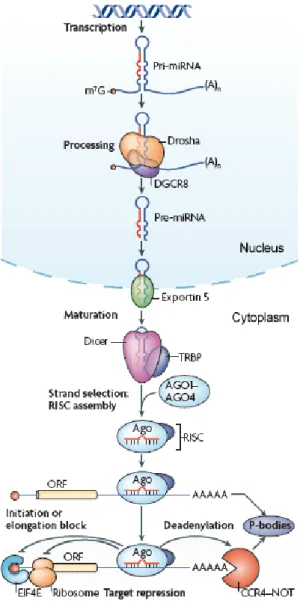
Figure 1.6. Schematic overview of miRNA biogenesis ... 29
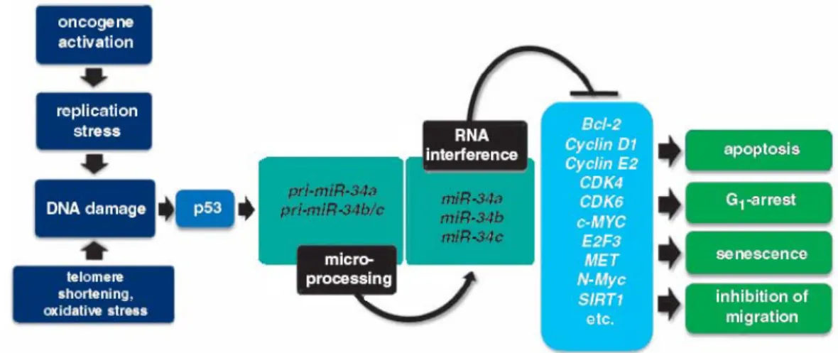
Figure 1.7. Involvement of members of miR-34 family in p53 network ... 33
Figure 2.1. Time course of neurogenesis and gliogenesis ... 47
Figure 2.2. The Akt/p-FOXO3A/Id1 signaling pathway is downregulated during mouse NSC differentiation ... 48
Figure 2.3. p53 activity and caspase-3 cleavage are modulated during mouse NSC differentiation. ... 50
Figure 2.4. Pan-caspase inhibition modulates p53 activity and interferes with the Akt/p-FOXO3A/Id1 pathway during mouse NSC differentiation ... 51
Figure 2.5. Inhibition of caspase activation delays neuronal and glial differentiation of mouse NSCs ... 52
Figure 2.6. Silencing of p53 decreases p-FOXO3A and Id1 expression levels during mouse NSC differentiation, without potentiating z-VAD.fmk effects ... 54
Figure 2.7. Downregulation of p53 delays neuronal and glial differentiation of mouse NSCs ... 55
Figure 3.1. Mouse NS cells have both neurogenic and gliogenic potential
in vitro ... 73
Figure 3.2. Apoptosis-associated miRNAs are modulated during mouse NS cell differentiation ... 77
Figure 3.3. Differentiation of mouse NS cells is not associated with increased cell death ... 79
Figure 3.4. Inhibition of apoptosis by TUDCA was not associated with a decrease in proapoptotic miRNAs expression... 80
Figure 3.5. miR-16, let-7a and miR-34a are increased during mouse ES cell differentiation ... 81
Figure 3.6. Differentiation of PC12 and NT2N cells were associated with modulated levels of miR-16, let-7a and miR-34a expression ... 83
Figure 3.7. miR-34a overexpression increases the ratio of postmitotic neurons in mouse NS cells ... 85
Figure 4.1. miR-34a modulates the proportion of NeuN-positive cells ... 98
Figure 4.2. SIRT1 expression levels decrease after miR-34a overexpression ... 100
Figure 4.3. miR-34a promotes the appearance of post-mitotic neurons by downregulating SIRT1 expression ... 101
Figure 4.4. miR-34a positively regulates the astrocytic population under resveratrol treatment ... 103
Figure 4.5. miR-34a overexpression increases p53-DNA binding activity. ... 105
Tables
Table 1.1. Function of mammalian caspases in cell differentiation. ... 20
Table 3.1. Proapoptotic miRs expression during mouse NS cell differentiation ... 75
1.1 Neural stem cells
Neural stem (NS) cells are self-renewing multipotent cell populations present in the developing and adult mammalian central nervous system (CNS) (Temple, 2001a). NS cells generate neurons and glia of the developing brain, and account for neuronal plasticity in the adult brain.
The process of generating functionally integrated neurons from progenitor cells, termed neurogenesis, was traditionally believed to occur only during embryonic stages in the mammalian CNS (Ramon y Cajal, 1913). However, almost forty years ago, pioneering work by Altman and Das suggested the existence of continuing neurogenesis throughout adulthood (Altman and Das, 1965). Significant progress has been made over the past few years and investigators have now firmly established that new neurons are indeed born in restricted regions of the adult mammalian CNS (Alvarez-Buylla and Garcia-Verdugo, 2002; Gage, 2000; Kempermann and Gage, 2000). Since then, stem cells have been isolated from many regions of embryonic and adult nervous systems (Fig. 1.1). The discovery of adult neurogenesis not only provides a unique model system to understand basic mechanisms of neural development in the mature CNS, but also may lead to strategies for neural tissue repair and cell-based replacement therapies of the nervous system.
Figure 1.1. Location of NS cells niches. Principal regions of embryonic and adult nervous
systems, from which neural stem cells have been isolated. SVZ, subventricular zone; PNS, peripheral nervous system. Adapted from Temple et al. 2001.
1.1.1 Neural stem cells during developing CNS
During brain development, predetermined programs give rise to different NS cell populations, specified in space and time (Alvarez-Buylla et al., 2001). Neurogenesis in mammals begins with the induction of neuroectoderm, which forms the neural plate, at embryonic day 7.5 (E7.5) in mice, and then folds to give rise to the neural tube. These structures are made up by a layer of neuroepithelial cells (Gotz and Huttner, 2005), which contact both apical (ventricular) and basal (pial) surfaces (Fig. 1.2). Initially, neuroepithelial cells divide symmetrically at the ventricular surface to increase the pool of stem cells. However, at a certain point, they start dividing asymmetrically, generating a stem cell that remains in the ventricular zone, and another cell that migrates radially outward (Haubensak et al., 2004). These cells are responsible for the first wave of neurogenesis in the neural tube, after which they give rise to radial glia (Conti and Cattaneo, 2010).
Figure 1.2. NS cells in the developing brain. NS cell (shown in blue) transformation from
neuroepithelial cells to radial glia, to astocyte-like cells of the lateral ventricular wall. Neural stem cells can produce progeny, either directly or via an intermediate progenitor (shown in green),
is constituted by the neural tube, which is composed by neuroepithelial cells. These cells can divide either symmetrically, at the ventricular surface, to expand the stem cell pool, or asymmetrically, to generate neurons. (b) Radial glial cells arise from neuroepithelial cells, and divide to generate neurons or glia, either directly or via an intermediate progenitor in the SVZ. The radial processes of radial glial cells support the migration of neuroblasts (shown in red). (c) In the adult brain, radial glia have transformed into astrocyte-like cells that have the capacity to generate olfactory bulb interneurons. The ventricular zone (VZ) also disappears, but SVZ remains into adulthood in some areas. Stri, striatum. Adapted from Merkle et al. 2006.
Radial glia arise in the neural tube at about E9.5 in mice. These cells originate from neuroepithelial cells at the beginning of neurogenesis, and represent the main cell type in the developing brain, where they act both as neural progenitors and as scaffolding for migrating newborn neurons (Conti and Cattaneo, 2010). Like neuroepithelial progenitors, radial glia are a transient population in the developing brain that exhibits definite morphological hallmarks. Their soma are located in the ventricular zone, and they extend a long process to the pial surface. In addition, radial glia exhibit several glial characteristics, such as glycogen granules (Choi and Lapham, 1978), and the expression of numerous glial antigens, including brain lipid binding protein (Feng et al., 1994), the glutamate transporter GLAST (Shibata et al., 1997), tenascin (Yuasa, 2001), and glial fibrillary acidic protein (GFAP) in some species (Choi and Lapham, 1978; Levitt and Rakic, 1980). In vivo, radial glia have the ability to generate different neural lineages, in a time-controlled fashion (Anthony and Heintz, 2008; Ogawa et al., 2005). A similar heterogeneity has also been observed in vitro. In mammals, radial glial cells disappear from the brain soon after birth, but in many species they persist into the adulthood (Merkle and Alvarez-Buylla, 2006). In mammals, the role of NS cells is filled by a closely related astrocyte-like cell.
Similarly to radial glia, adult mammalian NS cells have many astrocytic characteristics, such as the expression of GFAP (Doetsch et al., 1999; Garcia et al., 2004; Imura et al., 2003). In the adult brain, these astrocyte-like cells are mainly found in two regions: the subventricular zone (SVZ) of the lateral wall of the lateral ventricle, and the subgranular zone (SGZ) of the dentate gyrus of the hippocampus. The astrocytic nature of NS cells has been confirmed when GFAP+ cells were conditionally ablated in the adult mouse brain, resulting in an almost complete loss of neurogenesis (Garcia et al., 2004; Imura et al., 2003).
Since radial glial cells and SVZ astrocytes share many properties, it has been hypothesized that they belong to the same lineage (Tramontin et al., 2003). In particular, radial glial cells of the neonatal lateral ventricular wall occupy the same region as astrocytic stem cells of the adult SVZ. In summary, current evidence suggests that NS cells gradually transform from neuroepithelial cells to radial glial cells, to astrocyte-like cells (Merkle and Alvarez-Buylla, 2006).
1.1.2 Adult neurogenesis
New neurons are continuously added to neural circuits in the adult vertebrate brain (Doetsch, 2003). In mammals, including humans, neurons are added to restricted brain regions, the olfactory bulb and the hippocampus (for a review, see (Zhao et al., 2008a). The addition of new neurons to these regions represents a process by which the brain can make changes to its own functional circuitry, in addition to molecular, synaptic or morphological alterations in individual cells. Indeed, this cell-level renovation is not static or merely restorative, rather constitutes an adaptive response to challenges imposed by the environment and/or internal state of the animal (Lledo et al., 2006).
The identity of NS cells and adult neurogenesis have been most clearly defined in the SVZ of adult mice. The SVZ is a layer of dividing cells that extends along the lateral walls of the lateral ventricle. Neurons are born throughout the SVZ, and feed into a network of chains of tangentially migrating neuroblasts (Doetsch and Alvarez-Buylla, 1996) that coalesce to form the rostral migratory stream (RMS), leading to the olfactory bulb (Fig. 1.3). Here, neuroblasts, differentiate into two kinds of olfactory interneurons, granule and periglomerular cells (Doetsch and Scharff, 2001). The SVZ is separated from the lateral ventricle by a single layer of multiciliated ependymal cells. Chemorepulsive factor gradients produced by these cells guide neuroblast migration in the adult brain (Sawamoto et al., 2006). More than 30,000 neuroblasts exit the rodent SVZ for the RMS, each day (Alvarez-Buylla et al., 2001).
Three types of precursor cells exist in the SVZ: type B GFAP+ progenitors, type C transit amplifying cells, and type A migrating neuroblasts (Fig. 1.3). Type B cells are slowly dividing radial-glia like progenitors, which have been hypothesized to represent the primary NS cells in vivo (Mu et al., 2010). They are thought to generate rapidly dividing progenitors, type C cells, which typically have either no or very short processes, and are characterized by the expression of Dlx2, Mash1, and epidermal growth factor
receptor (EGFR). The majority of these intermediate progenitors subsequently give rise to type A cells that express both doublecortin (DCX) and poly-sialated neural cell adhesion molecule (PSA-NCAM). These cells migrate into the olfactory bulb, through the RMS, and differentiate into GABA-producing and dopamine-producing interneurons. Interestingly, the differentiation potential of SVZ progenitor cells appears to be limited, as the fate of the progeny is determined by the positional information established during early development of the CNS (Merkle et al., 2007).
Figure 1.3. Neurogenesis in the SVZ. Progenitor cells (A–C) in the SVZ are located next to the
ependymal cell (E) layer lining the lateral ventricles. A subset of slowly dividing GFAP+ radial
cells (type B cells) has the potential to serve as adult NS cells and generate rapidly dividing, transit-amplifying nonradial NS cells (type C cells), which in turn give rise to neuroblasts (type A cells) that migrate through the rostral migratory stream (RMS) toward the olfactory bulb (OB). Newborn neurons are integrated as granule neurons, in the granule cell layer (GCL), and as periglomerular neurons (not shown), in the glomerular layer (GL). Mi, mitral cell layer; EPL, external plexiform layer. Adapted from Zhao et al. 2008.
The hippocampus is other major site of neurogenesis in adult mammals. Granule neurons in the dentate gyrus are born locally in the SGZ, which lies between the granule cell layer and the hilus. In contrast to the extensive tangential migration undertaken by olfactory bulb neurons, hippocampal granule neurons move only a short distance into the granule cell layer (Fig. 1.4). On a superficial level, describing adult hippocampal neurogenesis is very straightforward. A dividing precursor cell gives rise to daughter cells, which migrate away from the site of division (Kuhn et al., 1996), and start to differentiate into neurons (Kempermann et al., 2003). They first extend dendrites toward the molecular layer, and later an axon that projects to area CA3 (Stanfield and Trice,
1988). Over four to seven weeks, these neurons become integrated into the local neuronal circuits (Jessberger and Kempermann, 2003).
Figure 1.4. Neurogenesis in the dentate gyrus. In the adult SGZ, a population of GFAP+ Sox2+
radial cells corresponds to quiescent NS cells (type 1 cells). They coexist with actively proliferating, GFAP- Sox2+ nonradial NS cells (type 2 cells) that generate both astrocytes and
neuroblasts. Neuroblasts then migrate into the granule cell layer, and predominantly differentiate into local glutamatergic dentate granule cells. Progenitor cells in the dentate gyrus are influenced by local astrocytes (not shown) and by the vasculature (red). GCL, granule cell layer; Mol, molecular layer. Adapted from Zhao et al. 2010.
Two types of neural progenitors can be identified in the SGZ, according to their specific morphologies and expression of unique molecular markers (Fig. 1.4). Type 1 hippocampal progenitors act as quiescent NS cells. They have a radial process, spanning the entire granule cell layer, and ramify in the inner molecular layer. These cells express nestin, GFAP, and the transcription factor sex-determining region Y-box 2, Sox2 (Fukuda et al., 2003; Garcia et al., 2004; Suh et al., 2007). Although expressing the astrocyte marker GFAP, these cells are morphologically and functionally different from mature astrocytes. They may generate actively self-renewing nonradial progenitors (type 2 cells), expressing Sox2 and nestin, but not GFAP. Type 2 cells, in turn, give rise to DCX+ neuroblasts that predominantly differentiate into local glutamatergic dentate
granule cells (Mu et al., 2010). A recent study showed that type 2 Sox2+ cells can self-renew, and that a single Sox2+ cell can give rise to neurons and astrocytes, providing the
first in vivo evidence of stem cell properties of hippocampal neural progenitors (Suh et al., 2007).
1.1.3 In vitro culturing of NS cells
For nearly 20 years, growth factor-based protocols have been developed, leading to NS cell expansion in both floating and adherent conditions (Kokovay et al., 2008). In addition, progress in cell culture technologies has enabled researchers to induce the neuralization of mouse and human embryonic stem (ES) cells, derived from the inner cell mass of the blastocyst, in vitro (Conti and Cattaneo, 2010). During neural differentiation, ES cells undergo progressive lineage restrictions, similar to those observed in normal fetal development (Pankratz et al., 2007), leading to the generation of a range of distinct neural precursor populations. These can be used to study the molecular and cellular events that occur during stage-specific transitions between different populations (Nishikawa et al., 2007).
Neurospheres are free-floating aggregates of neural progenitors, each potentially derived from a single NS cell (Reynolds et al., 1992). Their generation relies on tissue microdissection, followed by exposure to growth factors (Chojnacki and Weiss, 2008). Neurospheres and monolayer culturing systems of NS cells can be generated from ES cells, induced pluripotent stem (iPS) cells (derived from reprogrammed somatic cells) and from stem cell niches of the fetal and adult brain (Conti and Cattaneo, 2010). Both systems have the potential to give rise to neurons, astrocytes and oligodendrocytes. The different cellular compositions of the neurospheres (a mixed population, with only a fraction of the cells exhibiting NS cell properties) and monolayer cultures (with homogeneous composition) results in low and high neurogenic potential, respectively.
Although many efforts are being made to better understand cellular and molecular events associated with differentiation (Abranches et al., 2009), determining the best sources for the in vitro derivation of NS cells and optimizing protocols for stable, clonal proliferation are still central goals of stem cell research.
1.1.4 Potential therapeutic uses of NS cells
Despite the presence of NS cells in the adult mammalian brain, neurons are not replaced in most regions after injury or pathological conditions. However, evidence of neurogenic upregulation in the damaged brain has led to interest in therapeutic strategies
that exploit naturally existing responses. Such approaches could prove fruitful, not only for stroke and trauma patients, but also in patients with degenerative brain diseases, such as Parkinson’s disease, Huntington’s disease and multiple sclerosis (Curtis et al., 2003; Jin et al., 2004). Nevertheless, several problems must be solved before the potential attributed to stem cells becomes a realistic clinical strategy for cellular repair. The biggest single problem still to be solved is how to direct and control the differentiation of specific target phenotypes required for replacement and repair in each disease. Selection of appropriate starting cells and adequate parameters of in vitro manipulation are likely to be crucial factors in directing appropriate phenotypic differentiation. Further, a more comprehensive understanding of the molecular pathways controlling stem cell self-renewal and differentiation, in particular the apparent molecular link between programmed cell death and cell differentiation, would accelerate efforts to generate clinically relevant cell types from stem cells.
1.2 Apoptosis-associated proteins in cell differentiation
Apoptosis is a highly regulated form of cell death known to sculpt tissues during development, and maintain tissue homeostasis by eliminating unnecessary or harmful cells (Ellis et al., 1991). Apoptosis is a genetically predetermined mechanism. Genetic analysis of the nematode Caenorhabditis elegans provided the first insights into the molecular mechanism of apoptosis (Yuan et al., 1993). The canonical apoptotic pathways are well conserved in all metazoans from invertebrates to vertebrates. The best characterized and the most prominent are designated by extrinsic and intrinsic pathways (Kroemer et al., 2007). In the extrinsic pathway, also known as “death receptor pathway”, apoptosis is triggered by ligand-induced activation of death receptors at the cell surface. In the intrinsic pathway, also called “mitochondrial pathway”, apoptosis results from an intracellular cascade of events, mainly characterized by the mitochondrial membrane permeabilization (MMP).
In the extrinsic pathway of apoptosis, ligation of death receptors causes the recruitment and oligomerization of the adapter molecule Fas-associating death domain-containing protein (FADD) within the death-inducing signaling complex (DISC). Oligomerized FADD binds the initiator caspase-8 and -10, causing their dimerization and activation (Fig. 1.5) (Debatin and Krammer, 2004). As an alternative, the extrinsic
pathway can be activated by the so-called dependency receptors, which are believed to be connected to rapid caspase activation as well.
Fas-induced apoptosis has been shown to follow almost exclusively one of two different Fas signaling pathway (Scaffidi et al., 1998), mainly depending on the amount of active caspase-8 available in the cytosol. In type I cells, Fas-induced apoptosis ensures enough activation of caspase-8, so that it can go on to activate caspase-3 and -7. In contrast, in type II cells, apoptosis proceeds by an amplification loop through mitochondria. In this case, caspase-8 cleaves the inactive cytoplasmic protein Bid, a pro-apoptotic Bcl-2-family member, thus producing active truncated Bid (tBid) (Li et al., 1998). The carboxy-terminal fragment of Bid then translocates to the mitochondria leading to oligomerization of Bax or Bak and the consequent MMP, which in turn results in caspase-3 activation (Korsmeyer et al., 2000).
Most cell death in vertebrates proceeds via the intrinsic or mitochondrial pathway of apoptosis (Green and Kroemer, 2004). Here, the executioner caspases are cleaved and activated by initiator caspase-9, which is activated by multimerization on the adapter molecule apoptosis protease activating factor-1 (Apaf-1), within a multiprotein complex called “apoptosome”. Apaf-1 preexists in the cytosol as a monomer, and its activation depends on the presence of cytochrome c (Cyt c) and ATP/dATP (Cain et al., 2002). The release of Cyt c, which normally resides only in the inner mitochondrial space where it functions as an electron shuttle in the respiratory chain (Bernardi and Azzone, 1981), is rate-limiting for the generation of the apoptosome. Hence MMP is the critical event responsible for caspase activation in the intrinsic pathway. MMP can also commit a cell to die when caspases are not activated. This “caspase-independent death” (Chipuk and Green, 2005; Kroemer and Martin, 2005) can occur by either an irreversible loss of mitochondrial function or by the mitochondrial release of caspase-independent death effectors, including apoptosis-inducing factor (AIF) (Susin et al., 1999), endonuclease G (EndoG) (Li et al., 2001), and others (Chipuk and Green, 2005; Kroemer and Martin, 2005).
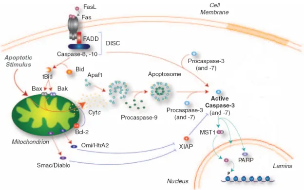
Figure 1.5. Intrinsic and extrinsic pathways of apoptosis in mammals. The intrinsic pathway
involves the mitochondria, which acts as an “intracellular death receptor”, receiving a variety of pro-apoptotic signals that trigger oligomerization of pro-apoptotic proteins (Bcl-2-associated protein, Bax, and Bcl-2-antagonist killer, Bak, to produce mitochondrial outer membrane permeabilization. This leads to the release of Cyt c, which activates Apaf-1, induction of apoptosome formation, recruitment/activation of procaspase-9, and direct processing and activation of procaspase-3 and -7. In the extrinsic pathway, the Fas receptor ligand (FasL) triggers the membrane-bound DISC, which recruits procaspase-8 and activates caspase-3 directly. In some cell types, caspase-8 can also cleave Bid to form tBid, which interacts with Bax/Bak to trigger mitochondrial outer membrane permeabilization, Cyt c release and apoptosome formation. The activation of caspase-3 and -7 is antagonized by inhibitor of apoptosis proteins (IAPs), which in turn can be inhibited by Smac/Diablo and Omi/HtrA2. Activation of caspase-3 and -7 orchestrates the demolition of the cell by cleavage of specific substrates, including poly (ADP) ribose polymerase (PARP), inhibitor of caspase-3-activated DNase (ICAD) and mammalian STE20-like kinase 1 (MST1). Adapted from D’Amelio et al. 2010.
A well-known observation in the apoptosis field is that differentiating cells are more refractory to apoptosis compared with less differentiated cells (Yi and Yuan, 2009). Next, it will be discussed the emerging evidence for nonapoptotic functions of apoptosis players in mediating cell fate specification and differentiation.
1.2.1 The tumor suppressor p53
The tumor suppressor p53 is a central protein in mammalian stress response. Owing to its ability to integrate many different signals controlling cell life and death, p53 has been named the “guardian of the genome” and plays a crucial role in maintaining genomic stability in somatic cells (Vousden and Prives, 2009). Under normal physiological conditions p53 remains inactive and unstable, but is rapidly stabilized and activated by upstream kinases in response to genotoxic and oncogenic stresses, leading to cell cycle arrest, apoptosis or senescence (Zhao and Xu, 2010). These functions of p53 can protect the genome from accumulating genetic mutations, by allowing the time for faithful repair of DNA damage or by eliminating cells with excessive DNA damage (Zhao and Xu, 2010).
p53 functions mainly as a DNA-binding, sequence-specific transcription factor that activates the expression of multiple genes, including p21, noxa and puma (Vousden and Prives, 2009), which in turn mediate p53 functions in cell cycle arrest, apoptosis and senescence. In addition, p53 regulates the expression of proteins that modulate its own activation and stability, such as Mdm-2, forming multiple positive and negative feedback loops (Harris and Levine, 2005). Posttranslational modifications of p53 are also important modulators of its function and stability (Bode and Dong, 2004). In fact, modifications have been detected on at least 30 different sites on the p53 protein. It has been proposed that the specific combination of modifications on p53 may provide selectivity toward a particular cell response.
The role of p53 in controlling genomic stability is crucial in stem cell populations. The accumulation of unrepaired DNA in ES cells could not only promote tumorigenesis in multiple cell lineages, but also pass these mutations to the progeny, leading to genetic instability in differentiated cells (Zhao and Xu, 2010). ES cells must have developed mechanisms to cope with various stresses, in particular, DNA damage insults. However, p53 checkpoint pathways are compromised in ES cells, which do not induce p53-dependent cell cycle arrest and apoptosis after certain types of physiological DNA damage, although p53 is expressed in abundant quantities in ES cells (Aladjem et al., 1998).
p53-mediated checkpoint controls can be activated during differentiation. Recent studies suggest that p53 may maintain genetic stability of ES cells by inducing differentiation. Following genotoxic stress, p53 can induce differentiation of ES cells by directly suppressing Nanog expression (Lin et al., 2005), which is the master
transcriptional factor that maintains self-renewal of ES cells. Therefore, the outcome of p53 activation in DNA-damaged ES cells is to differentiate ES cells into other cells types that can undergo efficient p53-dependent cell-cycle arrest or apoptosis (Aladjem et al., 1998; Chao et al., 2000), leading to the maintenance of genomic stability in self-renewing ES cells. Importantly, p53 was shown to suppress Nanog expression also after treatment with retinoic acid, suggesting that p53 may be important not only to maintain genomic stability but also to differentiate ES cells. Given that, p53 should be viewed not as a tumor suppressor protein, but rather as a crucial decision-maker molecule (Tedeschi and Di Giovanni, 2009; Vousden and Prives, 2009).
1.2.1.1 Role of p53 during neuronal differentiation
Before the link of p53 and Nanog had been established, mounting evidence was already suggesting a role for p53 in cell differentiation, including that of neuronal precursors. Initially, p53 was considered non-essential to development since the majority of p53-null mice were considered phenotypically normal at birth, with markedly increased tumor development later in life (Donehower et al., 1992). However, the discovery of low frequency neural tube defects in p53-/- mice (Armstrong et al., 1995; Sah et al., 1995) was an apparent paradox, and suggested that the role of p53 in development may be more complex than initially believed. Determination of p53 mRNA levels during mouse embryonic development showed that it peaked during the differentiation of several tissues, including early neuronal precursor cells of the brain, but strongly declines during terminal differentiation (Louis et al., 1988; Rogel et al., 1985; Schmid et al., 1991). In addition, evaluation of p53-dependent transcriptional activation during normal development in vivo indicated that p53 activity is at maximum during neuronal differentiation, and is clustered in areas that are not correlated with apoptosis (Gottlieb et al., 1997; Komarova et al., 1997).
Later work supported a possible link between p53 and cell differentiation of NS cells through the regulation of cell-cycle progression. One report showed that p53 negatively regulates the proliferation and survival of adult NS cells, without affecting their differentiation potential (Meletis et al., 2006). By comparing the transcriptome of adult NS cells from p53-null and wild-type mice, the authors identified altered expression of several cell cycle regulators, including downregulation of p21 expression, in the absence of p53. Of note, the expression of all well-known neuronal differentiation
markers was not affected. However, recent experiments of NS cells extracted from the olfactory bulb of wild-type and p53-null mouse embryos showed that p53 controls proliferation, chromosomal stability and differentiation pattern of mouse olfactory bulb stem cells (Armesilla-Diaz et al., 2009). In fact, p53-null NS cells have increased proliferation and a bias towards neuronal phenotype, with concomitant reduction of astrocytes. This effect had been previously reported in SVZ-derived stem cells from adult mice (Gil-Perotin et al., 2006).
In vitro models of neuronal differentiation also suggest a role for p53 during
differentiation. Studies in neuronal-like PC12 pheochromocytoma and neuroblastoma cells revealed that p53 gene expression is induced and required during neurotrophin-dependent neuronal differentiation and maturation (Hughes et al., 2000; Montano, 1997; Poluha et al., 1997; Zhang et al., 2006). Furthermore, p53 was shown to be crucial for correct neurite outgrowth and maturation of cortical neurons in vitro (Di Giovanni et al., 2006; Tedeschi et al., 2009). p53 knockdown reduced spontaneous and directed differentiation rate in human ES cells (Qin et al., 2007), while activation of p53 with the small-molecule activator nutlin led to rapid differentiation (Maimets et al., 2008). Still, although p53 protein levels were reduced after retinoic acid-induced ES cell differentiation, its transcriptional activity was increased (Lin et al., 2005). Finally, the involvement of p53 has also been shown in spermatogenesis (Rotter et al., 1993), eye development (Reichel et al., 1998), renal development (Saifudeen et al., 2009), osteogenesis (Lengner et al., 2006), immune development (Matas et al., 2004), lung development (Tebar et al., 2001) and muscle differentiation (Porrello et al., 2000).
1.2.1.2 Mechanism of p53 control of cell differentiation
Despite the supporting data on transcriptional activity of p53 protein in differentiation, the mechanism through which p53 acts in this cellular context is still a puzzling question. p53 was reported to play a critical role in nerve growth factor (NGF)-mediated neuronal differentiation of PC12 cells, in part via regulation of TrkA expression, which in turn activates mitogen-activated protein kinase (MAPK) pathways (Browes et al., 2001; Zhang et al., 2006). In the same study, p53 was reported to regulate cell cycle arrest by inducing p21 expression. Moreover, by genome-wide chromatin immunoprecipitation (ChIP), Brynczka and colleagues have recently identified new putative p53 target genes during NGF-mediated PC12 neuronal differentiation, among
which are wnt7b involved in dendritic development, and the tfcp214/grhl3 grainyhead homolog implicated in ectodermal development (Brynczka et al., 2007). Di Giovanni and co-workers showed that p53 regulates the expression of both actin-binding protein Coronin 1b and the GTPase Rab13, which are required for physiological neurite outgrowth in PC12 cells and dorsal root ganglion neurons (Di Giovanni et al., 2005). In this sense, within the PC12 neuronal context, p53 may have the ability to regulate two critical events for neuronal differentiation, cell cycle arrest and neurite outgrowth.
In ES cells, p53 is involved in the rapid down-regulation of Nanog expression during retinoic acid-induced differentiation (Lin et al., 2005). Furthermore, p53-dependent expression of p21, Mdm2 and Killer/DR5 were all also markedly induced indicating again a mechanism of action involving cell cycle regulation. In fact, it has been shown that p21 can negatively regulate the self-renewal of adult NS cells (Mori et al., 2001). Similar results were obtained in human ES cells (Maimets et al., 2008).
Integrated transcriptomic profiling, in silico promoter analysis and functional studies of murine NS cells established that dual, but not singular, inactivation of p53 and
Pten promotes an undifferentiated state, with high self-renewal potential, and drives
increased Myc protein levels (Zheng et al., 2008). Functional studies validated increased Myc activity as a potent contributor to impaired differentiation and enhanced self-renewal of NS cells doubly null for p53 and Pten.
Studies in neuronal cells have suggested that the interaction of p53 with the neuronal specific and pro-differentiation transcription factor Brn-3a facilitates a shift of p53 transcriptional activity from cell death to neuronal differentiation (Hudson et al., 2005). In fact, when forming a complex with Brn3a, p53 is not able to activate procell -death genes such as Bax and Noxa, but rather shows increased affinity for the pro-differentiation gene p21. Therefore, Brn-3a might play a crucial role in determining the pathway taken by p53, when co-expressed during development, and in controlling the cell fate.
Thus, p53 may generally contribute to neuronal differentiation by targeting cell cycle regulators, and particularly by controlling the expression of differentiation-specific genes in a cell-type specific manner. This could be directed by the interaction between Brn-3a and p53, which facilitates a shift of p53 transcriptional activity from cell death to neuronal differentiation.
1.2.1.3 p53 switch and multiple biological functions
Genomic elements regulated by p53 during neuronal differentiation may be unique, and different from those regulated in genotoxic stress and apoptosis. This is made possible by the multiple post-translational modifications that target p53 on its N- and C-termini which are not limited to phosphorylation, but may also include acetylation, sumoylation, neddylation, and ubiquitination (Lavin and Gueven, 2006). p53 post-translational modifications directly affect the transcriptional activity of p53 and regulate its affinity to diverse cofactors, which in turn regulate the occupancy of p53 specific promoters (Sims and Reinberg, 2008).
The best characterized p53 post-translational modifications are phosphorylation and acetylation. Kinases phosphorylate several serine and threonine residues, mainly in the p53 N-terminal region. Phosphorylation at Ser 15, Ser 20, Ser 33, Ser 46 and Thr 18 promotes p53 stabilization by preventing either its nuclear export, or its recruitment to specific promoters. Importantly, soon after the onset of differentiation of ES cells, phosphorylation of p53 at Ser 15, Ser 315 and Ser 392 is significantly increased, which is thought to activate p53 (Xu, 2003). p53 phosphorylation at Ser 315 was also shown to be particularly important for p53-dependent suppression of Nanog during differentiation of ES cells (Lin et al., 2005).
Acetylation targets mainly two distinct regions of the p53 C-terminus and, in neurons, involves the activity of at least two histone acetyltransferases, CREB-binding protein (CBP)/p300 and p300/CBP-associated protein (P/CAF). CBP/p300 acetylates Lys370, Lys372, Lys373 and Lys382, whereas P/CAF acetylates only a single residue, Lys320 (Brooks and Gu, 2003). Importantly, these kinase and acetyltransferase pathways are activated downstream from NGF and brain-derived nerve growth factor (BDNF) signaling during neuronal differentiation and axon outgrowth. Interestingly, the acetylation of p53 at Lys 320 by P/CAF and the highly homologous acetyltransferase CGN5 leads to increased transcriptional activation of the p21 promoter, which triggers G1/S arrest and promotes neuronal differentiation in PC12 cells (Wong et al., 2004). In addition, p53 acetylation in the same residue was reported to be involved in the promotion of neurite outgrowth (Di Giovanni et al., 2006). Furthermore, acetylated p53 at Lys372, Lys373 and Lys382 was reported to drive axon outgrowth and GAP-43 expression, and to bind specific elements on the neuronal GAP-43 promoter (Tedeschi et al., 2009).
The ability of p53 to modulate different transcriptional responses in neurons, depending on specific signaling and transcriptional contexts might be linked not only to the post-transcriptional modifications of p53, but also to the cross-talk between p53 and post-transcriptional modifications on histones. Histone modifications influence chromatin structure, which in turn influences the capacity of transcription factors to bind to specific promoters. A unique chromatin environment could regulate the affinity of p53 to specific promoters. Specific patterns of p53 codes might lead to different biological outcomes, depending on the transcriptional context in a given cell or tissue (Murray-Zmijewski et al., 2008). Understanding the role of such mechanisms in the regulation of the neuronal phenotype during development undoubtedly represents an exciting challenge for future investigations.
1.2.1.4 Role of p53 in limiting somatic cell reprogramming
The recent demonstration of in vitro reprogramming of somatic cells into iPS cells by Yamanaka and colleagues represents a major advance in the field of stem cells. This discovery raises hope for modeling of human diseases, especially those with complex genetic traits. In addition, iPS cells could become a renewable source of autologous cells for transplantation into human patients.
Reprogramming was initially achieved by transduction of four transcription factors (Oct3/4, Sox2, c-Myc, and Klf-4), but only a minority of donor somatic cells can be reprogrammed to pluripotency (Takahashi and Yamanaka, 2006). Although alternative and more efficient reprogramming protocols are being subject of extensive investigation, it was recently shown that the inhibition of the p53/p21 pathway increased cell division rate and resulted in accelerated kinetics of iPS cell formation, which was directly proportional to the increase in cell proliferation (Hanna et al., 2009). By comparing the reprogramming efficiency of wild-type and p53-deficient mouse and human fibroblasts, several studies have shown great increase in reprogramming frequency in p53-deficient cells, indicating that p53 is an effective barrier of induced pluripotency (Hong et al., 2009; Kawamura et al., 2009; Zhao et al., 2008b). Nevertheless, if inactivation of the p53 pathway is a prerequisite for successful reprogramming, these findings raise concerns on the genomic stability and tumorigenecity of iPS cells and their derivatives.
1.2.2 Caspases
Caspases are a family of cysteinyl aspartate-specific proteases that are highly conserved in multicellular organisms and function as central regulators of apoptosis. To date, 11 genes were found in the human genome to encode 11 human caspases, caspase-1 to -10 and caspase-14, whereas 10 genes were found in the mouse genome to encode 10 murine caspases, caspase-1, -2, -3, -6, -7, -8, -9, -11, -12 and -14 (for a review, see Li and Yuan, 2008). Human caspase-4 and -5 are functional orthologs of mouse caspase-11 and -12, whereas human caspase-10 is absent in the mouse genome. The remaining caspases with the same numbers in human and mouse are functional orthologs of each other. Members of the caspase family are subdivided into upstream “initiator” caspases (in mammals, caspase-1, -2, -4, -5, -8, -9, -10, -11, and -12), which respond to proapoptotic signals, and downstream “executioner” caspases (in mammals, caspase-3, -6, -7, and -14) (Yi and Yuan, 2009). Caspases are synthesized as zymogens and their activation requires allosteric conformational changes, specific cleavage after a selective aspartate residue, or both. To date, four pathways leading to caspase activation have been characterized in vertebrate organisms, including the receptor-mediated pathway, the mitochondria-mediated pathway, the granzyme B-mitochondria-mediated pathway, and the endoplasmic reticulum-mediated pathway (Nicholson, 1999). Extensive biochemical and structural analysis indicate that inflammatory and initiator caspases are recruited to larger complexes that induce caspase dimerization, autoproteolysis, and protease activation (Shi, 2004). Following activation, apoptotic initiator caspase-8 and -9 cleave executioner procaspase-3, -6, and -7. This leads to cleavage of more than 1,000 proteins (Dix et al., 2008; Mahrus et al., 2008) to produce the characteristic apoptotic phenotypes of membrane blebbing, nuclear condensation, DNA fragmentation, and ultimately phagocytosis by immune cells.
Although the critical function of caspases in apoptosis is firmly established, some pro-apoptotic caspases, namely caspase-3, -8 and -14 also regulate differentiation in certain cells types (Table 1.1). The cellular processes that stop caspases short of inducing apoptosis in diverse biological settings remain a mystery.
Table 1.1. Function of mammalian caspases in cell differentiation
Caspase Cell Type
1 Not determined
2 Not determined
3
Epithelial lens, erythroblasts, megakaryocytes, spermatids, NS and ES cells, osteoclasts, bone marrow stromal and skeletal muscle cells
4 Not determined
5 Not determined
6 Not determined
7 Not determined
8 Macrophages and trophoblasts
9 Not determined
10 Not determined
11 Not determined
12 Not determined
14 Keratinocytes
Adapted from Yi et al. 2009.
1.2.2.1 Caspase-3
The role of caspase-3 during terminal differentiation has been well established. Terminal differentiation of certain cell types, such as lens epithelial cells, erythroblasts, and megakaryocytes, requires the elimination of nuclei. The enucleation process of lens epithelial cells is prevented after inhibition of caspase activity by the pan caspase inhibitor, z-VAD.fmk, suggesting that caspases are involved in the specific elimination of nuclei as opposed to elimination of the whole cell (Ishizaki et al., 1998). The terminal maturation of erythroblasts into enucleate red blood cells also requires caspase activity. Caspase-3, together with caspase-9, -7 and -2 are transiently activated during erythroid differentiation induced by erythropoietin, and caspase inhibition completely inhibits erythroid differentiation at the basophilic erythroblast stage (Carlile et al., 2004; Zermati
et al., 2001). Caspase activation during enucleation is associated with the cleavage of lamin B and acinus, which are necessary for the maintenance of nuclear integrity and condensed chromatin, respectively (Zermati et al., 2001). Finally, during the differentiation of megakaryocytes into mature platelets, cytoplasmic fragmentation of megakaryocytes leads to the formation of enucleate cells, rather than removal of nucleus,
per se (Radley and Scurfield, 1980). Interestingly, activated caspase-3 was detected in
maturing megakaryocytes, while the pan-caspase inhibitor z-VAD.fmk, as well as more specific inhibitors of caspase-3 and -9, blocked platelet differentiation (De Botton et al., 2002).
Localized caspase activity has been also found to play an important role during sperm development. Dramatic removal of bulk cytoplasm occurs in the terminal differentiation of spermatid into individual sperm. Multiple fly caspases have been implicated in this highly specialized cellular event, known as spermatid individualization. It was found that, during this process, caspase-3 is detected in the individualization complex, a cytoskeletal membrane complex that moves along the length of the cyst to the sperm tail (Arama et al., 2003).
The function of caspase-3 in cell differentiation may extend beyond terminal differentiation. In the CNS, non-apoptotic active caspase-3 expression has been located in proliferating and differentiating neuronal cells of the VZ and external granular layer of the developing cerebellar cortex (Oomman et al., 2006). In addition, caspase-3 activation is also observed in glial cells within the developing cerebellum.
In the past decade, the cell autonomous pro-differentiation effects of caspase-3 have been noted in diverse cell types including neurons (Rohn et al., 2004), neural and glial progenitor cells (Fernando et al., 2005; Oomman et al., 2006), and also osteoclasts (Mogi and Togari, 2003), bone marrow stromal cells (Miura et al., 2004), and skeletal muscle cells (Fernando et al., 2002). Chemical or genetic blockade of caspase-3 activity leads to inhibition of differentiation.
Two recent studies implicate caspase-3 in controlling fate of both ES cells and adult hematopoietic stem cells. One study reported caspase-induced cleavage of Nanog in differentiating ES cells (Fujita et al., 2008). Unsurprisingly, stem cells lacking the
Caspase-3 gene showed marked defects in differentiation, while forced expression of
caspase cleavage-resistant Nanog mutant in ES cells strongly promoted self-renewal. This suggests that Nanog is the key caspase substrate for limiting the self-renewing capacity of ES cells. Curiously, caspase-3 was show to contribute to stem cell
quiescence, by dampening specific signaling events. Alteration in caspase-3 expression perturbs homeostasis of primitive hematopoietic cells (Janzen et al., 2008). Caspase-3-deficient stem cells displayed accelerated proliferation and retarded differentiation. At the molecular level, caspase-3 altered the sensitivity of primary hematopoietic cells to cytokine stimulation through modulation of specific signaling pathways, such as extracellular signal-related kinase (ERK) signaling. These data indicate a novel role for caspase-3 as a governor, downregulating stem cell responsiveness to environmental signals.
Interestingly, caspase-3 together with caspase-8 was recently shown to be involved in somatic reprogramming. Induction of iPS cells by transduction of Oct-4, a key iPS transcription factor, is associated with caspase-3 and -8 activation (Li et al., 2010a). Inhibition of both caspases in human fibrobast cells prevented the induction of iPS cells, suggesting that caspase-3 and -8 may play a role in the reprogramming process.
Finally, caspases are involved in dendritic pruning and synaptic plasticity. Two studies indicate that localized caspase activity is important for the selective removal of sensory neuron dendrites during fly metamorphosis (Kuo et al., 2006; Williams et al., 2006). The involvement of caspases in dendritic pruning suggests that caspase activity is specific to cellular compartments, and does not cause cell death. Furthermore, caspase-3 activity was shown to be regulated at postsynaptic sites in the brain following stimuli associated with memory (Huesmann and Clayton, 2006). In fact, caspase-3 activity is necessary to consolidate a persistent physiological trace of the song stimulus.
1.2.2.2 Caspase-8 and -14
Caspase-8 is involved in differentiation of macrophages. Caspase-8 deletion in cells of the myelomonocytic lineages led to arrest of differentiation into macrophages (Kang et al., 2004). In fact, caspase-8 activation is observed during differentiation of monocytes into macrophages, but not in dendritic cells (Netea et al., 2008; Sordet et al., 2002). An attempt to identify possible caspase substrates during macrophage differentiation was made, and several proteins associated with cytoskeleton rearrangements were identified (Cathelin et al., 2006). Expression and activity of caspase-8 are also necessary for the specialized cell fusion events that are necessary for the differentiation of human placental villous trophoblas (Black et al., 2004).
Caspase-14 is implicated in terminal differentiation of keratinocytes into enucleate cells that form the skin outer layer. Caspase-14 expression is highly specific for keratinocytes during the enucleation process (Lippens et al., 2000). Caspase-14-deficient mice present a unique phenotype of shiny and lichenified skin (Denecker et al., 2007). Although it was previously though that caspase-14 lacked protease activity (Van de Craen et al., 1998), caspase-14 was found to directly cleave filaggrin, an abundant protein in the epidermis, in vitro (Denecker et al., 2007). These findings indicate that caspase-14 plays a surprising role in maintaining the integrity of skin, although it is uncertain whether it directly regulates the enucleation process of keratinocytes.
1.2.2.3 Death versus differentiation phenotypes
It is still unclear how differentiating cells restrain active caspases without compromising cellular integrity. One explanation is that the choice between death and differentiation derives largely from caspase targeting and activation of specific substrates or cofactors that are unique to each event. In this regard, it was shown that during differentiation, but not during apoptosis, the chaperone protein Hsp70 protects the transcription factor GATA-1, a master regulator of erythroid maturation, from caspase-mediated proteolysis during the enucleation process of erythroids (Ribeil et al., 2007). It is unclear, however, whether the selective masking of caspase substrates by Hsp70 provides a universal control mechanism for substrate specificity. Clearly, a systematic investigation of each caspase substrate is needed to clarify whether these proteins maintain a regulatory role in both apoptosis and cell differentiation.
In the absence of substrate selectivity, a plausible explanation for death versus differentiation phenotypes may originate with the timing and intensity of signal pathway activation. Specifically, the degree of caspase activity required to effectively act on differentiation signals was lower than that observed to induce apoptosis (Weber and Menko, 2005). In addition to temporal kinetics, the final choice between death and differentiation may also depend on subcellular localization of activated caspases and their constituent regulatory pathways. Finally, negative control by inhibitory proteins of the inhibitor of apoptosis protein (IAP) family (Plenchette et al., 2004) may account for the maintenance of cell survival during caspase-mediated differentiation.