Trabalho Final do Mestrado Integrado em Medicina
2015/2016
Paracentral Acute Middle Maculopathy
Case report and literature review
Tiago Morais Sarmento
Clínica Universitária de Oftalmologia
CHLN - Hospital Santa Maria
Supervisor: Dr.ª Sara Vaz-Pereira
Coordinator: Prof. Doutor M. Monteiro-Grillo
Paracentral Acute Middle Maculopathy - Case report and literature review Tiago Morais Sarmento
Index
Resumo ... 1 Abstract ... 2 Introduction ... 3 Case Report ... 4 Discussion ... 8 Acknowledgments... 10 References ... 11Resumo
Introdução: Maculopatia média aguda paracentral (PAMM) designa lesões retinianas com
alterações na camada nuclear interna (INL) em tomografia de coerência óptica de domínio espectral (SD-OCT), sendo um achado tomográfico de isquémia retiniana profunda. PAMM associa-se a patologia vascular da retina. Descrevemos um caso clínico de PAMM, utilizando uma abordagem imagiológica multimodal.
Métodos: Relato de caso clínico e revisão de literatura, tendo sido realizado exame
oftalmológico e avaliação imagiológica com imagem de reflectância infravermelha (NIR), autofluorescência (FAF) e SD-OCT.
Resultados: Doente de 13 anos com queixas de escotoma paracentral agudo no olho direito
(OD) que se traduziu numa numa lesão paracentral hiporreflectiva no NIR e hipoautofluorescente. No SD-OCT, era evidente uma banda hiperreflectiva na INL. Após um mês, deu-se resolução do escotoma paracentral e da lesão na FAF, atenuação na NIR e atrofia subsequente da INL no SD-OCT. A pesquisa de factores de risco cardiovascular foi negativa.
Conclusão: Este caso demonstra a importância de suspeitar de PAMM perante queixas
visuais inespecíficas com exame objetivo normal. Este achado clínico deve ser considerado como complicação de vasculopatia retiniana ou de doença sistémica, podendo ser único sinal da sua presença. A constatação de PAMM deve motivar uma avaliação e investigação apropriadas. Na ausência de doença concomitante, a PAMM poderá ser considerada idiopática.
2
Paracentral Acute Middle Maculopathy - Case report and literature review Tiago Morais Sarmento
Abstract
Purpose: Paracentral acute middle maculopathy (PAMM) refers to retinal lesions with
changes in the inner nuclear layer (INL) on spectral-domain optic coherence tomography (SD-OCT), being a tomographic finding of deep retinal ischemia. PAMM is associated with retinal vascular pathology. We report a case of PAMM, using a multimodal imaging approach.
Methods: Case report and literature review. Ophthalmic examination and imaging study with
near infrared reflectance (NIR), fundus autofluorescence (FAF) and SD-OCT were performed.
Results: A 13-year-old female presented with an acute paracentral scotoma of the right eye
with a respective paracentral lesion hyporreflective on NIR and hypoautofluorescent. SD-OCT showed a hyperreflective band in the INL. At one-month, there was resolution of the paracentral scotoma and of the lesion in FAF, attenuation in NIR and subsequent INL thinning in SD-OCT. The patient did not present cardiovascular risk factors.
Conclusions: This case shows the importance of being suspicious of PAMM when faced
with nonspecific visual complaints with a normal examination. This clinical finding should be considered a complication of a retinal vasculopathy or systemic disease, possibly being the only sign to its presence. The recognition of PAMM should prompt an appropriate evaluation and investigation. In the absence of associated disease, PAMM might be considered idiopathic.
Introduction
Paracentral Acute Middle Maculopathy (PAMM) is a recently identified clinical condition characterized by a band-like, hyperreflective lesion on spectral domain-optical coherence tomography (SD-OCT) which affects the middle layers of the retina at the level of the inner nuclear layer (INL) and the outer plexiform layer (OPL). It courses with subsequent INL thinning and is a non-specific sign of retinal ischemia.1-4
It is now accepted that a vasculopathic cause is in the origin of PAMM, due to the relation between the location of these lesions and the deep capillary plexus. This plexus comprises the intermediate (ICP) and deep plexus (DCP) which are, respectively, located along the inner and outer aspects of the INL. The affected area in PAMM (INL and OPL layers) is actually a watershed-like region caused by a high macular oxygen demand (greatest concentration of photoreceptors) associated with a limited oxygen supply.1-8 Recent optical coherence tomography angiography (OCT-A) studies identified prunning, attenuation and low flow of the DCP in the zones of PAMM lesions, corroborating this hypothesis.9-11 Thus, it not surprising that PAMM lesions were more frequently identified in patients with local retinal vascular pathologies such as diabetic or hypertensive retinopathy, central retinal vein occlusion (CRVO), retinal artery occlusion (RAO), Purtscher's retinopathy, sickle cell retinopathy, inflammatory occlusive retinal vasculitis, but also in patients with systemic factors such as hypovolemia, migraine, medications, post H1-N1 vaccination, orbital compression injury and viral prodromes.2-11
However, while there is strong evidence of the causal relationship between PAMM and retinal ischemia, there is a subgroup of patients with PAMM that have distinct demographic and risk profiles (young, female or without any vasculopathic risk factors). They present without an accounted pathological cause and mechanism for the retinal ischemia and, as such, can be classified as having idiopathic PAMM. 5,8,11 This subgroup, in some descriptions, has
been interpreted as a new variant of acute macular neuroretinopathy (AMN) as AMN classically occurs in young female patients with a recent history of a hypotensive or hypertensive episode induced by febrile illness, trauma or medication.5,8,11
4
Paracentral Acute Middle Maculopathy - Case report and literature review Tiago Morais Sarmento
Case Report
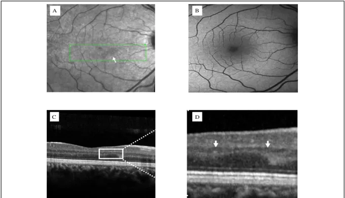
A 13-year-old, otherwise healthy white female presented with a 3-day history of a paracentral scotoma in the right eye (OD). Medical history was negative for ophthalmological pathologies, for flu-like symptoms and recent vaccination and her best-corrected visual acuity (BCVA) was 20/20 in both eyes. On examination, both anterior and posterior segments were unremarkable, with no signs of intraocular inflammation. Fundoscopy showed no significant changes in either eye. While multimodal imaging of OS was unremarkable, multimodal imaging of OD revealed a clear, wedge-like, dark grey lesion inferior and nasal to the fovea in both near-infrared reflectance (NIR) and fundus autofluorescence (FAF) (Figures 1A and 1B, respectively) which corresponded to a paracentral, hyperreflective, plaque-like band affecting the INL on SD-OCT (Figure 1C and 1D). These findings were compatible with PAMM. A cardiovascular work-up was performed, which was negative. No treatment was warranted.
Figure 1: Baseline multimodal imaging of the right eye. Near-infrared reflectance (A) revealed a subtle, wedge-like, dark grey lesion (arrow), which corresponded to a hypoautofluorescent paracentral lesion (B) (arrow). C- Spectral domain-optical coherence tomography revealed a hyperreflective plaque-like band involving the INL(box). D-Magnification of C, showing the affected area (between arrowheads).
A B
At one-week follow-up, there was attenuation of the scotoma and the lesion in NIR (Figure 2A) and FAF showed a less dense lesion (Figure 2B). SD-OCT examination revealed a more attenuated hyperreflective lesion at the level of the INL (Figure 2C and 2D). The ellipsoid zone was intact.
Figure 2: One-week follow-up multimodal imaging of the right eye. Attenuation of the hyporreflective lesion in both near-infrared reflectance (A) and fundus autofluorescence (B) (arrows). C- Spectral-domain optical coherence tomography showed a less hyperreflective lesion involving the inner nuclear layer (box). D- Magnification of C, showing the affected area (between arrowheads).
A B
6
Paracentral Acute Middle Maculopathy - Case report and literature review Tiago Morais Sarmento
Multimodal evaluation at one month demonstrated a complete resolution of the scotoma with a barely identifiable lesion in the NIR (Figure 3A). FAF was normalized (Figure 3B). Convalescent phase SD-OCT showed subsequent thinning of the INL (Figures 3C and 3D).
Figure 3: One-month follow-up multimodal imaging of the right eye. A- Near-infrared reflectance showed a barely defined lesion (arrowhead). B- Fundus autofluorescence showed resolution of the lesion. C- Spectral domain-optical coherence tomography revealed disappearance of the lesion with subsequent inner nuclear layer thinning (box). D- Magnification of C, demonstrating the affected area (between arrowheads).
A B
One-year follow-up demonstrated both an unremarkable NIR (Figure 4A) and FAF (Figure 4B). SD-OCT findings were the same as previously documented (Figures 4C and 4D).
Figure 4: One-year follow-up multimodal imaging of the right eye. A- Near-infrared reflectance showed no sign of the lesion. B- Fundus autofluorescence was unremarkable. C- Spectral domain-optical coherence tomography revealed subsequent inner nuclear layer thinning (box). D- Magnification of C, showing the affected area (between arrowheads).
A B
8
Paracentral Acute Middle Maculopathy - Case report and literature review Tiago Morais Sarmento
Discussion
PAMM is a band-like, hyperreflective lesion on SD-OCT which affects the middle layers of the retina at the level of the INL and the OPL. As this patient presented a lesion with these typical features plus an unremarkable clinical examination, a diagnosis of PAMM was established. Accordingly, in order to identify possible comorbidities, a clinical work-up was carried out following the flowchart in Figure 5, with negative results.
Figure 5: Paracentral Acute Middle Maculopathy (PAMM) differential diagnosis. AMN, acute macular neuroretinopathy; BRAO, branch retinal artery occlusion; CRVO, central retinal vein occlusion; CRAO, central retinal artery occlusion; R, retinopathy.
In the reported case, the patient denied any significant systemic or ophthalmological conditions, flulike illnesses and recent vaccination. Considering the age, gender, absence of any retinal disease, or any cardiovascular risk factor or relevant medical history, this case was labelled as idiopathic PAMM. Evaluation with OCT-A could have brought more insight, but it was not available in our clinic.8,11
No treatment was warranted as there are no recommended specific measures for the management of PAMM lesions.
Recently, it has been considered whether idiopathic PAMM lesions in young healthy individuals could be a new variant of AMN, mainly owing to the likely absence of retinal vascular disease or other risk factors of secondary PAMM.5,8,11 Nonetheless, it still remains to be seen whether the absence of risk factors for retinal ischemia heralds a new form of AMN or whether idiopathic PAMM is indeed a different entity with just epidemiological and risk factor similarities to AMN.8,11 Overall, while this doubt persists, this case emphasizes that, once in the presence of any scotoma with no pathological findings on clinical examination, it is of the utmost importance to conduct a multimodal imaging study to evaluate for a possible PAMM lesion. Whenever available, this multimodal approach should include NIR, SD-OCT and OCT-A. Additionally, if PAMM is confirmed, an exhaustive search for a contributing retinal or systemic vascular risk factor should be performed. If detected, their optimization should be guaranteed.
Ophthalmologists should recognize that PAMM is a clinical finding, which might be a complicating feature of an underlying retinal vasculopathic or systemic disease and that it might even be the only signal as to its presence. It is important to have a high index of suspicion for this condition in order to start an appropriate evaluation and work-up.
10
Paracentral Acute Middle Maculopathy - Case report and literature review Tiago Morais Sarmento
Acknowledgments
Firstly, I would like to thank Dr. ª Sara Vaz-Pereira for all the time, guidance and help offered throughout this project. I could have never done it without her wisdom and effort. I would like to thank Prof. Doutor M. Monteiro-Grillo, as Head of the Ophthalmology Department in CHLN, for allowing the use of the department facilities and the contact with the patient.
I would like to thank FMUL for all these years of medical and academic education and for all the opportunities to grow from a high school student to a medical student.
Lastly, I would like to thank my family and friends for all the support, enthusiasm and inspiration given to me ever since I first thought of becoming a Doctor.
References
1. Sarraf D, Rahimy E, Fawzi AA, et al. Paracentral acute middle maculopathy: a new variant of acute macular neuroretinopathy associated with retinal capillary ischemia. JAMA Ophthalmol. 2013;131:1275-87.
2. Chen X, Rahimy E, Sergott RC, et al. Spectrum of Retinal Vascular Diseases Associated With Paracentral Acute Middle Maculopathy. Am J Ophthalmol. 2015;160:26-34.
3. Tsui I, Sarraf D. Paracentral acute middle maculopathy and acute macular neuroretinopathy. Ophthalmic Surg Lasers Imaging Retina. 2013;44:S33-35.
4. Vaz-Pereira S. Paracentral acute middle maculopathy: a novel clinical finding. Oftalmologia. 2015;39:115-117.
5. Rahimy E, Sarraf D. Paracentral acute middle maculopathy spectral-domain optical coherence tomography feature of deep capillary ischemia. Curr Opin Ophthalmol. 2014;25:207-12.
6. Rahimy E, Sarraf D, Dollin ML, Pitcher JD, Ho AC. Paracentral acute middle maculopathy in nonischemic central retinal vein occlusion. Am J Ophthalmol. 2014;158:372-380.
7. Yu S, Pang CE, Gong Y, et al. The spectrum of superficial and deep capillary ischemia in retinal artery occlusion. Am J Ophthalmol. 2015;159:53-63.
8. Dansingani KK, Freund KB. Paracentral Acute Middle Maculopathy and Acute Macular Neuroretinopathy: Related and Distinct Entities. Am J Ophthalmol. 2015;160:1-3.
9. Christenbury JG, Klufas MA, Sauer TC, Sarraf D. OCT Angiography of Paracentral Acute Middle Maculopathy Associated With Central Retinal Artery Occlusion and Deep Capillary Ischemia. Ophthalmic Surg Lasers Imaging Retina. 2015;46:579-81.
10. Dansingani KK, Inoue M, Engelbert M, Freund KB. Optical coherence tomographic angiography shows reduced deep capillary flow in paracentral acute middle maculopathy. Eye (Lond). 2015. doi:10.1038/eye.2015.180.
11. Rahimy E, Kuehlewein L, Sadda SR, Sarraf D. Paracentral Acute Middle Maculopathy: What We Knew Then and What We Know Now. Retina. 2015;35:1921-30.