Faculdade de Engenharia da Universidade do Porto
Chitosan microspheres to remove Helicobacter
pylori adhesion in human gastric mucosa
Ana Patrícia Carvalho Henriques
Dissertação realizada no âmbito do Mestrado Integrado em Bioengenharia
Ramo de Engenharia Biomédica
Orientador: Inês C. Gonçalves, Instituto de Engenharia Biomédica (INEB)
ii
iii
Resumo
Helicobacter pylori (H. pylori), uma bactéria gram-negativa espiralada, é um dos agentes
infeciosos mais comuns em todo o mundo, colonizando a mucosa gástrica de mais de 50% da população mundial e cerca de 80-90% da população Portuguesa.
Devido à sua motilidade flagelar e capacidade para criar um microambiente favorável, a
H. pylori é capaz de persistir no ambiente acídico do estômago e aderir ao epitélio gástrico,
estabelecendo com sucesso a infeção. A adesão é mediada por moléculas na superfície bacteriana, denominadas adesinas, capazes de reconhecer glicanos expressos na superfície de células epiteliais gástricas e na camada de muco que reveste a mucosa gástrica. A longo prazo, a presença da bactéria aumenta significativamente o risco de desenvolver várias complicações gástricas, sendo uma delas o cancro gástrico.
Atualmente, as terapias convencionais contra a infeção causada pela H. pylori baseiam-se na administração combinada de dois antibióticos e um inibidor da bomba de protões. No entanto, o tratamento é ineficaz em 20% dos casos, deixando cerca de 140 milhões de doentes em todo o mundo sem tratamentos alternativos. As taxas de cura têm vindo a diminuir ao longo dos anos, principalmente devido à resistência bacteriana aos antibióticos e à baixa adesão dos pacientes.
Neste contexto, várias terapias alternativas estão sob investigação. O quitosano, um polímero catiónico natural e não tóxico, tem sido bastante investigado como uma ferramenta contra infeções gástricas, principalmente devido à sua biocompatibilidade e biodegradabilidade, juntamente com as suas propriedades anti- bacterianas e mucoadesivas. Apesar da crescente aplicação de sistemas de encapsulação baseados em quitosano, a utilização de microesferas de quitosano como um sistema de ligação à H. pylori foi também proposta, onde, após administração oral, as microesferas são capazes de capturar e remover bactérias do estômago, tirando partido da sua capacidade mucoadesiva/anti-bacteriana.
Embora encontradas livremente na camada de muco, as bactérias são também encontradas aderidas ao muco e à superfície de células epiteliais nas fovéolas gástricas (invaginações do estômago). A estabilidade das microesferas de quitosano em meio ácido, quando reticuladas com genipina, foi já demonstrada, bem como a sua capacidade para se ligar e remover a H. pylori aderente às células gástricas. Estudos em secções 2D de mucosa gástrica humana mostraram que as fovéolas gástricas têm aproximadamente 70 μm de largura, o que pode dificultar a penetração das microesferas anteriormente desenvolvidas, com diâmetro de cerca de 170 µm, e a remoção das bactérias instaladas no interior das fovéolas. É então sugerido que, de modo a alcançar as bactérias e removê-las do estômago, as microesferas devem apresentar um tamanho menor do que o descrito anteriormente.
iv
ratinho e humanas foi avaliada, bem como a sua capacidade de adesão à H. pylori.
Para esse propósito, microesferas de quitosano foram produzidas através de três sistemas diferentes, nomeadamente o sistema eletrostático, de pressão co-axial e aerodinâmico. Técnicas baseadas em microscopia ótica (Microscópio Ótico e IN Cell Analyzer) e na difração de laser (Mastersizer) foram usadas para caracterizar as microesferas de quitosano relativamente ao seu tamanho e morfologia.
Um modelo ex-vivo de estômagos frescos de ratinho foi utilizado para otimizar a marcação com fluorescência da mucosa gástrica. Diferentes marcadores foram testados, revelando DAPI (amostra fixa) e o marcador de membrana CellMaskTM Deep Red (fresco) com
bons marcadores para a visualização da mucosa gástrica. As microesferas de quitosano com um diâmetro médio de 20 µm foram produzidas com sucesso e incubadas com as mucosas gástricas de ratinhos e humana. Microscopia de confocal revelou a presença de microesferas em diferentes planos da mucosa, confirmando assim a sua capacidade de penetrar as fovéolas gástricas.
Além disso, a incubação da H. pylori com as microesferas revelou a sua capacidade de aderir à superfície das partículas.
Em conclusão, os resultados sugerem as microesferas de quitosano desenvolvidas como uma ferramenta promissora para explorar o tratamento de infeções causadas por H. pylori.
v
Abstract
Helicobacter pylori (H. pylori), a spiral-shaped gram-negative bacterium, is one of the most common infectious agents in the world, colonizing human gastric mucosa of over 50% of the world’s population and 80-90% of the Portuguese population.
Due to its flagellar motility and ability to create a favourable microenvironment, H. pylori is able to persist in the stomach acidic environment and attach to the gastric epithelium, establishing the infection. Its adherence is mediated by molecules (adhesins) on the bacterial surface able to recognize glycans expressed on the surface of gastric epithelial cells and mucus layer lining the gastric mucosa. Long-term carriage significantly increases the risk of developing several gastric-specific complications, going from gastritis to gastric cancer.
Current H. pylori infection conventional therapies rely on a concomitant administration of two antibiotics and proton pump inhibitor. However, the treatment is inefficient in 20% of the cases, leaving nearly 140 million patients worldwide without any alternative treatment option. The cure rates have been declining over the years, mostly due to bacterial resistance to antibiotics and poor patient compliance.
In this context, several alternative therapies are under investigation. Chitosan, a natural-nontoxic cationic polymer, has been thoroughly investigated as a tool against gastric infections, mainly due to its biocompatibility and biodegradability coupled with its anti-bacterial and mucoadhesive properties. Despite the growing application of chitosan-based encapsulation systems, the use of chitosan microspheres as a H. pylori binding system has also been proposed, where, after oral administration, microspheres are able to capture and remove bacteria from the stomach, taking advantage of their muco/bacterial adhesive capacity.
Although found free-swimming in the mucus layer, bacteria are also found adherent to the mucus layer and the surface of epithelial cells in gastric foveolae (stomach invaginations). The stability of chitosan microspheres in acidic environment, when crosslinked with genipin, has been demonstrated as well as their ability to bind and remove adherent H. pylori from gastric cells. Studies using 2D sections of human gastric mucosae have shown that human stomach foveolae are ~70 µm wide, which might hamper the penetration of previously developed chitosan microspheres, with a diameter around 170 µm, and the removal of the bacteria living within the foveolae. Therefore, it is suggested that the microspheres should be smaller than the previously developed in order to reach bacteria and remove them from the stomach.
vi
gastric foveolae as well as their ability to adhere onto H. pylori evaluated.
For this purpose, chitosan microspheres were produced by three different systems, namely high voltage electrostatic, co-axial air stream and aerodynamically driven systems. Techniques based on optical microscopy (Optical Microscopy, IN Cell Analyzer) and laser diffraction (Mastersizer) were used to characterize chitosan microspheres regarding size and morphology.
An ex-vivo model using fresh mice stomach samples was used to optimize the fluorescence labelling of gastric mucosa. Different dyes were explored, revealing DAPI (fixed sample) and CellMaskTM Deep Red plasma membrane (fresh) stain as good gastric mucosa
markers. Chitosan microspheres with an average diameter of 20 µm were successfully produced and incubated with mice and human gastric mucosa. Confocal microscopy revealed their presence in different plans of the mucosa, thus confirming their ability to penetrate the gastric foveolae.
Moreover, H. pylori incubation with chitosan microspheres has revealed their ability to adhere to the surface of the particles.
In conclusion, results suggest chitosan microspheres developed as a promising tool to explore as H. pylori infection treatment.
vii
Acknowledgements
My supervisor, Inês C. Gonçalves, for the constant trust, encouragement and support. For the guidance and knowledge shared throughout these last few months.
Cristina Martins and Bioengineered Surfaces Team for the support and shared ideas at the laboratory.
Paula Sampaio and Maria Lázaro for all the precious help on the confocal images acquisition and analysis.
Prof. Paulo Costa for the assistance with VarJ30 production system and Mastersizer equipment and for allowing their utilization throughout the project.
Cátia Lopes for the help with the cryostat and André Maia for the assistance with the IN Cell Analyzer acquisition and data analysis. Catarina Leitão regarding her help in the FACS equipment.
Last but not least, my family as well as my friends for all the support and encouragement during these five years.
This work was financed by FEDER funds through Programa Operacional Factores de Competitividade – COMPETE and by Portuguese funds through FCT – Fundação para a Ciência e a Tecnologia, in the framework of the project EXPL/CTM-BIO/0762/2013.
ix
Table of Contents
Resumo ... iii Abstract... v Acknowledgements ... vii Table of Contents ... ix Figure List ... xi Table List ... xvAbbreviations and Symbols ... xvii
Chapter 1 ... 1
Introduction ... 1
1.1 Stomach and its mucosal surface ... 1
1.2 Helicobacter pylori colonization ... 2
1.3 Current treatments ... 4
1.4 Reasons for unsuccessful treatments ... 5
1.5 Alternative therapies ... 6
1.6 Chitosan ... 7
1.7 Chitosan as encapsulation system ... 9
1.8 Chitosan as a binding agent ... 12
1.9 Active targeting to improve H. pylori treatment ... 13
Chapter 2 ... 15
Aim... ... 15
Chapter 3 ... 17
Materials and Methods ... 17
3.1 Chitosan microspheres preparation ... 17
3.1.1 Chitosan purification ... 17
3.1.2 Preparation of chitosan solution ... 17
3.2 Chitosan microspheres production ... 18
3.2.1 High voltage electrostatic system ... 18
3.2.2 Co-axial air stream system ... 18
3.2.3 Aerodynamically driven system ... 19
3.2.4 Variable conditions ... 20
3.3 Chitosan microspheres characterization ... 20
3.3.1 Size and morphology ... 20
3.3.2 Optical Microscopy... 20
3.3.3 IN Cell Analyzer ... 21
3.3.4 Mastersizer ... 21
x
3.5.2 Chitosan microspheres adhesion to mice gastric mucosa ... 24
3.5.3 Chitosan microspheres adhesion to human gastric mucosa ... 24
3.6 Helicobacter pylori adhesion to chitosan microspheres ... 24
3.6.1 H. pylori strain and culture conditions ... 24
3.6.2 Adhesion of H. pylori J99 strain to chitosan microspheres ... 24
Chapter 4 ... 27
Results and Discussion ... 27
4.1 Chitosan Microspheres Production and Characterization ... 27
4.1.1 High voltage electrostatic system ... 27
4.1.2 Co-axial air stream system ... 30
4.1.3 Aerodynamically driven system ... 40
4.2 Genipin crosslinking ... 44
4.2.1 Stability in acidic conditions ... 48
4.3 Chitosan microspheres emission spectrum ... 48
4.4 Chitosan microspheres adhesion to gastric mucosa ... 49
4.4.1 Optimization of gastric mucosa labelling ... 50
4.4.2 Chitosan microspheres adhesion to mice gastric mucosa ... 55
4.4.3 Chitosan microspheres adhesion to human gastric mucosa ... 59
4.5 Helicobacter pylori adhesion to chitosan microspheres ... 60
4.5.1 Adhesion of live H. pylori J99 strain to chitosan microspheres ... 61
4.5.2 Adhesion of fixed H. pylori J99 strain to chitosan microspheres... 62
Chapter 5 ... 65
Conclusions and future considerations ... 65
Conclusions ... 65
Future work ... 67
xi
Figure List
Figure 1 - Structures of human stomach and gastric mucosa [5]. ... 2
Figure 2 - Factors that influence the interactions of H pylori with human gastric mucosa [21]. The secretion of VacA protein by nonadherent bacteria can affect several cell types, including gastric epithelial cells and T cells [22]. Adhesins, namely BabA and SabA, mediate the binding of H. pylori to the gastric epithelial cells and mucus [16]. The adherent bacteria are able of assemble a type IV secretion system that allows the entrance of CagA protein into the gastric cells conducting to cellular alterations [20]. ... 4
Figure 3 - Chemical structure of chitosan [80]. ... 8
Figure 4 - High voltage electrostatic system. ... 18
Figure 5 - Droplet extrusion under co-axial air stream system. ... 19
Figure 6 – Aerodynamically driven system. ... 19
Figure 7 - Crosslinking reaction mechanism between chitosan and genipin [108]... 22
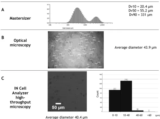
Figure 8 – Size distribution of chitosan microspheres in TPP after ionotropic gelation evaluated by Mastersizer (A), optical microscopy (B, scale bar 100 µm) and IN Cell Analyzer (C, scale bar 50 µm). ... 43
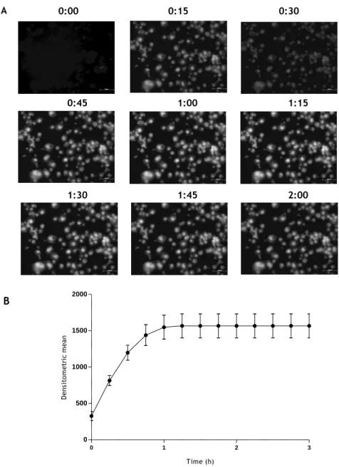
Figure 9 - Fluorescence microscopy images of chitosan microspheres crosslinked with 10 mM genipin (A). The time of crosslinking (h) is represented on the top of each image, scale bar 100 µm. Crosslinking kinetic of chitosan microspheres in the presence of 10 mM genipin (B). ... 45
Figure 10 - IN Cell Analyzer images of chitosan microspheres before (A) and after (B) lyophilisation. Scale bar 50 µm. ... 46
Figure 11 – Average diameter distribution of the individualized chitosan microspheres, after lyophilisation. ... 46
Figure 12 – Maximum chord of chitosan microspheres, after lyophilisation. ... 47
Figure 13 – Area (left graph) and form factor (right graph) of each chitosan microsphere, after lyophilisation. ... 47
Figure 14 – Optical microscopy images of chitosan microspheres in acidic conditions over 1 h. Scale bar 100 µm. ... 48
xii
Figure 16 – Fluorescence microscopy images of mice gastric mucosa mounted with
different mounting media. Scale bar 100 µm. ... 50
Figure 17 – Fluorescence microscopy images of mice gastric mucosa stained with nucleic
acid dyes (with two different concentrations, 1:100 and 1:1000), using two different mounting media. Scale bar 100 µm. ... 51
Figure 18 - Fluorescence microscopy images of mice gastric mucosa labelled with
different plasma membrane staining, at different concentrations and time of incubation (indicated above each image). Scale bar 100 µm. ... 52
Figure 19 – Auto-fluorescence of mice gastric mucosa in two ranges of emission
wavelength. CLSM images of the outer layer (Z=1) and deeper layers (Z=10 and Z=17) of mice gastric mucosa (ScanMode xyz; step size 2.9 µm). Scale bar 100 µm. ... 53
Figure 20 – Mice gastric mucosa fixed with PFA 4% and labelled with DAPI 1:100. CLSM
images of the outer layer (Z=5) and deeper layers (Z=7 to Z=15) of mice gastric mucosa (ScanMode xyz; step size 9.99 µm). Orthogonal views of two stacks (Z=9 and Z=15) are shown (ScanMode xzy). Scale bar 100 µm. ... 54
Figure 21 – Mice gastric mucosa cells labelled with CellMask™ Deep Red stain. CLSM
images of the outer layer (Z=7) and deeper layers (Z=12 to Z=36) of mice gastric mucosa (ScanMode xyz; step size 2.6 µm). Orthogonal views of stack Z=32 is shown (ScanMode xzy). Scale bar 100 µm. ... 55
Figure 22 - Fluorescence microscopy images of mice gastric mucosa alone and with
chitosan microspheres. Mucosa was fixed with PFA 4% followed labelling with DAPI 1:100 in both conditions. Scale bar 100 µm. ... 56
Figure 23 - Fluorescence microscopy images of mice gastric mucosa alone and with
chitosan microspheres. Mucosa was labelled with CellMask™ Deep Red in both conditions. Scale bar 100 µm. ... 56
Figure 24 – Chitosan microspheres (red) adhered to mice gastric mucosa fixed with PFA
4% and labelled with DAPI 1:100 (blue). CLSM images of the outer layer (Z=3) and deeper layers (Z=51 to Z=143) of mice gastric mucosa (ScanMode xyz; step size 0.17 µm). Orthogonal views of merged images are shown (ScanMode xzy). Scale bar 50 µm. ... 57
Figure 25 – Orthogonal view of chitosan microsphere (red) inserted into mice gastric
mucosa fixed with PFA 4% and labelled with DAPI 1:100 (blue) (ScanMode xzy). Scale bar 50 µm. ... 58
Figure 26 - Mice gastric mucosa cells labelled with CellMask™ Deep Red stain with
chitosan microspheres adhered (red). CLSM images of the outer layer (Z=20) and deeper layers (Z=28 to Z=59) of mice gastric mucosa (ScanMode xyz; step size 2.6 µm). Scale bar 100 µm. ... 58
Figure 27 - Human gastric mucosa labelled with CellMask™ Deep Red stain. CLSM images
of the outer layer (Z=13) and deeper layers (Z=19, Z=25 and Z=29) of human gastric mucosa (ScanMode xyz; step size 3.9 µm). Scale bar 100 µm. ... 59
Figure 28 - Human gastric mucosa labelled with CellMask™ Deep Red stain. CLSM images
of the outer layer (Z=1) and deeper layers (Z=8, Z=17 and Z=28) of human gastric mucosa (ScanMode xyz; step size 3.01 µm). Scale bar 100 µm. ... 60
xiii
Figure 29 – Fluorescence microscopy images of DAPI, Hoechst and Vectashield with DAPI
-labelled H. pylori adhered to chitosan microspheres. Scale bar 50 µm. ... 61
Figure 30 – Maximum projection of a chitosan microsphere with Vectashield with
DAPI-labelled bacteria. Images were obtained by CLSM. Scale bar 25 µm. ... 62
Figure 31 - FITC-labelled J99 strain (green) adhered to chitosan microspheres (red) under
pH 6.0 for five z-stacks (Z, step size of 2.98 µm). Images were obtained by CLSM. Scale bar 25 µm... 63
xv
Table List
Table 1 - Examples of methods for preparation of chitosan micro/nanoparticles [74]. ... 9
Table 2 – Factors influencing chitosan micro/nano particles features... 12
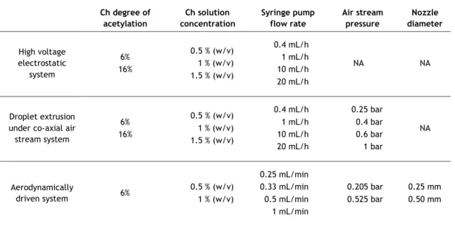
Table 3 – Conditions tested during Ch microspheres production... 20
Table 4 – Nuclear and plasma membrane stains evaluated. ... 23
Table 5 - Chitosan microspheres images obtained by optical microscopy (scale bar 200 µm). Different concentrations of chitosan solution (0.5%, 1% and 1.5% (w/v)) with DA of 6% and different flow rates (20, 10, 1 and 0.4 mL/h) are shown. Average diameter is indicated below each condition. ... 28
Table 6 - Chitosan microspheres images obtained by optical microscopy (scale bar 200 µm). Different concentrations of chitosan solution (0.5%, 1% and 1.5% (w/v)) with DA of 16% and different flow rates (20, 10, 1 and 0.4 mL/h) are shown. Average diameter is indicated below each condition. ... 29
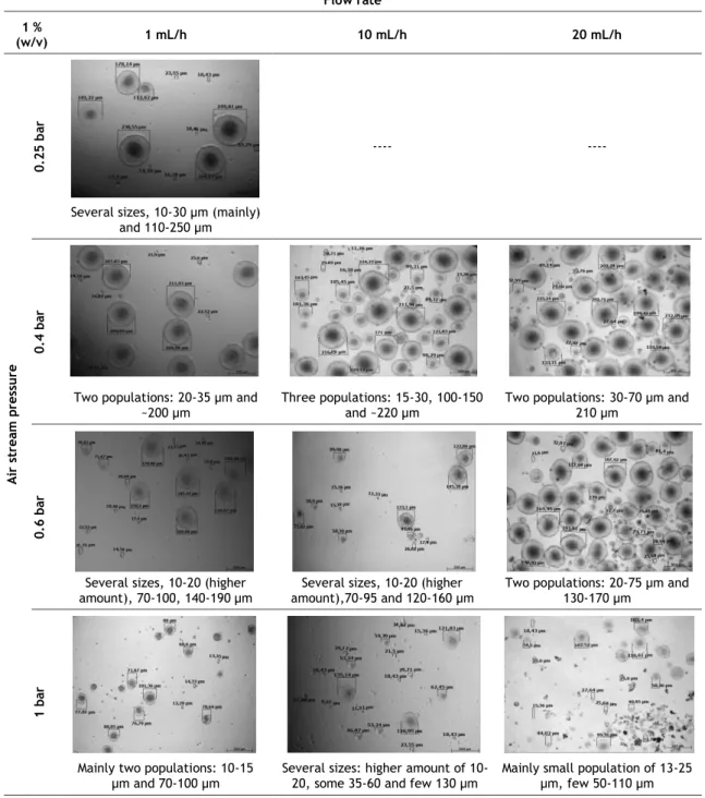
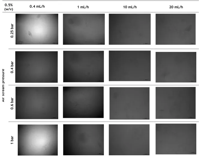
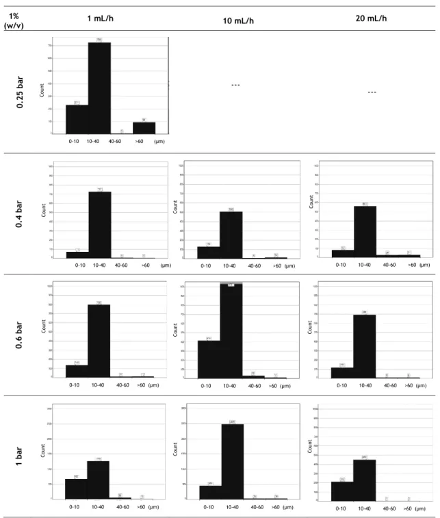
Table 7 - Chitosan microspheres images obtained by optical microscopy (scale bar 200 µm). Variation of the flow rate (20, 10, 1 mL/h) and air stream pressure (0.25, 0.4, 0.6 and 1 bar) regarding chitosan solution concentration of 0.5% (w/v)) and DA of 6% is shown. Average diameter is indicated below each condition. ... 31
Table 8 - Chitosan microspheres images obtained by optical microscopy (scale bar 200 µm). Variation of the flow rate (20, 10, 1 mL/h) and air stream pressure (0.25, 0.4, 0.6 and 1 bar) regarding chitosan solution concentration of 1 % (w/v)) and DA of 6% is shown. Average diameter is indicated below each condition. ... 32
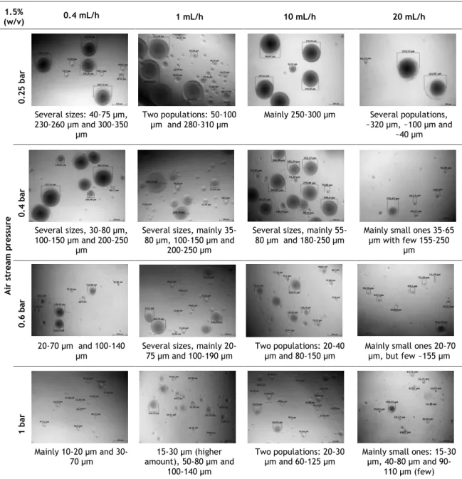
Table 9 - Chitosan microspheres images obtained by optical microscopy (scale bar 200 µm). Variation of the flow rate (20, 10, 1 and 0.4 mL/h) and air stream pressure (0.25, 0.4, 0.6 and 1 bar) regarding chitosan solution concentration of 1.5% (w/v)) and DA of 6% is shown. Average diameter is indicated below each condition. ... 33
Table 10 - Chitosan microspheres images obtained by optical microscopy (scale bar 200 µm). Variation of the flow rate (20, 10, 1 and 0.4 mL/h) and air stream pressure (0.25, 0.4, 0.6 and 1 bar) regarding chitosan solution concentration of 0.5% (w/v)) and DA of 16% is shown. Average diameter is indicated below each condition. ... 34
Table 11 - Chitosan microspheres images obtained by optical microscopy (scale bar 200 µm). Variation of the flow rate (20, 10, 1 and 0.4 mL/h) and air stream pressure (0.25, 0.4, 0.6 and 1 bar) regarding chitosan solution concentration of 1% (w/v)) and DA of 16% is shown. Average diameter is indicated below each condition. ... 35
xvi
and DA of 16% is shown. Average diameter is indicated below each condition. ... 36
Table 13 – Size distribution of chitosan microspheres produced with chitosan
concentration of 1% (w/v) and DA of 6%. Data obtained by IN Cell Analyzer analysis software. ... 38
Table 14 - Size distribution of chitosan microspheres produced with chitosan
concentration of 1.5% (w/v) and DA of 6%. Data obtained by IN Cell Analyzer analysis software. ... 39
Table 15 – Mastersizer analysis of size distribution of chitosan microspheres (DA 6%)
produced by aerodynamically driven system with nozzle diameter of 0.25 mm. Histogram (relating size classes and volume (%)) and corresponding volume percentiles (Dv10, Dv50 and Dv90) are shown. XX axis represents the size classes (µm - 10, 100 and 1000 marks are shown)... 40
Table 16 - Mastersizer analysis of size distribution of chitosan microspheres produced by
aerodynamically driven system with nozzle diameter of 0.5 mm. Histogram (relating size classes and volume (%)) and corresponding volume percentiles (Dv10, Dv50 and Dv90) are shown. XX axis represents the size classes (µm - 10, 100 and 1000 marks are shown). ... 41
Table 17 – IN Cell Analyzer size distribution of chitosan microspheres (DA 6%) produced in
the aerodynamically driven system with 0.25 mm nozzle under higher pressure (525 mBar). ... 42
Table 18 – Set of parameters applied in encapsulation system Var J30 for production of 50
xvii
Abbreviations and Symbols
BabA Blood group antigen binding adhesin CagA Cytotoxin-associated gene A
DA Degree of Acetylation DD Degree of Deacetylation DNA Deoxyribonucleic acid
FEUP Faculdade de Engenharia da Universidade do Porto FITC Fluorescein isothiocyanate
H. pylori Helicobacter pylori
INEB Institute of Biomedical Engineering MW Molecular Weight
mics Microspheres
OD600 Optical density at 600 nm
OM Optical Microscopy PPI Proton Pump Inhibitor SabA Sialic acid binding adhesin TPP Sodium Tripolyphosphate VacA Vacuolating cytotoxin A
Chapter 1
Introduction
1.1 Stomach and its mucosal surface
The digestive tube is a musculomembranous tube, with about 9 metres long, extending from the mouth to the anus, and lined throughout its entire extent by mucus membrane [1]. The stomach is the first intra-abdominal and the most dilated part of the gastrointestinal tract, and is situated between the end of the esophagus and the beginning of the small intestine, the duodenum [1].
It is a muscular, highly vascular, distensible bag-shaped organ, divided in four main different regions [1]: cardia, fundus, body and pylorus. The cardia, where the contents from the oesophagus are deposited, is the acute angle between the abdominal esophagus and the fundus of the stomach, the upper curvature. The body (corpus) corresponds to the bigger region of the stomach leading to the pyloric antrum, which is the lower section narrowing toward the pylorus, which occupies the distal one quart of the stomach, surrounded by the smooth muscle pyloric sphincter [2].
The wall of the stomach consists of four layers: serosa, muscularis, submucosa and mucosa, together with vessels and nerves (Figure 1) [1]. The outermost layer of the stomach is the serosa, a thin serous membrane made of simple squamous epithelial tissue and areolar connective tissue. The muscularis layer is composed by 3 layers of smooth muscle tissue arranged with its fibers running in 3 different directions: longitudinal external, circular media and oblique internal. The submucosa is made of various connective tissues, blood vessels, and nerves, and surrounds the mucosa, the innermost layer of the stomach. The stomach mucosa contains simple columnar epithelium tissue, a layer of loose connective tissue of lamina propria, and thin layer of smooth muscle, the muscularis mucosae. The surface of the epithelium is connected via the foveolae (gastric pits) and neck region to the deeper gastric glands [3]. These foveolae contain exocrine cells able to produce mucus and secrete digestive
enzymes and hydrochloric acid into the lumen of the stomach, creating an environment with an acidic pH, able to kill many of the bacteria present in the stomach [4].
The mucus layer is a biochemically complex medium, highly hydrated and rich in high molecular weight and heavily glycosylated glycoproteins known as mucins, antimicrobial peptides, immunoglobulins and other intestinal proteins [6]. The alkaline and viscous mucus is continuously secreted by the mucous superficial cells and neck cells, being its function to protect gastric epithelial cells against chemical, enzymatic, microbial and mechanical harm [7]. Mucins act as diffusion barrier to acidic HCl instilled into the lumen of the stomach and alkaline bicarbonate ions secreted by the gastric epithelium, causing the stomach pH to vary between pH 1.2–2.5 in the gastric lumen and pH ~7.4 near the epithelial surface [8].
1.2 Helicobacter pylori colonization
Helicobacter pylori (H. pylori), spiral-shaped gram-negative bacteria, is one of the most
common infectious agents, colonizing the gastric mucosa of over 50% of the human population [1]. H. pylori infection is the strongest known risk factor for gastroduodenal ulcer development, present in 60–80% of gastric ulcers and being as well causally (1-3%) related to gastric adenocarcinoma [2]. Infection induces an inflammatory response that does not eradicate the bacterial colonization, but which instead persists for the lifetime of the individual [3]. However, less than 20% of infected individuals have clinical symptoms [4].
The risk of serious clinical outcomes is related to interactions between the host, bacteria and environment [2]. In general, the host is able to eliminate the bacteria through gastric acidity, peristaltic movements and mucus continuously secreted from glands of the epithelial cell, which pushes bacteria toward the luminal surface, inhibiting the adhesion and colonization of the bacteria in the gastric mucus layer [5]. However, H. pylori have evolved intricate mechanisms to avoid the bactericidal acid in the gastric lumen and to survive near to, to attach to, and to subvert the human gastric epithelium and immune system [2]. The hostile environment features are overcome by virulence factors that create a micro
Helicobacter pylori colonization 3
environment favourable to its survival. The bacteria is then able to escape the acidic pH of the stomach (pH 1.2–2.5) crossing the mucus layer that covers and protects the gastric cells, reaching the gastric epithelium where the pH is more neutral (pH ~7.4). This is achieved due to its flagellar motility and secretion of urease, which converts endogenous urea into ammonia and carbon dioxide, thereby buffering gastric acid in the immediate vicinity of the organism [6].
Most H. pylori are frequently found moving in the mucus layer, but some bacteria actually adhere to the surface of gastric epithelial cells. Schreiber et al. [7] revealed that H. pylori colonizes mainly a thin mucus layer located 0-25 µm above the tissue surface, being the majority of H. pylori (88%) found within the first 15 µm, with 30% either swimming in the layer immediately adjacent to the epithelial cells (0-5 µm) or adhering to them.
Non-adherent H. pylori are able to cause a direct injurious effect on gastric epithelial cells, which is amplified by production and release of a vacuolating cytotoxin, VacA (Figure 2) [8]. This secreted protein is able to induce multiple structural and functional alterations in cells, such as the formation of large intracellular vacuoles [9,10] and the increase in membrane permeability [11]. This event occurs through its insertion in cell membranes, which in turn leads to the formation of anion selective channels [12]. In addition, VacA stimulates apoptosis in gastric epithelial cells [13], by inducing the release of cytochrome c from the mitochondria, therefore activating caspase 3. The inhibition of the expansion of T cells, thereby allowing H. pylori to evade the adaptive immune response, is also attributed to VacA protein action [14].
Nevertheless, attachment is a prerequisite for a successful microbial colonization of epithelial surfaces. Interaction between the bacteria and the cells is mediated by molecules on the bacterial surface, adhesins, which recognize proteins or glycoconjugates expressed on the surface of gastric epithelial cells and also in the mucus layer lining the gastric mucosa [6]. H. pylori express adhesins that confer intimate adherence to the gastric epithelium where the bacteria can gain easy access to nutrients from host tissues [15]. These adherence properties protect the bacteria from the extreme acidity of the gastric lumen and displacement from the stomach by forces such as those generated by peristalsis and gastric emptying [16]. Two carbohydrate structures in surface mucus cells serve as specific ligands for H. pylori adhesins: Lewis blood group antigens, such as Lewis b (Leb), mainly distributed
in the epithelium surface, and Lewis x (LeX), located deeper in the mucus [5]. The blood
group antigen-binding adhesin (BabA) was shown to recognize the Leb while sialic acid-binding
adhesin (SabA) mediates the adherence of H. pylori to inflamed gastric mucosa by binding sialylated carbohydrate structures such as sialyl Lewis x (sLex) [5,6].
Apart from adhesins and VacA protein, the cytotoxin-associated gene (cagA) is another genetic determinant involved in H. pylori virulence [17]. CagA antigen, gene inserted in the
H. pylori cag pathogenicity island (PAI), is an H. pylori strain-specific factor, which increases
the risk for development of distal gastric cancer [18], by inducing strong gastric inflammation [19]. Subsequent to epithelial cells adherence, H. pylori is able to assemble a type IV secretion system, encoded by the cag pathogenicity island (PAI), which translocates the CagA protein into gastric epithelial cells [19,20]. Once inside the former epithelial cells, CagA is tyrosine-phosphorylated, however both phosphorylated and nonphosphorylated CagA can cause numerous cellular alterations [20].
Figure 2 - Factors that influence the interactions of H pylori with human gastric mucosa [21]. The
secretion of VacA protein by nonadherent bacteria can affect several cell types, including gastric epithelial cells and T cells [22]. Adhesins, namely BabA and SabA, mediate the binding of H. pylori to the gastric epithelial cells and mucus [16]. The adherent bacteria are able of assemble a type IV secretion system that allows the entrance of CagA protein into the gastric cells conducting to cellular alterations [20].
H. pylori capability of expressing these aforementioned factors will conduct to strains
with different levels of pathogenicity, which will be determinant for the interaction between the bacteria and the human host [23].
1.3 Current treatments
H. pylori eradication treatments require not only antibiotics to kill the bacteria, such as
amoxicillin, clarithromycin or metronidazole, but also anti-acid medications, particularly proton pump inhibitors (PPI) such as omeprazole, rabeprazole, lansoprazole, to increase the environmental pH, therefore ensuring antibiotics stability within stomach [2].
Current available regimens to treat H. pylori infection rely on a triple treatment, which includes PPI-clarithromycin and amoxicillin or metronidazole [24–26]. Nevertheless, the most recent data have recognized lack of efficiency on the former treatment, often allowing the cure of only a maximum of 70% of the patients, which is less than the 80% rate aimed [26] and expected for an infectious disease [27]. The administration of the three antibiotics together with a PPI (non-bismuth quadruple therapy) has also been considered [28] as well as the bismuth-containing quadruple therapy following the development of a gallenic formulation including bismuth salts, tetracycline and metronidazole in the same pill [29].
The Maastricht IV/Florence Consensus Report [24] has stated that PPI-clarithromycin-containing triple therapy without prior susceptibility testing should be abandoned when the clarithromycin resistance rate in the region is more than 15-20%. Moreover, recommended regimens vary slightly between areas with low or high clarithromycin resistance. Regarding the former case, clarithromycin-containing treatments are recommended for first-line empirical treatment, with bismuth-containing quadruple therapy being also an alternative. In order to increase the efficacy of triple therapy, some modifications may be implemented: a higher dose (twice a day) of PPI can be used and extending the duration of PPI-clarithromycin-containing triple therapies from 7 to 10-14 days improves the eradication success by about 5% and therefore may be considered as well. PPI-clarithromycin-metronidazole (PCM) and PPI-clarithromycin-amoxicillin (PCA) regimens are equivalent, and therefore metronidazole can be used instead of amoxicillin as the second antibiotic. After failure of a PPI-clarithromycin-containing treatment, either a bismuth-containing quadruple
Reasons for unsuccessful treatments 5
therapy or levofloxacin-containing triple therapy is recommended. Concerning areas of high clarithromycin resistance, bismuth-containing quadruple therapies are recommended for first-line empirical treatment. If this regimen is not available, sequential treatment or a non-bismuth quadruple therapy is recommended. In case first line regimen fails, levofloxacin containing triple therapy is recommended. In both areas, after failure of second-line therapy and whenever possible, the treatment should be guided by antimicrobial susceptibility testing.
Particularly, for patients with penicillin allergy, in areas of low clarithromycin resistance, for a first-line treatment, a PPI-clarithromycin-metronidazole combination may be prescribed while in areas of high clarithromycin resistance, the bismuth- containing quadruple therapy should be advised [24].
1.4 Reasons for unsuccessful treatments
The low rate of success in H. pylori is manly related to: (a), poor penetration and (b) antibiotic degradation, (c) H. pylori resistance to the antibiotics, (d) recurrence of infection and (c) poor compliance.
The mucus membrane and H. pylori ability to survive in the deep gastric mucosa and in the intercellular space between epithelial cells have been proved to limit the access of the drugs to the site of action [5,30], causing poor penetration of the antibiotics [5], therefore reducing the concentration of antibiotics at the site of action.
Antibiotic degradation is also a problem leading to unsuccessful treatments, since to act effectively against H. pylori, the released antibacterial agents must remain stable in the acidic environment of the gastric lumen [31], in order to reach the site of infection in their active form [32]. Their proved instability in stomach environment [31–35], reduces the bioavailability of the antibiotics reducing their effect on H. pylori, therefore preventing the complete eradication of the bacteria [5,30], even in the presence of PPIs [33,36].
The difficulty of establishing a standard treatment regimen worldwide has also been referred as an obstacle to the successful treatment of H. pylori. A significant variation in the resistance to antibiotics in H. pylori has been reported [37], especially to clarithromycin, which global resistance rate has increased in Europe from 9% in 1998 [38] to 17.6% in 2008-9 [39]. Therefore, knowledge of previously prescribed antibiotics in the population and information about the presence of resistance in the region or other similar areas provides a basis for the prescription of the treatment, suggesting the use of some antibiotics over others. As H. pylori often becomes resistant when single antibiotics are used for other infections, discussion with the patient and identification of which antibiotics have been used in the past may be useful to gather information about possible resistance. Their prior use might exclude them from specific H. pylori therapy [2].
In addition, H. pylori treatment raises some concerns due to the possible recurrence of infection [37]. In fact, despite the fact that re-infection after eradication is rare in developed countries, in developing countries is still relatively high, around 13% [40].
Poor patient compliance due to the dosage regime [41] and due to adverse effects such as diarrhea, nausea, and retching [36] have also been considered as limited factors [24].
Overall, it has been estimated that eradication therapy is unsuccessful in nearly one in five patients [42], leaving potentially around 140 million people without an alternative
treatment [43]. Despite the large number of studies, an optimal therapeutic regimen for the treatment of H. pylori has not yet been defined, and therefore, alternative therapies are required.
1.5 Alternative therapies
Alternative therapeutic approaches able to overcome the aforementioned problems have been considered. Special attention has been given to the antimicrobial activity of certain non-antibiotic compounds, such as polyunsaturated fatty acids [44], vaccines developed against H. pylori [45], inhibitors of virulence factors [17,46] and other molecules, such as polyphenols [47], able to reduce the activity of the bacteria, therefore maximizing the success of the treatment. Some of the strategies may be applied as co-adjuvants of the current available therapies [48], leading to an improved outcome. Encapsulation of drugs for local delivery has been another extensively studied approach seeking to improve antibiotics effect against H. pylori[49].
Certain polyunsaturated fatty acids (PUFA) have been considered due to their inhibitory effect on bacterial growth [50]. Particularly, docosahexaenoic acid (DHA), an n-3 polyunsaturated fatty acid (n-3 PUFA), has been identified as an antibacterial agent, due to its ability to inhibit H. pylori growth in vitro and in vivo by reducing gastric mucosa colonization [48], by altering the bacterial membrane protein composition [44]. The recurrence of H. pylori infection in the mouse model was shown to decrease as a result of the combination of DHA with standard treatments.
Given the worldwide variation of H pylori infection prevalence, which in 2010 ranged between 7% and 87% [51], a vaccination strategy would be a valuable option to fight H. pylori infection [45,52,53]. However, despite several attempts to develop an H. pylori vaccine for humans, progress has been slow. Patent WO 2008/105740 A1 (A New immunoglobulin against
Helicobacter pylori) describes the preparation of antibodies against the H. pylori BabA
adhesin and suggests their application in the development of H. pylori passive vaccination. However, its action remains unproven.
Virulence factors inhibitors have also been considered in this sense. For instance, being urease essential for the survival of H. pylori [54], drugs such as acetohydroxamic acid (AHA), a specific urease inhibitor able to inhibit ammonia production [46], may also be valuable alternatives to fight H. pylori.
Several studies have shown that phenolic compounds found in cranberries, green tea, apple and wine, affect H. pylori [55,56]. A recent study showed that both gallic acid and catechin, two abundant phenolic compounds widely distributed among plants [57], display growth inhibitory effects in H. pylori [47].
The encapsulation process appears as a solution for several problems associated with the administration of the drugs alone. These systems are able to protect the drug from rapid degradation or clearance, extending their half-life and solubility, and reducing its immunogenicity [33,58–60]. Previous studies [61] revealed that local application of antibiotics to gastric mucosa resulted in better eradication compared to systemically available antibiotic. Encapsulation allows not only local drug delivery but also a controlled release of the drug [62], thus increasing the retention and concentration at the site of infection [63,64]. In fact, previous studies have reported that the efficacy in eradicating H. pylori infection
Chitosan 7
may be improved by delivering the antimicrobial agents from the gastric lumen into the mucus layer [31,32,65].
Different strategies have been tested including floating drug delivery systems, density-based approaches, mucoadhesive/bioadhesive systems and swelling systems for improving the gastric retention time of the system [5]. Several shapes and sizes can be acquired by these systems, including microspheres, nanoparticles, liposomes or other nano systems.
Particularly, micro/nano particle systems made from naturally occurring biodegradable polymers have been developed and applied to H. pylori treatment [59,66], seeking to overcome the limits of the conventional application of drugs for H. pylori treatment, characterized by limited effectiveness, poor biodistribution and lack of selectivity. These have been preferred over the conventional dosage forms like tablet and capsule because of their increased surface area, which by increasing the absorption of the drug reduces the dosing frequency, thus improving the patient compliance [5]. Moreover, because these tablets or capsules may fall to the base of the stomach from where they are readily emptied, little, if any, drug is delivered to the body or fundus of the stomach, being the main drug action through systemic effect [67].
In order to enhance the effect of the drug, further improvements on the micro/nano systems can be performed. Mucoadhesive polymers have been extensively used for gastric applications due to their ability to prolong the contact of the drug with the gastric mucosa [68], increasing residence time in the stomach [69] by adhering to the mucus layer [70]. Mucoadhesion is thought to occur due to electrostatic forces between the mucosal surface that is negatively charged and a positively charged polymer, followed by mechanical interlocking of the polymer chains, van der Waal’s force, hydrogen bonding and other forces [71,72]. This adherence allow micro/nano systems to more easily penetrate the gastric mucus barrier, which permit drug diffusion to occur without acidic degradation and at the desired local [5,30], therefore enhancing bioavailability and stability of the drug. Controlled release of a drug may lead to lower administration frequency [69], thus minimizing the resistance problems associated with systemic administration of antibiotics [73].
Mucoadhesive polymer should fulfil some requirements such as strong hydrogen bond– forming group, such as carboxylate or hydroxyl, strong anionic charge, high molecular weight, adequate chain flexibility, surface energy property favouring spreading onto the mucus and low or no toxicity [60]. Several materials have been considered for preparing these systems, including synthetic polymers, such as polylactic acid, copolymers of lactic, glycolic acids, poly(vinyl alcohol), and natural polymers such as chitosan [74].
1.6 Chitosan
Chitosan (Figure 3), a naturally occurring polysaccharide composed of D-glucosamine and N-acetyl-D-glucosamine [75], is obtained by alkaline deacetylation of chitin, which is the second most abundant polysaccharide after cellulose [76,77]. Chitin is the principal component of the exoskeleton of crustaceans such as shrimps, crabs, prawns and lobsters, cell walls of some fungi such as aspergillus and mucor and insects [78]. The conditions used for deacetylation determines the polymer molecular weight and the degree of deacetylation (DD), which will directly affect the chemical and biological properties of the polymer [79].
Figure 3 - Chemical structure of chitosan [80].
Chitosan is a polycationic, nontoxic, biodegradable and biocompatible polymer, stable in neutral conditions due to the strong inter and intra-molecular hydrogen bonds that the amine and hydroxyl groups on glucosamine unit are able to form [81]. Chitosan is a versatile polymer: its structure can vary considerably in size (average molecular weight; Mw) and DD [82], being this diversity exponentially increased by the numerous chemical modifications that are possible to perform [82].
Its cationic character, along with the presence of reactive functional groups, has demonstrated chitosan as a valuable component in the preparation of mucoadhesive formulations [71,83].
Chitosan mucoadhesive [79] and antimicrobial properties [84–86] are particularly relevant, namely regarding the treatment of H. pylori [74].
The mucoadhesive properties of chitosan result from the protonation of D-glucosamine residues at low pH, which leads to strong electrostatic interactions established between these charged free amines and gastric mucins, negatively charged at the acidic stomach pH [71,87,88]. He et al., [83] evaluated and demonstrated the excellent mucoadhesive properties of chitosan solution and chitosan microspheres. Turbidimetric measurement revealed a strong interaction between chitosan in aqueous solution and mucin, while in vitro studies with chitosan microspheres demonstrated similar results. The interaction between mucin and chitosan microspheres was suggested to be dominated by electrostatic attraction, which can be related to the effective surface charge. Mucoadhesion of chitosan microspheres in rat small intestine was also evaluated, revealing that not only chitosan microspheres adhere to mucins, they also can be adsorbed onto mucosal tissue. Factors such as ionic strength, surface charge and pH were found to influence interaction of the microspheres with mucins [83].
Regarding the antibacterial activity of chitosan, it is the consequence of the electrostatic interactions between the same cationic amino groups and the anionic groups on the bacterial wall, which leads to the inhibition of bacterial proliferation [88].
This polymer has been widely used in the pharmaceutical field as well as a carrier for drug delivery and as biomedical material [89], being commercially available in different forms, such as films, fibers, beads, scaffolds and micro/ nano particles [75].
Chitosan as encapsulation system 9
1.7 Chitosan as encapsulation system
Chitosan micro [66,90] and nano [91,92] particles have been used to provide controlled release of many drugs and to improve the bioavailability of degradable substances such as proteins or enhance the uptake of hydrophilic substances across the epithelial layers.
Chitosan micro/nanoparticles production methods
The production method selected for chitosan particles preparation is a relevant factor influencing its final features. The selection of the microencapsulation technique is primarily determined by the solubility of the drug and the polymer in various solvents systems [93,94], as well as by the particle size requirement [95]. To date, various methods and approaches have been proposed for the preparation of chitosan particles, such as ionotropic gelation, coacervation technique, spray drying, or emulsification/solvent evaporation (Table 1). Nevertheless, combination between the different methods can occur, depending on the purpose of the study and on the requirements established [96].
It is important to prepare uniform-sized particles, controlling the size for their application in drug delivery system. The reproducibility of microspheres between batches is relevant when selecting the production method since it might lead to poor repeatability of the release behaviour and efficacy of drug among doses [97].
Table 1 - Examples of methods for preparation of chitosan micro/nanoparticles [74].
Particles production method Particle Size Advantages (+)/Disadvantages (-)
Ionic gelation
Drop wise addition of chitosan solution (positively charged) under constant stirring into a polyanionic solution (negatively charged, generally TPP).
Complexation between oppositely charged species results in chitosan to precipitate as spherical particles.
Nanoparticles (nm) 20-200 [38] 550-900 [33] 152-376 [40] Microspheres (µm) ~170 [41] 100-400 [42]
+ Processing under mild conditions. + Organic solvent free.
+ Low toxicity impact of reagents. + No changes in drug chemistry. - Difficult entrapment of high molecular weight drugs.
- Poor stability in acidic conditions.
Precipitation/Coacervation
1. Addition of a solute (generally a salt) to chitosan solution, forming micro/nanoparticles due to a decrease in chitosan solubility.
2. Chitosan solution might also be blown into an alkali solution using a compressed air nozzle to form coacervate droplets.
Nanoparticles (nm) 100-250 [43] Microspheres (µm)
1.5-2.5 [44]
+ No complex apparatus needed. + Few purification steps required. + Organic solvent free.
+ High loading capacity combined with a sustained drug release.
- Poor stability in acidic conditions
Spray drying
Preparation of chitosan solution where a suitable cross-linking agent could be added (if desired). This solution or dispersion is then atomized in a stream of hot air. Atomization leads to the formation of small droplets, from which solvent evaporates instantly leading to the formation of free flowing particles.
Temperature and humidity might be regulated.
Microspheres (µm) 3-12 [45] 140-281 [46]
+ Simple, reproducible, and easy to scale up.
+ Low cost process. + Fast solvent removal. + Good sphericity. + Narrow size distribution.
+ Low dependency of the solubility of the drug and polymer.
- High temperatures required. - Size influenced by several parameters. - Possible difficulty in spraying fluid of high viscosity.
Supercritical anti-solvent precipitation
Spraying of the chitosan solution into a precipitation chamber with supercritical CO2 (anti-solvent), causing rapid
contact between the two media. A higher super-saturation ratio of the solution is generated, resulting in fast nucleation and growth.
Microparticles (µm) 1.0- 2.5 [47]
+ Processing under mild conditions.
+ Complete anti-solvent removal.
+ Non-toxic reagents.
+ Narrow size distribution. + No changes in drug chemistry.
Emulsion cross-linking
Chitosan aqueous solution is extruded into an oil phase, generally liquid paraffin (under intensive stirring), forming and water-in-oil (w/o) emulsion. Aqueous droplets are stabilized by adding a surfactant. The stable emulsion is cross-linked by using an appropriate cross-linking agent such as
glutaraldehyde.
Microspheres (µm) 60-100 [48] 100-330 [49] 350-690 [50]
+ Control of particle size. + Good sphericity. - Slow process.
- Chemical cross-linking agents used, possibly inducing chemical reactions with the drug.
- Difficult removal of the unreacted cross-linking agent.
Solvent Evaporation
Aqueous chitosan solution is added to an organic phase with vigorous stirring to form the primary water in oil emulsion (w/o). The latter is then added to a large volume of water containing a surfactant, forming a multiple emulsion (w/o/w). The double emulsion is then subjected to stirring until most of the organic solvent evaporates, leaving solid microspheres.
Microspheres (µm) 100-200 [51]
+ Processing under mild conditions. + Favorable for encapsulation of thermally sensitive drugs. + Indicated for delivery of small molecule drugs.
+ Good sphericity. - Organic solvents usage.
- Low drug encapsulation efficiency.
Reverse micellar method
The surfactant is dissolved in an organic solvent followed by the addition of chitosan, drug and cross-linking agent, under constant vortexing overnight. The organic solvent is evaporated, obtaining a transparent dry mass. The latter is dispersed in water and then a suitable salt is added to precipitate the surfactant out.
Nanoparticles (nm) ~100 [52]
+ Narrow size distribution. - Organic solvent usage.
Due to the high solubility of chitosan in the gastric fluids [75,98], it is important to consider mechanisms able to enhance its mechanical and physical properties during the micro/nano particles production, so that a gastric application, such as for the treatment of
H. pylori, is viable. When associated to drug delivery, the dissolution of the chitosan can be
restrictive, since due to the protonation of the glucosamine residues of the chitosan in the acidic pH, an extensive swelling of the microsphere is verified, followed by a faster release of the drug [31], which might not be desirable. As a consequence, low retention time and difficulty in crossing the mucus barrier have been observed as well [43].
Chitosan micro/nanoparticles stability under acidic conditions: crosslinking
In order to overcome this restriction and to preserve the stability and three dimensional structure of chitosan gel under gastric conditions or enzymatic degradation, physical and chemical modifications of chitosan have been used as a reinforcement strategy of the chitosan structure, improving its mechanical resistance and chemical stability in acidic solutions [75,81,95,98].
Tripolyphosphate (TPP), a non-toxic polyanion able to interact with chitosan via electrostatic forces, is conventionally used to form ionic crosslinked networks [98]. However, although TPP has fast gelling ability and higher stability in acid than the chitosan alone [98], it is difficult to accurately control the physical gel pore size, chemical functionalization, dissolution and degradation [79]. As an alternative, chemical crosslinking agents, mainly glutaraldehyde, have been considered [99,100]. Glutaraldehyde reacts with chitosan forming covalent bonds mainly with the amino groups of the polymer. However, glutaraldehyde, as well as other synthetic crosslinking reagents, is cytotoxic, which may impair the biocompatibility and biodegradability of the microspheres [101,102]. Biocompatible natural occurring crosslinking agents have therefore been thoroughly investigated, seeking for a less cytotoxic agent, able to form stable and biocompatible crosslinked products. Genipin is particularly effective for chemically crosslink polymers containing amino groups [79], forming secondary amides and heterocyclic amino linkages [101]. Sung et al. [103] have found that genipin-crosslinked networks are significantly less cytotoxic (about 5000–10000 times) than
Chitosan as encapsulation system 11
those crosslinked by glutaraldehyde [100,104], and also that they degrade slower than glutaraldehyde-crosslinked ones [105].
Conjugation of crosslinking methods [32] is also a valuable possibility in order to improve its usage for biomedical applications. Mi et al. [81] produced chitosan gel beads by the fixation of its amine groups with a co-crosslinking agent composed of TPP (ionic crosslinker) and genipin (chemical crosslinker). Simply, the negative charged TPP ions react with positive charged chitosan through electrostatic interactions, while genipin reacts with chitosan via covalent bonding [75,98]. However, this interaction was found to be dependent on the pH value of the co-crosslinker [81,106]. UV (Ultra Violet Spectroscopy), FTIR (Fourier Transform Infrared Spectroscopy) and EDAX (X- ray energy dispersion) analysis revealed that chemical crosslinking by genipin can be inhibited due to the presence of H+ in the co-crosslinking
process, significantly encouraging the ionic crosslinking reaction while diminishing the chemical crosslinking reaction. Depending on the pH, chitosan can bind more easily with TPP ions (low pH) or with genipin (neutral or alkaline pH), fact related to the presence of , , and on the solution. Different pH conditions will alter the
concentration of these ions and therefore crosslinkers relation with chitosan: ionic crosslinking will be the dominant reaction at lower pH, while the chemical crosslinking of chitosan with genipin will dominate the reaction of co-crosslinking at higher pH [81]. Shah et al., [32] developed drug containing microspheres prepared by ionic crosslinking (TPP) and precipitation. Chitosan microspheres were loaded with amoxicillin and metronidazole, capable of adhere to the mucus layer, releasing the contents locally at the site of infection.
Apart from crosslinking, alternative approaches can be considered, such as reacetylation for instance with acetic anhydride, strategy that has been shown to reduce chitosan solubility and improve drug release capacity of the microspheres [107].
Influence of crosslinking degree on chitosan particles properties
Nevertheless, chitosan modifications should be addressed carefully, since chitosan final properties depend on the extent of crosslinking reaction [95,108]. Its swelling ability, and consequently the release rate of the drug, as well as its mucoadhesive properties might be affected [101,109]. As previously said, the latter may be attributed to molecular attractive forces formed by electrostatic interaction between positively charged chitosan and negatively charged mucosal surfaces [106], and therefore by increasing the degree of crosslinking, the number of free primary amines will be reduced. Consequently, the possible ligand density and the polymer reactivity may also decrease as well as the accessibility to internal sites of the material, leading to a loss in the flexibility of the polymer chains [95]. Apart from crosslinking, ionic modification and salt formation can also affect the mucoadhesive properties of chitosan [110,111].
In order to control the crosslinking degree, several factors must be taken into account (Table 2), including not only concentration and volume of crosslinking agent, incubation time, temperature and stirring speed, but also MW and DD of chitosan [90,95]. When designing a particle for a specific application, all these factors should be taken into account during preparation [112], so that an optimized system can be developed.
Table 2 – Factors influencing chitosan micro/nano particles features.
Chitosan Size Charge Swelling
Molecular weight [113] Degree of deacetylation (%) [114] [113] Concentration (%w/v) [115] [115] Polymer-drug ratio [90] [90] [90] Crosslinking [83,90] [83,90] [75,109]
Regarding crosslinking, by increasing agent or volume of crosslinker, incubation time or temperature, crosslinking degree is increased. A high degree of deacetylation of chitosan favours crosslinking since it requires mainly deacetylated reactive units. The size of the particle is strongly dependent on the concentration of the solutions [33], and the higher the degree of crosslinking, the less irregular are the microspheres, and the smaller the particle size [83], due to the shrinking of the network observed. The charge of the particle, and therefore its mucoadhesive properties, is evaluated through potential zeta, and is reduced when the crosslinker volume, incubation time or stirring speed is increased [83,90]. The swelling ability influences the drug release profile, which is normally decreased when the incubation time and therefore the degree of crosslinking is increased [75,109]. Prolonged in
vitro drug release time is also associated with higher degrees of crosslinking [115].
All these parameters represent consequences regarding particles performance in vitro and in vivo, and therefore should be evaluated carefully. For instance, because the accumulated locations of the microspheres containing drug depend on the size of the particles, if the size distribution of microspheres is broad, the bioavailability of drug will be low and the side-effects of the drug will be increased [97].
1.8 Chitosan as a binding agent
Despite the growing application of chitosan particles as drug delivery vehicles, low retention of microspheres in the stomach, resistance to the delivered antibiotics, amongst other factors have prevented the development of efficient therapies against H. pylori, therefore persisting the need of alternative options for the treatment of H. pylori infection [74].
The antimicrobial activity of chitosan and its derivatives was observed against several bacteria [116,117], fungi [118] and parasites [119]. In liquid medium, chitosan was able to inhibit the growth of some spoilage bacteria such as Bacillus subtilis IFO 3025, Escherichia
coli RB, Pseudomonas fragi IFO 3458 and Staphylococcus aureus IAM 1011 [120].
Antimicrobial activity of chitosan or chitosan-based films has also been investigated, showing reduced microbial growth [117,121,122]. Leceta et al., [123] evaluated the antimicrobial activity of chitosan-based films and chitosan film forming solutions against E.
coli 0517H, and L. plantarum CECT748, observing that only chitosan film forming solutions
presented antibacterial properties, whereas chitosan-based films dried at room temperature only showed bacteriostatic properties. Particularly, chitosan ability to bind and kill H. pylori
Active targeting to improve H. pylori treatment 13
has been investigated and demonstrated [124], revealing new potential therapies against H.
pylori.
Nogueira et al. [124] produced chitosan thin films and evaluated its effect on H. pylori in a pH range that simulates gastric conditions (2.6, 4.0 and 6.0). Results revealed that chitosan films were able to induce cell death of more than 75% of the adherent bacteria, independently of pH. Therefore, the potential use of chitosan-based biomaterials as adjuvants in the elimination of H. pylori gastric infection might be a valuable option.
In fact, Fernandes et al. and Gonçalves et al. [43,108] produced chitosan microspheres that rather than being used as a vehicle for gastric drug delivery, were designed and applied as an H. pylori-binder system. The rational of the strategy consists on eliminating H. pylori present in the stomach by binding the bacteria and impairing its adhesion to host cells. As binding agent, it is important to prevent dissolution of microspheres while maintaining its mucoadhesive properties, so that binding can happen, followed by removal of the bacteria intact from the stomach, through the intestinal tract, after gastric mucosal turnover. Furthermore, the microsphere should have a size which allows it to effectively bind H. pylori bacteria. Bacteria contained in the stomach but not adhered to the gastric mucosa or mucus layer can also be bound by the microspheres [43]. Therefore, a diameter between about 70
m and about 200m would allow the microspheres not only to adhere to bacteria present on the mucus barrier, but also to bacteria adhered to foveolaes.
This system was further improved by directing the microspheres toward H. pylori. Glycosilated receptors that specifically bind to molecules displayed on the surface of H.
pylori bacteria were incorporated on the system [125], thereby adsorbing the bacteria so that
they can be removed from the gastric mucosa and/or mucus layer or prevented from binding to the gastric mucosa and/or mucus layer. The glycan receptors of H. pylori include fucosylated blood group antigens, such as Lewis B and/or Sialyl-Lewis X receptors. By removing bacteria from the stomach, colonisation of the gastric mucosa and mucus layer by
H. pylori bacteria is reduced, and re-colonisation can be reduced or prevented.
1.9 Active targeting to improve H. pylori treatment
Active targeting can provide the system specificity by directing it towards the mucosal surfaces or bacteria. Complex systems can combine bioadhesive properties, selective targets and delivery of the drugs, resulting in a system with improved ability to effectively kill and eradicate H. pylori. Any ligand/drug with a high binding affinity for mucins or for the bacteria can be covalently linked to the microspheres with the appropriate chemistry [5,59]. Depending on the purpose, lectins, bacterial adhesins, amino acid sequences or antibodies can be used, either to increase adhesion of bioadhesive microspheres to specific cell surface glycoproteins, mucins or bacteria wall [126].
Particularly, lectin-conjugated nanoparticle systems, which may bind to the carbohydrate residue present on the bacterial surface, have been proposed and play an effective tool for eradication of H. pylori. This may influence its adherence to the membrane of surface mucous cells [127].
A study performed by Ramteke et al. [33] demonstrated the efficacy of a more complex system on the delivery of antibiotics: chitosan-glutamic acid nanoparticles containing triple therapy were produced by ionotropic gelation. With a particle size ranging between 550nm
and 900nm, the nanoparticles showed to inhibit growth of H. pylori, however the drug was totally released within the first 5h. In order to improve the selectivity and efficacy of the nanoparticles, the authors functionalized the chitosan nanoparticles with α(L)-fucose, verifying an improvement on the antibacterial effect when comparing to non-conjugated ones. The fucose-conjugated formulations showed strong agglutination with H. pylori, confirming the presence of lectin type receptors on the surface of H. pylori that can selectively bind with the specific ligand present on nanoparticles. In vitro antibacterial studies revealed a higher eradication rate for monotherapy when functionalized nanoparticles were used when comparing to non-functionalized. Triple therapy showed once again to have better effects when applied with functionalized nanoparticles, with an eradication rate of 97.17% against 91.01% for non-conjugated chitosan nanoparticles and 81.32% for plain triple therapy. These results show superior targeting potency toward lectin receptors on H. pylori surface. Chitosan nanoparticles without antibiotics, both functionalized and non-functionalized, were shown to possess antimicrobial activity against H. pylori, with a maximum % of growth inhibition of 5.13% for non-functionalized nanoparticles and 7.9% for functionalized ones. In vivo tests demonstrated the H. pylori elimination from the stomach of the mice after administration of the nanoparticles. An eradication of 100% was found for functionalized nanoparticles, confirmed by the negative results of the Gram-staining and urease test. Non-functionalized ones presented an eradication rate of 50% or less, while plain triple therapy presented even lower values.
To notice that functionalization leads to an increase in the particle size [33], which depending on the application, may not be desired.
![Figure 1 - Structures of human stomach and gastric mucosa [5].](https://thumb-eu.123doks.com/thumbv2/123dok_br/15155032.1013221/20.893.186.734.204.473/figure-structures-human-stomach-gastric-mucosa.webp)
![Figure 2 - Factors that influence the interactions of H pylori with human gastric mucosa [21]](https://thumb-eu.123doks.com/thumbv2/123dok_br/15155032.1013221/22.893.180.672.101.306/figure-factors-influence-interactions-pylori-human-gastric-mucosa.webp)