Characterization and modulation of survivin in an experimental model of
pulmonary arterial hypertension
Porto, 2013
Master Degree Course in Cardiovascular Pathophysiology
V
ACKNOWLEDGMENTS
The elaboration of this master thesis would not have been possible without those who, with their support, dedication and above all friendship, motivated and helped me overcome the most difficult battles. To all of you I express my sincere gratitude:
To Professor Adelino Leite-Moreira, head of the Department of Physiology and Cardiothoracic Surgery for trusting me and give me the opportunity to grow as student, as scientist and mostly as an individual.
To my supervisor Professor Tiago Henriques Coelho, thank you for all the guidance, scientific experience and support you gave me in all these years. Your enthusiastic spirit and competence were crucial to made me see you as an example to follow. Words are not enough to express how thankful I am.
To my buddy, you have been my coach, my scientific “big brother” and my example during this journey. Thank you for all the help, support and courage you gave me. Thank you for all the laughs we shared but also for all the “I told you so...” that I needed to hear. Hope our friendship remains for the upcoming years even knowing that I am your “qualified slave”. Everybody should know a person like you.
To my friend Nádia Gonçalves, I know you don’t like acknowledgements but I am so thankful for all the friendship you provided me in such short period of time. Thank you for all advises and encouragement words you gave me, they were crucial for me to grow. Also thank you for all the excessive hours of work we had together, they were full of amazing treasures that I will never forget. To Manuel Pinto, for all the hours of work and dedication to this project we had together. It has been an amazing journey with you. Even when we don’t agree we make the best team.
To Joana Justino, Paulo Silva and Sara Pinto you make the lab a special place to be. Thank for all the hours of fun and work we have spent together.
To Rita Ferreira and Ana Padrão for all the amazing work we have developed.
To all the staff in the Physiology department with special thanks to Marta Oliveira, Mizé Mendes, Miss Rosinha, Miss Francelina and Miss Margarida.
And lastly…
À minha família, em especial aos meus pais por todo o apoio, carinho e motivação que me deram durante mais esta etapa. Obrigado por caminharem ao meu lado e acreditarem nos meus sonhos mesmo com todas as dificuldades que a vida nos coloca. Tenho os melhores pais do mundo!
Na última década, estudos sobre a fisiopatologia da Hipertensão Arterial Pulmonar (HAP) demonstraram o papel crucial da remodelagem vascular, observando-se uma mudança do interesse da comunidade científica para a pesquisa de terapias anti-remodelagem. A remodelagem vascular pulmonar pode refletir um desequilíbrio entre mitógenios e inibidores de crescimento, com consequente desregulação da apoptose e proliferação celular. A fase inicial da HAP é geralmente caraterizada por apoptose a nível endotelial enquanto que a fase tardia é caraterizada por resistência à apoptose a nível da intima e da média. Deste modo, os doentes com HAP em fases precoces beneficiariam de terapias anti-apoptóticas, enquanto que doentes em estadios avançados beneficiariam de estratégias pró-apoptóticas. Neste sentido, avaliar o estado apoptótico de doentes com HAP ajudaria a individualizar terapias. Desta forma, a presente dissertação teve como objetivo caraterizar a expressão das proteínas da via intrínseca da apoptose, survivina e Smac/DIABLO, durante as fases precoce e tardia da HAP induzida pela monocrotalina (MCT). Avaliamos também in vivo os efeitos de uma estratégia pró-apoptótica usando terameprocol (TMP), um antagonista da survivina, num modelo experimental de HAP induzido por MCT. Os resultados obtidos demonstram que o aumento da expressão de survivina e a diminuição de Smac/DIABLO, tanto a nível cardíaco como pulmonar, precedem as manifestações hemodinâmicas da HAP, sugerindo fortemente uma causa neurohumoral ao invés de uma dependente da carga. Verificamos ainda que o tratamento com TMP resultou na reversão dos parâmetros hemodinâmicos e histológicos nos animais com hipertensão pulmonar. Concluindo, os dados deste estudo sugerem que a modulação da apoptose pela via da survivina poderá ter um duplo efeito benéfico através da reversão da remodelagem vascular pulmonar e da hipertrofia cardíaca.
VII
ABSTRACT
Over the last decade, research on the pathophysiology of Pulmonary Arterial Hypertension (PAH) highlighted the crucial role of vascular remodeling in its pathophysiology, with a shift in the interest of scientific community to seek anti-remodeling therapies rather than new vasodilators. According to this recent paradigm, pulmonary vascular remodeling might reflect an imbalance between mitogens and growth inhibitors with a deregulation on apoptosis and cell proliferation. Early-stages of PAH are characterized by endothelial apoptosis and late-stages of PAH by apoptosis resistance in both intimal and medial layers. Therefore, patients in early-stages of PAH would benefit from anti-apoptotic therapies whereas patients in advanced stages might benefit from pro-apoptotic strategies. In this context, assessing the apoptosis status would help on individualizing therapies for PAH patients. The present work aimed to characterize the expression of intrinsic apoptotic pathway proteins survivin and Smac/DIABLO during early and late stages of monocrotaline (MCT)-induced PAH. We also evaluated the in vivo effect of a pro-apoptotic strategy using terameprocol (TMP), a survivin antagonist, in a MCT-induced model of PAH. Results demonstrate that survivin upregulation and Smac/DIABLO downregulation at cardiac and pulmonary levels preceded hemodynamic manifestations of PAH, strongly suggesting a neurohumoral cause rather than a load-dependent one. We also verified that TMP treatment resulted in reversion of hemodynamic and histological parameters in pulmonary hypertensive rats. In conclusion, our data suggest that targeting survivin in PAH could have dual beneficial effects, by reversing pulmonary vascular remodeling as well as cardiac hypertrophy.
ACKNOWLEDGMENTS ... V RESUMO ... VI ABSTRACT ... VII FIGURE INDEX ... X TABLE INDEX ... XII LIST OF ABBREVIATIONS ... XIII
Part I – Introduction ... 15
1. Pulmonary arterial hypertension ... 16
1.1. Definition ... 16 1.2. Classification ... 16 1.3. Epidemiology ... 18 1.4. Pathophysiology... 18 1.4.1. Genetics ... 19 1.4.2. PAH as panvasculopathy ... 19 1.4.3. Inflammation ... 21
1.4.4. Right ventricular hypertrophy ... 21
1.5. Experimental models ... 22
1.5.1. Monocrotaline model ... 22
2. Apoptosis in PAH ... 23
2.1. The role of apoptosis in pulmonary vascular remodeling ... 25
2.2. Apoptosis as therapeutic target for PAH ... 25
2.2.1. Survivin ... 26
2.2.1.1. Terameprocol ... 28
Part II – Aims ... 29
Part III – Material and Methods ... 45
3. Experimental Design ... 33
3.1. Survivin expression in the progression of MCT-induced PAH ... 33
3.2. Terameprocol in vivo study ... 33
4. Hemodynamic analysis ... 33
5. Tissue Preparation ... 34
6. Morphometric analysis ... 34
IX
9. Statistical Analysis ... 36
Part IV – Results ... 37
10. Progression of MCT-induced pulmonary arterial hypertension ... 38
10.1. Right ventricle hemodynamic evaluation ... 38
10.2. Morphometric analysis ... 38
10.3. Survivin and Smac/DIABLO expression ... 42
11. Terameprocol in vivo study ... 45
11.1. Regression of RV dysfunction in MCT animals treated with TMP ... 45
11.2. Terameprocol reverts pulmonary and cardiovascular remodelling ... 46
Part V – Discussion ... 49
Part VI – Conclusions ... 52
Figure 1. PAH as panvasculopathy. In PAH several alterations occur in the 3 layers (adventitia, media and intima) of pulmonary arteries. PAH: Pulmonary Arterial Hypertension; SMC: Smooth muscle cell; EC: Endothelial cell; TGF: Transforming Growth Factor; PDGF: Platelet-derived Growth Factor. ... 20 Figure 2. Apoptosis pathways. Two major pathways are present during apoptosis: the extrinsic (or death receptor pathway) and the intrinsic (or mitochondrial pathway). In the extrinsic pathway the activation of death receptors leads to the formation of death inducing signalling complex (DISC) and activation of procaspase-8, initiating the caspase cascade. In the intrinsic pathway, the death stimuli leads to opening of the mitochondrial permeability transition pore (MPT) and thus to the release of cytochrome c and Smac/DIABLO. Cytochrome c will, together with Apaf-1 and procaspase-9, form the apoptossome and activate caspase cascade while Smac/DIABLO will suppress inhibitor of apoptosis proteins (IAPs) thus contributing to caspase activation and apoptosis. Finally the balance between pro and anti-apoptotic Bcl proteins will determine the mitochondrial response to death stimuli. FADD: Fas-associated death domain protein; TRADD: Tumor necrosis factor receptor type 1-associated death domain protein. ... 23 Figure 3. Roles of survivin. Survivin is the only inhibitor of apoptosis protein (IAP) known for its double function in inhibiting apoptosis and regulating cell division. Survivin is capable of inhibiting caspases either directly or indirectly (in association with XIAP). Also Smac/DIABLO release suppresses survivin activity. In respect to cell cycle, survivin can be located in the mitotic spindle and the centromeres where in association with proteins Aurora B, Borealin and INCENP regulate chromosome segregation. ... 27 Figure 4. Experimental design. Progression of MCT-induced PAH and Terameprocol "in vivo" studies. MCT: Monocrotaline; PAH: Pulmonary arterial hypertension; sc: subcutaneous; DMSO: Dimethyl sulphoxide hybri-max; TMP: Terameprocol; ip: intraperitoneal. ... 32 Figure 5. Pulmonary arterial hypertrophy. A) Histological appearance of small pulmonary arteries stained with hematoxylin and eosin; B) Percentage of arterial medial layer hypertrophy. Sham: Sham group; MCT: monocrotaline group. Data are mean±SEM; *p < 0.05 vs. Sham of the same day, bp < 0.05 vs. D7 of the same treatment group, cp < 0.05 vs. D3 of the same treatment group, dp < 0.05 vs. D1 of the same treatment group. ... 41 Figure 6. Pulmonary survivin and Smac/DIABLO expression (A and B, respectively) evaluated by western blot. Sham: sham group, MCT: monocrotaline group. Data are mean±SEM. *p < 0.05 vs. Sham of the same day, bp < 0.05 vs. D7 of the same treatment group. ... 42 Figure 7. Survivin expression in the right ventricle evaluated by immunohistochemistry (A and B) and
XI
monocrotaline group. Data are mean±SEM. *p < 0.05 vs. Sham of the same day, bp < 0.05 vs. D7 of the same treatment group, dp < 0.05 vs. D1 of the same treatment group. ... 43 Figure 8. Smac/DIABLO expression in the right ventricle evaluated by immunohistochemistry (A and B) and by western blot (C) during the progression of pulmonary arterial hypertension. Sham: Sham group, MCT: monocrotaline group. Data are mean±SEM. *p < 0.05 vs. Sham of the same day, dp < 0.05 vs. D1 of the same treatment group. ... 44 Figure 9. Terameprocol effects on RV maximal pressure and cardiac output (A and B, respectively). Sham: Sham group, MCT: monocrotaline group, TMP: Terameprocol, CO: cardiac output, Pmax: maximum pressure. Data are mean±SEM. αp < 0.05 vs. Sham+V, βp < 0.05 vs. Sham+TMP and γp <0.05 vs. MCT+V. ... 45 Figure 10. Terameprocol effects on the right ventricle structure demonstrated by right ventricle/body weight ratio (RV/BW) and cardiomyocyte cross sectional area (CSA) (A and B respectively). Sham: Sham group, MCT: monocrotaline group, TMP: Terameprocol. Data are mean±SEM. αp < 0.05 vs. Sham+V, βp < 0.05 vs. Sham+TMP and γp<0.05 vs. MCT+V. ... 47 Figure 11. Pulmonary response to Terameprocol evaluated by lung weight/body weight ratio (L/BW) and by the percentage of medial hypertrophy of pulmonary arteries (B, A and C respectively). Sham: Sham group, MCT: monocrotaline group, TMP: Terameprocol. Data are mean±SEM. αp < 0.05 vs. Sham+V, βp < 0.05 vs. Sham+TMP and γp<0.05 vs. MCT+V. ... 48
Table 1. WHO classification of pulmonary hypertension, Dana Point, 2008 ... 17
Table 2. Functional classification of pulmonary arterial hypertension (adapted7) ... 18
Table 3. Right ventricle hemodynamic evaluation parameters. ... 39
Table 4. Morphometric progression of MCT-induced PAH. ... 40
Table 5. Terameprocol effects on cardiac hemodynamics ... 46
XIII
LIST OF ABBREVIATIONS
AT - Angiotensin
AVD - Apoptotic volume decrease BIR - Baculovirus IAP repeat BMP - Bone morphogenetic protein
CTEPH - Chronic thromboembolic pulmonar hypertension DCA - Dichloroacetate
DISC - Death-inducing signalling complex DMSO - Dimethyl sulphoxide hybri-max ECG - Electrocardiogram
EC - Endothelial cell ET-1 - Endothelin-1
EPR-1 - Effector cell proteasereceptor-1 GLUT4 - Glucose transporter protein 4 HAP - Hipertensão Arterial Pulmonar HF - Heart failure
HIF-1α - Hypoxia inducible factor 1-α IAP - Inhibitor of apoptosis
Ip - Intraperitoneal
IPAH - Idiopathic pulmonary arterial hypertension Kv1.5 - O2-sensitive K+ channel
LV - Left ventricle MCT - Monocrotaline MH - Medial Hypertrophy
MPT - Mitochondrial permeability pore NDGA - Nordihydroguairetic acid
NFAT - Nuclear factor of activated T cells NO - Nitric oxide
PA - Pulmonary artery
PAEC - Pulmonary artery endothelial cell PAH - Pulmonary arterial hypertension PAP - Pulmonary arterial pressure
PASMC - Pulmonary artery smooth muscle cell PCH - Pulmonary capillary hemanglomatosis PDE5 - Phosphodiesterase 5
PDK - Pyruvate dehydrogenase kinase PH - Pulmonary hypertension
PPH - Primary pulmonary hypertension PVOD - Pulmonary veno-occlusive disease PVR - Pulmonary vascular resistance ROS - Reactive oxygen species RV - Right ventricle
RVH - Right ventricle hypertrophy Sc - Subcutaneous
SNP - Single nucleotide polymorphisms SSc - Schistosomiasis
TGF - Transforming growth factor
TMP - Tetra-O-methyl nordihydroguaiaretic acid (terameprocol) VSMC - Vascular smooth muscle cell
Part I – Introduction
1. Pulmonary arterial hypertension
1.1. Definition
Pulmonary arterial hypertension (PAH) is clinically defined as mean pulmonary arterial pressure (PAP) elevation above 25 mmHg at rest or above 30 mmHg with exercise with a mean pulmonary capillary wedge pressure of less than 15 mmHg1. PAH is also characterized by vascular growth and proliferation, leading to increased vascular resistance and right heart failure2.
1.2. Classification
In 1981 German physician and pathologist Ernest Van Romberg described a case of right heart failure (HF) without a reason for pulmonary arteriosclerosis1 but it was only in 1951 that Dresdale and colleagues3 first used the term primary pulmonary hypertension (PPH). An epidemic increase of pulmonary arterial hypertension (PAH), attributed to the excessive use of appetite suppressing drug aminorex fumerate, led to the first World Health Organization (WHO) symposim in 1973 in Geneva4, where PH was divided in two categories: PPH, a rare disease with unknown cause and secondary PH, when a disorder is due to known underlying cause and risk factors5. Since then, several conferences on PH have been held. The “Evian classification” (2nd WHO symposium, Evian, France) in 1998 attempted to classify PH according to its pathological, clinical and therapeutic features. Later in 2003, the 3rd WHO symposium was held in Venice, Italy, where the most notable difference was the replacement of PPH in favour of idiopathic pulmonary arterial hypertension (IPAH), familial PAH (when exists a family history of PAH) or associated PAH (when another cause is present such presence of human immunodeficiency virus)6. The most recent classification was achieved in 2008 at the 4rd WHO symposium (Dana Point, California) where familial PAH was replaced by heritable PAH and left heart disease was subdivided in systolic and diastolic dysfunction as well as valvular disease. More recently, in March 2013 the 5th WHO Symposium was realized in Nice (France) however the new guidelines have not been published yet.
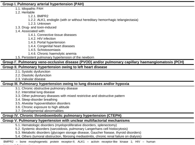
Currently, PH is clinically classified in: i) PAH; ii) pulmonary veno-occlusive disease (PVOD) and/or pulmonary capillary hemanglomatosis (PCH); iii) PH owing to left heart disease; iv) PH owing to lung diseases and/or hypoxia; v) chronic thromboembolic PH (CTEPH); vi) PH with unclear multifactorial mechanisms (Table 1).
Characterization and modulation of survivin in an experimental model of pulmonary arterial hypertension Part I – Introduction
17
Cardiovascular Pathophysiology | Ana Filipa Silva
Table 1. WHO classification of pulmonary hypertension, Dana Point, 2008
Group I. Pulmonary arterial hypertension (PAH) 1.1. Idiopathic PAH
1.2. Heritable
1.2.1. BMPR2
1.2.2. ALK1, endoglin (with or without hereditary hemorrhagic telangiectasia) 1.2.3. Unknown
1.3. Drug- and toxin-induced 1.4. Associated with:
1.4.1. Connective tissue diseases 1.4.2. HIV infection
1.4.3. Portal hypertension 1.4.4. Congenital heart diseases 1.4.5. Schistosomiasis
1.4.6. Chronic haemolytic anemia
1.5. Persistent pulmonary hypertension of the newborn
Group I’. Pulmonary veno-occlusive disease (PVOD) and/or pulmonary capillary haemangiomatosis (PCH) Group II. Pulmonary hypertension owing to left heart disease
2.1. Systolic dysfunction 2.2. Diastolic dysfunction 2.3. Valvular disease
Group III. Pulmonary hypertension owing to lung diseases and/or hypoxia 3.1. Chronic obstructive pulmonary disease
3.2. Interstitial lung disease
3.3. Other pulmonary diseases with mixed restrictive and obstructive pattern 3.4. Sleep-disorder breathing
3.5. Alveolar hypoventilation disorders 3.6. Chronic exposure to high altitude 3.7. Developmental abnormalities
Group IV. Chronic thromboembolic pulmonary hypertension (CTEPH) Group V. Pulmonary hypertension with unclear multifactorial mechanisms
5.1. Hematologic disorders (myeloproliferative disorders, splenectomy) 5.2. Systemic disorders (sarcoidosis, pulmonary Langerhans cell histiocytosis)
5.3. Metabolic disorders (glycogen storage disease, Gaucher fisease, thyroid disorders) 5.4. Others (tumoral obstruction, fibrosing mediastinitis, chronic renal failure on dialysis)
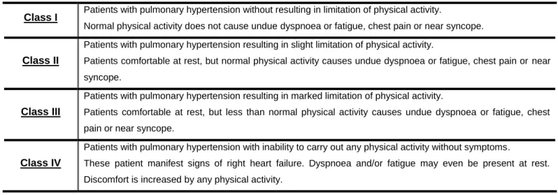
Besides de clinical classification, PH patients can also be classified according to their functional performance (Table 2.). This classification has been adopted from the New York Heart Association (NYHA) for left heart disease and together with clinical classification can be useful for diagnosis, prognosis and therapy of PH.
BMPR2 – bone morphogenetic protein receptor-II; ALK1 – activin receptor-like kinase 1; HIV – human immunodeficiency virus
Part I – Introduction
Table 2. Functional classification of pulmonary arterial hypertension (adapted7)
Class I Patients with pulmonary hypertension without resulting in limitation of physical activity.
Normal physical activity does not cause undue dyspnoea or fatigue, chest pain or near syncope.
Class II
Patients with pulmonary hypertension resulting in slight limitation of physical activity.
Patients comfortable at rest, but normal physical activity causes undue dyspnoea or fatigue, chest pain or near syncope.
Class III
Patients with pulmonary hypertension resulting in marked limitation of physical activity.
Patients comfortable at rest, but less than normal physical activity causes undue dyspnoea or fatigue, chest pain or near syncope.
Class IV
Patients with pulmonary hypertension with inability to carry out any physical activity without symptoms.
These patient manifest signs of right heart failure. Dyspnoea and/or fatigue may even be present at rest. Discomfort is increased by any physical activity.
1.3. Epidemiology
The incidence and prevalence of PAH, respectively, are estimated at 2.4-7.6 cases per million per year and 15-26 cases per million in large population studies8, 9. Young adults are the most affected being more than two thirds women10. This female predominance might be explained by the low survival rate of male fetuses with PPH11. However, it is hard to appraise the global prevalence because it depends on the method of diagnosis and the specific groups of population studied. The worldwide prevalence of PAH is likely greater than is recognized given the newer associations with dialysis12 and metabolic syndrome13 as well as with the developing world diseases that are risk factors for PAH such as HIV, schistosomiosis and sickle cell disease14. However, non-PAH are increasingly far more common.
1.4. Pathophysiology
The normal pulmonary circulation is a low-pressure, low-resistance and high-capacitance system (pulmonary vascular resistance being less than one-tenth of systemic vascular resistance) where arteries are compliant structures with few muscle fibers. PAH is characterized by excessive pulmonary vasoconstriction and vascular remodeling that commonly affects all vessel layers (intima, media and adventitia) rising pulmonary vascular resistance (PVR) and PAP ultimately increasing right ventricular afterload. The histological findings in PAH include intimal thickening, medial hypertrophy (MH), adventitial proliferation/fibrosis, arterial occlusion, thrombosis in situ and infiltration of inflammatory/progenitor cells15, 16. In the later stage of the disease, the formation of a vessel “neointima” is characterized by augmented deposition of extracellular matrix and myofibroblasts. Moreover, located downstream from occluded arteries, plexiform lesions can predominate and express growth factors typically observed in
Characterization and modulation of survivin in an experimental model of pulmonary arterial hypertension Part I – Introduction
19
Cardiovascular Pathophysiology | Ana Filipa Silva angiogenesis17. It is still unknown the exact processes that initiate the pathological changes observed in PAH however the interaction of predisposing conditions and exogenous stimuli may represent the causes.
1.4.1. Genetics
The study of genetic predisposition of PAH is of utmost importance for the understanding pathogenesis of this disease. Mutations in bone morphogenetic protein (BMP) receptor-2 (BMPR2) are present in more than 70% of familial PAH and 10%-40% of IPAH leading to loss of Smad signalling and therefore to increased proliferation and decreased differentiation of pulmonary artery smooth muscle cells (PASMCs) 18-20. In spite of the autosomal dominant inheritance of BMPR2 gene mutations, this disease has a low penetrance since only approximately 20% of individuals carrying the mutation will develop PAH 20.
More research is currently being performed to find possible epigenetic mechanisms that enhanced PAH susceptibility, namely single nucleotide polymorphisms (SNP). SNP variants, including KV1.521, transient receptor potential (Trp) channels22 and serotonin (5-HT) transporters23, may predispose to PAH.
1.4.2. PAH as panvasculopathy
Accompanied by the histological features previously described, PAH is currently viewed as a panvasculopathy (Figure 1). In PAH the vasodilator/vasoconstrictor ratio is decreased in the endothelium 24-26
whereas prothrombotic factors, such as tissue factor27, are increased. Apoptosis in early stages of PAH may generate apoptosis-resistant endothelial cells (ECs) that cross-talk with PASMCs through growth factors (e.g. transforming growth factor-β, TGF-β) leading to EC and fibroblast transdifferentiation and PASMCs proliferation28, eventually forming plexiform lesions29. Besides growth factors, several other phenomena drive PASMCs into excessive proliferation including: mitochondrial abnormalities29, increased expression/activity of platelet-derived growth factor (PDGF) receptor30 and serotonin receptor (SERT)31, 32, tyrosine kinase activation33 and decreased expression of voltage-gated O2-sensitive potassium channel, KV1.534. Moreover, metalloproteinase activation causes disruption of the adventitia which allows cell migration and generates mitogenic peptides35. Furthermore, adventitial fibroblasts show a hyperproliferative phenotype in PAH, displaying increased sensitivity to 5-HT36.
Part I – Introduction
Several observations indicate that PAH shares a mitochondrial-metabolic abnormality with cancer, the “Warburg phenotype”, a shift in glucose metabolism from oxidative phosphorylation to glycolysis (despite adequate oxygen supply) that enhances proliferation and prevents apoptosis37. The downstream consequences of this mitochondrial-metabolic abnormality include mitochondrial hyperpolarization, reduced production of reactive oxygen species (ROS), normoxic-activation of hypoxia inducible factor-1α (HIF-1α), overexpression of pyruvate dehydrogenase kinase (PDK) and decreased expression KV1.5 channels37. Other groups demonstrated that these mitochondrial abnormalities can be partially reverted by Dichloroacetate (DCA), a PDK inhibitor and KV1.5 channel opener. Indeed, DCA activates pyruvate dehydrogenase (PDH), increasing glucose oxidation, restoring mitochondrial membrane potential and reversing normoxic HIF-1α activation37.
Figure 1. PAH as panvasculopathy. In PAH several alterations occur in the 3 layers (adventitia, media and intima) of pulmonary arteries. PAH:
Pulmonary Arterial Hypertension; SMC: Smooth muscle cell; EC: Endothelial cell; TGF: Transforming Growth Factor; PDGF: Platelet-derived Growth Factor.
Characterization and modulation of survivin in an experimental model of pulmonary arterial hypertension Part I – Introduction
21
Cardiovascular Pathophysiology | Ana Filipa Silva 1.4.3. Inflammation
Increasing attention is being focused on several inflammatory mechanisms that play a key role in experimental models of PAH and in human PAH. These include the presence of T and B cells monocytes, macrophages and dendritic cells in plexiform lesions38, 39, the detection of autoantibodies on ECs and fibroblasts40, raised cytokine and chemokine levels41 and the association of PAH with certain infections such as human herpes virus 8 (HHV-8)42 and HIV43. The nuclear factor of activated T cells (NFAT) is known for increasing the transcription of multiple inflammatory mediators such as interleukins (IL) and tumour necrosis factor-α (TNF-α)44
. Activation of NFAT in PAH causes the downregulation of KV1.545 and regulates the transcription of several genes that control mitochondrial function46. Moreover, NFAT was also found to be activated in PASMCs isolated from patients with PAH and that inhibition of its predominant isoform, NFATc2, would contribute to attenuate monocrotaline(MCT)-induced PAH47.
Finally, PAH is associated with latent viral infections (such as HIV and HHV-8) and schistosomiasis (SSc). Nonhuman primates infected with chimeric simian HIV-nef virus demonstrated lung vascular remodeling characteristic of PAH48. Moreover, the HIV-nef gene was also implicated in plexiform lesions in pulmonary arteries of HIV-infected patients with PAH49. SSc, a parasitic infection that causes inflammation and pulmonary vascular disease, has also been implicated in PAH. Infiltration of T and B cells together with increased levels of IL-6, CX3CL1 and RANTES have been reported in the pulmonary vascular lesions of PAH patients with SSc6. Additionally mice chronically infected with SSc presented extensive pulmonary vascular remodeling in the absence of PH and correlated with cytokine IL-13 levels50.
1.4.4. Right ventricular hypertrophy
One of the consequences of PVR increasing is right ventricular overload leading to right ventricular hypertrophy (RVH) and failure. According to the Laplace relationship an elevated intraluminal pressure leads to an increase in wall stress, unless the chamber wall thickness is increased or the internal radius of the chamber is reduced. To compensate the high pressure registered in PAH, the right ventricle (RV) adapts by increasing wall thickness and assuming a more rounded shape51. The increase in ventricular mass is due to myocyte hypertrophy, sarcolemmal and contractile protein synthesis, altered calcium homeostasis, a shift in gene expression and extracellular matrix remodelling52. Although is known that the best approach to reduce RVH is to treat the underlying pulmonary arterial alterations, recent data suggests that RV can be an independent target in experimental PAH53. For exemple, inhibition of phosphodiesterase 5 (PDE5) (e.g. sildenafil) improved RV contractility53, without altering left ventricle (LV) function.
In normal conditions the RV is capable of adjusting its substrate utilization from fatty acids to glucose. In RVH the myocardium is reliant on anaerobic glucose metabolism due to PDK activation and it has hibernating properties, demonstrating augmented glucose oxidation and contractility in response to DCA54.
Part I – Introduction Studies in MCT-induced PAH have demonstrated an upregulation of glucose transporter protein 4 (GLUT4)55.
Moreover alterations in neurohumoral signalling, formation of ROS as well as nitrogen species and exaggerated inflammatory responses can also lead to right heart failure51. Still, more research is needed on therapy agents with effects on both RV and pulmonary vasculature.
1.5. Experimental models
Even though animal models do not mimic all the characteristics of human pathology, they have allowed the study on several diseases and its therapeutics56. Until now, no animal model has been able to mimic all the biochemical and histopathological features of PAH57. MCT and hypoxia are the most frequently used animal models in the study of this disease56. Although MCT model has been used for over than 50 years, the molecular mechanisms underlying MCT-induced PAH have yet to be better clarified58.
1.5.1. Monocrotaline model
Derived from the seeds of Crotalaria spectabilis, monocrotaline (MCT) is a toxic pyrrolizidine alkaloid that can be administrated by intraperiotoneal (60 mg/Kg), subcutaneous (60 mg/Kg), or intravenous injection (1-5 mg/Kg) inducing vascular injury after hepatic generation of its pyrrolic derivative by cytochrome P4(1-50 3 A59. With a single injection of MCT rats develop PAH after approximately 3 weeks and die within 6-8 weeks60, making this a very simple and thus technically appealing animal model available to a wide spectrum of investigators. Unfortunately, MCT represents a significant limitation for long-term survival studies because it can injury other organs like the liver61 and the kidney62. Another limitation is the different sensitivity that rat strains present to MCT63 which may be related to the pharmacokinetics of this molecule59. MCT primarily affects the pulmonary arterial bed because lungs are the first major vascular bed after the liver64. In fact, at the pulmonary level, the ECs are the first site of damage. After only 4h of MCT administration rats develop platelet thrombi in small arteries65 and within 4 days EC toxicity by increasing the number of swollen mitochondria and decreasing the proportion of microfilaments66, 67. Approximately 7 days after MCT injection there is a peripheral extension of SMCs into small nonmuscularized PAs66 and an increase in oxygen consumption and cardiac index68. The medial hypertrophy of small PAs is described 12 days post MCT injection accompanied by an increase in PAP. RVH is only present later in the disease68 (~21 days after injection) together with increased RV systolic and diastolic pressures and ultimately RV failure69-71.
Characterization and modulation of survivin in an experimental model of pulmonary arterial hypertension Part I – Introduction
23
Cardiovascular Pathophysiology | Ana Filipa Silva
2. Apoptosis in PAH
The pathogenesis of PAH is a complicated, multifactorial process. The current vasodilator therapies are limited and research is now pursing strategies that could reverse structural remodeling in the pulmonary arterial bed, thus providing a more significant decrease in PVR. Even though growth is the classical mechanism associated with vascular remodeling, it has increasingly been acceptable that apoptosis may influence the extent of alterations that occurs72. Indeed, a reduction in apoptosis has been implicated in severe PAH while induction of this mechanism seems to promote the regression of pulmonary vascular remodeling.
Figure 2. Apoptosis pathways. Two major pathways are present during apoptosis: the extrinsic (or death receptor pathway) and the intrinsic (or
mitochondrial pathway). In the extrinsic pathway the activation of death receptors leads to the formation of death inducing signalling complex (DISC) and activation of procaspase-8, initiating the caspase cascade. In the intrinsic pathway, the death stimuli leads to opening of the mitochondrial permeability transition pore (MPT) and thus to the release of cytochrome c and Smac/DIABLO. Cytochrome c will, together with Apaf-1 and procaspase-9, form the apoptossome and activate caspase cascade while Smac/DIABLO will suppress inhibitor of apoptosis proteins (IAPs) thus contributing to caspase activation and apoptosis. Finally the balance between pro and anti-apoptotic Bcl proteins will determine the mitochondrial response to death stimuli. FADD: Fas-associated death domain protein; TRADD: Tumor necrosis factor receptor type 1-associated death domain protein.
Part I – Introduction Apoptosis, or programmed cell death, is an important biological process of normal tissue development and function that involves the genetically determined elimination of cells. It is characterized by a distinct series of morphological and biochemical alterations such as cell shrinkage, chromatin condensation, DNA fragmentation, caspase activation, formation of apoptotic bodies and membrane blebbing73-75. The apoptotic events are initiated when the cells loss their volume upon the receiver of apoptotic signals (Figure 2). Then mitochondrial membrane potential is depolarized, cytochrome c (cyt c) is released and caspases are activated. Finally, the last phase is when the DNA degradation occurs, apoptotic bodies are formed, the nuclear lamina and cytoskeleton are degraded and the internal phosphatidyl serine is exposed to the external environment.
Until now, two major apoptotic pathways are known: the intrinsic or mitochondrial pathway and the extrinsic or death receptor pathway. Both pathways converge on the same execution phase that involves the activation of specific cysteinyl aspartic acid-proteases (caspase-3, caspase-6 and caspase-7). These activate cytoplasmic endonucleases and proteases that degrade nuclear and cytoskeletal material76. The extrinsic pathway initiates apoptosis through transmembrane receptor-mediated interactions involving death receptors and ligands that are members of the TNF receptor gene superfamily77 like FasR/FasL, TNFR1/TNF-α/, DR3/Apo3L/, DR4/Apo2L and DR5/Apo2L78-82. Upon ligand binding cytoplasmic adapter proteins, such as FADD and TRADD, are recruited and associated with procaspase-8 via dimerization of the death effector domain. This leads to the formation of a death-inducing signalling complex (DISC) that activates procaspase-883. After that, the execution phase begins with the activation of caspase-8. The intrinsic pathway can be initiated through either negative signalling (absence of factors that suppress death programs such as growth factors, hormones and cytokines) or positive signalling (radiation, toxins hypoxia, hyperthermia, viral infections and free radicals). Both signals lead to loss of the mitochondrial transmembrane potential, formation of the mitochondrial permeability transition pore (MPT) and release of pro-apoptotic proteins such as cytochrome c and Smac/DIABLO from the intermembrane space to the cytosol84. Once released into cytosol, cytochrome c binds and activates Apaf-1 and procaspase-9 to form the “apoptosome”85, 86
which then activates caspase-9 and lead to chromatin degradation and apoptosis. Although the exact mechanisms are yet to be discovered, thoughts are that Bcl-2 family of proteins is the responsible for the regulation of apoptotic mitochondrial events87. This family of proteins is constituted by either pro-apoptotic proteins (Bcl-10, Bax, Bak, Bid, Bad, Dim, Bik and Blk), that upon mitochondrial transmembrane potential depolarization regulate cytochrome c release88, and anti-apoptotic proteins (Bcl-2, Bcl-x, Bcl-XL, Bcl-XS, Bcl-w and BAG) that can promote mitochondrial hyperpolarization and prevent both the opening of MPT and release of cytochrome c89.
In addition, ion channels are known to perform an essential role in apoptosis. High concentration of intracellular K+ ([K+]i) is needed to maintain a normal cell volume. Opening of plasma membrane voltage-gated K+ channels increases the loss of cytoplasmic K+ inducing apoptotic volume decrease (AVD). On the other hand, closure or downregulation of K+ channels contributes to the maintenance of [K+]i attenuating
Characterization and modulation of survivin in an experimental model of pulmonary arterial hypertension Part I – Introduction
25
Cardiovascular Pathophysiology | Ana Filipa Silva apoptosis90, 91. Moreover [K+]i is also required for the suppression of caspases and nucleases in the final apoptotic stage91.
Finally, the last component of apoptosis is the phagocytic uptake of apoptotic bodies where the externalization of phosphotidylserine onto the outer leaflet facilitates noninflammatory phagocytic recognition leading to an early uptake and disposal92.
2.1. The role of apoptosis in pulmonary vascular remodeling
Recent research demonstrated that apoptosis has an important pathophysiological role in PAH by eliminating unwanted cells such as cells migrated into the vascular lumen and hypertrophied cells accumulated in the pulmonary vasculature93.
Apoptotic modulators in the vasculature are diverse and include ROS94, NO95, angiotensin type 2 (AT2) receptors96 and the endothelin system97. For exemple, ROS are involved in PAH and more specifically in H2O2 induced-apoptosis of vascular cells by PKC dependent mechanism98.
Animal studies demonstrated that inducing apoptosis of hypertrophied PASMC in intact pulmonary vasculature can prevent the progression of the medial hypertrophy93, 99, 100.
Moreover, apoptosis of hypertrophied PASMCs has been related to a regression in medial hypertrophy while inhibition of apoptosis is related to progression of pulmonary vascular medial thickening100-102. This assumption is based on the theory for a spatio-temporal diversity within the vascular wall as PAH progresses. In this theory, an inherited or acquired alteration in BMP axis promotes the apoptosis of pulmonary artery endothelial cells (PAECs) and consequently loss of small capillaries (constituted essentially by PAECs tubes) leading to the increase of the flow and shear stress in the remaining vessels. Intima cell proliferation and plexiform lesions are formed with the emergence of apoptosis-resistant PAECs expressing survivin29. Simultaneously, the loss of PAECs expose PASMCs to circulating growth factors such PDGF that has been shown to induce the expression of survivin103 and thus a resistance to apoptosis. Although all of this knowledge, the role of apoptosis in the pulmonary vascular remodeling needs further research. Studies on the mechanisms and biochemical markers of apoptosis will help in the understanding the role of apoptosis has a treatment for PAH.
2.2. Apoptosis as therapeutic target for PAH
Discover that normal vascular smooth muscle cells (VSMCs) possess the machinery to undergo apoptosis allowed the assumption that this phenomenon may be the one responsible for regulating cell number in the vessel wall.104 While early PAH is characterized by increased apoptosis in the endothelial layer, late disease is characterized by suppressed apoptosis and increased proliferation both in intima and media. Therefore an anti-remodeling treatment may be a potential therapeutic option against various types of PAH.
Part I – Introduction Patients in early stages of PAH may benefit from an antiapoptotic strategy, whereas the ones in late forms of PAH may receive a pro-apoptotic approach.
2.2.1. Survivin
Inhibitors of apoptosis (IAPs) are a family of proteins related with apoptosis. Several mammalian IAP family members have been identified including, NIAP105, c-IAP1 and 2, X-IAP106, apollon, TX-XIAP, livin , survivin and bruce. 107-109
Survivin is a IAP known for its double function as inhibitor of apoptosis and regulator of cell division110 (Figure 3.). This protein was first discovered in 1997 by Ambrosini and collegues while hibridization screening of a human genomic library with cDNA of the effector cell protease receptor-1 (EPR-1)111. Survivin presents a high homology to EPR-1 and has been mapped to chromosome 17 at band 25 encoding a protein of 142 amino acids, with a molecular weight of approximately 16.3 kDa111. The initial studies revealed a strong survivin expression in fetal tissues whereas little or no survivin was detected in normal adult tissues110, 112-115. Interestingly, this protein is known to be expressed in most cancers and it is described as the fourth most significant transcriptome expressed in human tumors116. The feature that defines apoptosis in the IAP protein family members is the presence of baculovirus IAP repeat (BIR) in at least one copy. Inhibition of proteolytic maturation and enzyme activity of caspases by IAPs may be due to a physical association with initiator and effector caspases114, 117. In fact, several IAPs BIRs act as caspases-interacting regions. The dual function that separates survivin from the other IAP family members is ensured by its unique structure. Sense this protein presents only one copy of a modified BIR domain similar to the three-dimensional structure BIR3 domain of XIAP. Survivin is thought to be able to interact with XIAP, protecting it from ubiquitination and increasing its stability leading to caspase-9 inhibition118, 119. This interaction was also studied through HeLa cells in which loss of phosphorylation of threonine 34 by p34cdc2 -cyclin B1 leads to dissociation of an immunoprecipitable survivin-caspase-9 complex on the mitotic apparatus and thus caspase-9-dependent apoptosis119. Moreover, expression of the T34A dominant-negative mutant has shown a depletion of procaspase-9 in culture tumor cell lines120.
Despite direct binding between survivin and the caspases has not been confirmed, survivin may also inhibit caspase activity indirectly through the mitochondrial pathway of apoptosis.
Mitochondrial pro-apoptotic dimeric protein Smac/DIABLO is known for increasing Apo-2L/TRAIL-induced caspase-3 activity and thus suppress the activity of survivin and other IAPs such XIAP and cIAP1121. During mitochondrial apoptosis pathway Smac/DIABLO along with cytochrome c are released from mitochondria into the cytosol promoting apoptosis. This is due to the suppression of inhibitor effects of the IAP proteins122. Concluding, the binding of Smac/DIABLO to survivin leads to its delayed release in the cytosol, which in turn results in prolonged cell survival123.
Characterization and modulation of survivin in an experimental model of pulmonary arterial hypertension Part I – Introduction
27
Cardiovascular Pathophysiology | Ana Filipa Silva Most IAP family members are considered anti-apoptotic proteins, however the gene that encodes survivin (BIRC5) generates five major isoforms of the transcript by alternative splicing: survivin-2α and survivin-2B that favour induction of apoptosis as well as survivin-ΔEx3, survivin-3B and wild type (WT)-survivin that appear to be cytoprotective124-126. Survivin is the only IAP protein capable of regulating cell division by interacting with the mitotic apparatus by binding to microtubules through its C-terminal domain127. In fact its expression is present on centromeres at prophase/metaphase and in the spindle midzone during anaphase/telophase128, 129. Survivin recognizes phosphorylated histone H3 associating with the chromosome passenger proteins Aurora B, INCENP and the neoformed complex is recruited to the centromeres to ensure a proper chromosomal segregation130. Moreover, survivin is present in centrossomes of dividing cells where it binds to Cdk1 which after activation allows cells to enter mitosis119. It has been proposed that survivin has multiple compartmentalizations in the cells that include the nucleus, the cytoplasm and the mitochondria118, 131, 132. Nuclear localization seems to be connected to the ability of survivin to regulate cell cycle by facilitating progression and inducing exit of the cells from G1 checkpoint
Figure 3. Roles of survivin. Survivin is the only inhibitor of apoptosis protein (IAP) known for its double function in inhibiting apoptosis and
regulating cell division. Survivin is capable of inhibiting caspases either directly or indirectly (in association with XIAP). Also Smac/DIABLO release suppresses survivin activity. In respect to cell cycle, survivin can be located in the mitotic spindle and the centromeres where in association with proteins Aurora B, Borealin and INCENP regulate chromosome segregation.
Part I – Introduction arrest and subsequent entry into S phase133-135. The greatest stability of survivin happens in the mitochondria where it is associated with oncogenic transformation, possibly increasing protection from apoptosis in cancer cells. This survivin confers resistance to apoptosis by inhibiting activation of effector caspases118, 132. Once apoptosis is initiated survivin exit the mitochondria and remains in cytoplasm where it loses its cytoprotective ability, probably because posttranslational modifications occur136. Finally, it was recently described that stress stimuli leads to a release of survivin to the cytosol, retaining both anti-apoptotic and proliferative actions137-139. It is believe that the increased release of mitochondrial survivin to cytoplasm and sequestering of Smac/DIABLO are of the utmost importance for the apoptosis resistant phenotype of neoplasic cells. This findings lead to the thought that use of molecular antagonists of survivin to increase cell death and prevent pathological resistance to apoptosis might hold therapeutic potential.
2.2.1.1. Terameprocol
Terameprocol (tetra-O-methyl nordihydroguairetic acid, M4N, EM1421, TMP) is a hydrophobic molecule derivated of the nordihydroguairetic acid (NDGA) isolated from the plant Larrea tridentata140, 141, known for obliterating survivin gene expression142, 143 by binding to the transcription factor Sp1. In vitro studies demonstrate that terameprocol induced apoptosis and growth arrest of various human cancer cell lines 142-148. In vivo experiments with murine models of human xenograft solid tumors demonstrated the anti-tumoral action of this compound with no relevant systemic toxicity143-147. Moreover, clinical trials are already initiated to evaluate the potential of TMP in diverse types of neoplasias and also in the prevention of sexually transmitted viruses.
Part II – Aims
Considering the concepts reviewed before, the aim of the present study was to investigate the contribution of apoptosis to the pathogenesis of PAH through the analysis and modulation of the anti-apoptotic protein survivin in an animal model of MCT induced-PAH.
In order to achieve the objective the work was divided in two parts, namely: a) Survivin expression in the progression of MCT-induced PAH
Pulmonary and cardiac patterns of controls and animals with MCT-induced PAH were related with expression of survivin and Smac/DIABLO proteins throughout the hemodynamic and morphometric progression of disease;
b) Terameprocol in vivo study
The effects of terameprocol in vivo administration were characterized through morphometric and hemodynamic evaluation of sham and pulmonary hypertensive rats.
Part III – Material and
Methods
Part III – Material and Methods
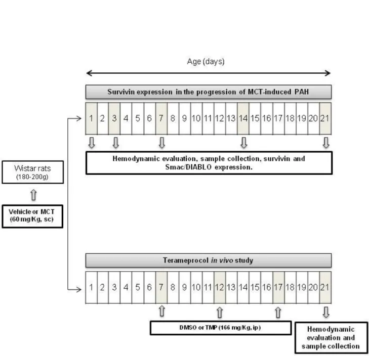
Figure 4. Experimental design. Progression of MCT-induced PAH and Terameprocol "in vivo" studies. MCT: Monocrotaline; PAH: Pulmonary
Characterization and modulation of survivin in an experimental model of pulmonary arterial hypertension Part III – Material and Methods
33
Cardiovascular Pathophysiology | Ana Filipa Silva
3. Experimental Design
Animal experiments were performed according to the Portuguese law for animal welfare and conform to the National Institutes of Health Guide for the Care and Use of Laboratory Animals (NIH Pub. No. 85-23, Revised 2011). Figure 4. represents the studies performed.
3.1. Survivin expression in the progression of MCT-induced PAH
Male Wistar Han rats (Charles River Laboratories, Barcelona, Spain) weighing 180-200 g were housed in groups of 5 animals/cage, in a controlled environment under a 12:12-h light-dark cycle at a room temperature of 22ºC, with free supply of food and water. Rats randomly received a subcutaneous injection of MCT (60 mg/kg, Sigma, Barcelona, Spain) (MCT groups, n=15/time point) or an equal volume of vehicle (1 mL/kg of 0.9% NaCl) (SHAM groups, n=10/time point). In order to evaluate the progression of the disease, hemodynamic assessment was performed on days 1, 3, 7, 14 or 21 after MCT/vehicle injection followed by sample collection.
3.2. Terameprocol in vivo study
Animals were randomly submitted to four different protocols: i) SHAM injected Dimethyl sulphoxide hybri-max (DMSO, 1 mL/Kg, intraperitoneal (ip); Sigma, Barcelona, Spain) (Sham+Vehicle, n=10), ii) SHAM injected with tetra-O-methyl nordihydroguairetic acid (Terameprocol or TMP, 166 mg/Kg, ip; Cayman Chemical, Michigan, USA) (Sham+TMP, n=10) iii) MCT injected with DMSO (MCT+Vehicle, n=15) and iv) MCT injected with TMP (MCT+TMP, n=15). DMSO or TMP were administrated at day seven after MCT or vehicle injection, a time point where significant morphological and molecular alterations were already noted in the first study, and at every five days until hemodynamic evaluation (21 days after MCT or vehicle).
4. Hemodynamic analysis
Animals were anesthetized by inhalation of mixture of sevoflurane (4%) and oxygen, intubated for mechanical ventilation (Dual Mode, Kent Scientific, Connecticut, USA
)
and placed over a heating pad (body temperature was maintained at 37°C). Under binocular surgical microscopy (Wild M651.MS-D, Leica; Herbrugg, Switzerlad), the right jugular vein was cannulated for fluid administration (prewarmed 0.9% NaCl solution) to compensate for perioperative losses. The heart was exposed by a median sternotomy and the pericardium was widely opened.Bi-ventricular hemodynamic function was measured with pressure-volume (PV) catheters (PVR-1045 for RV and PVR-1035 for LV, Millar instruments, Houston, USA). Data was continually acquired (MPVS 300, Millar Instruments, Houston, USA) and digitally recorded at 1000HzPart III – Material and Methods (ML880 PowerLab 16/30, Millar TM instruments, Houston, USA). After complete instrumentation, the animal preparation was allowed to stabilize for 15 min. Hemodynamic recording was made under basal conditions and under vena cava, ascending aorta or pulmonary artery occlusion with respiration suspended at end-expiration. Parallel conductance values were obtained by the injection of approximately 100 µl of 10% NaCl into the right atrium. Calibration from relative volume units (RVU) conductance signal to absolute volumes (µl) was undertaken using a previously validated method of comparison to known volumes in Perspex wells149. Heart rate (HR), RV and LV peak systolic pressure (Pmax), end-diastolic pressure (EDP), peak rate for pressure rise (dP/dtmax), peak rate of pressure decline (dP/dtmin), constant time of isovolumetric pressure decline (Tau), ejection fraction (EF), cardiac output (CO) and maximal elastance (Ea) were obtained and analysed using PVAN 3.5 and LabChart 7.0 (Millar Instruments, Houston, USA).
5. Tissue Preparation
The heart (H), lungs and right gastrocnemius muscle were excised and weighted. The right tibia was also excised and its length was measured with a millimetric ruler. Under binocular magnification (x3.5, Wild M651.MS-D, Leica; Herbrugg, Switzerlad), the RV free wall was dissected from the left ventricle + septum (S) and weighted separately. Heart, lungs, RV and LV+S weights were normalized to body weight (BW) and gastrocnemius weight was normalized to tibial length. Samples from heart and lung were fixed and included in paraffin for light microscopy, or frozen with liquid nitrogen for molecular studies.
6. Morphometric analysis
Samples of RV, LV (midway between the apex and base) and lung were fixed in 4% (v/v) buffered paraformaldehyde followed by dehydration with graded ethanol, diaphanization with xylene and included in paraffin blocks. Serial sections (4 µm of thickness) of paraffin blocks were cut by a microtome (RM2125RTS, Leica, Nussloch, Germany) and mounted on silane-coated slides. The slides were dewaxed in xylene, hydrated through graded alcohols and stained for haematoxylin-eosin by immersing slides in Mayer’s haematoxylin solution for 5 min. followed by immersion in aqueous eosin solution for 5 min. Slides were after submitted to graded alcohols and xylene and mounted with Entellan. Studied samples were observed at light microscopy (Dialux 20, Leitz, Wetzlar, Germany), photographed with a digital camera (XC30, Olympus, California, USA) and measured with a digital image analyzer (cell^B life science basic imaging software, Olympus, California, USA). Five images of random microscopic fields (magnification of x400) were obtained from each section to compensate for variations within sections. Only round to ovoid muscle fibers with a nuclear profile were counted to measure the cardiomyocytes surface area (CSA) being 500 cardiomyocytes/group/time point analyzed. On pulmonary specimens, external diameter and medial
Characterization and modulation of survivin in an experimental model of pulmonary arterial hypertension Part III – Materials and Methods
35
Cardiovascular Pathophysiology | Ana Filipa Silva Orthogonal intercepts were used to generate eight random measurements of external diameter (distance between the external lamina) and sixteen random measurements of medial thickness (distance between the internal and external lamina). For each artery medial hypertrophy was expressed as follows: % wall thickness = [(medial thickness x 2) / (external diameter)] x 100.
7. Imunohistochemistry
Imunohistochemistry was performed to determine survivin and Smac/DIABLO expression in the RV. Sections (4 µm) were placed on SuperFrost®Plus slides followed by deparaffinization, rehydration, and subjected to heat induced antigen retrieval by immersion in 10 mM sodium citrate buffer (C6H5Na3O7.2H2O, pH 6.0) in the microwave for 30 min. Endogenous peroxidase activity was blocked by 3% hydrogen peroxide for 10 min. at room temperature (RT). Blockage of non-specific binding was performed with 5% normal goat serum (NGS) in TBS (100 mM Tris, 1.5 mM NaCl, pH 8.0) 0.1% Tween-20 (TBS-T) for 1 hour, RT flowed by 3 washes, 5 min. each in TBS-T. Sections were encircled with a pap pen (Vector Laboratories, California, USA) to prevent splitting leakage of the flowing incubation solutions. Endogenous avidin-biotin expression was blocked using an endogenous avidin + biotin blocking system, (ab3387, abcam, Cambridge, UK), according to manufacturer’s instructions, being followed by incubation with the primary antibodies (1:500 dilution; rabbit anti-survivin, ab469; dilution 1:250; rabbit anti-Smac/DIABLO, ab8115, abcam, Cambridge, UK) overnight at 4°C. After incubation with primary antibodies, slides were washed 3 times, 5 min each with TBS-T, RT and incubated with goat anti-rabbit IgG secondary antibody (1:250 dilution; ab6720, abcam, Cambridge, UK) for 2 hours, RT. Slides were submitted to another 3 washes, 5min each in TBS-T, RT prior being incubated with streptavidin protein, HRP (1:1000 dilution; ab7403, abcam, Cambridge, UK). To visualize the peroxidase activity in sections 3,3-diaminobenzidine (DAB, ab94665, abcam, Cambridge, UK) was used. Finally, slides were counterstained with Mayer’s haematoxylin, submitted to graded alcohols and xylene and mounted with Entellan. Negative control reactions included omission of the primary antibody. The slides were observed and photographed with a microscope (Dialux 20, Leitz, Wetzlar, Germany) under x400 magnification. Survivin and Smac/DIABLO expression was qualitatively determined as positive (cytoplasmatic staining) or negative (500 cardiomyocytes/group/time point).
8. Western Blotting
RV, LV and lung samples (n=6 animals/group/time point) previously frozen with liquid nitrogen were homogenised in phosphate buffer (13 mM KH2PO4, 54mM NaHPO4, pH 7.4) (in the proportion of 1:20) with a Bio-Gen PRO200 homogeniser (Pro 200, Pro Scientific, Connecticut, USA). Total protein concentration was spectrophotometrically determined with the colorimetric method RC-DC protein assay (Bio-Rad,
Part III – Material and Methods California, USA). The optic density was determined at 750nm in a microplate reader (UVM340, Asys, Cambridge, UK). Simultaneously, a calibration curve was performed using different concentrations of bovine serum albumin (BSA).
Equivalent amounts of total protein from the homogenized RV, LV and lung of each group were electrophoresed on a 12.5% SDS-PAGE at 200V at room temperature as described by Laemmli150. Gels containing total proteins were transferred to a nitrocellulose membrane (0.2 µm, Bio-Rad, California, USA) in 25 mM Tris, 192 mM and 20% methanol at 200 mA. Equal loading of membranes was confirmed by staining the membranes with Ponceau S and nonspecific binding was blocked with 5 % (w/v) dry non-fat milk in TBS-T. Membranes were incubated with primary antibody (1:1000 dilution; rabbit anti-survivin, ab469; 1:500 dilution, rabbit anti-Smac/DIABLO, ab8115, abcam, Cambridge, UK) overnight at 4°C with agitation. Afterwards, blots were washed in TBS-T and incubated with a secondary antibody (1:15000, LI-COR IRDye® 800CW, Nebraska, USA). Immunoreactive bands were observed under fluorescence using an Odyssey system (LI-COR Odyssey, Nebraska, USA) and the results were analyzed with Quantity One software v. 4.6.3 (Bio-Rad, California, USA).
9. Statistical Analysis
Statistical analysis was performed using Graph Pad Prism software (version 5.0, Graph Pad software, California, USA). All data are presented as mean ± SEM and were compared using Two Way ANOVA. When treatments were significantly different, Students-Newman Keuls post-hoc test was selected to perform pairwise multiple comparisons. Results were considered significantly different when p<0.05.
Part IV – Results
10.
Progression of MCT-induced pulmonary arterial hypertension
10.1. Right ventricle hemodynamic evaluation
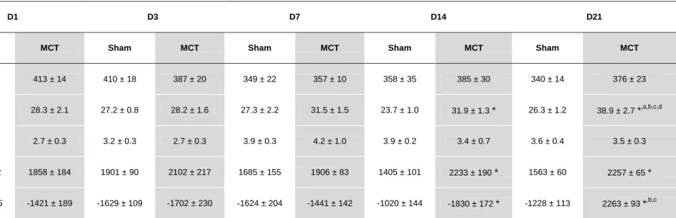
Table 4. represents the results from RV hemodynamic evaluation. RVPmax, dP/dtmax and dP/dtmin were significantly different between Sham and MCT groups 14 and 21 days after injection. Also, in MCT groups and at day 21, RVPmax was significantly augmented when compared with the previous time-points. MCT treatment did not induce any alterations in heart rate and end-diastolic pressure.
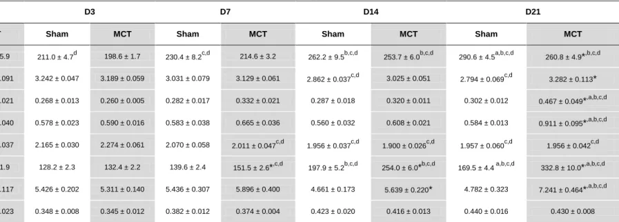
10.2. Morphometric analysis
Table 5. summarizes the morphometric progression of MCT-induced PAH. In Sham groups there is a significant increase in body weight throughout the protocol. In contrast, MCT animals had a lower weight gain being significantly different at day 21 after injection. No variation in the relation HT/BW was observed in MCT group, while it was reduced in the Sham group on days 14 and 21. MCT administration induced hypertrophy as expressed by the indexes RV/(LV+S) and RV/BW, both significantly increased on D21. Also, MCT-treated animals developed hypertrophy at cellular level represented by a significant increase in RV cardiomyocyte cross sectional area since day 7 after injection and progressively until day 21. Neither groups presented any changes in the left ventricle ((LV+S)/BW parameter).
Table 3. Right ventricle hemodynamic evaluation parameters.
D1 D3 D7 D14 D21
Sham MCT Sham MCT Sham MCT Sham MCT Sham MCT
Heart rate (bpm) 378 ± 23 413 ± 14 410 ± 18 387 ± 20 349 ± 22 357 ± 10 358 ± 35 385 ± 30 340 ± 14 376 ± 23
Pmax(mmHg) 28.0 ± 1.9 28.3 ± 2.1 27.2 ± 0.8 28.2 ± 1.6 27.3 ± 2.2 31.5 ± 1.5 23.7 ± 1.0 31.9 ± 1.3 * 26.3 ± 1.2 38.9 ± 2.7 *,a,b,c,d
EDP (mmHg) 3.9 ± 0.5 2.7 ± 0.3 3.2 ± 0.3 2.7 ± 0.3 3.9 ± 0.3 4.2 ± 1.0 3.9 ± 0.2 3.4 ± 0.7 3.6 ± 0.4 3.5 ± 0.3
dP/dtmax (mmHg/sec) 1671 ± 152 1858 ± 184 1901 ± 90 2102 ± 217 1685 ± 155 1906 ± 83 1405 ± 101 2233 ± 190 * 1563 ± 60 2257 ± 65 *
dP/dtmin (mmHg/sec) -1544 ± 355 -1421 ± 189 -1629 ± 109 -1702 ± 230 -1624 ± 204 -1441 ± 142 -1020 ± 144 -1830 ± 172 * -1228 ± 113 2263 ± 93 *,b,c
Pmax: maximum pressure; EDP: end-diastolic pressure; dP/dtmax: peak rate of pressure rise; dP/dtmin: peak rate of pressure fall. Data are mean±SEM. Sham: sham group; MCT: monocrotaline
group. *p < 0.05 vs. Sham of the same day; ap < 0.05 vs. D14 of the same treatment group, bp < 0.05 vs. D7 of the same treatment group; cp < 0.05 vs. D3 of the same treatment group; dp < 0.05 vs. D1 of the same treatment group.
Table 4. Morphometric progression of MCT-induced PAH.
D1 D3 D7 D14 D21
Sham MCT Sham MCT Sham MCT Sham MCT Sham MCT
Body weight (g) 194.1 ± 5.1 195.4 ± 5.9 211.0 ± 4.7d 198.6 ± 1.7 230.4 ± 8.2c,d 214.6 ± 3.2 262.2 ± 9.5b,c,d 253.7 ± 6.0b,c,d 290.6 ± 4.5a,b,c,d 260.8 ± 4.9*,b,c,d
HW/BW (g/Kg) 3.271 ± 0.028 3.243 ± 0.091 3.242 ± 0.047 3.189 ± 0.059 3.031 ± 0.079 3.129 ± 0.061 2.862 ± 0.037c,d 3.025 ± 0.051 2.794 ± 0.069c,d 3.282 ± 0.113*
RV/(LV+S) (g/g) 0.264 ± 0.014 0.265 ± 0.021 0.268 ± 0.013 0.260 ± 0.005 0.282 ± 0.017 0.332 ± 0.021 0.287 ± 0.018 0.320 ± 0.011 0.302 ± 0.012 0.467 ± 0.049*,a,b,c,d
RV/BW (g/Kg) 0.578 ± 0.025 0.591 ± 0.040 0.578 ± 0.023 0.590 ± 0.016 0.583 ± 0.038 0.665 ± 0.036 0.560 ± 0.032 0.608 ± 0.021 0.584 ± 0.013 0.911 ± 0.095*,a,b,c,d
(LV +S)/BW (g/Kg) 2.197 ± 0.037 2.240 ± 0.037 2.165 ± 0.030 2.274 ± 0.061 2.070 ± 0.058 2.011 ± 0.047c,d 1.956 ± 0.037c,d 1.900 ± 0.026c,d 1.957 ± 0.060c,d 1.956 ± 0.042c,d
RV CSA (µm2) 131.2 ± 2.4 134.1 ± 1.9 128.2 ± 2.3 132.4 ± 2.2 139.6 ± 2.4 151.5 ± 2.6*,c,d 197.9 ± 5.2b,c,d 254.0 ± 6.0*b,c,d 169.5 ± 4.4 a,b,c,d 332.8 ± 10.0*,a,b,c,d
L/BW (g/Kg) 5.731 ± 0.260 5.592 ± 0.117 5.426 ± 0.202 5.311 ± 0.140 5.436 ± 0.307 5.896 ± 0.400 4.661 ± 0.173 5.639 ± 0.220* 4.782 ± 0.323 7.241 ± 0.464*,a,b,c,d
G/Tib (g/cm) 0.341 ± 0.008 0.337 ± 0.023 0.348 ± 0.008 0.345 ± 0.012 0.382 ± 0.012 0.374 ± 0.004 0.423 ± 0.020 0.416 ± 0.013 0.440 ± 0.016 0.430 ± 0.008
HW/BW: heart weight/body weight; RV/(LV+S): right ventricle/(left ventricle+septum); RV/BW: right ventricle/body weight; (LV+S)/BW: (left ventricle+septum)/body weight; CSA: cross sectional area; L/BW: lung/body weight; G/Tib: gastrocnemius/tibia; Sham: sham group; MCT: monocrotaline group Data are mean±SEM. *p < 0.05 vs. Sham of the same day; ap < 0.05 vs. D14 of the same treatment group, bp < 0.05 vs. D7 of the same treatment group; cp < 0.05 vs. D3 of the same treatment group; dp < 0.05 vs. D1 of the same treatment group.
Characterization and modulation of survivin in an experimental model of pulmonary arterial hypertension Part IV – Results
41
Cardiovascular Pathophysiology | Ana Filipa Silva
D1 D3 D7 D14 D21 0 20 40 60 80 100
Sham
MCT
*,b,c,d
*,b,c,d
Day after injection
% M e d ia l H yp e rt ro p h y (< 5 0 m)
Normalized lung weight was significantly increased in MCT groups compared with Sham groups in days 14 and 21. Also in MCT treated animals, at D21, the L/BW parameter was significantly higher when compared to the other time-points evaluated. As demonstrated in Figure 5. pulmonary arterial wall suffered a significant thickening from day 14 and forward in MCT groups.
A
B
Figure 5. Pulmonary arterial hypertrophy. A) Histological appearance of small pulmonary arteries stained with hematoxylin and eosin; B)
Percentage of arterial medial layer hypertrophy. Sham: Sham group; MCT: monocrotaline group. Data are mean±SEM; *p < 0.05 vs. Sham of the same day, bp < 0.05 vs. D7 of the same treatment group, cp < 0.05 vs. D3 of the same treatment group, dp < 0.05 vs. D1 of the same treatment group.
Part IV – Results 10.3. Survivin and Smac/DIABLO expression
Lung survivin and Smac/DIABLO expression are shown in Figures 6, A and B. Survivin expression is augmented in MCT groups in days 7 and 21 when compared with Sham groups. Moreover, MCT groups presented a significant reduction from day 7 to day 21. No differences were noted between days in Sham groups. In contrast, pulmonary Smac/DIABLO expression decreases in MCT groups compared to Sham groups in both time-points.
Figure 7. (A, B and C) shows survivin expression in the right ventricle. When in comparison with Sham group, MCT treated animals survivin expression is barely present on day 3 after injection and significantly rises on day 7, further increasing throughout days 14 and 21. Confirmation by western blotting (C) demonstrated a significantly increase in protein expression in MCT groups between day 7 and 21 after injection. Sham groups do not suffer any alteration between time points.
Contrarily, MCT groups presented a lower Smac/DIABLO expression since D3 and forward when compared with Sham animals. (Figure 8. A, B and C)
D7 D21 0 500 1000 1500
*
*
Sham MCTDay after injection
S
m
a
c
/DI
A
BL
O
(
O
D-A
U
)
D7 D21 0 20 40 60 80 100 *,b *Day after injection
S
u
rv
iv
in
(
O
D-A
U
)
A
B
Figure 6. Pulmonary survivin and Smac/DIABLO expression (A and B, respectively), evaluated by western blot. Sham: sham group, MCT: