D
ESIGN
,
DEVELOPMENT AND
CHARACTERIZATION OF INNOVATIVE
LIPID NANOCARRIER
-
BASED
EPIGALLOCATECHIN GALLATE
DELIVERY SYSTEM FOR PREVENTIVE
AND THERAPEUTIC SUPPLEMENTATION
I
ÚRIA
NDRÉT
EIXEIRAF
RIASDissertação submetida para satisfação parcial dos requisitos do grau de
MESTRE EM BIOENGENHARIA —ESPECIALIZAÇÃO EM BIOTECNOLOGIA MOLECULAR
Orientadora: Professora Doutora Maria de La Salette de Freitas Fernandes Hipólito Reis Dias Rodrigues
Coorientadora: Doutora Marina Barroso Pereira Pinheiro
M
ESTRADOI
NTEGRADO EMB
IOENGENHARIA2013/2014
DEPARTAMENTO DE ENGENHARIA QUÍMICA Tel. +351-22-557 4199
Editado por
FACULDADE DE ENGENHARIA DA UNIVERSIDADE DO PORTO Rua Dr. Roberto Frias
4200-465 PORTO Portugal Tel. +351-22-508 1400 Fax +351-22-508 1440 [email protected] http://www.fe.up.pt
i AGRADECIMENTOS
Estou profundamente agradecido à professora Salette Reis, por me ter dado a oportunidade de realizar este projeto inserido no seu grupo de trabalho e por todos os conselhos, encorajamento e boa disposição transmitidos.
Estou sinceramente agradecido à doutora Marina Pinheiro que me orientou ao longo desta tese. Admiro todo o trabalho, dedicação e paciência que demonstrou durante momentos difíceis e prazos apertados. Estou profundamente arrependido de por vezes ter não ter estado à altura em certas ocasiões e por ter sido um grande chato. Se pudesse voltar atrás, mudaria algumas coisas, é certo (menos a parte do chato). Por todas essas razões, o trabalho não se teria concretizado sem ti.
Gostaria de agradecer à Nini por toada a boa disposição, conhecimento e ajuda transmitidos durante os fatídicos, morosos e desesperantes ensaios celulares. Com ela aprendi a fazer ensaios celulares sem que eles causem um esgotamento. Certamente, a boa disposição e gargalhadas frequentes ajudaram ao término com sucesso destes ensaios. Sem ela, parte este trabalho não seria o mesmo.
Gostaria de agradecer a todos os meus colegas de laboratório por me terem integrado tão bem, especialmente ao José com o qual partilhei procedimentos, protocolos, piadas, gargalhadas e ensaios celulares. Com toda a certeza, tornaste o infindável trabalho celular mais fácil e divertido.
Estou profundamente agradecido aos meus pais por todo o apoio, incentivo e educação prestados e por terem possibilitado a minha continuação nos estudos.
Estou também profundamente agradecido à Rita Santos, por todo os incentivos, sermões, puxões de orelhas, conselhos, sorrisos e por todo o amor e carinho dados. Estiveste sempre presente quando precisei de ti e sem dúvida que este trabalho não seria o mesmo sem ti, Rita.
iii ABSTRACT
Nowadays the society is facing a large health problem with the rising of new diseases like heart diseases, diabetes, neurodegenerative diseases, and obesity. For many years, these diseases were only found in the elders and with low incidence, but nowadays they are emerging in a more aggressive way, in a larger and younger group of people. Thus, it is important to invest in substances that enhance the health of the population. In this context, epigallocatechin gallate (EGCG) is a flavanol naturally found in many plants, especially in tea, being a molecular modulator of various inflammatory and metabolic pathways and possessing high antioxidant activity. Several studies support that EGCG shown several benefits in fighting cancer, diabetes and heart diseases, among others. Nevertheless, the intestine poorly absorbs EGCG, which is the main drawback to be used as a supplement or as a therapeutic drug.
In this work, solid lipid nanoparticles and nanostructured lipid carriers were developed and studied as transporters and bioavailability enhancers of EGCG. The mean diameter of the nanoparticles was found to be around 325 nm, which is suitable to be used by oral route. Moreover, EGCG was effectively encapsulated and with a remarkable efficiency of encapsulation of 80% and 90% for SLN and NLC, respectively. In addition, high storage stability for the formulations is expected since they maintain the initial characteristics for 10 weeks. These nanoparticles present high stability in simulated gastric medium with minimal EGCG release and an moderate stability in simulated intestinal fluid with controlled released of EGCG. The viability (MTT) and cytotoxicity (LDH) assays demonstrated that the nanoformulations possess a low toxicity, having potential to be used in vivo.
KEYWORDS: bioavailability, epigallocatechin gallate (EGCG), lipid nanoparticles, nanocarrier system, nutraceutical, supplementation.
v RESUMO
Atualmente a sociedade está a enfrentar um grande problema de saúde com o surgimento de novas doenças, como doenças cardíacas, diabetes, doenças neurodegenerativas e obesidade. Durante muitos anos, estas doenças eram encontradas com baixa incidência e surgiam maioritariamente em pessoas idosas, mas hoje em dia elas estão a surgir de uma forma mais agressiva, num grupo maior e mais jovem de pessoas. Assim, é importante estudar substâncias que melhoram a saúde geral da população. Neste contexto, o galhato de epigalhocatequina (EGCG) é um flavanol encontrado naturalmente em muitas plantas, especialmente no chá, sendo um modulador molecular de várias vias inflamatórias e metabólicas e tem uma elevada atividade antioxidante. Vários estudos suportam que o EGCG demonstra vários benefícios no combate ao cancro, diabetes e doenças cardiovasculares, entre outras. No entanto, a absorção intestinal de EGCG é muito baixa, sendo a principal dificuldade para ser utilizada como um suplemento ou como uma substância terapêutica.
Neste trabalho, nanopartículas lipídicas sólidas (SLN) e carregadores lipídicos nanoestruturados (NLC) foram desenvolvidos e estudados como possíveis transportadores e sistemas que aumentem a biodisponibilidade do EGCG. O diâmetro médio das nanopartículas foi de aproximadamente 325 nm, o que é adequado para serem utilizadas por via oral. Além disso, EGCG foi encapsulado com uma eficiência de encapsulação elevada de 80% e 90% para as SLN e NLC, respetivamente. Além disso, as formulações apresentam elevada estabilidade, mantendo as características iniciais ao longo de 10 semanas a 25 ºC. Estas nanopartículas apresentam uma elevada estabilidade em meio gástrico simulado, apresentando uma libertação mínima de EGCG e uma estabilidade moderada, apresentando uma libertação controlada em meio intestinal simulado. Os ensaios de viabilidade (MTT) e citotoxicidade (LDH) demonstraram que as nanoformulações possuem uma baixa toxicidade e como tal poderão ter potencial para serem utilizadas in vivo.
PALAVRAS-CHAVE: biodisponibilidade, epigalocatequina galato (EGCG), nanopartículas lipídicas, nanosistemas de transporte, nutracêutico, suplementação.
vii CONTENT AGRADECIMENTOS ... i ABSTRACT ... iii RESUMO ... v
1. INTRODUCTION
... 1 1.1.MOTIVATION ... 11.2.NATURAL SOURCES OF EGCG ... 2
1.2.1.TEA ... 2
1.3.CHEMICAL STRUCTURE OF CATECHINS ... 3
1.4.PHARMACOKINETICS ... 4 1.4.1.ABSORPTION ... 5 1.4.2.DISTRIBUTION ... 5 1.4.3.METABOLIZATION ... 5 1.4.4.ELIMINATION ... 6 1.4.5.TOXICITY ... 6
1.5.THERAPEUTIC AND PREVENTION USE OF EGCG ... 7
1.5.1.CANCER ... 7
1.5.2.CARDIOVASCULAR DISEASES ... 8
1.5.3.NEURODEGENERATIVE DISEASES ... 8
1.5.4.INFECTIOUS DISEASES ... 9
1.5.5.OBESITY ... 9
1.5.6.CHRONIC INFLAMMATORY DISORDER ... 10
2. NANOTECHNOLOGY
... 132.1.LIPID NANOPARTICLES ... 13
2.2.POLYMERIC NANOPARTICLES ... 15
2.3.GOLD NANOPARTICLES ... 18
2.4.LIPOSOMES ... 19
Design, development and characterization of innovative lipid nanocarrier-based epigallocatechin gallate delivery system for preventive and therapeutic supplementation
viii
3.1.MATERIALS ... 21
3.2.INITIAL FORMULATION ... 22
3.3.DRUG LOADING ... 23
3.4.CHOICE OF SOLID LIPID FOR IMPROVEMENT OF NANOFORMULATION STABILITY ... 23
3.5.CHARACTERIZATION ... 24
3.5.1.PARTICLE SIZE MEASUREMENT ... 24
3.5.2.PARTICLE ZETA POTENTIAL MEASUREMENT ... 24
3.5.3.ENCAPSULATION EFFICIENCY (EE) AND LOADING CAPACITY (LC) OF EGCG ... 25
3.5.4.PARTICLE MORPHOLOGY ... 25
3.5.5.STABILITY STUDY... 26
3.5.6.RELEASE STUDY ... 26
3.6.CELL CULTURE ... 26
3.7.CELL VIABILITY ASSAY ... 27
3.8.CELL TOXICITY ASSAY ... 27
3.9.STATISTICAL ANALYSIS ... 28
4. RESULTS AND DISCUSSION
... 294.1.CHOSEN FORMULATION ... 29
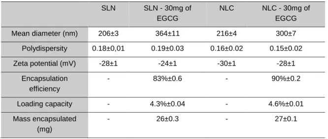
4.2.NANOPARTICLE CHARACTERIZATION ... 29
4.3.STABILITY STUDY ... 30
4.4.PARTICLE MORPHOLOGY CHARACTERIZATION ... 32
4.5.Release study ... 33
4.6.CELL VIABILITY AND CYTOTOXICITY ASSAY ... 34
5. CONCLUSIONS
... 395.1.FUTURE PROSPECTS ... 40
ix LIST OF FIGURES
Figure 1 - Chemical structure of the four major catechins present in tea. a) (-)-epigallocatechin gallate
(EGCG); b) (-)-epigallocatechin; c) (-)-epicatechin; d) (-)-epicatechin gallate. ... 4
Figure 2 - Schematic representation of the matrix for SLN and NLC and drug release ... 14
Figure 3 - A schematic representation of the steps to produce lipid nanoparticles ... 23
Figure 4 – Synthetized nanoformulations: a) SLN; b) SLN+EGCG; c) NLC; d) NLC+EGCG ... 29
Figure 5 - Evolution of SLN mean diameter and polydispersity ... 31
Figure 6 - Evolution of NLC mean diameter and polydispersity ... 31
Figure 7 - Evolution of zeta potential of SLN ... 31
Figure 8 - Evolution of zeta potential of NLC ... 31
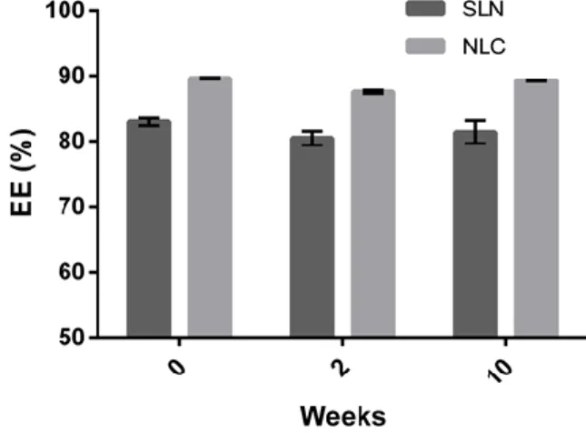
Figure 9 – Evolution of EE ... 31
Figure 10 – Cryo-SEM images for: a) SLN; b) SLN+EGCG; c) NLC; d) NLC+EGCG. Scale bar of 2 µm ... 32
Figure 11 – TEM images for: a) SLN; b) SLN+EGCG; c) NLC; d) NLC+EGCG. Scale bar of 0.5 µm . 33 Figure 12 – EGCG release in simulated gastrointestinal. ... 34
Figure 13 – Morphology of confluent Caco-2 cells by inverted microscope observation with 100X magnification ... 35
Figure 14 – MTT results for Caco-2 cell line (Mean ± SD) ... 36
Figure 15 – LDH results for Caco-2 cell line (Mean ± SD) ... 36
Figure 16 – IC50 graph for MTT assay ... 37
xi LIST OF TABLES
Table 1 - Natural sources of EGCG ... 2
Table 2 - Lipid nanoparticles used as EGCG carriers ... 15
Table 3 - Polymeric nanoparticles used as EGCG carriers. ... 17
Table 4 - Gold nanoparticles used as EGCG carriers. ... 19
Table 5 - Liposomes used as EGCG carriers. ... 20
Table 6 – Quantitative composition of stearic acid lipid NP prepared... 22
Table 7 - Quantitative composition of Precirol® ATO 5 lipid nanoparticles. ... 23
Table 8 – Composition of the EGCG-loaded nanoformulations. ... 24
xiii ABBREVIATIONS AND SYMBOLS
AD – Alzheimer’s disease BBB – blood brain barrier COX-2 – cyclooxygenase-2 DLS – dynamic light scattering
DMEM – Dulbecco’s modified Eagle’s medium DMSO – dimethyl sulphate
EC – epicatechin
ECG – epicatechin gallate EE – encapsulation efficiency EGC – epigallocatechin
EGCG – epigallocatechin gallate FBS – fetal bovine serum
GRAS – Generally Recognized as Safe HIF-1α – hypoxia-inducible factor 1-α
IC50 – half maximum inhibitory concentration IL-1β – interleukin-1β
LC – loading capacity
LDH – lactase dehydrogenase MMPs – matrix metalloproteinase
MTT – thiazolyl blue tetrazolium
NF-kB – nuclear factor kappa-light-chain-enhancer of activated B cells
NLC – nanostructured lipid carriers Papp – apparent permeability
PBS – phosphate buffered saline PD – Parkinson disease
PEG – poly(ethylene glycol) PhC – phosphatidylcholine PLA – polylatic acid
PLGA – poly (lactide-co-glycolide) acid p53 – tumour suppressor p53
SD – standard deviation
SEM – Scanning Electron Microscopy SLN – solid lipid nanoparticles
TEM – transmission Electron Microscopy TNFα – tumour necrosis factor α
VEGF – vascular endothelial growth factor
Denc – mass of EGCG encapsulated
Dlost – mass of EGCG non encapsulated
Dused – mass of EGCG
1
1
1.
INTRODUCTION
1.1. MOTIVATION
In the last years, the modern society is facing a large health problem with the rising of new diseases like heart diseases, diabetes, neurodegenerative diseases and obesity. For many years, these diseases were only found in the elders and with low incidence, but nowadays they era emerging in a more aggressive way, in a larger and younger group of people. For the above-mentioned reason, it is important to invest in a healthier and more holistic life style.
Green tea is an infusion of the tea plant, which is consumed for centuries in China, associated with health benefits. This beverage is highly concentrated in antioxidants, namely polyphenols, such as epigallocatechin (EGC), epicatechin (EC), epicatechin gallate (ECG) and epigallocatechin gallate (EGCG) (1). Recent studies found that these polyphenols have numerous benefits in the prevention and treatment of cancer, vascular and degenerative diseases, diabetes, obesity and other health concerns. Of the former compounds, EGCG is the most abundant and therapeutically active(2, 3). The worldwide population consumption of EGCG would be of high interest, being the use of a nutraceutical or supplement EGCG-based since it may prevent the appearance of severe health concerns. Notwithstanding, EGCG has an extremely low intestinal absorbance and a high degradation rate in the intestinal environment(4, 5). For the former reasons, the use of dietary natural sources of EGCG in the dietary nutrition seems to be insufficient to reach therapeutic concentrations of EGCG, and consequently health benefits.
The use of nanotechnology in the medicine, more specifically the use of nanocarrier systems is well known, and currently several nanoformulations are already in the market. More specifically, nanoparticles are used to enhance permeability, transport and in some cases to target a specific tissue for the treatment of a certain illness(6).
In this context, in this work two nanocarrier systems for the delivery of EGCG were synthetized, developed and characterized. The chosen nanocarriers were lipid nanoparticles, more specifically solid lipid nanoparticles (SLN) and nanostructured lipid carriers (NLC). The nanocarriers were designed to be biocompatible, and to improve the stability and bioavailability of EGCG. In the future, the developed nanocarrier-based EGCG delivery systems may be exploited in vivo for preventive and therapeutic supplementation of EGCG.
Design, development and characterization of innovative lipid nanocarrier-based epigallocatechin gallate delivery system for preventive and therapeutic supplementation
2
1.2. NATURAL SOURCES OF EGCG
Catechins are compounds widely distributed in nature, being well dispersed through the plant kingdom. Specifically, EGCG has been identified in a large number of plants, being found in a variety of edible vegetables. In the human diet, most of the source of catechins is obtained by the consumption of tea, and more specifically green tea. A list of plants containing EGCG is presented in the Table 1.
Table 1 - Natural sources of EGCG
Plant Scientific name Concentration of EGCG (mg/100g)
Organ Ref.
Strawberry Fragaria X ananassa 0.11 Fruit (7)
Apple Malus domestica 0.11 – 1.93 Fruit (7)
Avocado Persea amaricana 0.15 Fruit (7)
Pear Pyrus communis 0.17 Fruit (7)
Onion Allium cepa 0.20 Bulb (7)
Peach Prunus persica 0.30 Fruit (7)
Pistachio nut
Pistacia vera 0.40 Seed (7)
Plum Prunus, spp. 0.40 – 0.48 Fruit (7)
Raspberry Rubus, spp. 0.54 Fruit (7)
Kiwifruit Actinidia deliciosa 0.55 Fruit (7)
Blackberry Rubus, spp. 0.68 Fruit (7)
Cranberry Vaccinium macrocarpum
0.97 Fruit (7)
Hazelnut Corylus, spp. 1.06 Seed (7)
Pecan Carya illinonensis 2.30 Seed (7)
Tea Camellia sinensis 70.20 Leafs (7)
Carob Ceratonia siliqua 109.46 Seed pod with
seeds
(7)
N/A Salacia reticulata N/A Leafs (8)
1.2.1. TEA
Tea is an infusion made from the leaves of the tea plant (Camelia sinensis) originated in China. Depending of the process used to dry the leaves, the tea will gain different aspects, tastes, chemical compositions and therapeutically properties. There are three principal types of tea: green tea, black tea and oolong. Green tea is prepared by steaming the tea leaves after the harvest. This process will prevent the enzymatic oxidation of the compounds found in the green leaf, mainly tea catechins, preserving the fresh state. After the initial process, leafs are dried and stored. By opposition, the black
3
tea is prepared by allowing the fresh picked leafs to ferment in a humid place at the room temperature for a day after drying. This allows the natural enzymes present in the leaf to oxidize the catechins in to complex polymeric products: thearubigins and theaflavins. These compounds change the taste, color and therapeutic properties of the tea. The oolong tea is an intermediate between green and black tea, where the leaves are allowed to partially ferment and dried. The composition of oolong is like the process itself, an intermediate between the two others, comprising a mixture of monomeric polyphenols and higher weight theaflavins(1).
Tea is a very popular beverage in Asia for centuries. Nowadays, tea is the second most consumed beverage after water and this beverage has gained a large interest recently for its promising health benefits in several fields, including weight loss, diabetes and cancer prevention. The health benefits of tea are mainly attributed to green tea and its polyphenol’s content, in particular, tea catechins(2, 3). Traditionally, tea is consumed in the form of infusion. This ancient way of consuming tea provides a substantial source of polyphenols, but for achieve the target benefits for the treatment or prevention of a particular pathology, several cups of tea would be needed to ingest the necessary amount of EGCG. In another perspective, would be interesting to use the most therapeutic compound of the green tea for supplementing food or in the form of a pharmaceutical supplement to be orally taken and surpass the low bioavailability issue. With this approach a higher amount of EGCG may be administered in a convenient way. Indeed, the bioavailability of tea catechins is very low, being in order of 1-2% (9). Therefore, technologies that enhance the stability and bioavailability of the catechin would be of high interest, providing a more effective use and cost of EGCG.
1.3. CHEMICAL STRUCTURE OF CATECHINS (EGCG)
Green tea has a great amount of polyphenols divided in three major groups: flavanols, flavones and flavonols. Flavanols comprises the majority of these content, being about one third alone of the dry mass of green tea leafs, and are mainly distributed by four molecules: epicatechin (EC), (-)-epicatechin gallate (ECG), (-)-epigallocatechin (EGC) and (-)-epigallocatechin gallate (EGCG). The chemical structures of these compounds are illustrated in Figure 1. The last one, EGCG is the most abundant catechin present in green tea, being 65% of the total flavanols and one-third of the tea dry mass.
Design, development and characterization of innovative lipid nanocarrier-based epigallocatechin gallate delivery system for preventive and therapeutic supplementation
4
a) b)
c) d)
Figure 1 - Chemical structure of the four major catechins present in tea. a) (-)-epigallocatechin gallate (EGCG); b) (-)-epigallocatechin; c) (-)-epicatechin; d) (-)-epicatechin gallate.
Epigallocatechin-3-gallate (EGCG) is a complex molecule formed by a flavanol core (flavan-3-ols) structure with a gallocatechol group and a gallate ester. These two gallocatechol rings confer the potent antioxidant and chelating properties to EGCG. Each of the gallocatechol rings is capable of directly capture free radicals from the environment with high efficiency(10). Previous studies, have shown, that EGCG possesses the stronger antioxidant capacity comparing with the others green tea catechins, and it is also demonstrated that EGCG is more efficient in radical scavenging than vitamin E and C(11).
In the human body, catechins are capable of reducing the amount of free radicals by chelating metal ions, specially the iron ion. These metal ions are known to produce free radicals by the Fenton’s reaction. They accomplish this sequestration by biding the ion to the catechol or the galloyl groups found in the structrure of the catechins. The numbers of these groups that are present in the catechin strongly influence the ion binding capacity. The catechins with only one of the group’s, epicatechin and epigallocatechin, are only capable to bind one ion per identity. In the other hand, the two catechol rings in epicatechin gallate and the two galloyl rings in EGCG are spatially distant from one another, which allows then to independently chelate two ions per molecule(12).
1.4. PHARMACOKINETICS
EGCG has various health benefits. However, EGCG has very low bioavailability and undergoes through extensive degradation in the intestine. For above-mentioned reasons, a daily supplementation of the diet with this molecule would be of high interest for the society health care.
5 1.4.1. ABSORPTION
The absorption of a drug is a key component to achieving good bioavailability and ensuring that the drug is able to reach the systemic circulation. For oral drugs, the majority of drug absorption occurs in the small intestine where the presence of villi and microvilli greatly increase the surface area for optimal absorption. Drug absorption in the small intestine is greatly influenced by multiple interacting factors, including drug properties (e.g., solubility, formulation, and amount), gastrointestinal properties (e.g., pH, food intake, region of the small intestine), metabolism, permeability, and active transport across the intestinal epithelial membrane(13).
Permeability studies demonstrated that EGCG has a very low apparent permeability (Papp) of 3,5x10
-7
cm2/s (4). Comparing the apparent permeability of EGCG with manitol, which is a poorly permeable compound with an apparent permeability of 6 to 10x10-6 cm2/s is clear that the intestinal mucosa is a highly impermeable barrier to EGCG. Therefore, the Papp of the catechin is almost ten times lower in
comparison with manitol (14). One of the major reasons of the lower permeation through the intestinal barrier is the high hydrophility of the catechin, associated with a lack of transporters to help the uptake and the presence of efflux transporter that actively excretes EGCG to the intestinal lumen(13)
Although the low bioavailability, EGCG is rapidly absorbed after oral administration, being readily detected in the blood circulatory system. The maximum blood concentration of EGCG is accomplished after 1 to 2 hours after a single oral dose administration (15, 16).
To achieve therapeutic concentrations of EGCG, a larger dose or alternatively the use of a delivery system can be exploited to overcome the low bioavailability of this catechin
1.4.2. DISTRIBUTION
After the absorption across the gastrointestinal tract, the drug will be distributed through the entire body. This process can be influenced by several factors, such as the solubility of the drug, the protein binding properties, the bioaccumulation in non-target organs and the clearance rate. EGCG has the capacity to bind to plasma proteins, especially to fibronectin, fibrinogen and histidine-rich glycoprotein (17). In addition, EGCG shows affinity to some tissues of the body, being accumulated in several organs, such as the lung, intestine, liver and prostate gland (9, 18).
1.4.3. METABOLIZATION
Metabolization comprises the life-sustaining chemical processes that occur within the cells of living organisms. Humans have several metabolization pathways that can be allocated in two major categories; the phase I reactions and the phase II reactions. In the phase I reactions, typically the xenobiotic is activated by a sort of enzymes like the cytochrome P-450, modifying the molecule into a more reactive intermediate. This allows the chemical reactions with the phase II enzymes. The phase II enzymes bind the activated molecule to a functional group including among others, sulphate and acetate. These conjugates are in general less active and less toxic than the original molecules and are more easily eliminated(19).
EGCG like other molecules can be metabolized. There are three major metabolites of EGCG that can be found in plasma namely, the glucuronide, the methylated and the sulphated forms (16). These metabolites are present at lower concentration in the serum. Moreover, these metabolites are inert, with no biological relevance with the exception of the methylated EGCG. The methylated EGCG
Design, development and characterization of innovative lipid nanocarrier-based epigallocatechin gallate delivery system for preventive and therapeutic supplementation
6
seems to accumulate in the prostate tissues and could be helpful in the treatment of prostate cancer(20).
Another metabolization pathway of the EGCG in the human saliva was described recently (16). Chung and co-workers described the presence of a degalloylate esterase that hydrolysed the ester bound between the epigallocatechin core and gallate group(21).
Another mechanism is the metabolization of EGCG by the gut flora in the intestine, which seems to be also responsible for the low bioavailability of EGCG(5). A study performed by Kohri et al using radioactive EGCG found a spike radioactivity in the mouse urine 24 hours after the initial oral dosecontrairlly to the found in literature. So the presence of radioactivity was confirmed to be resultant of by-products of intestine flora metabolization. They found that mouse pre-treated with antibiotics show no significative clearance of radioactivity trough mouse urine. Based in this results they conclude that EGCG is metabolised by the gut flora and the resultant metabolites reabsorbed by the intestine to the blood current(22). This could be one major cause of the poor bioavailability of EGCG.
1.4.4. ELIMINATION
In the human body, there are two major elimination mechanisms. The most common is the elimination of EGCG metabolites through the urine. The kidney is an organ specialised in the filtration of the blood, eliminating board spectrum of unwanted substances and excreting then in the urine. According previous studies, only small amounts of EGCG are detected in the urine (18, 23).
The second most common elimination pathway in the human body is the elimination through faeces. In this case, the xenobiotic is captured by the liver and excreted to the bile, being posteriorly released in intestinal tract and excreted in the faecal matter. This elimination pathway is the principal elimination route of EGCG (9, 18). In lesser extent, the clearance of some substances can be helped by mechanisms of active transport in the intestinal mucosa through efflux transporters. This is more relevant when the drug is first absorbed by the epithelial cell and then is actively transported to the intestinal lumen again. With lesser extent, EGCG can be cleared by the last mechanism via efflux transporters bellowing to the P-glycoprotein family. The sum of the eliminations mechanisms leads to a rapid clearance of EGCG (24).
In summary, the conjugation of a low absorption in the gut, and high rate of degradation in the gut environment, the high extension of the metabolization and elimination processes makes the overall bioavailability of EGCG extremely low.
1.4.5. TOXICITY
Green tea is an ancient beverage, being consumed worldwide and never was reported any case of toxicity. However, the same cannot be extrapolated to the larger dosages founded in EGCG supplements. These new supplements possess doses of EGCG that are virtually not possible to obtain with only the tea consumption. EGCG seems to be well-tolerated by the human body and to the date, none adverse effects are described. Nonetheless, no studies that analysed the long-term use of high doses of EGCG in humans were founded (25). In France, a green tea supplement was withdrawn from the marked, with suspicions of liver toxicity. A fulminant liver hepatitis was reported after the consumption of a green tea extract in the form of weight loss supplement (26). This emphasizes the necessity of long-term studies to help to understand the risks of the supplementation with EGCG.
7
Another interesting property of EGCG is the ability to block the folate transporter in the intestine. The inhibition of the uptake of folate can lead to depletion of the folate levels in the human body, which may result in permanent brain damages(27).
1.5. THERAPEUTIC AND PREVENTION USE OF EGCG
For many years, the consumption of green tea was associated with numerous health benefits. These properties can be directly linked with the polyphenol content of tea, more specifically with EGCG. For these reason, the study of EGCG is of high interest because this compound seems to prevent and also be useful in the treatment of numerous diseases like cancer, cardiovascular and neurodegenerative diseases and also obesity. EGCG is a powerful antioxidant, anti-inflammatory, antibacterial, antiviral and is capable to modulate some metabolic pathways like the metabolism of lipids(2, 3).
1.5.1. CANCER
Cancer is the end of several steps of cellular growth lesions, namely hyperplasia, metaplasia, dysplasia and neoplasia. Each of the presented conditions is a progression in the cancer formation, culminating in the neoplasia malignant known as cancer(28). The development of cancer can be induced by several factors, including among others, diet, drugs and smoking. In the disease process, the cells evade the immune system and have the capacity to undergo through successful mitosis without external repression. Nowadays, most modern therapies currently available for treating cancer are very expensive, toxic, and have low effectiveness in treating the disease. Therefore, is necessary investigate natural compounds like EGCG derived from green tea, described traditionally, for the prevention and treatment of cancer and other diseases(28).
According to previous studies, EGCG is a promising molecule in the prevention and treatment of cancer. Some anti-cancer properties of EGCG are attributed to its free radical scavenging properties, avoiding the damage of the cell structures induced by the free radicals. Besides being antioxidant, EGCG has the ability to bind and modulate the activity of several signalling molecules related with mitosis, survival and cellular death, moderating the cellular responses present in cancer. Previous works demonstrated that EGCG is able to inhibit all of the processes involved in carcinogenesis: initiation, promotion and progression (20, 29).
EGCG has the ability to bind with some proteins associated in molecular pathways that are miss-regulated in cancerous cells. Indeed, EGCG modulates the suppression of two important transcription factors, tumour suppressor p53 (p53) and nuclear factor kappa-light-chain-enhancer of activated B cells (NF-kB), leading to a regression of the tumours (20, 29, 30).
To assist the growth of the tumour, new capillaries are needed to ensure the requirements of the cells in oxygen and nutrients. The grow process of new blood vessels is called angiogenesis. To promote formation of new capillaries, the tumour secrete to the surrounding tissues signalling molecules, especially vascular endothelial growth factor (VEGF). VEGF is directly influence by the activity of hypoxia-inducible factor 1-α (HIF-1α) and NF-kB factors, which are modulate by the presence of EGCG. For these reason, EGCG is capable of diminishing the tumour angiogenesis and stall growth (31).
In addition, there is strong evidence that EGCG is capable of diminishing migration and metastasis formation of tumours. Previous studies report that EGCG promotes a reduction in the migration and
Design, development and characterization of innovative lipid nanocarrier-based epigallocatechin gallate delivery system for preventive and therapeutic supplementation
8
metastasis formation of tumoral cells with tumour size reduction, accomplishing a more reliable and efficient chemotherapy(32).
Although the single use of EGCG in chemotherapy is unlikely due its inefficacy in eliminating the disease completely, it would be very interesting using EGCG as adjuvant of the cytostatic drugs. This synergism may be useful to reduce the amount of the necessary cytostatic drugs, which will reduce the side-effects. Moreover, the health benefits of EGCG would be advantageous in enhancing the overall condition of the patients (28, 30).
1.5.2. CARDIOVASCULAR DISEASES
Cardiovascular diseases have a high incidence, mainly in the developed world due to a sedentary life style, poor nutrition and ambient factors. A diet rich in cholesterol, fat and sugar causes can lead to coronary diseases like arteriosclerosis and ischemia. These diseases are characterised by a deposition of cholesterol in the vascular vessels forming platelets, which can lead to thrombosis. These depositions can interfere and obstruct the normal flow of blood, and in some cases can lead to serious complications like strokes and myocardial infarctions(33, 34).
Recent studies show that EGCG can enhance the capillary circulation dilating the capillaries, diminishing inflammation and interfere with the lipid absorption and digestion(34-36).
On the other hand, EGCG interferes directly with the lipid emulsion process in the lipid digestion. This is achieved by directly interference in the micelle formation and by inhibiting the phospholipase A2, being this enzyme of high importance in the lipid digestion (37). The junction of the two processes can limit the absorption of lipids and consequently, lowering the amount of plasmatic lipids and cholesterol (37). In addition, EGCG can lower cholesterol even more, stimulating his excretion through the bile. Moreover, EGCG will further improve the lipid profile by enhancing the lipid metabolism. This catechin can also modulate the process of the platelets formation, from the macrophage recruiting to the macrophage up take of cholesterol. This effect is internally modulated in the macrophage and externally helped by the anti-inflammatory response caused by EGCG. Previous studies demonstrated that the administration of EGCG is capable of prevent the growth and also to reduce the size of existing platelets. The action mechanism responsible for the anti-inflammatory property of EGCG is the direct inhibition of the phospholipase A2(35).
1.5.3. NEURODEGENERATIVE DISEASES
The cause of neurodegenerative diseases like Parkinson (PD) and Alzheimer’s (AD) diseases are still unknown, being various theories proposed. Both diseases present clinic features, like the oxidative damage to neurons and accumulation of iron in specific brain areas. Another relevant aspect is the accumulation of missfolded proteins in deposits, such as the β-amyloid peptide in AD that interfere with the survival of the neurons, leading to a premature apoptosis(38).
Special interest has been assigned to the therapeutic role of antioxidants in such neurodegenerative diseases. The neuroprotective properties of EGCG agent are related with its properties as antioxidant, anti-inflammatory and iron chelating. In addition, the blood brain barrier (BBB) is permeable to EGCG (39). The mechanism behind the passage of this hydrophilic compound through the BBA remains unknown.
9
In the literature it is described that EGCG is more efficient in radical scavenging than vitamin C and E, being its iron chelating ability useful to significantly improve the symptoms of these neurodegenerative diseases(11, 40).
According to the above-mentioned, EGCG is also a cellular modulator that interacts with various pathways. In neuronal cells, this catechin promotes cell survival responses and the inhibition of cell death signals, leading to an enhancement of neuronal health. The modifications in the cell signalling also promote the non-amyloid α-secretase pathway, diminishing the production of β-amyloid peptides(40).
Several research studies confirm that EGCG has neuroprotective properties in humans, being the degree of cognition enhanced after the oral administration of EGCG. These studies confirm that EGCG promotes an overall increase in the cerebral activity and calmness. All of these aspects are important in neurodegenerative diseases, being the administration of EGCG proven to help in maintaining the normal levels of cognition, diminishing agitated stages and mod shifts(41).
1.5.4. INFECTIOUS DISEASES
Viruses are small infectious agents that replicates only in living cells. They are composed by nucleic acid (DNA or RNA) and some enzymes encapsulated in a proteic sell and may have a lipid membrane covering the structure. To multiplied, they enter a living cell and redirect the cell bioquemistry to start producing virus components. Generally, viruses are dificul to treat due to its strong intricacy with cellular host mechanisms to replicate(42).
Nowadays, the main strategy to fight viruses is the immunization. Unfortunately, several viral infections lack one efficient vaccine, being the most mediatic the HIV infection. In the last viral infection, several studies were performed with some encouraging results. Fassina et al, shown a strong HIV inhibition caused by EGCG in cell cultures in a dose dependent manner(43). Later, Li S. et al also proven that EGCG inhibit the reverse transcriptase, and act synergistically with another reverse transcriptase inhibitor, namely azidothymidine(44). Some studies also described that EGCG is capable to bind to the CD4, preventing the virus to anchor and enter the cells(43).
EGCG is also useful in the inhibition of other viruses, such as Enterovirus 71, Hepatitis C, Adenovirus, Herpes simplex and Influenza. One of the molecular targets that seem to be deregulated by the viral infection is the NF-kB and the MAP-kinases pathway(45-49).
1.5.5. OBESITY
Obesity is a medical condition characterized by an excess accumulation of fat in the body in an extension that may have negative effects in the overall health condition and may lead to the development of diseases, such as diabetes and arteriosclerosis, among others. Obesity is mainly attributed to the diet and lifestyle (50). Sedentary habits and high caloric intake commonly lead to fat accumulation in the adipose tissue. The fat percentage can alter significantly the biochemistry of the human body, leading to hormonal problems, high oxidative stress, circulatory complications and other conditions. The main treatment of obesity is the lifestyle re-education, including diet modification. However, sometimes it is difficult to achieve the desirable results and in some cases, drugs and supplements are needed to help the losing weight process(50).
Design, development and characterization of innovative lipid nanocarrier-based epigallocatechin gallate delivery system for preventive and therapeutic supplementation
10
As previously stated, EGCG interferes directly with the lipid digestion by the inhibition of the phospholipase A2, and interfering with the lipid/cholesterol emulsion in gut (37, 51). The lipid blocking capacity of EGCG can be highly relevant in the loss of weight and weight managing protocols. In addition, EGCG is capable to enhance the lipid metabolism, leading to a more caloric burn and consequent fat loss. EGCG can also interfere with the digestion of starch by inhibition of α-amilase(52).
Besides the above-mentioned, the ingestion of EGCG during a weight loss programme is very useful because its administration is strong linked with the circulation improvement, free radical scavenging and mod enhancer (10, 34, 41, 49).
1.5.6. CHRONIC INFLAMMATORY DISORDER
The inflammation is a body response to foreign structures to the human body and damage in the tissues. This response is highly important to the normal health of an individual due to the capacity to combat diseases and assisting heals of damage tissues like cuts and bruises. The inflammatory process is characterised by an increase of the capillary diameter lead to an increase in swelling, redness and pain. The inflammation response also recruit immune cell to eliminate any foreign body and damage tissue. However in chronic inflammatory disorders, this inflammatory response is continuously active leading to the destruction of healthy tissues causing all the above mentioned symptoms. These conditions can be incurable and cause major discomfort to the patients(53).
Rheumatoid arthritis is one chronic inflammatory disorder characterized by cellular infiltration and proliferation of the synovium, leading to the progressive destruction of the joints through the interaction between infiltrating cells and mediators(54, 55). These injuries lead to chronic pain affecting the life quality of the patients. In this disease, the cartilage cells (i.e., chondrocytes) enter in apoptosis in response to inflammatory cytokines interleukin (IL-1β) and tumour necrosis factor α (TNFα) and oxidative stress. The same cytokines also lead to the increase of bone resorption and the differentiation of osteoclasts(54).
IL-1β is an inflammatory cytokine that is over expressed in arthritis promoting imbalance between excessive cartilage destruction and repair processes. In addition, IL-1β is also capable of increasing the amounts of reactive oxygen species via overexpression of inducible nitric oxide synthase and increases the inflammation by an overexpression of cyclooxygenase-2 (COX-2)(56). The presence of IL-1β can also activate the expression of matrix metalloproteinases (MMPs) responsible for the matrix degradation. TNFα also plays an important role in the bone turn over. In arthritis, there is an overexpression of TNFα who is responsible the differentiation and activity of osteoclasts. The long term activation of this cells lead to the bone erosion and fragility(57).
The normal treatment in arthritis is the administration of methotrexate, which combines a good efficacy and low toxicity. This drug is widely used, however in some patients’ it is ineffective and recent studies shown that the drug tends to lose efficacy over time. To attenuate the symptoms is also used analgesics and nonsteroidal anti-inflammatory drugs (58).
Although the efficacy of the current treatment, the complete remission of the disease is often not achieved and for this reason new therapies are needed. EGCG has high antioxidant activity and also capacity to decrease the inflammation response in the body. In cartilage cell cultures, EGCG showed a marked inhibition of IL-1β inducible nitric oxide synthase COX-2 expression and activity. The
11
expressions of both enzymes are mediated by the NF-kB, which is supressed in the presence of EGCG (59).
13
2
2.
NANOTECHNOLOGY
Nanotechnology is the science and technology of small things and particularly materials with dimensions between 1 and 1000 nanometers(6). Nanoparticles are structures that have at least one of the dimensions in the nanoscale range. Due to their high surface area to volume ratio, nanoparticles present chemical, physic and biological properties distinct from conventional materials(60). The size and the different properties of the nanoparticles can be used for medical purposes. The small scale of nanoparticles makes then excellent drug carriers, and because they can be modified in various factors like size, chemical composition, outer layer and others they are very versatile. The physical properties can also be useful in diagnostic techniques, such as tomography imaging. In addition, nanoparticles can modify the pharmacokinetics and the stability of some drugs(60). This is specially truth in the case of EGCG, where the nanotechnology can be used to increase the bioavailability of this catechin (61). Another interesting characteristic of the nanoparticles is the possibility of enhance the cellular uptake by modifying the outer layer with different ligands to assign specificity to certain cells and/or structures. This technology may increase the bioavailability and stability of EGCG, enhancing the health potential of this compound. To the date, no studies are described related with the use of nanoparticle-based formulations with EGCG in humans(62).
There are several nanosystems used in drug delivery, including lipid nanoparticles liposomes, polymeric nanoparticles, nanocrystals, gold nanoparticles, nanotubes and magnetic nanoparticles.
2.1. LIPID NANOPARTICLES
Lipid nanoparticles were introduced in the early 1990s, being one of the most currently used nanosystems to delivery drugs. These nanoparticles are prepared from a lipid matrix, with final particle size ranging between 1 and 1000 nm(6). The combination of particle size, use of non-toxic materials, physical stability, controlled release properties, high drug load and excellent tolerability make them important colloidal carriers (63). Their synthesis requires three components, namely the hydrophobic lipid phase, one emulsifier and the hydrophilic aqueous phase. If their final destination use is for oral and topical administration, all components used need to hold a Generally Recognized as Safe (GRAS) status. The lipids used, normally have a melting point above the body temperature and can be mono, di and triglycerides alone or in mixtures, waxes and other types of lipids(64).
There are two main types of lipid nanoparticles, namely solid lipid nanoparticles (SLN) and nanostructured lipid carriers (NLC). These two nanoparticles differed in the type of lipid used, being
Design, development and characterization of innovative lipid nanocarrier-based epigallocatechin gallate delivery system for preventive and therapeutic supplementation
14
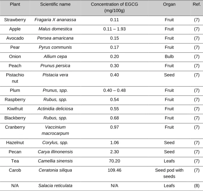
in case of SLN used only solid lipids and NLC are composed of a mixture of both solid and liquid lipids. Due to their distinctive lipid crystalline structure, these nanoparticles have different encapsulation and release behaviours. The SLN lipid matrix fully crystallised tend to have lower drug loads, since there are fewer gaps in which the dug can enter. It also tends to expulse the drug content during long storage due to crystalline rearrangement. Instead, NLC have a higher drug loading and storage stability due to the larger number of imperfection in the lipid core(64) The structure and drug interaction is schematised in Figure 3.
Figure 2 - Schematic representation of the matrix for SLN and NLC and drug release
Lipid nanoparticles have been used to enhance oral bioavailability of various drugs, by enhancing the gastrointestinal stability and/or by increasing the permeability. Only one study was found related with the encapsulation of EGCG in lipid nanoparticles. In this context, Zhang et al synthetized NLCs with a mixture of two lipids (glyceryl tridecanoate and glyceryl tripalmitate) and two surfactants (soy lecithin and Kolliphor® HS15). The formulation was designed to enhance bioavailability of EGCG by oral administration and safely transport the catechin through the blood stream, targeting THP-1-drived macrophages. THP-1-drived macrophages are responsible for the accumulation of cholesterol in arteries and therefore the primarily target for EGCG. In addition, the authors synthetized the lipid nanoparticles coated with a layer of chitosan to enhance the nanoparticles permeability and targeting to the macrophages. The nanoparticles exhibited a low release (i.e., 4.43%) of the EGCG content after 9 hours of incubation in phosphate buffered solution (PBS) at pH 5.0, being the degradation rate of encapsulated EGCG in neutral pH significantly lower that the non-encapsulated EGCG. At 37ºC, the EGCG was completely degraded in the course of three hours; meanwhile 65% of the encapsulated EGCG was still unchanged. Cell toxicity was found to be very low, leading to a 9-fold decrease in the accumulation of cholesterol in macrophage cultures in combination with a diminishing secretion of inflammatory factors. Despite the promising results presented by Zhang et al., the study lacks the permeation study in caco-2 monolayer to estimate the permeation of EGCG (65).
15 Table 2 - Lipid nanoparticles used as EGCG carriers
Nanoparticles composition Loading capacity (%) Loading efficiency (%) Size (nm) Administration route
In vitro/in vivo results Ref.
Glyceryl tridecanoate, glyceryl tripalmitate, soy lecithin and Kolliphor® HS15, chitosan
3% 99% 50 Oral High stability in both acidic and neutral environments.
In vitro studies performed in
THP-1-drived macrophages showed decrease in inflammation and accumulation of cholesterol. (65) 2.2. POLYMERIC NANOPARTICLES
Polymeric nanoparticles are made from polymers that can be natural or synthetic. These kinds of nanoparticles have been extensively studied for drug transport due to their high biocompatibility and easy synthesis. In polymeric nanoparticles, the drug is transported in the polymer matrix, being protected from the outside conditions. To enhance the pharmacokinetics profile of the drug, or in some cases, to target some specific tissue, the outer layer of the polymer can be functionalized with molecular markers. Some reports of EGCG enhancement uptake have been proposed and tested in vitro and in vivo with promising results(66).
Chitosan is a popular choice to encapsulate EGCG regarding its biocompatibility, biodegradability and low toxicity. In addition, chitosan is mucoadhesive and can enhance the permeability of the intestine by opening the tight junctions(67). Dube et al used chitosan nanoparticles to enhance the intestinal absorption of EGCG in vitro. Moreover, the same authors proved that chitosan nanoparticles also enhance the EGCG uptake in vivo (14, 68).
Khan et al used EGCG loaded chitosan nanoparticles in athymic mice implanted with prostate cancer cells and found a reduction in the growth of the tumour comparing to the two controls, one that receive void nanoparticles and the other that received the free EGCG. The results also indicate a dose dependent relation between the amount of encapsulated EGCG and the size of the tumour (69).
Hu et al synthetized chitosan cross-linked with casein phosphopeptides. The developed nanoparticles enhance the stability of EGCG through the digestive system (70). In addition, the permeability of EGCG was increased in caco-2 cells monolayers (71).
Polylatic acid (PLA) and poly (lactide-co-glycolide) acid (PLGA) are two of the most extensively studied polymers to create nanoparticles because of their versatility and high biocompatibility. The outer layer of these nanoparticles can be easily modified with functional groups to specifically target certain cells and/or structures. In addition, the degradation time of these nanoparticles can also be manipulated and they can be have high encapsulation rates and controlled release properties(66, 72). Sanna et al synthetized PLGA-based nanoparticles coated with poly(ethylene glycol) (PEG) and they modified their surface with a pseudomimeticdipeptideN-[N-[(S)-1,3-dicarboxypropyl]carbamoyl]-(S)-lysine to act as a molecular target to a prostate cancer membrane protein (i.e., prostate-specific membrane antigen), which is overexpressed. The nanoparticles were tested in prostate cancer cells
Design, development and characterization of innovative lipid nanocarrier-based epigallocatechin gallate delivery system for preventive and therapeutic supplementation
16
lines with promising results (73). Siddiqui et al, also have encouraging results but instead of PLGA-PEG they used PLA-PLGA-PEG nanoparticles (74). The drawback is that both PLA and PLGA polymers are sensitive to the gastric pH and the majority of the studies focus in an intravenous administration method, which is an invasive route of administration (73, 74).
Srivastava et al used a PLGA EGCG loaded nanoparticle to prevent DNA damage caused by 7,12-dimethylbenzanthracene (DMBA). In this study, mouse was treated topically with EGCG and PLGA EGCG nanoparticles prior to topical of DMBA. The pre-treatment with the EGCG resulted in a protection of DNA damage in the order of 28%. However, the pre-treatment with the loaded nanoparticles result in a 63% protection in DNA. Additionally, EGCG loaded nanoparticles, showed significant induction of DNA repair genes and inhibiting inflammatory genes.(75). Other types of polymers have been studied; in this context, Li et al used ovalbumin-dextran conjugates nanoparticles to enhance the stability and absorption of EGCG through the digestive system. The results revealed that the ovalbumin-dextran nanoparticles were stable and the EGCG absorption profile was improved (76).
17 Table 3 - Polymeric nanoparticles used as EGCG carriers.
Nanoparticles composition Loading capacity (%) Loading efficiency (%) Size (nm) Administration route In vitro/in vivo results Ref.
Chitosan 0.4 N/A 440±37 Oral Enhancement of
the gastrointestinal permeation of EGCG in mice’s.
(14, 68)
Chitosan N/A ~10 ~150 Oral Reduction of
human prostate tumors in mice.
(69)
Chitosan, casein and peptides
N/A N/A 150 oral Bioavailability of
EGCG increment in caco-2 monolayers.
(70)
Chitosan casein phosphopeptides
N/A N/A 150±4.3 oral Enhancement of
the intestinal permeation of EGCG using caco-2 monolayers.
(71)
PLGA 5.76 26 127 topical Inhibition of DNA
damage.
(75)
PLGA and PEG ~0.4 ~9.5 80±15.0 Intravenous Inhibition of the growth of cultured cancerous cells.
(73)
PLA PEG N/A N/A N/A Intravenous Reduction in the
size of the implanted tumor in mice. (74) Ovalbumin and dextran 20.9 30.0 339 oral Enhancement of the intestinal stability and improvement of apparent permeability in Caco-2 models. (76)
Design, development and characterization of innovative lipid nanocarrier-based epigallocatechin gallate delivery system for preventive and therapeutic supplementation
18
2.3. GOLD NANOPARTICLES
Gold nanoparticles constitute a recent approach to the enhancement of EGCG permeability and cytotoxicity to the cancer cells. Gold nanoparticles are made from a gold core covered with a film of functional molecules, which are capable to transport complex cargos like drugs and targeting molecules. It is possible to synthetize gold nanoparticles covered with EGCG due to the capacity of this catechin to combine with metals to form complexes by the gallate ring (77). Although gold EGCG complexes present high stability in acidic pH conditions, they start decomposing in more pH basic conditions. Shukla et al and Chen et al solved this disadvantage by using an intratumoural injection (78, 79). Shukla et al synthetized radioactive gold nanoparticles coated with EGCG, to evaluate the chemotherapy efficacy. In this case, EGCG serves as chemoadjuvant and targeting molecule simultaneously, due to its high affinity to prostate cancer cells and anti-cancer properties(20, 29). The results obtained were very promising and a high efficacy of the formulation was achieved in reducing the tumour size (78). On the other hand, Chen et al. synthetized nonradioactive gold nanoparticles coated with EGCG to test only the capacity of EGCG as a chemoadjuvant or chemotherapeutical in cancer therapies(79). These authors found a promising cytotoxic effect both in vitro and in vivo using a murine melanoma cancer model.
Although the promising results, the two gold nanoformulations above-mentioned were administered by intratumoural injection, which is an invasive route of administration.
Hseig et al synthetized gold nanoparticles with the main goal to enhance gastrointestinal stability, increasing the release time in the intestine. The nanosystems obtained, revealed to be stable in the gastrointestinal environment with a sustained release of EGCG over 2 hours. The authors also demonstrated that the formulation shown a preferential toxicity to cancerous cells and an in vivo efficiency in reducing the growth of implanted tumours in mice. The sustained release and the high cytotoxicity to cancerous cells make these nanoparticles promising future nanoformulations to use in cancer therapy (80).
19 Table 4 - Gold nanoparticles used as EGCG carriers.
Nanoparticles composition Loading capacity (%) Loading efficiency (%) Size (nm) Administration route In vitro/in vivo results Ref.
Gold N/A N/A 20-
1200
Intratumoural Noticeable
reduction in bladder tumor in mice.
(77)
Gold 2% 27% 68 Intratumoural High cytotoxicity in melanoma cell culture and in mice.
(79)
Gold N/A 27% 50 oral Nanoformulation
stable in neutral pH with a sustained release of 2 hours. The in vitro and in
vivo experiments revealed that the nanoformulation is highly effective in the cancer therapy.
(80)
Radioactive gold
N/A N/A N/A Intratumoural Noticeable
reduction of tumor size achieved after 28 days with a single
administration of the formulation and
with minor
radioactive leakage to other organs.
(78)
2.4. LIPOSOMES
Liposomes are artificial vesicles composed by a lipid bilayer and an aqueous milieu. Liposomes are versatile nanocarriers because of their unique structure with both hydrophilic and hydrophobic environments, which enable the transport of all type of drugs. The size of liposomes can be finely adjusted and their surface can be chemically modified to target specific tissues or evading the immune system. The intrinsic stability of liposomes can also be modified by the lipid content. External factors, such as temperature, pH, light and enzyme presence can trigger an abrupt release of their content by interfering with the membrane stability, causing the lipid layer to collapse(81, 82). Although the promising results of liposomes as drug carriers, only two studies are described using liposomes as EGCG carriers. Both studies are focused in enhancing the bioavailability of EGCG. Luo et al synthetized phosphatidylcholine (PhC)/cholesterol liposomes to enhance the stability of EGCG in the gut. The liposomes obtained present a high stability in gastric conditions with small cargo loss around
Design, development and characterization of innovative lipid nanocarrier-based epigallocatechin gallate delivery system for preventive and therapeutic supplementation
20
20%, which increases slightly in the intestinal fluid to 40%. Although, cellular uptake studies are lacking, the results of the intestinal release by itself are promising in a future use as an EGCG formulation (83).
Song et al. synthetized liposomes with non-ionic surfactants and cholesterol to improve the stability of EGCG in the gut and enhance cellular uptake. Liposomes shown to be stable at pH=7.4 with minimal loss of the catechin. The authors also investigated the permeability of caco-2 cell monolayers to the EGCG entrapped liposomes in comparison with free EGCG. The liposomes were capable of significantly enhance the apparent permeability of EGCG (84). These promising results stimulate the investigation of a nanoformulation liposome-based to enhance the bioavailability of EGCG.
Table 5 - Liposomes used as EGCG carriers.
Particle Loading capacity Loading efficiency Size (nm) Administration rout In vitro/in vivo results Ref. PhC and cholesterol
N/A 85.8±1.65 180 Oral Favorable
release profile in the gastrointestinal fluids. (83) Sorbitan monostearate and cholesterol
N/A 40 100 Oral The
nanoformulation presents a high stability in neutral pH and enhances the cellular permeability in caco-2 cell monolayer. (84)
21
3
3.
MATERIALS AND METHODS
3.1. MATERIALS
The lipid Precirol® ATO 5 was a gift from Gattefossé (Gattefossé, France). The lipid Miglyol® 812 was purchased from Acofarma® (Terrassa, Spain). Tween® 60, TritonTM X-100, Thiazolyl Blue Tetrazolium (MTT), Trypan Blue powder, dimethyl sulfoxide ≥99.9%, acetic acid ≥99.8%, potassium phosphate monobasic, and Dulbecco’s Phosphate Buffered Saline pH 7.4 (PBS) and (-)-epigallocatechin gallate (EGCG) ≥80% (HPLC) from green tea were obtained from Sigma-Aldrich®. Sodium phosphate monobasic monohydrated was acquired from Fluka® (Germany). Sodium chloride was purchased from Panreac® (Spain). Sodium hydroxide was obtained from VWR International (Belgium). Acetonitrile (Lichrosolv®) and Stearic acid were obtained from Merck (Germany). SIF Powder was obtained from Biorelevant.com (Croydon, Surrey, UK). LDH Cytotoxicity Detection Kit was from Takara Bio Inc. (Shiga, Japan). Dulbecco’s Modified Eagle’s Medium (DMEM) + GlutaMAXTM-I, 0.25% Trypsin-EDTA (1X), Penicillin-Streptomycin and Heat Inactivated Fetal Bovine Serum (FBS) (origin: South America) were purchased from Gibco® by Life TechnologiesTM (UK).
All the weighting measurements were performed using a Kern ACS-80-4 digital analytical balance (Kern & Sohn; Balingen, Germany). The pH measurements were executed using a Crison pH meter GLP22 with a Crison 52-02 tip (Crison; Barcelona, Spain). Particle size and seta potential was measured using a Dynamic Light Scattering (DLS) (Brookhaven Instruments Corporation; Software: Particle Sizing v.5 Brookhaven Instruments; Holtsville, NY, USA and PALS Zeta Potential Analyser v.5, Brookhaven Instruments; Holtsville, NY, USA). Microplate measurement were performed in a microplate reader BioTek Instruments Inc., Synergy HT, Software: Gen5 v1.08.4, BioTek Instruments Inc.; Winooski, USA. The Spectrophotometer used was (Jasco Corporation, Software: Spectra Manager v.2, Jasco Corporation; Easton, MD, USA) (Jasco V-660).
Ultrapure water was purified by an Ultra-pure water system (Milli-Q, Sartorius, Arium® pro, Sartorius Weighing Technology; Gettingen, Germany, Filters: Sartorius Arium® Cartige 1 and 2, Sartorius Stedin Biotech; Gettingen, Germany) by a reverse osmosis process.
Seringe filters Ministart®, pore size 0.8 µm, Satorius Stedin Biotech; Goettingen, Germany. Centrifugal filters Amicom® Ultra Centrifugal Filter Devices 50kD pore size (50000 NMWL) (Merk Milipore, Ltd; Cork, Ireland). Dialysis was performed using Cellu.Sep®T1, 3500 NMWCO,
Design, development and characterization of innovative lipid nanocarrier-based epigallocatechin gallate delivery system for preventive and therapeutic supplementation
22
Membrane Filtration Products, Inc.; (Seguin, TX, USA). UV-Vis microplate (UV flat bottom Microliter® plates, Thermo Electron Corporation; Vantaa, Finland).
The Cryo-SEM was performed in X-Ray Microanalysis and Cryo-SEM experimental facilities with (JEOL JSM 6301F/ Oxford INCA Energy 350/ Gatan Alto 2500). TEM imaging was performed with JEM-1400 Transmission Electron Microscope (JEOL Ltd., USA).
Cell cultures were incubated in a Unitherm CO2 Incubator 3503 (Uniequip; Planegg, Germany). Cell
counting were performed using an Improved Neubauer Bright-line (Boeco; Germany). Cells were cultured in Tissue culture flasks 250 mL (75 cm2), 0.2 μm vented plug seal cap, Falcon® (Becton Dickson; England).
MTT and LDH were performed in Tissue Culture OrPlates, surface treated flat bottom, Orange Scientific; Belgium) 96 well microplates
Microplates were centrifuged in a 3k-2 Microplates Centrifuge (Sigma). Sonication’s were performed using an sonication probe (Sonics Vibra-cell, with CV18 probe).
To simulate the gastric fluid was used Fasted State Simulated Gastric Fluid (FaSSGF: NaCl/HCl solution, pH 1.2 with SIF® Powder). To simulate the intestinal fluid (FaSSIF: buffer solution containing potassium dihydrogen phosphate, pH 6.5 with SIF® Powder).
3.2. INITIAL FORMULATION
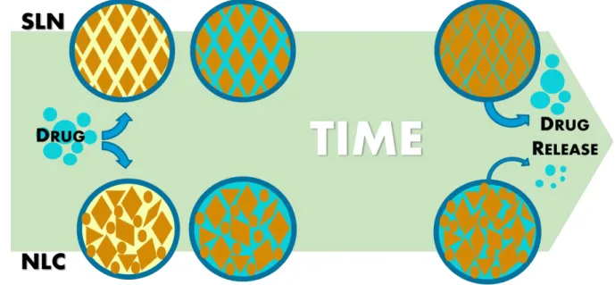
NLCs were initially prepared with stearic acid (C18H36O2; M=284.48 g/mol) as the solid lipid, and
Mygliol® 812 as the liquid lipid. Tween® 60 (C64H126O26; M=1310 g/mol) was used as a surfactant, to
stabilize the emulsion. In SLN preparation, mygliol was not used in the formulation and his mass was substituted by the solid lipid. Quantities used of each component are descried in Table 6.
Table 6 – Quantitative composition of stearic acid lipid NP prepared. Stearic acid (mg) Mygliol® (mg) Tween® 60 (mg)
SLN 290 - 60
NLC 200 90 60
The method used in this work to produce the lipid nanoparticles was an ultra-sonication method. In detail, the lipids and the surfactant were heated in a water bath up to 70ºC, temperature at all the lipids and the surfactants are in the liquid state. When the solid lipid was fully melted, 4.4 mL of ultrapure water heated at same temperature was added to the lipid. Mixture then went through ultraturrax (Ystral X10/20 E3) at 7000 rpm for 30 s to produce an emulsion, followed by a sonication at 70% power for 5 min to reduce the diameter of the lipid particles. The nanoformulation was left to cool at room temperature and stored.
23 Figure 3 - A schematic representation of the steps to produce lipid nanoparticles
3.3. DRUG LOADING
The aim of this thesis was to successfully encapsulate EGCG (C22H18O11; M=458.372 g/mol) in lipid
nanoparticles.
A total of 10mg of EGCG was dissolved in the aqueous phase and then added to melted lipid followed by ultraturrax and sonication, as previously described. The equivalent mass of the EGCG was subtracted to the solid lipid mass.
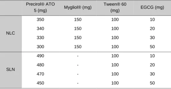
3.4. CHOICE OF SOLID LIPID FOR IMPROVEMENT OF NANOFORMULATION STABILITY
Due to a complete instability of the stearic acid nanoformulations with catechin, another solid lipid Precirol® ATO 5 was tested with the same amount of EGCG and a larger amount of lipid. The quantities are presented in the following table.
Table 7 - Quantitative composition of Precirol® ATO 5 lipid nanoparticles.
SLN NLC Precirol® ATO 5 500 350
Mygliol® - 150
Tween® 60 100 100
Although a notorious improvement of the formulations’ stability, the SLN obtained were not completely stable. To solve this issue, the sonication time was reduced from the initial 5 min to only 30 s using the same sonication’ potency.
In order to obtain the leader formulation, four different amounts of EGCG were added and a qualitative solubility test was performed. The quantities of the formulation constituents are presented in the following table.