Purpose: To raise awareness of potential severe ocular complications associated with autoimmune diseases and to describe the surgical technique of crescent-shaped anterior lamellar keratoplasty to treat a corneal perforation in peripheral ulcerative keratitis.
Methods and Materials: Case report.
Results: A female patient with 59 years old, with history of longstanding rheumatoid arthritis, presented with redness and photophobia in the right eye. As past ocular history, the patient had a severe keratoconjunctivitis sicca and a corneal ulcer in the right eye. Clinical examination revealed an epithelial defect with a significant inferior corneal thickness decrease. Topical steroids, antibiotics and a therapeutic contact lens were prescribed. One month later, a corneal perforation was observed and a crescent-shaped deep anterior lamellar keratoplasty was then performed.
Conclusion: The clinical course of peripheral ulcerative keratitis depends on the associated systemic pathology, being essential a multidisciplinary approach by ophthalmologists and rheumatologists to allow systemic control of the autoimmune disease, avoiding possible complications such as eye perforation.
Keywords: deep anterior lamellar keratoplasty; peripheral ulcerative keratitis; rheumatoid arthritis; ocular autoimmune diseases.
RESUMO
Objetivo: Alertar para possíveis complicações oculares graves associadas a doenças autoimunes e descrever a técnica cirúrgica da queratoplastia lamelar anterior em forma de crescente como opção terapêutica nas perfurações de córnea associadas à queratite ulcerativa periférica.
Métodos e materiais: Descrição de um caso clínico.
Resultados: Uma doente do sexo feminino, 59 anos, com antecedentes de artrite reumatoide, apresentou queixas de hiperemia e fotofobia no olho direito. Como antecedentes oculares, é de referir uma queratoconjuntivite sicca grave e úlcera de córnea no olho direito. Apresentava um defeito epitelial paracentral com diminuição significativa da espessura inferior da córnea. Iniciou cortico e antibioterapia tópica, e foi aplicada uma lente de contato terapêutica. Um mês depois, foi observada perfuração da córnea na região perilímbica inferior tendo sido submetida a uma queratoplastia lamelar anterior profunda em forma de crescente.
Conclusão: A evolução da queratite ulcerativa periférica depende da patologia sistémica associada, sendo fundamental uma abordagem multidisciplinar por oftalmologistas e reumatologistas para permitir o controlo sistémico da doença autoimune, evitando assim possíveis complicações como a perfuração ocular.
Keywords: queratoplastia lamelar anterior, queratite ulcerativa periférica, artrite reumatoide, doenças oculares autoimunes.
INTRODUCTION
The most severe ophthalmological manifestation of autoimmune diseases is peripheral ulcerative keratitis (PUK).1
PUK consists in a crescent-shaped area of inflammation at the margin of the corneal stroma with progressive necrosis and thinning, leading to ocular perforation.2 Almost 50% of
the cases are the initial manifestation of underlying systemic autoimmune diseases.3 The three most frequent underlying
diseases are rheumatoid arthritis in 34–42%, systemic lupus erythematous and granulomatosis with polyangiitis.4 Ocular
causes, such as infection, eyelid malposition, lagophthalmos, or neurotrophic defects can also be related to PUK.2
Clinical manifestations of PUK include ocular irritation, pain, redness, photophobia, and corneal opacity and may lead to corneal perforation and vision loss1 Although the pathogenesis of PUK associated with rheumatoid arthritis has not been completely elucidated, many studies suggested that both humoral and cellular immunity are involved.5-7
To minimize the risk of complications, PUK requires early diagnosis and treatment. The recommended systemic therapy for patients with PUK associated with rheumatoid arthritis is the use of nonsteroidal anti-inflammatory drugs
(NSAIDs), corticosteroids and immunosuppressive
chemotherapy.5 New systemic treatments, such as etanercept,
infliximab and adalimumab have also been successfully employed. They inhibit the proinflammatory cytokine TNF-α and lead to a decrease of matrix metalloproteinases, which would stop the progression of corneal stromal lysis.5
Rituximab, a monoclonal antibody directed against the CD20 molecule expressed in B cells, inhibits not only antigen presentation but also antibody and cytokine production. Rituximab is an alternative option when anti-TNF α drugs are not effective in controlling the disease.5
Topical treatment for PUK includes lubricant eye drops, AINEs and corticosteroids.2,8 In case of corneal perforation,
surgical intervention employing cyanoacrylate glue,
conjunctival flap, lamellar patch flap, amniotic membrane grafting, conjunctival resection of the inflamed area,
penetrating keratoplasty or deep anterior lamellar keratoplasty may be necessary.5 If corneal perforation is present, the visual
prognosis may be severely compromised.9
This case aims to raise awareness of potential severe ocular complications associated with rheumatoid arthritis resistant to systemic therapy and to describe the surgical technique of crescent-shaped anterior lamellar keratoplasty to treat a corneal perforation in PUK.
CLINICAL CASE
A 59 years old female presented with redness, discomfort and photophobia in the right eye. The patient had past ocular history of severe keratoconjunctivitis sicca and corneal ulcer in the right eye. Left eye was eviscerated after 5 unsuccessful penetrating keratoplasties to treat a corneal perforation due to PUK associated with rheumatoid arthritis.
The patient had past medical history of rheumatoid arthritis, secondary Sjögren's syndrome, latent pulmonary tuberculosis, pleural and pericardial effusion, Mediterranean spotted fever; pulmonary aspergillosis and depression.
The patient’s medication to rheumatoid arthritis included naproxen 500mg 2id, prednisolone 7.5 mg id and rituximab 1000 mg (2 injections every 6 months). Previously, the patient was medicated with methotrexate 10mg/week which was discontinued by unsuccessful disease control.
At ophthalmological evaluation, right eye visual acuity was 20/200. In the biomicroscopy of the right eye, the patient
presented a paracentral epithelial defect in the inferior region associated with a significant decrease in corneal
thickness, a central leukoma, perilimbic corneal
neovascularization and a dense cataract. There was no anterior chamber reaction. Intraocular pressure was 14 mmHg. At fundoscopy, optic disk head had normal dimensions and was well delimited, the foveal reflex was preserved, the retinal vessels presented normal dimensions and distribution. The patient was prescribed with a preservative free eye drop with trehalose (3%)/hyaluronic acid 2/2h and a combination of tobramycin and dexamethasone drops 5id. A therapeutic contact lens was also placed on the right eye to ease the epithelial defect closure. Ophthalmological manifestations were discussed with the attending rheumatologist to clarify the possibility of changing the systemic therapy in order to improve ocular manifestations and systemic control of disease. Although ocular disease was progressing, systemic therapy remained the same. One month later, we verified a progressive inferior crescent-shaped area of corneal thickness decrease with a corneal perforation and descemetocele in the perilimbic inferior region.
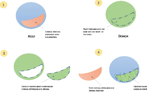
The patient was submitted to a crescent-shaped deep anterior lamellar keratoplasty in the right eye. During surgery, we performed an anterior lamellar dissection of corneal stroma from the center to the inferior limbic area. The donor graft was prepared with a crescent-shape, by copying the host lesion size and shape (Figure 1). Fibrin glue was used to improve apposition between the graft and residual stroma.
After surgery, prednisolone plus chloramphenicol ointment 5id, tobramycin plus dexamethasone drops 3id, preservative free eye drops of trehalose (3%) and hyaluronic acid 1/1h, ofloxacin drops 5id, prednisolone 20 mg id, acetazolamide 250mg 2id and doxycycline 100 mg 1id were prescribed.
In the first day after surgery, the graft presented some temporal softening, anterior chamber was formed without significant inflammatory reaction. An intumescent white cataract was also observed.
One month later, the patient was submitted to cataract surgery. After surgery, visual acuity was counting fingers at 1 meter. In the right eye, she presented a superficial punctate keratitis (grade 4 in Oxford classification), the graft was well-positioned without signs of rejection (Figure 2).
Figure 2 - Crescent-shaped deep anterior lamellar keratoplasty. One month after surgery, the graft was well-positioned without signs of rejection.
The anterior segment tomography (OCULUS Pentacam®) of
right eye demonstrated a significant irregularity of the anterior and posterior corneal surfaces (Figure 3).
Figure 3 - Anterior segment tomography (OCULUS Pentacam®) showed a significant
irregularity of the anterior and posterior corneal surfaces.
The patient was medicated with prednisolone plus
chloramphenicol ointment 3id, tobramycin plus
dexamethasone drops 3id, ofloxacin drops 5id and preservative free eye drops of trehalose (3%) and hyaluronic acid 1/1h. Six months later, no signs of graft rejection were seen.
DISCUSSION
The clinical history along with the findings on slit-lamp examination were consistent with the diagnosis of PUK secondary to rheumatoid arthritis.
Patients suffering from peripheral keratitis and ulceration must have a detailed personal and family history investigation because PUK is often secondary to autoimmune systemic diseases. Symptoms and signs, biomarkers and other auxiliary examinations should be used to clarify the diagnosis.
Ocular complications associated with autoimmune diseases, including PUK, may threaten vision and require timely diagnosis and treatment. Despite improvements in new immunomodulatory therapy, the outcome of PUK depends on the systemic disease control, as well as timely diagnosis and treatment. The patient’s refractory response to treatment was due to a delay in starting aggressive treatment. To conclude, the clinical course of PUK depends on the associated systemic pathology, being essential a multidisciplinary approach by ophthalmologists and rheumatologists to allow systemic control of the autoimmune disease, avoiding possible complications such as eye perforation.
REFERENCES
1. Clewes AR, Dawson JK, Kaye S, Bucknall RC.
Peripheral ulcerative keratitis in rheumatoid arthritis: successful use of intravenous cyclophosphamide and comparison of clinical and serological characteristics. Annals of the rheumatic diseases. 2005;64(6):961-2. 2. Yagci A. Update on peripheral ulcerative keratitis.
Clinical ophthalmology (Auckland, NZ). 2012;6:747-54.
3. Lohchab M, Prakash G, Arora T, Maharana P, Jhanji V, Sharma N, et al. Surgical management of peripheral
corneal thinning disorders. Survey of ophthalmology. 2018.
4. Levitt AE, McManus KT, McClellan AL, Davis JL, Goldhardt R, Galor A. Ocular Inflammation in the Setting of Concomitant Systemic Autoimmune Conditions in an Older Male Population. Cornea. 2015;34(7):762-7.
5. Cao Y, Zhang W, Wu J, Zhang H, Zhou H. Peripheral
Ulcerative Keratitis Associated with Autoimmune Disease: Pathogenesis and Treatment. Journal of ophthalmology. 2017;2017:7298026.
6. Maseda D, Bonami RH, Crofford LJ. Regulation of B lymphocytes and plasma cells by innate immune mechanisms and stromal cells in rheumatoid arthritis.
Expert review of clinical immunology.
2014;10(6):747-62.
7. Galor A, Thorne JE. Scleritis and peripheral ulcerative keratitis. Rheumatic diseases clinics of North America. 2007;33(4):835-54, vii.
8. Huerva V, Ascaso FJ, Grzybowski A. Infliximab for peripheral ulcerative keratitis treatment. Medicine. 2014;93(26):e176.
9. Fondi K, Wozniak PA, Schmidl D, Bata AM,
Witkowska KJ, Popa-Cherecheanu A, et al. Effect of
Hyaluronic Acid/Trehalose in Two Different
Formulations on Signs and Symptoms in Patients with Moderate to Severe Dry Eye Disease. Journal of ophthalmology. 2018;2018:4691417.
CONTACT
Sónia Torres da Costa
Department of Ophthalmology
Centro Hospitalar Universitário de São João, Alameda Prof. Hernâni Monteiro
4200-319 Porto, Portugal
E-mail: [email protected]
Conflicts of Interest
The authors declare that there are no conflicts of interest regarding the publication of this article.
The authors declare that this work has not been previously published and assign the copyright to the SPO.