ISSN 2179-8087 (online)
Original Article
Silviculture
Creative Commons License. All the contents of this journal, except where otherwise noted, is licensed under a Creative Commons Attribution License.
Maturation and Desiccation Tolerance in Seeds of
Sesbania virgata
(Cav.) Pers.
Fabrício Palla Teixeira
1, José Marcio Rocha Faria
1,
Wilson Vicente Souza Pereira
1, Anderson Cleiton José
11Laboratório de Sementes Florestais, Programa de Pós-graduação em Engenharia Florestal, Departamento de Ciências
Florestais, Universidade Federal de Lavras – UFLA, Lavras/MG, Brasil
ABSTRACT
The maturation process of Sesbania virgata seeds was investigated through fruits and seeds at different maturation stages, where they were described morphologically. For the seeds, fresh and dry mass and moisture were determined and structural analysis of the tegument under a light microscope was performed. Germination tests in the presence and absence of drying and the electrophoretic profile of heat resistant proteins were also performed. The results showed that the seeds reached physiological maturity at 44 days after anthesis (DAA), evidencing a secondary cell wall. The acquisition of desiccation tolerance was observed at 44 DAA, with the seeds responding to the drying treatment. Protein bands were observed from 32 DAA, which remained constant until the end of maturation. These results allowed the characterization of the development and acquisition of desiccation tolerance in S. virgata seeds.
1. INTRODUCTION
Sesbania virgata is a shrub species from the Fabaceae family, native to Brazil, which can be found in the South, Southeast and Center-West regions, reaching 6 m in height, 25 cm in diameter at breast height (DBH) and with the crown reaching up to 5 m in diameter (Araújo et al., 2004). It is a plant with a short life cycle, being able to compete with forage species and develop naturally in moist terrains, in addition to being well-adapted to floodable areas. Due to its rapid growth and hardiness, it is a potential species for plantations aimed at recovering degraded areas and in riparian forests (Delarmelina et al., 2014). Nevertheless, the literature still lacks studies on its development, mainly regarding the maturation of its seeds.
There are several studies related to the morphology of seeds from agronomic species, such as soybean (Marcos, 1980), maize (Fancelli & Dourado-Neto, 1997) and rice (Fonseca et al., 2015). However, the seed development of many native forest species has not yet been characterized, with this knowledge being relevant for studies in diverse areas. The identification of species present in the soil seed bank through knowledge of morphoanatomy can contribute to a better understanding of plant regeneration and succession in ecosystems. Additionally, the morphological aspects are necessary for identification and for certification of seed quality, as well as to understand its maturation process (Beltrati, 1984).
The development of orthodox seeds, i.e., those that tolerate desiccation, can be divided into three stages: embryogenesis; deposition of reserves and desiccation. Development comprises an orderly series of genetically controlled changes (Bewley et al., 2013). These modifications provide rapid growth of internal structures and deposition of reserve material, which may be carbohydrates, proteins and lipids, and there are also physiological adaptations that may culminate with induction of embryo to dormancy. In the desiccation stage, there is the action of protective and repair mechanisms, preserving the integrity of the cellular compounds, avoiding irreversible damage, keeping the seeds healthy until dispersion (Bewley et al., 2013).
The maturation process in seeds is generally well-described in the literature, however, in order to estimate the physiological maturity of a particular
seed, it is important to study the physiological and anatomical changes that occur during its development (Walters et al., 2013). Anatomical changes at the structural level, such as changes in the cell wall during seed maturation, may provide better understanding of developmental mechanisms, and even knowledge of survival strategies (Moura et al., 2010), such as the characteristic of desiccation tolerance.
Desiccation tolerance can be defined as the cellular ability of an organism to tolerate water loss at extreme levels without irreversible damage to cells through metabolic reduction and, in the presence of water, being able to return to normal functioning (Buitink & Leprince, 2008). Seeds can be classified in relation to desiccation and storage tolerance into two distinct groups: orthodox, which tolerate very low water content levels and can be stored for long periods at subzero temperatures, and a recalcitrant group that presents the opposite behavior (Roberts, 1973). There are also seeds with intermediate behavior, i. e. those that do not tolerate water loss at very low levels and generally can be stored for short periods at temperatures above zero (Ellis et al., 1990).
Tunnacliffe & Wise (2007) stated that the species show variations in the degree of desiccation tolerance. This is due to the differences that occur in the physical structure of their internal matrices, which involve interactions of organic acids, amino acids, sugars and proteins that are produced during maturation, being associated with several macromolecules, preventing denaturation (Berjak & Pammenter, 2008). Heat-resistant proteins have been reported in studies related to protection against stress damage. Among the classes of these proteins are the Late Embryogenesis Abundant (LEA) (Farrant & Moore, 2011). Studies regarding seed development can contribute to the understanding of its maturation process, knowing its possible metabolic changes and survival strategies.
The aim of this research was to study morphological, physiological and molecular aspects during the maturation of Sesbania virgata seeds.
2. MATERIAL AND METHODS
of Lavras, south of the State of Minas Gerais, Brazil, at 21°23’S latitude, 44°98’W longitude and 889.37 m altitude. A total of 16 trees were randomly selected for the experiment, with their flowers being marked with dated tapes for the anthesis record, in sufficient numbers to obtain seeds in an amount compatible with experimental requirements. A photographic record was made continuously from the anthesis to follow the development of fruits and seeds. The characterization of fruits and seeds was based on Araújo et al. (2004), and the coloring based on the color chart DIN 6164 (Biesalski, s.d.).
Throughout the maturation process, fruits were collected at different maturation stages, days after anthesis (DAA). Based on tests of moisture content and fresh mass and on descriptions made by Araújo et al. (2004), fruits and seeds at 85 DAA were defined as the final maturation stage. Seeds collected at 24, 36, 44, 58, 75 and 85 DAA were used for quantification of fresh and dry mass, moisture, structural analyses and physiological tests. After the collection of fruits, the seeds were removed from the fruits manually and weighed on an analytical scale (0.0001 g) to obtain the fresh weight. Subsequently, they were placed in an oven at 105 ± 3 °C for 24 h and then weighed again. A total of 20 seeds per collection point were used in four replicates of five seeds (Brasil, 2009), cut in half to enhance drying due to seed coat thickness. For structural analysis, the material was fixed in FAA50 (10% solution of 40% formaldehyde + 5% glacial acetic acid + 50% v/v ethyl alcohol) for 48 h. Afterwards, the cross sections were performed free-hand in the region opposite the hilum. The plant fragments were stained with 0.05% toluidine blue. The slides were sealed with Entellan and visualized on Olympus BX 51 optical microscope at 10, 20, and 40x magnification. The images were scanned using a video camera coupled to the microscope and processed by computer through Image - Pro-plus 5.1 software. To characterize desiccation tolerance acquisition in the seeds, preliminary tests based on the germination of seeds dried and non-dried on silica until reaching 7% water content, were performed. Germination was not observed at points 24 or 36 with or without drying, thereby fixing maturation stages studied at 44, 58, 75, and 85 DAA. At each studied maturation stage, the seeds were divided into two groups, the first consisted of non-dried seeds. In the second group, the seeds were weighed and subsequently dried
(with the exception of 85 DAA, because they already had the desired moisture) in plastic boxes containing approximately 100 g of silica gel. For this experiment, the protocol developed by Hong & Ellis (1996) pdated in 2000 with modifications was used as a reference. Seeds were arranged uniformly on a screen, not in contact to the silica. The boxes were kept in a drying room at 20 °C with constant monitoring of seed weight until they reached a hygroscopic equilibrium (approximately 7% moisture). The seeds were pre-humidified after drying to avoid possible imbibition damage. Both seed groups were mechanically scarified using No. 40 sandpaper to overcome physical dormancy (Silva et al., 2011) and later placed in a germinator at 25 °C in moist paper rolls containing four replicates of 20 seeds per treatment. The germination percentages of seeds with primary root ≥ 2 mm were evaluated. The readings occurred daily until the 20th day of counting. The experiment was based on a factorial design with additional treatment (3x2+1), with three stages of seed development (44, 58, and 75 DAA), in the presence and absence of drying. The additional treatment was composed of seeds at 85 DAA because they were fully mature, and presented desired water content. Data analysis of variance was performed using the F test. The averages were compared by Tukey test at 5% probability.
following which, the gels were fixed for 30 min in acetic acid solution plus methanol. The gels were stained with Coomassie Brilhant Blue 0.05% CBB-G250 for 48 h, followed by discoloration for 30 min in 1.2% (p/v) Tris base solution pH 6.5, and later washed in water for three days. The images of gels were made using a Umax scanner (PowerLook model 1120).
3. RESULTS AND DISCUSSION
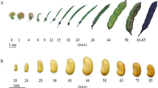
After the anthesis, the S. virgata flowers began a gradual dehydration process, losing their petals between the fourth and sixth days after the anthesis (DAA). From there, it was possible to observe the fruit at the beginning of its development. According to the DIN6164 color chart, the fruits showed a green color until reaching 44 DAA. From this point on, the green color became darker, reaching brown at the end of maturation (Figure 1A).
When mature, S. virgata fruits were characterized as undescended and nucleoid, in oblong format with the corrugated edge, with a suture from the peduncle extending to the apex. The peduncle is woody in appearance, with the pericarp being thin and the endocarp smooth and presenting a grayish-white color. Araújo et al. (2004) also described the S. virgata fruits as having a dry, rough surface, with significant tapering at the base and at the extremity, which is
pointed. The coloration of the fruits, together with other factors, can aid in determining the ideal collection stage, making field work faster and more efficient.
The seeds showed a beige/yellowish coloration in the initial stages until reaching the mid-point of the maturation process, at 44 DAA, gaining a brownish tone as they developed (Araújo et al., 2004). The mature seed is reniform, with a polished and water-impermeable forehead tegument (Figure 1B).
At 24 DAA, the internal structures became evident. The endosperm already occupied a large part of the seed interior, presenting a shiny and gelatinous aspect. The cotyledons were green and elongated, and about 2 mm long. At 36 DAA, all internal structures were visible, its being possible to observe hypocotyl attached to cotyledons and showing a gain in size (Figure 2).
The largest seed size was observed at 44 DAA due to the maximum gain of dry mass and accumulation of water, with the cotyledons occupying most of the internal space of seeds along with the fully developed hypocotyl, both embedded by the endosperm. From this stage, the seed entered the desiccation phase, where all its internal structures began to lose water, acquiring a yellowish coloration for the whole embryo (Figure 2).
At the beginning of the maturation process, the seeds showed high water content, which decreased simultaneously with the increase in dry mass. The maximum value of both fresh and dry mass was
reached at 44 DAA, indicating that this is the point of physiological maturity (Figure 3). The high water content in the seeds helps in intense cell divisions, as
Figure 2. Sections of Sesbania virgata (C) seeds at different maturation stages. CT = cotyledons; EN = endosperm; HP = hypocotyl; TG = tegument. Numbers indicate days after anthesis (DAA).
Figure 3. Water content, fresh mass and dry mass in
Sesbania virgata seeds at different maturation stages. Numbers indicate days after anthesis (DAA).
Figure 4. Photomicrography of integument region cross sections on Sesbania virgata seeds at different maturation stages, stained with toluidine blue. CT = cuticle; EN = endosperm; LL = lucid line; MC = macrosclereid, OS = osteosclereid; Pq = parenchyma; CO = cotyledon. A = 24; B = 36; C = 44, D = 58, E = 75; F = 85 days after anthesis (DAA). Bar at A = 10 μm; B, C, D, and E = 20 μm; F = 50 μm.
well as in the promotion of interactions between growth hormones (Lulsdorf et al., 2013). After 44 DAA, there was a significant reduction in the water content of seeds, followed by a decrease in the fresh mass values. The dry mass remained constant until the end of the desiccation process (Figure 3).
The behavior of S. virgata seeds during maturation followed the classic orthodox pattern, where the reduction of water content is continuous, being more pronounced at the end of the maturation process. Similar to these results, Carvalho et al. (2008) observed initial water content of approximately 90% in Handroanthus serratifolia (Vahl) Nich seeds at 10 DAA, followed by a drop to about 30% at 53 DAA. The development pattern during maturation is characterized by the rapid increase in seed size, reaching a maximum value that may coincide with half of its maturation process (Carvalho & Nakagawa, 2012), in addition to showing internal structural changes that continue until the end of maturation.
In the presence of thin wall and secondary wall, it shows respectively blue and red tones. At the developmental stages of the tegument, the plant tissue color changed drastically, acquiring blue tones, which can be observed until the seeds reach their DAA (Figure 4B-D). In the last two maturation stages (75 and 85 DAA), the presence of lignin is characterized by the red color (Figure 4E and F), and the spaces among the osteosclereids became larger. In the first stage, the newly-split cells showed at first only a primary wall composed of cellulose (Buchanan et al., 2000), which justifies the blue staining found in S. virgata seeds in the early stages. After the formation of the primary wall, the formation of the secondary wall begins, which is rich in lignin, making the cell walls much thicker.
Concomitantly, it was also possible to observe the cuticle and the lucid line more externally (Figure 4C), which together with the other structures, became more rigid and gradually impermeable, due to desiccation, forming a barrier for water entering the seeds until reaching complete dormancy. S. virgata seeds show physical dormancy, characterized by tegument impermeability (Silva et al., 2011). This dormancy is certainly related, amongst other things, to the lignin deposition at the end of the maturation process, associated with the decrease in water content, aiding in the stiffening of the macrosclereid layer. The components that promote the achievement of seed dormancy are both structural and chemical, being present in the macrosclereid layer (Baskin et al., 2000), very frequent in seeds from the Fabaceae family (Baskin et al., 2000), especially in species with orthodox seeds.
In Erythrina speciosa seeds, Molizane et al. (2013) observed pronounced spaces in the osteosclereid region and parenchyma cells during final stages of maturation, due to the drastic drop in water content, which was also observed for S. virgata seeds in the present study, observing that during desiccation, the cells reduce their size due to water loss and the intercellular spaces become visible between the osteosclereids
In the absence of drying, there was an increase in germination values as the seeds advanced throughout the developmental stages until their maximum at 85 DAA. (Figure 5). Brito et al. (2015) observed an increase in germination as Jatropha curca L. seeds advanced through the maturation stages. Delgado & Barbedo (2007) also observed this in seeds of Eugenia species,
and Carvalho et al. (2008) in Handroanthus serratifolia (Vahl) Nich seeds. The same was found in seeds that underwent drying, with the exception of those at 75 DAA, where there was a decrease in this value (Figure 5), with interaction between DAA and drying being observed.
The acquisition of desiccation tolerance in seeds was evidenced initially at 44 DAA, where after reaching 7% of its water content, it was possible to obtain 49% germination. S. virgata seeds at 44 and 58 DAA, after being submitted to artificial drying, showed a higher germination percentage in relation to non-dried seeds. In orthodox seeds, drying at the final maturation stage is a start for the seed to redirect its metabolism towards germination (Bewley et al., 2013). However, to prepare seeds for desiccation, desiccation tolerance mechanisms begin to be activated gradually throughout the maturation process (Walters et al., 2013). The ability of these seeds to tolerate the natural reduction of their water content is related to complex mechanisms involving the accumulation of soluble sugars, LEA proteins and the activation of antioxidant systems, all regulated by a hormonal balance (Berjak & Pammenter, 2008).
Desiccation survival is conditioned by the activation of systems that guarantee the integrity of membranes, the presence of antioxidant mechanisms, the dedifferentiation of organelles, and the formation of the vitreous state, among other mechanisms, which together promote
Figure 5. Percentage of germination for Sesbania virgata
desiccation tolerance in seeds (Berjak & Pammenter, 2008). Delgado & Barbedo (2007) state that the drying speed is closely linked to the desiccation tolerance limit in seeds. The faster the drying process, the greater the degree of dehydration tolerated by the seeds, reducing the possibility of damage from the process. When slow dried, the seeds remain for a longer period in a desiccation range favorable to damage, including the action of free radicals, which cause great damage to the membrane structures (Berjak & Pammenter, 2000). Corsato et al. (2012) studied the germination of Annona emarginata and obtained values higher than 70% germination in seeds at advanced maturation stages after drying to 5%.
Seeds at 75 DAA after drying, showed lower performance than non-dried seeds (Figure 5). At 75 DAA, the decrease in germination values of dried seeds in relation to non-dried seeds may be related to damage suffered during the imbibition process, due to its low water content. At this point, even with pre-humidification, the S. virgata seeds may have undergone an increase in the solute leaching rate when subjected to rapid hydration, resulting in the transition from the gel phase to the liquid-crystalline phase of the membrane phospholipids (Berjak & Pammenter, 2008), leading to damage that may have compromised germination performance.
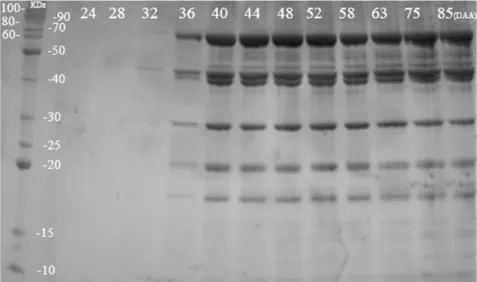
Based on the protein electrophoretic profile, bands were observed from 32 DAA, showing molecular weight
between 30 and 70 KDa, showing an increase in the band intensity up to 44 DAA, as well as observing the appearance of bands with low molecular weight. S. virgata seeds in initial stages of maturation showed reserve structures still at the beginning of their development, consequently presenting little accumulated material, thereby justifying the absence of bands in the first points. The material quantified in the spectrophotometer may have been basically composed of enzymes that, after the quantification process may have undergone degradation and, therefore, did not appear in the gel.
It was verified through the profile that the accumulation of reserve material in S. virgata seeds occurs over a short time interval, between 32 and 44 DAA. From this last stage onwards, the intensity of bands remained constant, with little variation, and molecular weight ranging from 15 to 90 KDa (Figure 6).
Contrary to Hand et al. (2006) who stated that heat-resistant proteins would only occur at the end of the maturation process, this accumulation was still in the early stages of maturation of S. virgata seeds, attesting to their early presence. This protein profile correlates with the desiccation tolerance results, which were acquired by the seeds at 44 DAA when the bands became more evident, reinforcing the relationship of these proteins with desiccation tolerance.
In the seeds of some species from the Fabaceae family, proteins are the main reserve components. In these seeds, part of these proteins will be stored
Figure 6. Electrophoretic profile on SDS-PAGE discontinuous polyacrylamide gel of heat-resistant proteins in
after accumulation during maturation to nourish the embryo in the seedling phase. Another fraction of proteins has a protective function, avoiding damage caused by environmental stresses (Santini & Martorell, 2013), and assisting in the assembly and maintenance of cellular structures (Hand et al., 2011).
Heat-resistant proteins act to prevent denaturation and promote the aggregation of other proteins. They can also act in stress situations, increasing their abundance and preparing the seed for desiccation (Berjak & Pammenter, 2008), until achieving stability during maturation. Moore et al. (2009) related the participation of heat-resistant proteins in Annona emarginata seeds with the achievement of desiccation tolerance, as well as observed in S. virgata seeds.
Several factors promote an increase in heat-resistant proteins. One of them is the synthesis of abscisic acid (ABA), a plant hormone present in seeds and related to the synthesis and accumulation of substances that allow water reductions at the end of maturation without damaging the membrane system, demonstrating the complexity of these interactions (Finkelstein, 2002).
These results further reinforce the relationship between heat-resistant proteins and the acquisition of desiccation tolerance, which, together with other factors, provided orthodox seeds with efficient survival strategies.
4. CONCLUSION
S. virgata seeds reached the physiology maturity point at 44 DAA.
It was possible to describe the internal structures of seeds and observe the deposition of lignin at the end of maturation.
Physiologically, desiccation tolerance was acquired in seeds from 44 DAA.
Acquisition of desiccation tolerance is related to the expression of heat-resistant proteins.
ACKNOWLEDGEMENTS
The authors would like to thank the Federal University of Lavras for the support and the infrastructure for conducting the experiments and CAPES for granting the scholarship.
SUBMISSION STATUS
Received: 28 mar., 2017 Accepted: 26 sep., 2017
CORRESPONDENCE TO
Fabrício Palla Teixeira
Departamento de Ciências Florestais, Universidade Federal de Lavras – UFLA, Av. Sylvio Meniccuci, CEP 37200-000, Lavras, MG, Brasil
e-mail: [email protected]
FINANCIAL SUPPORT
Coordenação de Aperfeiçoamento de Pessoal de Nível Superior, Conselho Nacional de Desenvolvimento Científico e Tecnológico (CNPQ), Grant / Award number: 310225/2015-9).
REFERENCES
Araújo EC, Mendonça AV, Barroso DG, Lamônica KR, Silva RF. Caracterização morfológica de frutos, sementes e plântulas de Sesbania virgata (Cav.) Pres. Revista Brasileira de Sementes 2004; 26(1): 105-110. http://dx.doi. org/10.1590/S0101-31222004000100016.
Baskin JM, Baskin CC, Li X. Taxonomy, anatomy and evolution of physical dormancy in seeds. Plant Species Biology 2000; 15(2): 139-152. http://dx.doi.org/10.1046/ j.1442-1984.2000.00034.x.
Beltrati CM. Morfologia e anatomia das sementes de
Trichilia elegans A. Juss (Meliaceae). Naturalia 1984; 9: 35-42.
Berjak P, Pammenter NW. What ultrastructure has told us about recalcitrante seeds. Revista Brasileira de Fisiologia Vegetal 2000; 12: 22-55.
Berjak P, Pammenter NW. From Avicennia to Zizania: seed recalcitrance in perspective. Annals of Botany 2008; 101(2): 213-228. http://dx.doi.org/10.1093/aob/mcm168. PMid:17704237.
Bewley JD, Bradford JD, Hilhorst HWM, Nonogaki H. Seeds: Physiology of development, germination and dormancy. 3rd ed. New York: Springer; 2013. http:// dx.doi.org/10.1007/978-1-4614-4693-4.
Bradford MM. A rapid and sensitive method for the quantitation of microgramquantities of protein utilizing the principle of protein-dye binding. Analytical Biochemistry
Brasil. Ministério da Agricultura, Pecuária e Abastecimento. Secretaria de Defesa Agropecuária. Regras para análise de sementes. Brasília; 2009. 399p.
Brito CD, Loureiro MB, Souza AP Jr, Fernandez LG, Castro RD. Perfil morfofisiológico da maturação de frutos e sementes de Jatropha curcas L. Semina: Ciências Agrárias
2015; 36(6): 3615-3628. http://dx.doi.org/10.5433/1679-0359.2015v36n6p3615.
Buchanan BB, Gruissem W, Jones RL. Biochemistry & molecular biology of plants. Rockville: Americcan Society of Palnt Physiologists; 2000.
Buitink J, Leprince O. Intracellular glasses and seed survival in the dry state. Comptes Rendus Biologies 2008; 331(10): 788-795. http://dx.doi.org/10.1016/j.crvi.2008.08.002. PMid:18926493.
Carvalho MLM, Nery MC, Oliveira LM, Hilhorst HWM, Guimarães RM. Morphophysiological development of
Tabebuia serratifolia Vahl Nich. seeds. Scientia Agrícola
2008; 65(6): 643-651. http://dx.doi.org/10.1590/S0103-90162008000600012.
Carvalho NM, Nakagawa J. Sementes: ciência, tecnologia e produção. 5. Ed. Jaboticabal: Funep; 2012.
Corsato JM, Ferreira G, Barbedo CJ. Desiccation tolerance in seeds of Annona emarginata (Schldtl.) H. Rainer and action of plant growth regulators on germination. Brazilian Journal of Plant Physiology 2012; 24(4): 253-260. http:// dx.doi.org/10.1590/S1677-04202012000400004. Delarmelina WM, Caldeira MVW, Faria JCT, Gonçalves EO, Rocha RLF. Diferentes Substratos para a Produção de Mudas de Sesbania virgata.Floresta e Ambiente 2014; 21(2): 224-233. http://dx.doi.org/10.4322/floram.2014.027. Delgado LF, Barbedo CJ. Tolerância à dessecação de sementes de espécies de Eugenia.Pesquisa Agropecuária Brasileira 2007; 42(2): 265-272. http://dx.doi.org/10.1590/ S0100-204X2007000200016.
Ellis RH, Hong TD, Roberts EH. An intermediate category of seed storage behaviour? I. Coffee. Journal of Experimental Botany 1990; 41(9): 1167-1174. http:// dx.doi.org/10.1093/jxb/41.9.1167.
Fancelli AL, Dourado-Neto D. Fenologia do milho. Piracicaba: Publique; 1997. Tecnologia da produção de milho; p. 131-140.
Farrant JM, Moore JP. Programming desiccation-tolerance: from plants to seeds to resurrection plants. Current Opinion in Plant Biology 2011; 14(3): 340-345. http:// dx.doi.org/10.1016/j.pbi.2011.03.018. PMid:21511516. Finkelstein R. Abscisic acid synthesis and response. The Arabidopsis Book/The American Society of Plant Biologists
2002;11.https://doi.org/10.1199/tab.0058.
Fonseca JR, Vieira EHN, Pereira JA, Cutrim VA. Descritores morfoagronômicos e fenológicos de cultivares tradicionais de arroz coletados no Maranhão. Ceres 2015; 51(293)
Hand SC, Jones D, Menze MA, Witt TL. Life without water: expression of plant LEA genes by an anhydrobiotic arthropod. Journal of Experimental Zoology. Part A, Ecological Genetics and Physiology 2006; 307A(1): 62-66. http://dx.doi.org/10.1002/jez.a.343. PMid:17109393. Hand SC, Menze MA, Toner M, Boswell L, Moore D. LEA protein during water stress: not just for plants anymore.
Annual Review of Physiology 2011; 73(1): 115-134. http:// dx.doi.org/10.1146/annurev-physiol-012110-142203. PMid:21034219.
Hong TD, Ellis RH. A protocol to determine seed storage behaviour. Vol. 1. Rome: International Plant Genetic Resources Institute; 1996.
Lulsdorf MM, Yuan HY, Slater AMH, Vandenberg A, Han X, Zaharia LI et al. Endogenous hormone profiles during early seed development of C. arietinum and C. anatolicum. Plant Growth Regulation 2013; 71(2): 191-198. http:// dx.doi.org/10.1007/s10725-013-9819-2.
Marcos J Fo. Maturidade fisiológica de sementes de soja.
Pesquisa Agropecuária Brasileira 1980; 15(4): 447-460. Molizane DM, Kanashiro S, Tavares AR, Barbedo CJ. Seed maturation of Aechmea bromeliifolia and Vriesea paraibica (Bromeliaceae). Hoehnea 2013; 40(4): 619-625. http://dx.doi.org/10.1590/S2236-89062013000400005. Moore JP, Le NT, Brandt WF, Driouich A, Farrant JM. Towards a systems-based understanding of plant desiccation tolerance. Trends in Plant Science 2009; 14(2): 110-117. http://dx.doi.org/10.1016/j.tplants.2008.11.007. PMid:19179102.
Moura EF, Ventrella MC, Motoike SY. Anatomy, histochemistry and ultrastructure of seed andsomatic embryo of Acrocomia aculete (Arecaceae). Scientia Agrícola 2010; 67(4): 399-407. http://dx.doi.org/10.1590/ S0103-90162010000400004.
Roberts EH. Predicting the storage life of seeds. Seed Science and Technology 1973; 1(4): 499-514.
Santini BA, Martorell C. Does retained‐seed priming drive the evolution of serotiny in drylands? An assessment using the cactus Mammillaria hernandezii. American Journal of Botany 2013; 100(2): 365-373. http://dx.doi.org/10.3732/ ajb.1200106. PMid:23345416.
Silva PEM, Santiago EF, Daloso DM, Silva EM, Silva JO. Quebra da dormência em sementes de Sesbania virgata (Car.) Pres. Idesia 2011; 29(2):39-45.
Tunnacliffe A, Wise MJ. The continuing conundrum of the LEA proteins. Naturwissenschaften 2007; 94(10): 791-812. http://dx.doi.org/10.1007/s00114-007-0254-y. PMid:17479232.