RESUMO.- [Lesões placentárias associadas a abortos e natimortos em cabras naturalmente infectadas pelo
Neospora caninum.] Neospora caninum é descrito como um parasito que causa problemas reprodutivos esporádicos em cabras. Muitos aspectos da patogênese da neosporose
em cabras naturalmente infectadas ainda precisam ser estabelecidos. Os objetivos deste trabalho foram caracterizar as lesões placentárias em cabras naturalmente infectadas por N. caninum e avaliar as técnicas diagnósticas para a detecção efetiva do protozoário na placenta. Algumas placentas deste estudo são originárias de abortos e natimortos, nas quais havia lesões graves. As lesões foram classificadas principalmente por necrose envolvendo o mesênquima das vilosidades coriônicas e células trofoblásticas, geralmente associadas a infiltrado inflamatório mononuclear e em alguns casos infiltrado neutrofílico. O DNA do N. caninum foi detectado nestas placentas, porém estruturas parasitárias não foram visualizadas na imuno-histoquímica (IHQ). Entretanto, cinco das onze placentas de cabras infectadas, que deram à luz ABSTRACT.- Mesquita L.P., Costa R.C., Nogueira C.I., Abreu C.C., Orlando D.R., Ascari Jr. I.,
Peconick A.P. & Varaschin M.S. 2018. Placental lesions associated with abortion and stillbirth in goats naturally infected by Neospora caninum. Pesquisa Veterinária Brasileira 38(3):444-449. Setor de Patologia Veterinária, Departamento de Medicina Veterinária, Universidade Federal de Lavras, Campus Universitário, Cx. Postal 3037, Lavras, MG 37200-000, Brazil. E-mail: msvaraschin@dmv.ufla.br
Neospora caninum has been described as a parasite that sporadically causes reproductive problems in goats. Several aspects of the pathogenesis of neosporosis in naturally infected goats remain to be established. The aims of the present study were to characterize the placental lesions in goats naturally infected by N. caninum and to evaluate several diagnostic techniques for effective detection of this protozoan in the goat placenta. Some placentas in this study originated from abortion and stillbirth in which there were severe lesions. The lesions were characterized mainly by necrosis involving the mesenchyme of the chorionic villi and trophoblast cells often alongside mononuclear inflammation and in some cases with neutrophilic infiltration. N. caninum DNA was detected in these placentas, but parasite structures were not visualized through immunohistochemistry (IHC). However, five of 11 placentas from N. caninum-infected goats that gave birth to healthy kids had histological lesions characterized by mononuclear inflammation. Of these 11 placentas, N. caninum DNA was detected in seven, and N. caninum tachyzoites were detected in only one of these seven placentas using IHC. The present study demonstrates that severe lesions in the placenta are associated with abortion and stillbirth in caprine neosporosis and the placental alterations are likely involved in abortion pathogenesis. Moreover, the results highlight the importance of using more than one diagnostic technique for the detection of the protozoan in placentas because N. caninum cannot be reliably detected by histological and immunohistochemical tests.
INDEX TERMS: Placental lesions, abortion, stillbirth, goats, Neospora caninum, caprine, immunohistochemistry, transplacental infection, neosporosis, necrotizing placentitis, protozoan.
Vet 2519 pvb-4598 LD
Placental lesions associated with abortion and stillbirth
in goats naturally infected by
Neospora caninum
1Leonardo P. Mesquita2, Rafael C. Costa2, Clayton I. Nogueira3, Camila C. Abreu3,
Débora R. Orlando3, Ivan Ascari Junior4, Ana Paula Peconick3 and Mary S. Varaschin3*
1 Received on February 15, 2016.
Accepted for publication on June 07, 2017.
2 Departamento de Patologia, Faculdade de Medicina Veterinária e Zootecnia, Universidade de São Paulo (USP), Avenida Prof. Dr. Orlando Marques de Paiva 87, Cidade Universitária, São Paulo, SP 05508-270, Brasil.
3 Departamento de Medicina Veterinária, Universidade Federal de Lavras (UFLA), Campus Universitário, Cx. Postal 3037, Lavras, MG 37200-000, Brazil. *Corresponding author: msvaraschin@dmv.ufla.br
a conceptos saudáveis, apresentaram lesões histológicas caracterizadas por infiltrado inflamatório mononuclear. Destas 11 placentas, foi detectado DNA de N. caninum em sete e taquizoítos foram encontrados em apenas uma por meio de imuno-histoquímica. O presente estudo demonstra que abortos e natimortos na espécie caprina, causados pelo N. caninum estão associados a lesões acentuadas nas placentas, sendo que as mesmas estão envolvidas na patogênese do aborto. Os resultados também ressaltam a importância do uso de mais de uma técnica diagnóstica para a detecção do protozoário em placentas, pois o N. caninum não pode ser confiavelmente detectado somente pelos exames de histopatologia e imuno-histoquímica.
TERMOS DE INDEXAÇÃO: Aborto, natimorto, cabras, Neospora caninum, caprinos, imuno-histoquímica, infecção transplacentária,
neosporose, placentite necrotizante, protozoário.
INTRODUCTION
Neospora caninum is an obligate intracellular protozoan, recognized worldwide as a main cause of abortion in cows (Dubey et al. 2007). In goats, N. caninum is known to cause reproductive problems, but the extent of the effects is still unknown (Dubey 2003, Dubey & Schares 2011). In goats, few cases of abortion and stillbirth related to neosporosis have been described (Barr et al. 1992, Dubey et al. 1992, 1996, Eleni et al. 2004, Moreno et al. 2012, Mesquita et al. 2013). Other reported cases include those in which newborn goats presented neurologic abnormalities (Corbellini et al. 2001, Varaschin et al. 2012), those in which goat kids showed congenital infection despite being clinically normal, and cases in which goat kids were born free of infection (Mesquita et al. 2013).
The pathogenesis of abortion in neosporosis is not yet completely understood. In cows, N. caninum can cause severe placental and fetal lesions that result in abortion (Maley et al. 2006, Gibney et al. 2008). Placental lesions in cows experimentally infected with N. caninum are characterized by necrosis of the mesenchymal and trophoblast cells associated with a mixed inflammatory infiltrate (Caspe et al. 2012). Similarly, in experimentally infected goats with the Nc-Spain7 strain, multifocal necrotic placentitis has been described in different gestational ages (Porto et al. 2016). However, placental lesions in naturally infected goats are poorly characterized. Only inflammatory and sometimes mild lesions are reported in natural cases of abortion and stillbirth due to N. caninum in goats (Barr et al. 1992, Dubey et al. 1992). Therefore, the aim of the present study was to characterize the placental lesions in goats naturally infected by N. caninum and to evaluate different techniques to detect the parasite in the placentas.
MATERIALS AND METHODS
Goats and sampling. Placental samples were collected from a caprine herd composed of 15 pregnant goats from a previous study (Mesquita et al. 2013). The herd was held at the Departamento de Medicina Veterinária (DMV), Universidade Federal de Lavras (UFLA). The goats became pregnant by natural mating and the entire pregnancy of each goat was monitored monthly by transrectal ultrasound to verify foetal viability. The herd was composed by multiparous Saanen,
Pardo-Alpina, and mixed-breed goats. Thirteen goats naturally infected by Neospora caninum, verified by the presence of specific antibodies
against N. caninum through IgG immunofluorescence antibody test
(IFAT), and two uninfected goats were used as negative controls. The goats were also tested for Toxoplasma gondii using an IFAT test as previously described (Mesquita et al. 2013) at Setor de Patologia Veterinária, DMV, UFLA. Also, the goats were tested for specific antibodies against Brucella spp., using the buffered plate antigen test according to Programa Nacional de Controle e Erradicação de Zoonoses (Brasil 2001), at Laboratório de Microbiologia, DMV, UFLA. Additionally, placental samples were submitted to PCR for Coxiella burnetii, as similar as previously described (Herrin et al. 2011) at
Departamento de Patologia, Faculdade de Medicina Veterinária e Zootecnia, Universidade de São Paulo, and PCR for Chlamydophila
spp., performed at Instituto Biológico de São Paulo. The diagnoses of the abortion and stillbirth that occurred in some goats of the herd were previously described, and N. caninum infection was associated in these cases (Mesquita et al. 2013). All experimental procedures were approved by the Ethics Committee of Animals Use (CEUA) from Universidade Federal de Lavras (UFLA) under the protocol number 013/2011.
The placentas of all goats that were normally expelled during parturition or abortion, and many samples of the cotiledonary and intercotiledonary areas were collected and fixed in 10% buffered formalin for histological and immunohistochemical analysis. Each one of the samples from cotiledonary areas was aliquoted, frozen and stored at -20oC for PCR detection of N. caninum and
T. gondii DNA.
Histological and immunohistochemical analysis. The placental samples fixed in 10% buffered formalin were routinely processed for histological analysis and were embedded in paraffin. Sections of 5μm were deparaffinized, rehydrated, and stained with hematoxylin and eosin (HE). The histological lesions were classified as: absent (-), mild (+), moderate (++), or severe (+++). These samples were also submitted to IHC for detection of N. caninum and T. gondii. The IHC
was performed using the streptavidin-biotin-peroxidase method, as described by Varaschin et al. (2012). Sections of cerebellum and brainstem containing N. caninum cysts (Varaschin et al. 2012) and sections of cerebellum from the Veterinary Pathology Sector of UFLA containing T. gondii cysts were used as positive controls. For negative controls, the primary antibodies were replaced by non-immunoreactive sera.
DNA extraction and polymerase chain reaction (PCR) for detection of Neospora caninum and Toxoplasma gondii. The DNA was extracted from 40mg of tissue from cotiledonary areas of the placentas using a commercial kit (Wizard SV Genomic DNA Purification System, Promega, Madison, USA) according to the manufacturer´s instructions. The extracted DNA was then quantified by spectrophotometry and stored at -20oC until PCR analysis.
The ITS-1 region from the ribosomal DNA (rDNA) was used as a target for amplification to detect N. caninum and T. gondii. The primers used were Lav 1 (Forward) 5’-CGG AAG GAT CAT TCA CAC G-3’ and Tim 11 (Reverse) 5’ -CCC ACT GAA ACA GAC GTA CC-3’, which amplify an expected fragment of 588bp between the 4 and 592 positions of the rDNA of N. caninum and T. gondii (Payne & Ellis 1996, Santos et al.
2011).
Carlsbad, USA) and were visualized using an ultraviolet transilluminator. Band sizes were determined by comparison to a 100bp or 1Kbp ladder (DNA ladder, Promega, Madison, USA).
To differentiate N. caninum from T. gondii, the amplicons of positive samples and positive controls were quantified by spectrophotometry and sequenced using the dideoxy technique (Sanger et al. 1977). The sequenced samples were compared with those deposited in GenBank using BLAST (NCBI).
RESULTS
Outcome of pregnancy and differential diagnosis Eleven seropositive and two seronegative goats displayed healthy conceptuses. One seropositive goat aborted four fetuses at 87 days of pregnancy (Goat 1), while another seropositive goat (Goat 2) generated two stillbirths that were expelled at 148 days of pregnancy.
The goats were serologically negative for Brucella spp. and Toxoplasma gondii. In addition, the placentas of these goats were negative on PCR for other agents that may cause abortion in goats such as Coxiella burnetii and Chlamydophila spp.
Histological, immunohistochemical and PCR analysis of the placentas
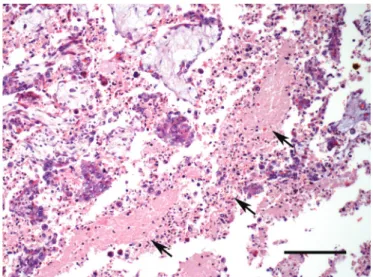
Macroscopic lesions were not observed in any of the placentas. Histologically, lesions were visualized in seven placentas from N. caninum seropositive goats (Table 1). The lesions were more severe in the goats that had reproductive disorders (Goat 1 and 2). In the placenta of Goat 1, severe, multifocal, sometimes locally extensive, necrosis was observed in placental cotyledons. The necrosis was localized mainly in the chorionic villi involving the trophoblastic cells and mesenchyme (Fig.1 and 2). These necrotic areas were characterized by loss of tissue architecture,
eosinophilia, cellular debris associated with rare calcification foci, nuclear pyknosis and mild neutrophilic infiltration. Multifocal placentitis was observed in the interstitium below the chorionic epithelium and often around vessels, and was composed mainly by plasma cells, lymphocytes, macrophages and rare neutrophils. In the placenta of Goat 2, the lesions were similar to those described for Goat 1, but the neutrophilic infiltration was moderate and there were numerous multifocal areas of calcification. In these two placentas, the N. caninum DNA was detected, but in IHC, immunolabeled protozoan structures were not observed.
N. caninum DNA was detected in seven of the 11 (63.6%) seropositive goats’ placentas with healthy conceptuses (Table 1). The molecular sequence obtained from the placenta of Goat 8 was deposited in GenBank (accession number HQ323749.1)
Table 1. Histopatological, molecular and immunohistochemical analysis for Neospora caninum
in 15 goat placentae
Placentas PCR and genetic sequencing *
Immunohistochemistry
(IHC)* Histopathological lesions c
1 a + - +++
2 a + - +++
3 + + ++
4 + - +
5 + - +
6 + - +
7 + - +
8 + -
-9 + -
-10 - -
-11 - -
-12 - -
-13 - -
-14 b - -
-15 b - -
-a Goats that had reproductive disorders, b Seronegative goats, c Intensity
of histopatological lesions: + mild, ++ moderate, +++ severe, - absent; * PCR and IHC results: + positive, - negative.
Fig.1. Neosporosis in goats. Goat 1, placenta, extensive area of necrosis characterized by eosinophilia and cellular debris (arrows). HE, bar = 150μm.
(Varaschin et al. 2012). Histological lesions were observed in five of the seven (71.4%) placentas that were positive for N. caninum DNA. In Goat 3, multifocal moderate placentitis composed by lymphocytes, plasma cells and macrophages was visualized mainly in chorionic mesenchyme. Rare necrotic areas associated with calcification foci were also observed. In this placenta (Goat 3), groups of tachyzoites in the interstitium were visualized and were strongly immunolabeled for N. caninum in IHC (Fig.3). In two placentas (Goat 4 and 5), a mild, multifocal, mononuclear inflammatory infiltrate were observed, and in two other goats (Goat 6 and 7), rare foci of mononuclear inflammatory infiltrates were observed. In the other placentas (Goat 8 to 15), histological lesions were not observed. In the placentas of seronegative goats (Goat 14 and 15), the N. caninum DNA was not detected.
DISCUSSION
Inflammatory lesions, sometimes mild, are the only type of lesion that has been described in placentas of goats naturally infected with Neospora caninum (Barr et al. 1992, Dubey et al. 1992). In contrast, the present study describes severe lesions that were observed in the placentas of Goat 1 and 2. In addition to the inflammatory infiltrate observed in Goat 1, the lesions were characterized mainly by multifocal, sometimes locally extensive, necrotic areas that were located predominantly in the chorionic villi involving the trophoblast cells and mesenchyme. Necrotic areas associated with extensive calcification and a moderate neutrophilic infiltrate were visualized in Goat 2. Similarly, non-purulent necrotic placentitis was described in goats experimentally infected (Porto et al. 2016). However, these lesions differ in intensity from those presented here, since they were described as mild lesions (Porto et al. 2016). The differences in the intensity of these placental lesions could be due to differences regarding the N. caninum strains, since Porto et al. (2016) used a strain derived from cattle in Spain, and the goats from the present study were naturally
infected with strains circulating in Brazil. In addition, the inoculation route and number of inoculated parasites may explain the differences regarding placental lesions observed in the present study and those reported by Porto et al. (2016). The placental lesions in experimentally infected goats were more prominent in early and mid-gestational stage (Porto et al. 2016). Similarly, placental lesions were found in one goat during mid-gestational age (87 days) from the present study. However, the severe placental lesions described here also occurred in the late gestational age in one goat. Placental lesions in neosporosis have also been described in bovine that were naturally (Barr et al. 1991) and experimentally infected by N. caninum (Macaldowie et al. 2004, Maley et al. 2006, Gibney et al. 2008) and in experimentally infected sheep (Buxton et al. 1998, Arranz-Solís et al. 2015).
Although the pathogenesis of abortion in neosporosis is not completely understood, inflammatory and necrotic lesions in the placenta and in the fetus are correlated with fetal death in cattle (Maley et al. 2006, Gibney et al. 2008). In the present study, the necrotizing placentitis visualized in Goat 1 is also likely to have contributed to abortion. The insufficient oxygen supply resulting from placental lesions can induce the release of fetal adrenocorticotropic hormone with consequent rising of fetal cortisol and placental prostaglandin. This mechanism may also be responsible for the occurrence of late abortion or for the birth of premature calves infected by N. caninum (Dubey et al. 2006); similarly, the fetal lesions could be responsible for the occurrence of the stillbirth. Severe necrotizing placentitis, which could be associated with the pathogenesis of abortion and stillbirth in the present study, did not occur in the seropositive goats that displayed to healthy conceptuses. In the placentas of five goats naturally infected by N. caninum that had healthy conceptuses, four showed mild placental lesions and one showed moderate placental lesions. Other study indicates that placental lesions along with fetal lesions with high loads of parasite within these tissues might contribute to abortion in ovine neosporosis (Arranz-Solís et al. 2015)
The N. caninum DNA was detected in the placentas of Goat 1 and 2, but immunolabeling for N. caninum antigens was not found in the analyzed IHC sections of these tissues. Nevertheless, in one goat (Goat 3) that was naturally infected by N. caninum and that gave birth to a healthy, but congenitally infected conceptus, groups of tachyzoites could be observed on histological sections and were evidenced by IHC. These groups of tachyzoites were located mainly in the interstitium of chorionic villi and were strongly immunolabeled by the N. caninum antibody. In the placentas of cattle and sheep experimentally infected with N. caninum, it was possible to visualize structures consistent with tachyzoites and antigens through IHC (Buxton et al. 1998, Macaldowie et al. 2004, Maley et al. 2006, Gibney et al. 2008). In contrast, the present study demonstrates that N. caninum can be seen in the placenta of naturally infected goats through histology and IHC, but at a low frequency. This low frequency of positive results on IHC for N. caninum in placental tissue sections of the present study is probably due to an absence of protozoan structures (tachyzoites or cysts) in tissue sections. N. caninum can be detected by PCR within the placenta of infected goats in all gestational stages, including late gestational age (Porto et al. 2016). In the present study, seven of the 11 placentas from
the goats that gave birth to healthy but congenitally infected conceptuses were positive for N. caninum DNA. Similarly, N. caninum was found through bioassays in the placenta of cattle that generated healthy, but congenitally infected conceptuses (Fioretti et al. 2003). As with those observed in the seronegative goats, three of four seropositive goats in which the N. caninum DNA was not found in the placentas had uninfected conceptuses. Out of all the 13 placentas from seropositive goats, the N. caninum DNA was detected in nine of them, while only one was positive in IHC testing for N. caninum. In another previous study, the PCR assay for N. caninum also presented a higher sensitivity when compared to IHC (Van Maanen et al. 2004).
CONCLUSIONS
The present study demonstrates that severe necrotizing placentitis was associated with abortion and stillbirth in caprine neosporosis.
The polymerase chain reaction (PCR) was efficient in detecting Neospora caninum DNA in the placentas of goats naturally infected, while N. caninum structures were successfully visualized through IHC in only one case.
Acknowledgements.- The authors would like to thank FAPEMIG (Fundação
de Amparo à Pesquisa do Estado de Minas Gerais) for the financial support, and CAPES (Coordenação de Aperfeiçoamento de Pessoal de Nível Superior)
for the Master´s grant.
REFERENCES
Arranz-Solís D., Benavides J., Regidor-Cerrillo J., Fuertes M., Ferre I., Ferreras M.C., Collantes-Fernández E., Hemphill A., Pérez V. & Ortega-Mora L.M.
2015. Influence of the gestational stage on the clinical course, lesional development and parasite distribution in experimental ovine neosporosis. Vet. Res. 2015(46):19. PMid:25884945.
Barr B.C., Anderson M.L., Dubey J.P. & Conrad P.A. 1991. Neospora-like protozoal
infections associated with bovine abortions. Vet. Pathol. 28(2):110-116. http://dx.doi.org/10.1177/030098589102800202. PMid:2063512.
Barr B.C., Anderson M.L., Woods L.W., Dubey J.P. & Conrad P.A. 1992. Neospora
-like protozoal infections associated with abortion in goats. J. Vet. Diagn.
Invest. 4(3):365-367. http://dx.doi.org/10.1177/104063879200400331. PMid:1515507.
Brasil 2001. Programa Nacional de Controle e Erradicação da Brucelose e da
Tuberculose Animal. Ministério da Agricultura, Pecuária e Abastecimento,
Secretaria de Defesa Animal, Brasília. 192p. Disponível em <http://www. adapar.pr.gov.br/arquivos/File/GSA/PECEBT/MANUAL_PNCEBT.pdf> Buxton D., Maley S.W., Wright S., Thomson K.M., Rae A.G. & Innes E.A.
1998. The pathogenesis of experimental neosporosis in pregnant sheep.
J. Comp. Pathol. 118(4):267-279. http://dx.doi.org/10.1016/S0021-9975(07)80003-X. PMid:9651804.
Caspe S.G., Moore D.P., Leunda M.R., Cano D.B., Lischinsky L., Regidor-Cerrillo
J., Álvarez-García G., Echaide I.G., Bacigalupe D., Ortega Mora L.M., Odeón
A.C. & Campero C.M. 2012. The Neospora caninum-Spain 7 isolate induces placental damage, fetal death and abortion in cattle when inoculated in early gestation. Vet. Parasitol. 189(2-4):171-181. http://dx.doi.org/10.1016/j. vetpar.2012.04.034. PMid:22621962.
Corbellini L.G., Colodel E.M. & Driemeier D. 2001. Granulomatous encephalitis
in a neurologically impaired goat kid associated with degeneration of
Neospora caninum tissue cysts. J. Vet. Diagn. Invest. 13(5):416-419. http://
dx.doi.org/10.1177/104063870101300509. PMid:11580064.
Dubey J.P. & Schares G. 2011. Neosporosis in animals-The last five years. Vet.
Parasitol. 180(1/2):90-108. http://dx.doi.org/10.1016/j.vetpar.2011.05.031. PMid:21704458.
Dubey J.P. 2003. Review of Neospora caninum and neosporosis in animals.
Korean J. Parasitol. 41(1):1-16. http://dx.doi.org/10.3347/kjp.2003.41.1.1.
PMid:12666725.
Dubey J.P., Acland H.M. & Hamir N.A. 1992. Neospora caninum (Apicomplexa) in a
stillborn goat. J. Parasitol. 78(3):532-534. http://dx.doi.org/10.2307/3283661.
PMid:1597802.
Dubey J.P., Buxton D. & Wouda W. 2006. Pathogenesis of bovine neosporosis. J.
Comp. Pathol. 134(4):267-289. http://dx.doi.org/10.1016/j.jcpa.2005.11.004. PMid:16712863.
Dubey J.P., Morales J.A., Villalobos P., Lindsay D.S., Blagburn B.L. & Topper M.J. 1996. Neosporosis-associated abortion in a dairy goat. J. Am. Vet. Med.
Assoc. 208(2):263-265. PMid:8567387.
Dubey J.P., Schares G. & Ortega-Mora L.M. 2007. Epidemiology and control of
neosporosis and Neospora caninum. Clin. Microbiol. Rev. 20(2):323-367. http://dx.doi.org/10.1128/CMR.00031-06. PMid:17428888.
Eleni C., Crotti S., Manuali E., Costarelli S., Filippini G., Moscati L. & Magnino
S. 2004. Detection of Neospora caninum in an aborted goat foetus. Vet. Parasitol. 123(3/4):271-274. http://dx.doi.org/10.1016/j.vetpar.2004.06.017. PMid:15325053.
Fioretti D., Pasquali P., Diaferia M., Mangili V. & Rosignoli L. 2003. Neospora caninum infection and congenital transmission: serological and parasitological
study of cows up to the fourth gestation. J. Vet. Med. B Infect. Dis. Vet. Public Health 50(8):399-404. http://dx.doi.org/10.1046/j.1439-0450.2003.00686.x.
PMid:14633211.
Gibney E.H., Kipar A., Rosbottom A., Guy C.S., Smith R.F., Hetzel U., Trees A.J. & Williams D.J.L. 2008. The extent of parasite-associated necrosis
in the placenta and foetal tissues of cattle following Neospora caninum
infection in early and late gestation correlates with foetal death. Int. J.
Parasitol. 38(5):579-588. http://dx.doi.org/10.1016/j.ijpara.2007.09.015. PMid:18021783.
Herrin B., Mahapatra S., Blouin E.F. & Shaw E.I. 2011. Growth of Coxiella
burnetii in the Ixodes scapularis-derived IDE8 tick cell line. Vector Borne
Zoonotic Dis. 11(7):917-922. http://dx.doi.org/10.1089/vbz.2010.0126. PMid:21254834.
Macaldowie C., Maley S.W., Wright S., Bartley P., Esteban-Redondo I., Buxton D. & Innes E.A. 2004. Placental pathology associated with fetal death in
cattle inoculated with Neospora caninum by two different routes in early
pregnancy. J. Comp. Pathol. 131(2/3):142-156. http://dx.doi.org/10.1016/j.
jcpa.2004.02.005. PMid:15276854.
Maley S.W., Buxton D., Macaldowie C.N., Anderson I.E., Wright S.E., Bartley P.M., Esteban-Redondo I., Hamilton C.M., Storset S.K. & Innes E.A. 2006.
Characterization of the immune response in the placenta of cattle experimentally infected with Neospora caninum in early gestation. J. Comp.
Pathol. 135(2/3):130-141. http://dx.doi.org/10.1016/j.jcpa.2006.07.001. PMid:16997005.
Mesquita L.P., Nogueira C.I., Costa R.C., Orlando D.R., Bruhn F.R., Lopes P.F., Nakagaki K.Y., Peconick A.P., Seixas J.N., Bezerra Jr P.S., Raymundo D.L. &
Varaschin M.S. 2013. Antibody kinetics in goats and conceptuses naturally infected with Neospora caninum. Vet. Parasitol. 196(3/4):327-333. http:// dx.doi.org/10.1016/j.vetpar.2013.03.002. PMid:23537945.
Moreno B., Collantes-Fernández E., Villa A., Navarro A., Regidor-Cerrillo J. &
Ortega-Mora L.M. 2012. Occurrence of Neospora caninum and Toxoplasma gondii infections in ovine and caprine abortions. Vet. Parasitol. 187(1/2):312-318. http://dx.doi.org/10.1016/j.vetpar.2011.12.034. PMid:22260901.
Payne S. & Ellis J. 1996. Detection of Neospora caninum DNA by polymerase chain
reaction. Int. J. Parasitol. 26(4):347-351.
Porto W.J.N., Regidor-Cerrillo J., Kim P.C.P., Benavides J., Silva A.C.S., Horcajo P., Oliveira A.A.F., Ferre I., Mota R.A. & Ortega-Mora L.M. 2016. Experimental
caprine neosporosis: the influence of gestational stage on the outcome of infection. Vet. Res. 47(1):29. http://dx.doi.org/10.1186/s13567-016-0312-6. PMid:26864744.
Sanger F., Nicklen S. & Coulson A.R. 1977. DNA sequencing with chain-terminating inhibitors. Proc. Natl. Acad. Sci. USA 74(12):5463-5467. http://
dx.doi.org/10.1073/pnas.74.12.5463. PMid:271968.
Santos D.S., Andrade M.P., Varaschin M.S., Guimarães A.M. & Hirsch C. 2011.
Neospora caninum in bovine fetuses of Minas Gerais, Brazil: genetic
characteristics of rDNA. Revta Bras. Parasitol. Vet. 20(4):281-288. http://
dx.doi.org/10.1590/S1984-29612011000400005. PMid:22166381.
Van Maanen C., Wouda W., Schares G., von Blumröder D., Conraths F.J., Norton R., Williams D.J.L., Esteban-Redondo I., Innes E.A., Mattsson J.G., Björkman C., Fernández-García A., Ortega-Mora L.M., Müller N., Sager H. & Hemphill A.
2004. An interlaboratory comparison of immunohistochemistry and PCR methods for detection of Neospora caninum in bovine foetal tissues. Vet. Parasitol. 126(4):351-364. http://dx.doi.org/10.1016/j.vetpar.2004.08.016. PMid:15567040.
Varaschin M.S., Hirsch C., Wouters F., Nakagaki K.Y., Guimarães A.M., Santos D.S., Bezerra Jr P.S., Costa R.C., Peconick A.P. & Langohr I.M.
2012. Congenital neosporosis in goats from the state of Minas Gerais,
Brazil. Korean J. Parasitol. 50(1):63-67. http://dx.doi.org/10.3347/