Biofilm formation by Vibrio parahaemolyticus on different surfaces
and its resistance to sodium hypochlorite
Formação de biofilme por Vibrio parahaemolyticus em diferentes superfícies
e resistência ao hipoclorito de sódio
Janaina Viana da Rosa1* Natália Volpato da Conceição1 Rita de Cássia dos Santos da Conceição1 Cláudio Dias Timm1
ISSNe 1678-4596
INTRODUCTION
Fish has high nutritional value. It is composed of proteins, unsaturated lipids, vitamins, and minerals. However, their microbiota is closely linked to the microbiota of the water where they live
which makes fish susceptible to contamination by
several microorganisms (JAY, 2005) including some species of the genus Vibrio.
Bacteria of the genus Vibrio account for
a significant number of cases of human infections
caused by the consumption of raw or undercooked crustaceans (THOMPSON, 2004). There are three species of Vibrio that are pathogenic to humans: V. cholerae, V. parahaemolyticus, and V. vulnificus.
V. parahaemolyticus is a halophilic bacterium
reported mainly during summer months when water temperatures exceed 15°C (SU & LIU, 2007) in
1Faculdade de Veterinária, Universidade Federal de Pelotas (UFPel), 96010-900, Campus Capão do Leão, Pelotas, RS, Brasil. E-mail:
janavrosa@yahoo.com.br. *Corresponding author.
ABSTRACT: Vibrio parahaemolyticus is an important pathogen for both fish industry and consumers. It forms biofilm which makes it difficult
to eliminate this microorganism using sanitizers. This study aimed to assess biofilm formation on different surfaces and effect of biofilm on resistance to sanitizers. Eight isolates of biofilm-forming V. parahaemolyticus were tested for the ability to form biofilms on a number of surfaces including high density polyethylene, stainless steel, glass, exoskeleton of Farfantepenaeus paulensis (Pink Shrimp), and operculum of Micropogonias furnieri (Whitemouth Croaker). Efficiency of sanitizer sodium hypochlorite against the bacteria was evaluated in the biofilms formed on the surface of the materials used; out the eight strains analyzed four formed biofilm on different surfaces. The present study shows that there are variations between surfaces in terms of biofilm formation, with more than one bacterial strain being able to form biofilm on the surface of the operculum of M. furnieri and on high density polyethylene as well. One isolate formed biofilm on glass, and one isolate formed biofilm on stainless steel. Sanitizers reduced biofilm formation on all surfaces. Based on our findings, we concluded that V. parahaemolyticus isolates have different ability to form biofilm on different surfaces. No isolates formed biofilm on shrimp shells. Results of this study also showed
that sodium hypochlorite eat a concentration of 20 parts per million (20ppm) of Cl2, albeit not able to eliminate bacteria reported in biofilms,
is still capable of reducing bacterial populations.
Key words: bacterial contamination, sanitizers, food safety, fish, shrimp.
RESUMO: Vibrio parahaemolyticus é uma bactéria patogênica importante tanto para a indústria como para os consumidores de pescados, uma
vez que pode formar biofilme, dificultando a sua eliminação por sanitizantes. Este estudo teve como objetivo verificar a formação de biofilme em diferentes superfícies e o efeito do biofilme sobre a resistência a sanitizante. Oito isolados de V. parahaemolyticus formadores de biofilme foram testados quanto à capacidade de formar biofilme em superfícies de polietileno de alta densidade, aço inoxidável, vidro, exoesqueleto de Farfantepenaeus paulensis (Camarão-rosa) e opérculo de Micropogonias furnieri (Corvina). A eficiência do sanitizante hipoclorito de sódio foi avaliada frente às bactérias nos biofilmes formados sobre a superfície dos materiais utilizados. Das oito cepas analisadas, quatro foram consideradas formadoras de biofilme em diferentes superfícies. Os resultados mostraram variação entre as superfícies, sendo que mais de uma cepa formou biofilme na superfície do opérculo de M. furnieri e do polietileno de alta densidade. Um isolado formou biofilme em vidro e um em aço inoxidável. Nenhum isolado formou biofilme na carapaça de camarão. O sanitizante reduziu a formação do biofilme em todas as superfícies. Conclui-se que os isolados de V. parahaemolyticus apresentam distinta capacidade de formar biofilme em diferentes superfícies e que o hipoclorito de sódio na
concentração de 20 partes por milhão (20ppm) de Cl2, embora não elimine as bactérias que se encontram em biofilme, reduz a sua população.
Palavras-chave: contaminação bacteriana, sanitizantes, segurança alimentar, peixe, camarão.
coastal and estuarine waters (HEITMANN et al., 2005; OLIVER & KAPER, 2001).
V. parahaemolyticus may be reported in planktonic or sessile state, that is, within a microbial
community, forming biofilm. Microbial biofilm is an
association of bacterial cells that attach to biotic or abiotic surfaces, and are surrounded by a complex extracellular matrix (MCCARTER, 1999).
A number of studies mentioned the
ability of many bacteria to form biofilm on different
surfaces. MILAN et al. (2015) evaluated the
biofilm-forming ability of two different isolates of
Salmonella enterica subsp. enterica on high density
polyethylene, stainless steel, and glass surfaces. These authors demonstrated that both isolates had the ability to attach to all the three surfaces tested. In another study carried out by QUATRIN et al. (2015),
researchers evaluated the bacterial biofilm formation
by Pseudomonas aeruginosa on metal, stainless steel,
acrylic, glass, polyethylene terephthalate (PET), high
density polyethylene (HDPE), and Teflon surfaces. This microorganism formed biofilms in different
densities on all surfaces tested. These studies showed that isolates of each bacterial species has different
biofilm formation ability on different surfaces.
According to FLACH et al. (2015),
the organization of microorganisms in biofilms
may provide protection against dehydration and resistance to sanitizers. This increased resistance results from the formation of a barrier consisting of an exopolysaccharide matrix which prevents or
decreases contact between microbial biofilms and
antimicrobial agents (SREY et al., 2013).
Resistance may occur due to the use of different sanitizers with similar mechanisms of action, which increases the risk of resistance
development, particularly in biofilms (BRAOUDAKI
& HILTON, 2004) or even resistance transmission
between bacteria within the biofilm (BORGES et al.,
2013). Due to all these factors, research should be carried out in order to broaden our knowledge on the resistance of V. parahaemolyticus against sanitizers in
biofilms. Surfaces in the food industry are susceptible
to bacterial adhesion and are; therefore, potential
sources of contamination if microbial biofilms
are formed (ROSSONI & GAYLARDE, 2000).
If bacterial isolates from biofilms become more
resistant, the concentration of product or contact time
used during hygiene procedures may be insufficient
to eliminate microorganisms from a contaminated surface (ANTONIOU & FRANK, 2005). Sodium hypochlorite induces changes in bacterial cell permeability and interferes with the enzymatic
processes of these microorganisms (EVANGELISTA, 2000). Biocidal and oxidizing activities of this compound increased with the formation of hypochlorous acid (HClO) in its undissociated form when in pure aqueous solution (EMMANUEL et al., 2004). The present study aimed to assess the
formation of biofilm by V. parahaemolyticus on different surfaces as well as evaluating resistance of
these microbial biofilms against sodium hypochlorite.
MATERIALS AND METHODS
We tested eight previously obtained
isolates which were considered biofilm-forming
strains by ROSA et al. (2017) as follows: one of
Micropogonias furnieri (Whitemouth Croaker), four of Mugil platanus (Lebranche Mullet) and three of
Farfantepenaeus paulensis (Pink Shrimp). These
isolates were tested for their ability to form biofilm
on different surfaces, according to the technique used
by MILAN et al. (2015) with minor modifications in
order to adapt this method for V. parahaemolyticus. We used high density polyethylene (HDPE) plastic coupons, stainless steel coupons, and 4cm² sterile
glass vials with flat surfaces; 1cm² coupons of F.
paulensis exoskeletons and M. furnieri opercula that were prepared according to CASTRO-ROSAS & ESCARTÍN (2002) were also used in this study. Exoskeletons and opercula were manually removed
from fish specimens and cut into the appropriate
size after being subjected to the following washing steps: (1) 30 seconds under running water. After washing, samples were shaken in order to remove any residual liquid and soft tissue; (2) 30 seconds in 70% alcohol to remove possible contaminants; (3) samples were then washed again under running water. After washing, these samples were stored at -20°C until use. Coupons were placed in Petri dishes containing 100ml of Alkaline Peptone Water (APW) with 1% NaCl (APW-1% NaCl, Himedia, Mumbai, India) and 2ml of culture from each isolate recovered in APW-1% NaCl for 24 hours. Plates were then incubated at 37°C. At each 48 hours of incubation, coupons were gently washed twice with APW-1% NaCl to remove unbound cells and were again placed in Petri dishes with 100ml APW-1% NaCl without the inoculum and incubated at 37°C.
After five repetitions of this procedure, sterile swabs
Lansing, Michigan, USA). An isolate unable to form
biofilm as determined by ROSA et al. (2017) was
used as a negative control. Isolates which counts were statistically different compared to those of the control
isolate were considered competent biofilm formers. The efficiency of the sanitizer sodium
hypochlorite (solution containing 20ppm of Cl2) was
evaluated against bacteria in biofilms formed on the
surface of the different materials used according to the technique described by MILAN et al. (2015) with
some modifications. The same procedure described above was repeated for the biofilm-forming isolates and biofilm-forming materials. After the last wash, the biofilm coupons were immersed in vials containing
the sanitizer for 10 minutes. Once the established contact time was reached, the coupons were immersed in a neutralizing solution (0.1M Na2S2O3) for 30 seconds. After washing with APW-1% NaCl, a sterile swab was scrubbed on the surface of each coupon and
counting was performed in PCW-2% NaCl. Biofilms
formed by the same isolates were used as controls and counted in PCW-2% NaCl before coming into contact with the sanitizer.
All experiments were repeated three times independently with new bacterial cultures
and new coupons. In order to assess biofilm
formation on different surfaces and bacterial resistance to sanitizers, analysis of variance of V. parahaemolyticus counts was performed. Results were evaluated by Tukey’s test (P<0.05) using the software Statistix® (2003).
RESULTS AND DISCUSSION
In the present study, four out of the eight
isolates analyzed were considered biofilm formers on
different surfaces (Table 1). Our results showed that
there are variations in biofilm formation between the surfaces tested. More than one isolate formed biofilm
on the surface of the operculum of M. furnieri and on a high density polyethylene (HDPE) surface. Over time, plastic surfaces may become rough. Cracks may form as a result of the wear and tear of these surfaces which may in turn harbor residues that protect bacteria; and therefore, favor the formation
of biofilms (SHI & ZHU, 2009). This highlights the importance of our findings in such material. HAN et
al. (2016) tested the ability of different isolates of
V. parahaemolyticus to form biofilm in shrimp and
crab shells. The authors observed that these isolates
were able to form biofilm on these surfaces. Their
results differ from the results obtained in our study since none of the isolates that we tested formed a
biofilm in shrimp shells. However, we observed
that V. parahaemolyticus is able to form biofilm
in opercula, which may hinder the elimination of
the microorganism from the surface of fish. This
makes this microorganism a potential source of
contamination for other fish, utensils, and equipment both in vessels and in the fish industry. ABDALLAH
et al. (2009) and HAN et al. (2016) carried out studies in which they observed that isolates of V. parahaemolyticus are able to form biofilm in glass
and stainless steel, respectively. Similar findings
were obtained in our study with some isolates on both surfaces. When analyzing the results of our study as well as comparing these results with those obtained in the afore mentioned studies, we noticed that there is a great variation among isolates regarding their
ability to form biofilm on different surfaces. This
variation may occur due to the bacterial extracellular matrix. This matrix may vary even within the same
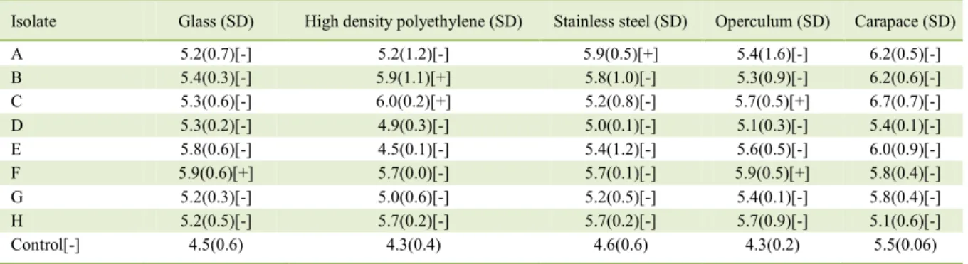
Table 1 - Counts and assessment of biofilm formation by V. parahaemolyticus on different surfaces (Log/cm²).
Isolate Glass (SD) High density polyethylene (SD) Stainless steel (SD) Operculum (SD) Carapace (SD)
A 5.2(0.7)[-] 5.2(1.2)[-] 5.9(0.5)[+] 5.4(1.6)[-] 6.2(0.5)[-]
B 5.4(0.3)[-] 5.9(1.1)[+] 5.8(1.0)[-] 5.3(0.9)[-] 6.2(0.6)[-]
C 5.3(0.6)[-] 6.0(0.2)[+] 5.2(0.8)[-] 5.7(0.5)[+] 6.7(0.7)[-]
D 5.3(0.2)[-] 4.9(0.3)[-] 5.0(0.1)[-] 5.1(0.3)[-] 5.4(0.1)[-]
E 5.8(0.6)[-] 4.5(0.1)[-] 5.4(1.2)[-] 5.6(0.5)[-] 6.0(0.9)[-]
F 5.9(0.6)[+] 5.7(0.0)[-] 5.7(0.1)[-] 5.9(0.5)[+] 5.8(0.4)[-]
G 5.2(0.3)[-] 5.0(0.6)[-] 5.2(0.5)[-] 5.4(0.1)[-] 5.8(0.4)[-]
H 5.2(0.5)[-] 5.7(0.2)[-] 5.7(0.2)[-] 5.7(0.9)[-] 5.1(0.6)[-]
Control[-] 4.5(0.6) 4.3(0.4) 4.6(0.6) 4.3(0.2) 5.5(0.06)
bacterial species as previously mentioned. It is
the matrix that forms biofilms, which components
include bacterial cells, exopolysaccharides, proteins, nucleic acids, glycoproteins, phospholipids, debris, and inorganic matter (SUTHERLAND, 2001). Results of the present study show that we should not generalize about the genus and species of this bacterium. Differences between the isolates of V. parahaemolyticus according to each surface tested showed that they may present different features
regarding the ability to form biofilm.
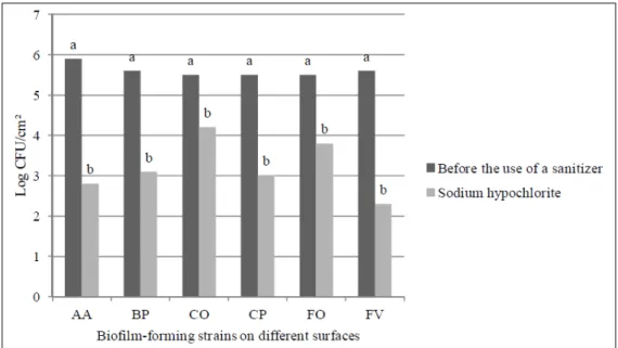
To evaluate the efficiency of the
sanitizer, the surfaces in which the isolates formed
biofilm were immersed in flasks containing sodium
hypochlorite. The analysis of variance showed effect only for the use of the sanitizer which reduced
the bacterial population in the biofilms from all
surfaces tested (Figure 1). However, this sanitizer was unable to eliminate the microorganisms. This result raises concern since the solution containing 20ppm sodium hypochlorite for 10 minutes that
was used in our study is commonly used in the fish
industry. To obtain the effective elimination of V. parahaemolyticus in biofilms it would be necessary
to increase the exposure time or the concentration of the sanitizer in the solution.
BELTRAME et al. (2015) and
BELTRAME et al. (2016) tested the efficiency of sodium hypochlorite against biofilms formed by
Listeria monocytogenes and Escherichia coli in coupons made of high density polyethylene. These authors noted that the use of a solution containing 10ppm of sodium hypochlorite for 10 minutes fully removed E. coli from these coupons. In contrast,
L. monocytogenes was fully removed from these coupons only when the concentration of 40ppm of this sanitizer was used. Based on the results of our study, we may infer that each bacterium from a
biofilm reacts differently when coming into contact
with a sanitizer. According to MEYER (2003), sodium hypochlorite acts not only on bacteria
in a planktonic state but also acts in biofilms by
removing exopolysaccharides from surfaces which
makes difficult for new bacteria to attach (SINDE &
CARBALLO, 2000).
CONCLUSION
Some isolates of V. parahaemolyticus are
able to form biofilm a number of surfaces including
glass, high-density polyethylene (HDPE), stainless steel, and operculum of M. furnieri. Isolates of V.
Figure 1 - Effect of sodium hypochlorite on biofilm-forming isolates of V. parahaemolyticus (A, B, C, and F) on
parahaemolyticus display a singular ability to form
biofilm on different surfaces.
Sodium hypochlorite solutions at the concentration of 20ppm of Cl2 is able to reduce the bacterial population of V. parahaemolyticus
in biofilm but fails to eliminate the bacteria. These findings serve as an eye alert for those involved in the fish industry fish as microbial biofilms may form on a
number of different surfaces. Hygiene and sanitation procedures may be reevaluated.
ACKNOWLEDGEMENTS
The authors acknowledge the Coordenação de Aperfeiçoamento de Pessoal de Nível Superior (CAPES) through scholarships.
DECLARATION OF CONFLICTING INTERESTS
The authors declared no potential conflicts of interest with
respect to the research, authorship, and/or publication of this article.
AUTHORS’ CONTRIBUTIONS
All authors contributed equally for the conception and writing of the manuscript. All authors critically revised the
manuscript and approved of the final version.
REFERENCES
ABDALLAH, F.B. et al. Adherence assays and slime production of Vibrio alginolyticus and Vibrio parahaemolyticus. Brazilian
Journal of Microbiology, v.40, p.394-398, 2009. Available from: <http://www.scielo.br/scielo.php?script=sci_arttext&pid =S1517-83822009000200033>. Accessed: Dec. 11, 2017. doi: 10.1590/S1517-83822009000200033.
ANTONIOU, K.; FRANK, J.F. Removal of Pseudomonas putida
biofilm and associated extracellular polymeric substances from
stainless steel by alkali cleaning. Journal of Food Protection, v.68, n.2, p.277-281, 2005. Available from: <http://jfoodprotection. org/doi/pdf/10.4315/0362-028X-68.2.277>. Accessed: Dec. 11, 2017. doi: 10.4315/0362-028X-68.2.277.
BELTRAME, C.A. et al. Adhesion of Listeria monocytogenes to cutting board surfaces and removal by different sanitizers. Journal
für Verbraucherschutz und Lebensmittelsicherheit, v.10, p.41-47, 2015. doi: 10.1007/s00003-014-0923-7.
BELTRAME, C.A. et al. Effectiveness of sanitizing agents in inactivating Escherichia coli (ATCC 25922) in food cutting board surfaces. Removal E. coli using different sanitizers. Italian
Journal of Food Science, v.28, n.1, p. 148-154, 2016. doi: 10.14674/1120-1770/ijfs.v468.
BORGES, A. et al. Antibacterial activity and mode of action of ferulic and gallicacids against pathogenic bacteria. Microbial
Drug Resistance, v.19, n.4, p.256-65, 2013. doi: 10.1089/ mdr.2012.0244.
BRAOUDAKI, M.; HILTON, A.C. Adaptive resistance to biocides in Salmonella enterica and Escherichia coli O157 and cross-resistance to antimicrobial agents. Journal of Clinical
Microbiology, v.42, p.73–78, 2004. Available from: <https:// www.ncbi.nlm.nih.gov/pmc/articles/PMC321691/pdf/0976.pdf>. Accessed: Dec. 11, 2017. doi: 10.1128/JCM.42.1.73-78.2004.
CASTRO-ROSAS, J.; ESCARTÍN, E.F. Adhesion and colonization of Vibrio cholera O1 on shrimp and crab carapaces.
Journal of Food Protection, v.65, n.3, p.492–498, 2002. Available from: <http://jfoodprotection.org/doi/pdf/10.4315/0362-028X-65.3.492>. Accessed: Dec. 11, 2017.
EMMANUEL, E.; et al. Toxicological effects of disinfestations using sodium hypochlorite on aquatic organisms and its contribution to AOX formation in hospital wastewater.
Environment International, v.30, p.891-900, 2004. doi: 10.1016/j.envint.2004.02.004.
EVANGELISTA, J. Tecnologia de Alimentos. São Paulo: Atheneu, 2000. 652p.
FLACH, J. et al. Biofilm formation from milk in contact with raw material: virulence factors involved. Acta Scientiae
Veterinariae, v.33, n.3, p.291-296, 2005. Available from: <http:// www.lume.ufrgs.br/bitstream/handle/10183/20189/000531682. pdf?sequence=1>. Accessed: Dec. 11, 2017.
HAN, N. et al. Biofilm formation by Vibrio parahaemolyticus on food and food contact surfaces increases with rise in temperature.
Food Control, v.70, p.161-166, 2016. Available from: <http:// www.sciencedirect.com/science/article/pii/S0956713516302997>. Accessed: Dec. 11, 2017. doi: 10.1016/j.foodcont.2016.05.054.
HEITMANN, I.G. et al. Revisión y recomendaciones para el manejo de diarrea por Vibrio parahaemolyticus. Revista Chilena
de Infectologia, v.22, n.2, p.131-140, 2005. Available from:
< http://www.scielo.cl/scielo.php?script=sci_arttext&pid=S0716-10182005000200003&lng=en&nrm=iso&tlng=en>. Accessed: Dec. 9, 2017. doi: 10.4067/S0716-10182005000200003.
JAY, J.M. Microbiologia de Alimentos. Porto Alegre: Artmed, 2005. 711p.
MCCARTER, L. The Multiple Identities of Vibrio parahaemolyticus.
Journal of Molecular Microbiology and Biotechnology, v.1, n.1, p.51-57, 1999. Available from: <https://www.caister.com/jmmb/v/ v1/v1n1/08.pdf>. Accessed: Dec. 12, 2017.
MEYER, B. Approaches to prevention, removal and killing of
biofilms. International Biodeterioration & Biodegradation, v.51, n.4, p.249-253, 2003. doi: 10.1016/S0964-8305(03)00047-7.
MILAN, C. et al. Sanitizer resistance of biofilm-forming
Salmonella isolated from meat products. Arquivo Brasileiro de
Medicina Veterinária e Zootecnia, v.67, n.2, p. 642-646, 2015. Available from: <http://www.scielo.br/scielo.php?script=sci_artte xt&pid=S0102-09352015000200642>. Accessed: Dec. 12, 2017. doi: 10.1590/1678-7298.
OLIVER, J.D.; KAPER, J.B. Vibrio species. In: DOYLE, M.P.; BEUCHAT, L.R.; MONTVILLE, T.J.(Eds). Food microbiology: Fundamentals and frontiers. Washington: ASM Press, 2001. p.263-300.
QUATRIN, P.M. et al. Evaluation of different substrates for in vitro
Scientia. Série: Ciências da Saúde, v.16, n.2, p.191-203, 2015. Available from: <https://www.periodicos.unifra.br/index.php/ disciplinarumS/article/view/1003/947>. Accessed: Dec. 13, 2017.
ROSA, J.V. et al. Formação de biofilme por Vibrio parahaemolyticus isolados de pescados. Pesquisa Veterinária Brasileira, v.37, n.4, p.339-345, 2017. Available from: <http://www.scielo. br/scielo.php?pid=S0100-736X2017000400339&script=sci_ abstract&tlng=pt>. Accessed: Dec. 13, 2017. doi: 10 1590/s0100-736x2017000400007.
ROSSONI, E.M.M.; GAYLARDE, C.C. Comparison of sodium hypochlorite and peracetic acid as sanitising agents for stainless steel food processing surfaces using epifluorescence microscopy. International Journal of Food
Microbiology, v.61, n.1, p.81-85, 2000. doi: 10.1016/S0168-1605(00)00369-X.
SHI, X.; ZHU, X. Biofilm formation and food safety in food
industries. Trends in Food Science & Technology, v.20, n.9, p.407-413, 2009. doi: 10.1016/j.tifs.2009.01.054.
SINDE, E.; CARBALLO, J. Attachment of Salmonella spp. and Listeria monocytogenes to stainless steel, rubber, and
polytetrafluorethylene: The influence of free energy and the effect
of commercial sanitizers. Food Microbiology, v.17, n.24, p.439-447, 2000. doi: 10.1006/fmic.2000.0339.
SREY, S. et al. Biofilm formation in food industries: A food safety
concern. Food Control, v.31, p. 572-585, 2013. doi: 10.1016/j. foodcont.2012.12.001.
SU, Y.C.; LIU, C. Vibrio parahaemolyticus: a concern of seafood safety. Food Microbiology, v.24, p.549-558, 2007. doi: 10.1016/j. fm.2007.01.005.
SUTHERLAND, I.W. The biofilm matrix: an immobilized but
dynamic microbial environment. Trends Microbiology, v.9, n.5, p.222-227, 2001. doi: 10.1016/S0966-842X(01)02012-1.
THOMPSON, F.L. et al. Biodiversity of Vibrios. Microbiology