2017
UNIVERSIDADE DE LISBOA
FACULDADE DE CIÊNCIAS
DEPARTAMENTO DE QUÍMICA E BIOQUÍMICA
Alpha-Synuclein Phosphorylation Role on Parkinson's Disease
Carolina da Silva Madaleno
Mestrado em Bioquímica
Especialização em Bioquímica
Dissertação orientada por:
Sandra Isabel Nogueira Tenreiro, PhD
III
Acknowledgments
In this gesture, I would like to mention and recognize the ones involved in this journey.
First and foremost, to my supervisor Dra. Sandra Tenreiro, whose dedication, patience and optimism were crucial during the development of this research project. I am sincerely grateful for the precious knowledge she transmitted, for her trust, for her effort in keeping me motivated and valuable guidance throughout this time, as well as for the treasured tools she acquainted me with, that made it possible to grow as a person and as a scientist. To my co-supervisor, Dr. Francisco Pinto, for his time and assistance during this project.
To Cell and Molecular Neuroscience group, to Dr. Tiago Fleming Outeiro, for this wonderful research opportunity in his lab group, for welcoming me and for his words of advice in the pursuing of a science path. Likewise, I am thankful to the members of the group I worked more closely with, Inês Brás, for her friendship, encouragement and help and to Dr. Hugo Miranda, for his insightful comments and availability.
To CEDOC community, for the sympathy shown by all researchers, for the interesting seminars and discussions organized and for the passion for science transmitted. A special gratitude goes for the kindness and help of the technical team and of the Flow Cytometry Unit.
To FCUL, for the assistance during the entire master’s degree and for the nicely carried incorporation in the faculty.
To my friends for the endless emotional support and motivation given when I needed it the most, a special thank you to Vera Xistra, Mariana Caetano, Mariana Morais and Ricardo Morais.
For last but not the least important, to my family. To my parents, Salomé Madaleno and Manuel Madaleno, my brother Romeu Madaleno and my sister-in-law Marília Fernandes, for always supporting me in the dream of becoming a scientist, for believing in me, for their daily caring and unconditional support during this stage and throughout my entire life.
With these words I hope to return the affection and dedication you have always offered me during this time, each one of you shed a special light during my path.
IV
Resumo
A incorreta aquisição da estrutura final e consequente agregação de proteínas são a principal causa para o desenvolvimento de patologias neuro-degenerativas, tais como a doença de Parkinson (DP), de Alzheimer (DA) e Huntington (DH).
A DP é a segunda mais comum doença neuro-degenerativa, afetando entre 1 a 2% da população acima dos 60 anos de idade nos países industrializados, sendo a Europa o continente mais afetado. Clinicamente, esta doença é caracterizada por tremor e rigidez musculares e instabilidade postural e de marcha. Eventualmente, há ainda o aparecimento de sintomas não-motores, tais como psicose e demência. Os sintomas motores, apenas detetados no momento do diagnóstico, ocorrem quando já uma significativa parte da população neuronal foi afetada e atualmente, a estratégia de tratamento é ainda paliativa, em vez de curativa. Patologicamente, a DP caracteriza-se pela acumulação da proteína α-Synucleína (αSyn), que forma inclusões intracelulares designadas por corpos de Lewis (CLs), causando a perda progressiva de neurónios dopaminérgicos na região cerebral da Substantia Nigra (SN) pars compacta e, em estádios mais avançados da doença, espalhando-se às restantes populações neuronais. Atualmente, é aceite que o papel pato-fisiológico da αSyn reside na sua agregação, no entanto, é ainda desconhecido quais os fatores que desencadeiam esse mesmo evento, assim como quais os mecanismos celulares e quais os intermediários proteicos responsáveis pela neuro-toxicidade.
A DP tem duas variantes, que têm como elo comum a agregação da αSyn: a esporádica e a familiar (menos de 10% dos casos). Assim, o estudo de uma das formas contribui para a compreensão do mecanismo patológico da outra. A forma esporádica da doença é desencadeada principalmente pela idade avançada e/ou fatores ambientais, enquanto a forma familiar ocorre prematuramente e com um fenótipo mais severo e é caracterizada pela existência de predisposições e mutações genéticas.
No caso da αSyn, mutações que originam duplicações e triplicações do gene que codifica esta proteína (gene SNCA), assim como as substituições de aminoácidos A30P, E46K, H50Q, G51D A53T e A53E na sequência da αSyn foram identificadas como associadas à forma familiar de DP. Adicionalmente, modificações pós-traducionais (MPTs) da αSyn, tais como a fosforilação do resíduo de serina S129, que em 90% da αSyn nos CLs se encontra fosforilada, em contraste com os 4% em condições fisiológicas, e recentemente, a identificação de aumento da fosforilação do resíduo de serina S87, estão associadas a ambas as formas esporádica e familiar da doença. No entanto, os mecanismos celulares e a toxicidade intrínseca pelos quais as mutações familiares contribuem para o papel neuropatológico da αSyn são ainda desconhecidos, assim como o papel neuro-protetor ou prejudicial da fosforilação da αSyn é ainda alvo de debate, já que diferentes modelos celulares apresentam diferentes efeitos na toxicidade da αSyn, quando fosforilada no resíduo S129. Em relação ao papel patológico da fosforilação do resíduo S87, este ainda não foi totalmente explorado, principalmente devido a duas principais limitações: este resíduo não é conservado entre espécies, o que dificulta o estabelecimento de conclusões de modelos de ratinhos para humanos, e é apenas fosforilável em humanos, o que evidencia um papel importante deste resíduo no desenvolvimento da DP.
A levedura Saccharomyces cerevisiae (S. cerevisiae) é um organismo unicelular que partilha um elevado grau de conservação de várias vias celulares básicas com outros eucariotas superiores, tais como o ser humano, como por exemplo o controlo de qualidade proteico. Este organismo foi estabelecido como um modelo celular para a compreensão de diversos mecanismos biológicos, assim como no estudo de doenças neuro-degenerativas. Nomeadamente, o estudo dos processos de fosforilação e agregação da αSyn, assim como a pato-biologia desta proteína são passíveis de ser estudados neste
V
organismo, uma vez que em levedura foi previamente demonstrado que a expressão da αSyn humana induz a formação de inclusões citoplasmáticas e confere toxicidade às células, de forma semelhante ao que ocorre não só noutros modelos biológicos, como ratinho e linhas celulares de mamífero, como também nos cérebros de pacientes com DP. Adicionalmente, a utilização deste organismo confere várias vantagens, como o a sua facilidade de manipulação, o curto tempo de geração, o genoma estar sequenciado e devidamente anotado em bases de dados e ser facilmente manipulável geneticamente.
Assim, utilizando a levedura como modelo celular, este projeto teve como objetivos abordar a pato-biologia da αSyn em ambas as vertentes esporádica e familiar da DP, nomeadamente A) na caracterização fenotípica da mais recentemente mutação identificada como associada a DP familiar, a mutação da αSyn A53E e B) o estudo da interação da fosforilação entre os resíduos S87 e S129. Para atingir os objetivos propostos, o fenótipo da mutação A53E foi caracterizado recorrendo à avaliação da viabilidade celular, formação de inclusões, expressão e fosforilação da αSyn, solubilidade dos agregados de αSyn e avaliação das alterações da rede mitocondrial. O efeito da fosforilação nos resíduos S129 e S87 foi estudado utilizando estirpes de levedura com o gene que codifica a proteína αSyn humana integrado no genoma, usando diferentes versões mutadas da αSyn, nomeadamente, mutantes que mimetizam e bloqueiam a fosforilação no resíduo S129, os mutantes S129D e S129A, respetivamente, o mutante que mimetiza a fosforilação no resíduo S87 (S87E) e um duplo mutante que mimetiza a simultânea fosforilação no resíduo S87 e S129 (S87E_S129D). Para este grupo de fosfo-mutantes foram avaliados os parâmetros de viabilidade celular, formação de inclusões, expressão, fosforilação e remoção intracelular da αSyn e a função dos mecanismos de manutenção da qualidade celular (função proteossomal e autofágica).
Resultante desta análise, foi observado que a expressão da proteína mutante A53E αSyn não resulta em diferenças significativas no fenótipo dos parâmetros avaliados, quando comparado com a expressão da proteína nativa (WT). De facto, a expressão da mutação A53E induz toxicidade e formação de inclusões, mas de forma equivalente à proteína WT, embora com tendência para diminuir o número de inclusões e ainda, relativamente à natureza das inclusões, a solubilidade das inclusões aparenta ser semelhante à proteína WT. Estes resultados estão em parte em concordância com estudos in vitro realizados, que descrevem um fenótipo da A53E de toxicidade celular semelhante à proteína WT.
Relativamente ao estudo da interação da fosforilação entre os resíduos S87 e S129, foi observado que quando a fosforilação no resíduo S87 é mimetizada (mutante S87E), a formação de inclusões e toxicidade conferidas pela αSyn aumentam significativamente (aproximadamente 80% de células com inclusões), causando a perda de função proteossomal e ativando a autofagia, quando comparando com a expressão da proteína WT. Surpreendentemente, quando a mutação S87E foi acoplada à mutação que mimetiza a fosforilação no resíduo S129 (mutante S87E_S129D), o fenótipo de toxicidade induzindo pelo mutante S87E é inexistente, prevalecendo um suposto efeito protetor da fosforilação no resíduo S129 (mutante S129D). Adicionalmente, é possível observar que o mutante S87E_S129D tem menos formação de inclusões e apresenta menos toxicidade que a expressão da proteína WT, o que permite inferir que não só prevalece o efeito protetor da mutação S129D sobre o efeito tóxico da mutação S87E, como também poderá existir um efeito sinérgico neuro-protetor quando ambos os resíduos se encontram fosforilados simultaneamente.
Explorar os mecanismos específicos com outros organelos assim como os eventos iniciais da agregação, nomeadamente aquando da presença de mutações familiares, é crucial para a determinação de como a αSyn induz toxicidade celular no caso da forma familiar de DP. Em relação ao papel da fosforilação na patogénese de PD, o presente estudo evidencia a importância do estudo de padrões de ativação/desativação de diferentes resíduos. Embora o presente trabalho suporte a ideia de um papel neuro-protetor da fosforilação do resíduo S129 da αSyn, assim como quando esta fosforilação é acoplada
VI
à fosforilação no resíduo S87, outras abordagens relativamente a outros padrões de MPTs e outros resíduos de fosforilação, como os restantes resíduos de tirosina, assim como o efeito destes padrões em organelos celulares específicos devem ser seguidos no futuro.
As contribuições do presente trabalho, assim como um esforço futuro para a compreensão do efeito de alterações da αSyn no seu comportamento e na proteostase celular, são cruciais para o desenvolvimento de novas estratégicas terapêuticas, no contexto da DP e de outras synucleinopatias.
Palavras-chave: Doença de Parkinson, α-Synuclein, Agregação, Mutações Familiares,
VII
Abstract
Protein misfolding and aggregation is a pathological hallmark in neurodegenerative disorders, including the currently incurable and second most common age-related neurodegenerative disease, Parkinson’s disease (PD). In both sporadic and familial cases (<10%) of PD, the protein α-Synuclein (αSyn) accumulates in intracellular inclusions, known as Lewy bodies (LBs), causing the progressive loss of dopaminergic neurons in the Substantia Nigra (SN) pars compacta and, ultimately spreading to non-dopaminergic neurons at later stages of the disease. In LBs, 90% of αSyn is phosphorylated at serine 129 (S129), in contrast with the 4% under physiological conditions. Nevertheless, whether this phosphorylation has a protective or detrimental effect is unclear and recently, additional phosphorylation sites of this protein, as serine 87 (S87) and three tyrosine residues (Y125, Y133 and Y136), have been identified. Furthermore, duplications and triplications of the αSyn coding gene (SNCA gene) and six missense mutations of αSyn (A30P, E46K, H50Q, G51D A53T and A53E) are associated with familial forms of PD, but how these mutations affect αSyn phosphorylation profile, as well as the overall cell mechanisms by which αSyn induces toxicity are not completely understood.
Given the high degree of conservation in basic cellular pathways between the budding yeast Saccharomyces cerevisiae (S. cerevisiae) and higher eukaryotes, yeast is an established model to study the process of αSyn phosphorylation and aggregation, as well as its pathobiology.
Therefore, the aims of this project were A) to phenotypically characterize the most recent PD familial linked mutation of αSyn, A53E and B) to study the phosphorylation interplay between the serine residues 129 and 87 on αSyn inclusion formation and toxicity. To achieve these objectives, the mutation A53E in αSyn was studied regarding cell viability, inclusion formation, protein expression and phosphorylation, solubility of aggregates and mitochondrial network alterations. The effect of phosphorylation at S129 and S87 was assessed using integrative transformed yeast strains encoding αSyn phospho-mutants, which were evaluated regarding cell viability, inclusion formation, protein expression, phosphorylation and clearance and proteasome and autophagy functions.
It was observed that the expression of the αSyn A53E mutant did not show any emerging phenotypic differences in yeast cells on the parameters evaluated, when compared to the wild-type (WT) protein expression. It was also found that mimicking phosphorylation at the S87 residue (S87E mutant), significantly increased αSyn toxicity and inclusion formation, caused proteasome impairment and up-regulated autophagy, when compared to the WT αSyn expression. Interestingly, when the S87E mutant was coupled with the S129 phosphomimic mutation (S87E_S129D mutant), the phenotype was lost and αSyn induced toxicity decreased, prevailing a supposedly protective effect of the phosphorylation at the S129 residue (S129D mutant).
Exploring the mechanism of αSyn toxicity to cells, including the impact of mutations and post-translational modifications (PTMs), are pivotal to understand PD etiology and new therapeutic strategies.
Overall, this study shed a light into the effect of both the phosphorylation state and missense point mutations of αSyn linked to early onset of PD, in its toxicity and aggregation tendency, as well as the impact in protein clearance and degradation systems and general cell homeostasis.
VIII
Communications of Intervention
Paper
Diana F Lázaro*, Mariana Castro Dias*, Anita Carija, Susanna Navarro, Carolina Silva
Madaleno, Sandra Tenreiro, Salvador Ventura, Tiago F Outeiro, The effects of the novel A53E
alpha-synuclein mutation on its oligomerization and aggregation, Acta Neuropathologica Communications, 2016 Dec 9, 4(1):128
*equal contribution
Abstract
Carolina Madaleno*, Inês Brás*, Sandra Tenreiro, Tiago F. Outeiro, Molecular insights into
the role of alpha-synuclein phosphorylation in synucleinopathies. 1st CEDOC Symposium on Chronic Diseases (July 2016), NOVA Medical School, New University of Lisbon, Lisbon, Portugal.
IX
Index
Acknowledgments ... III Resumo ... IV Abstract ... VII Communications of Intervention ... VIII List of Tables ... XII List of Figures ... XIII List of Abbreviations ... XV
1. Introduction ... 1
1.1. Parkinson’s disease and synucleinopathies ... 2
1.2. Etiology of PD ... 4
1.2.1. Sporadic PD ... 4
1.2.2. Familial PD ... 4
1.3. αSyn ... 11
1.3.1. Biology, structure and physiological function of αSyn ... 11
1.3.2. Pathophysiological role in PD ... 12
1.4. PTMs of αSyn ... 16
1.4.1. Phosphorylation of αSyn ... 16
1.5. Cellular organelles and pathways cross-talk with αSyn in PD ... 22
1.5.1. αSyn degradation systems: UPS and ALP ... 22
1.5.2. αSyn and mitochondria wellness ... 25
1.6. Yeast as a model organism to study αSyn pathobiology ... 27
2. Objectives... 29
3. Materials and Methods ... 31
3.1. Molecular biology and genetics using E. coli ... 31
3.1.1. E. coli cell culture ... 32
3.1.2 E. coli competent cells transformation ... 32
3.1.3. E. coli plasmid DNA extraction and purification ... 32
3.1.4. Glycerol stocks construction for plasmid DNA storage ... 32
3.2. Gel electrophoresis ... 33
3.2.1. Purification of DNA fragments from an agarose gel ... 33
3.3. Molecular biology and genetics using yeast S. cerevisiae ... 33
X
3.3.2. Growth media and spotting assays ... 34
3.3.3. Yeast cells transformation ... 35
3.3.4. Yeast genomic DNA extraction ... 36
3.4. Construction of a yeast plasmid containing the αSyn familial linked PD-linked mutation A53E ... 36
3.4.1. Design of primers ... 36
3.4.2. Site-directed mutagenesis reaction ... 38
3.4.3. DNA sequencing ... 39
3.5. Protein Extraction ... 39
3.5.1. Yeast protein extraction using the TCA-MURB approach ... 39
3.5.2. Yeast protein extraction for TritonX-100 soluble and insoluble protein fractions separation... 40
3.6. Western Blot analysis ... 40
3.6.1. Sample preparation ... 40
3.6.2. Gel preparation ... 41
3.6.3..SDS-PAGE and transference to a nitrocellulose membrane ... 41
3.6.4. Immunostaining analysis ... 41
3.6.5. Image editing and signal quantification... 42
3.7. Fluorescence microscopy ... 42
3.7.1. αSyn inclusion formation evaluation ... 43
3.7.2. Cellular mitochondrial network and nuclei localization evaluation ... 43
3.8. Flow cytometry ... 43
3.8.1. Cell viability evaluation ... 43
3.8.2. Cellular mitochondrial network integrity evaluation ... 44
3.9. Confirmation of the identity of integrative transformed yeast strains ... 44
3.10. Data representation and statistical analysis ... 49
4. Results ... 50
4.1. Molecular insights into the effect of the αSyn familial PD-linked mutation A53E ... 51
4.1.1. Phenotypic characterization of the αSyn familial PD-linked mutation A53E in Yeast ... 51
4.1.2. A53E effect on αSyn toxicity and inclusion formation ... 52
4.1.3. A53E effect on αSyn protein levels and S129 phosphorylation levels ... 55
4.1.4. A53E effect on protein solubility ... 56
4.1.5. A53E effect on mitochondrial network integrity ... 56
4.2. αSyn phosphorylation role on PD – The interplay between S87 and S129 phosphorylation .. 59
XI
4.2.2. S87 and S129 phospho-mutations effect on αSyn protein levels and S129 phosphorylation
levels ... 65
4.2.3. S87 and S129 phospho-mutations effect on αSyn protein clearance ... 66
4.2.4. S87 and S129 phospho-mutations effect on cell degradation systems: UPS function evaluation and ALP activity assessment ... 67
4.2.5. Confirmation of the phenotype of S87E αSyn ... 70
5. Discussion ... 75
6. General Conclusions and Future Perspectives ... 81
7. References ... 83
XII
List of Tables
Table 1.1. Genes and chromosomal loci identified and linked to familial forms of PD, PARK1 to
PARK18 ... 5
Table 1.2. Major pathological and clinical features of the PD familial linked αSyn mutations and SNCA gene duplications and triplications ... 7
Table 1.3. Key points and state of the art of phenotypical and functional characterization of the mutants A53E, G51D and H50Q of αSyn, when compared to WT protein, using different study systems ... 8
Table 1.4. Key points and state of the art of phenotypical and functional characterization of the effect of phosphorylation at the S129 residue of αSyn using different study systems ... 17
Table 1.5. Key points and state of the art of phenotypical and functional characterization of the effect of phosphorylation at S87 residue of αSyn using different study systems ... .20
Table 3.1. Primers for the PCR of the site directed mutagenesis to obtain the mutant A53E αSyn ... .37
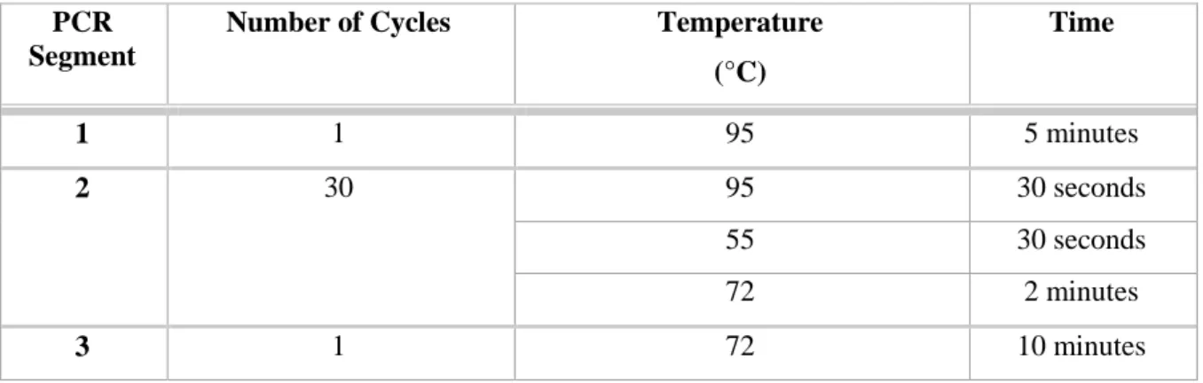
Table 3.2. PCR program used in the site directed mutagenesis reaction ... 38
Table 3.3. Primer used for DNA sequencing of αSyn coding gene sequence ... .39
Table 3.4. Primers used for amplification of αSyn coding gene sequence by PCR ... .45
Table 3.5. PCR program for auxotrophies presence confirmation and αSyn coding gene sequence amplification ... 46
Table 3.6. Primers used for the amplification of the yeast genomic auxotrophic markers ... 48
Table 8.1. List of yeast plasmids used in this study ... 104
Table 8.2. List of yeast strains used in this study ... .104
XIII
List of Figures
Figure 1.1. Schematic representation of PD pathology ... 3
Figure 1.2. Schematic representation of αSyn protein and PD familial linked mutations. ... 6
Figure 1.3. Schematic model of αSyn aggregation. ... 13
Figure 1.4. Schematic representation of the cellular events that control αSyn protein levels ... 13
Figure 1.5. Schematic representation of the established αSyn pathological mechanisms in the cell .... 15
Figure 1.6. Schematic representation of αSyn protein and its phosphorylation sites ... 16
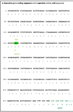
Figure 3.1. Schematic representation of the WT αSyn coding gene sequence (in black) fused to GFP (in green) ... 37
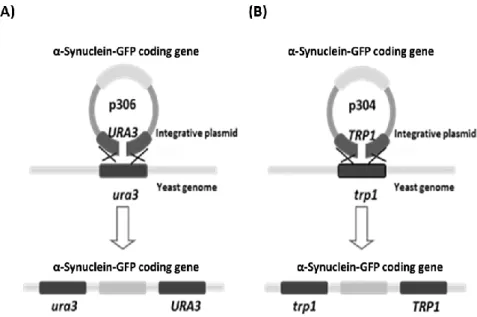
Figure 3.2. Schematic representation of yeast genomic integration of integrative plasmids that contain the protein coding gene of interest ... 45
Figure 3.3. Schematic representation of the sites of hybridization of the set of primers P15/P99 within the yeast genome transformed with the integrative plasmids (A) p306 (URA3 mark) or (B) p304 (TRP1 mark) ... 46
Figure 3.4. Amplification of the αSyn coding gene sequence ... 47
Figure 3.5. Schematic representation of the sites of hybridization of the set of primers within the yeast genome ... 47
Figure 3.6. Amplification of the auxotrophic markers ura3 and trp1 ... 48
Figure 3.7. Evaluation of yeast growth in SC media ... 49
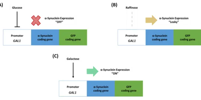
Figure 4.1. Schematic representation illustrating αSyn expression modulation in yeast cells by activity control of the GAL1 promoter ... 52
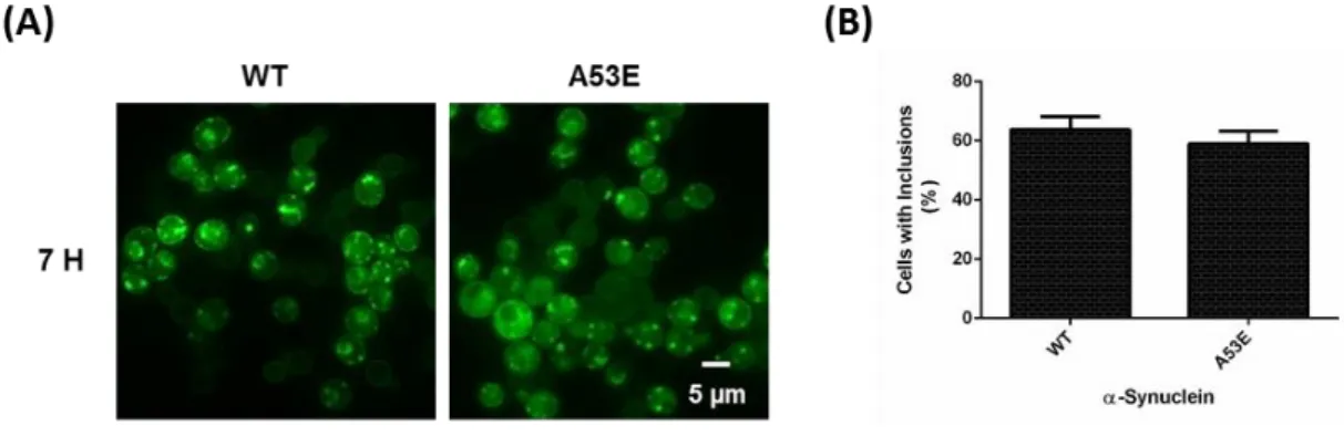
Figure 4.2. Inclusion formation evaluation for the A53E αSyn mutant expression in yeast cells ... .53
Figure 4.3. Spotting assay of the indicated yeast cells (Empty, WT and A53E) ... 53
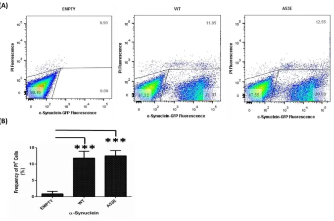
Figure 4.4. Toxicity assessment of the A53E αSyn mutant expression for yeast cells ... .54
Figure 4.5. Effect of the A53E mutation on total and phosphorylated at S129 αSyn protein levels ... 55
Figure 4.6. Evaluation of A53E mutation on αSyn solubility ... 56
Figure.4.7..Intracellular localization of the WT or A53E αSyn-GFP and mitochondrial network assessment by MitoTracker Deep Red staining ... 57
Figure 4.8. Evaluation of the effect of the A53E mutation on mitochondrial network integrity ... 58
Figure 4.9. Spotting assay of the indicated yeast cells containing two copies of the indicated αSyn forms (Empty, WT, S129A, S129D, S87E and S87E_S129D) integrated on the genome ... 59
Figure 4.10. Toxicity assessment of the αSyn phospho-mutations expression for yeast cells ... 61
Figure 4.11. Inclusion formation evaluation for the αSyn phospho-mutants expression in yeast cells in SC media ... 62
XIV
Figure 4.12. Inclusion formation evaluation for the αSyn phospho-mutants expression in yeast cells in
YEP media ... 64
Figure 4.13. Effect of the phospho-mutations on total and phosphorylated at S129 αSyn protein levels
... 65
Figure 4.14. Effect of the phospho-mutations on total and phosphorylated at S129 αSyn protein levels
during protein clearance ... 67
Figure 4.15. Effect of the phospho-mutations on uGFP levels, for proteasome function evaluation .... 68 Figure 4.16. Effect of the phospho-mutations on Atg8 levels, for autophagy function evaluation ... 70 Figure 4.17. Spotting assay of the yeast cells expressing either the WT or the S87E αSyn from a
multi-copy plasmid ... 71
Figure.4.18. Inclusion formation evaluation for the S87E αSyn mutant expression in yeast cells
harboring a multi-copy plasmid ... 72
Figure 4.19. Effect of the S87E mutation on total and phosphorylated at S129 αSyn protein levels .... 73 Figure 4.20. Inclusion formation evaluation for the S87E αSyn mutant expression in yeast cells ... 74 Figure 8.1. Identity confirmation of the multi-copy yeast plasmid encoding for A53E αSyn by DNA
sequencing ... 106
Figure 8.2. Identity confirmation of the phospho-mutant yeast strains encoding for S87E αSyn and
S87E_S129D αSyn ... 107
Figure 8.3. Validation of the results for the toxicity induced by the A53E αSyn mutant... 109 Figure 8.4. Validation of the results for toxicity assessment of the αSyn phospho-mutations expression
for yeast cells ... 110
Figure 8.5. Validation of the results for the effect of the phospho-mutations on uGFP levels, for
XV
List of Abbreviations
(E)GFP (Enhanced) Green fluorescent protein
˚C Degrees Celsius
αSyn α-Synuclein/Alpha-synuclein
µg Micrograms
µl Microliters
A Alanine
A30P Alanine replacement to proline at the amino acid position 30
A53E Alanine replacement to glutamic acid at the amino acid position 53
A53T Alanine replacement to threonine at the amino acid position 53
ALP Autophagy-lysosome pathway
APS Ammonium persulphate
Ar Argon
Atg Autophagy related protein
ATP Adenosine 5’ triphosphate
BCA Bicinchoninic acid assay
bp Base pairs
BSA Bovine serum albumin
C Cytosine
C. elegans Caenorhabditis elegans
Ca2+ Calcium
CaM Calmodulin
CCD Charged coupled device
cDNA Complementary deoxyribonucleic acid
CEDOC Chronic Diseases Research Center
CG Cytosine-Guanine Content
cHsc70 Cytosolic heat shock protein 70
CK1/2 Casein kinase 1/2
CMA Chaperone-mediated autophagy
CTSD Cathepsin D
Cu2+ Copper
D. melanogaster Drosophila melanogaster
DLB Dementia with Lewy bodies
DNA Deoxyribonucleic acid
E Glutamic acid
E. coli Escherichia coli
E46K Glutamic acid replacement to lysine at the amino acid position 46
ECL Enzyme linked chemiluminescence
ER Endoplasmic reticulum
FACS Fluorescence-activated cell sorting
FCUL Faculty of Sciences – University of Lisbon
G Guanine
G51D Glycine replacement to aspartic acid at the amino acid position 51
GAL(1) Galactose inducible promoter (1)
XVI
HSPs Heat shock proteins
JNK c-Jun N-terminal kinases
Kbp Kilo base pairs
kDa Kilo Daltons
LAMP2A Lysosome-associated membrane protein type 2A
LB Luria Broth
LBDs Dementias with Lewy bodies
LBs Lewy bodies
lHsc70 Lysosome-associated heat shock protein 70
LiAc Lithium acetate
LN Lewy neurites
M Molar
mA Miliampers
MAMs Mitochondria-associated endoplasmic reticulum membranes
MFI Mean fluorescence intensity
ml Milliliters
mM Milimolar
mTOR Mammalian target of rapamycin
mW Miliwatts
NAC Non-amyloid-β component
ng Nanograms
nm Nanometers
NMS-UNL Nova Medical School - New University of Lisbon
OD Optical density
OD600nm Optical density at 600 nm wavelength
ON Overnight
PAGE Sodium dodecyl sulfate polyacrylamide gel electrophoresis
PBS Protein buffer sample
PBS 1X Phosphate-buffered saline solution
PCR Polymerase chain reaction
PD Parkinson’s disease
PDD Parkinson’s disease with dementia
PEG Polyethylene glycol
PGK Phosphoglycerate kinase
PI Propidium iodide
PI+ Propidium iodide positive cells
PI+GFP+ Propidium iodide positive and GFP positive cells
PKCδ Protein kinase C δ
PLK2 Polo-like kinase 2
PP2A Protein phosphatase 2
pS129 Phosphorylated S129 residue
pS87 Phosphorylated S87 residue
PTMs Post-translational modifications
PUFA Polyunsaturated fatty acid
ROS Reactive Oxygen Species
Rpm Revolutions per minute
S129 Serine 129
XVII
S129D Serine replacement to aspartic acid at the amino acid position 129
S87 Serine 87
S87E Serine replacement to glutamic acid at the amino acid position 87
SC Selective synthetic minimum media
SDS Sodium dodecyl sulfate
SN Substantia Nigra (pars compacta)
TAE Tris-acetate-EDTA
TBS Tris-buffered saline solution
TCA Trichloroacetic acid
TE Tris-EDTA buffer
TEMED Tetramethylethylenediamine
TH Tyrosine hydroxylase
TI Insoluble protein fraction
Tm Temperature of melting
TOM20 Translocase of the outer membrane 20
TP Total protein fraction
TRP/TRP1/trp1 Tryptophan/Tryptophan prototrophy/Tryptophan auxotrophy
TS Soluble protein fraction
uGFP Unstable green fluorescent protein
UPRs Unfolded protein response
UPS Ubiquitin-proteasome system
URA/URA3/ura3 Uracil/Uracil prototrophy/Uracil auxotrophy
V Volume V Volt WT Wild-type Y125 Tyrosine 125 Y133 Tyrosine 133 Y136 Tyrosine 136
YEP Yeast extract peptone media
1
2
1.1. Parkinson’s disease and synucleinopathies
Parkinson’s disease (PD) is the second most common age-related neurodegenerative disorder, has an average of onset of 60 years old [Marques et al, 2011] and according to the Parkinson’s Disease Foundation it affects between 7 to 10 million people worldwide. The prevalence of PD in industrialized countries is approximately 1 to 2% of the population over 60 years of age, which increases to 3 to 5% in people above 85 years old [de Lau and Breteler, 2006], being the most affected men, in Europe, North America and Australia [Pringsheim et al, 2014].
This slow and progressive neurodegenerative disorder is clinically characterized by the following motor symptoms: slow and decreased movement (bradykinesia), rest tremor or rigidity, gait and postural instability [Weintraub et al, 2008]. These motor symptoms, detected only at the time of diagnosis, occur when patients already have undergone significant neurodegeneration (approximately 70% of nigral dopaminergic neurons are lost) [Chaudhuri et al, 2016], [Huang et al, 2004]. Furthermore, at later stages of the disease, PD is widespread to non-dopaminergic neurons and other non-motor symptoms arise, such as depression, dementia, psychosis, constipation, neurogenic bladder disturbance, fatigue, insomnia, apathy, anxiety and excessive daytime sleepiness [Todorova et al, 2014], [Chaudhuri et al, 2016].
The current treatment for this disease is palliative rather than curative and its effectiveness is still far from satisfactory, since it is based on the administration of L-DOPA, by which the non-motor symptoms are neglected [Chaudhuri et al, 2016], [Taschenberger et al, 2012]. Thus, tremendous efforts are underway to elucidate the causes underlying this disorder and to find a cure as well as a viable early-stage biomarker [Chaudhuri et al, 2016].
In the past decades, α-Synuclein (αSyn) protein has been the center of focus in understanding the etiology of a group of overlapping neurodegenerative disorders called Lewy body diseases (LBDs), in which PD, PD with dementia (PDD) and dementia with Lewy bodies (DLB) are included [Kim et al, 2014]. LBDs are mainly characterized pathologically by the progressive cytoplasmic accumulation of the protein αSyn in eosinophilic intracellular inclusions composed of amyloid-like fibers, known as Lewy bodies (LBs) or Lewy neurites (LNs) [Weintraub et al, 2008], [Lashuel et al, 2013]. Hence LBDs are also referred to as Synucleinopathies or α-Synucleinopathies [Lashuel et al, 2013]. These protein aggregates lead to degeneration of neocortical, limbic and nigrostriatal circuitries [Braak and Braak, 2000], [Vekrellis et al, 2011] and, in the case of PD, patients show loss of dopaminergic neurons in the substantia nigra (SN) pars compacta and the consequential attenuation of dopaminergic innervation to the striatum, with the surviving neurons bearing signs of Lewy pathology and exhibiting abrogated levels of neuromelanin pigmentation (Figure 1.1) [Simuni and Sethi, 2008].
3
Figure 1.1. Schematic representation of PD pathology. On the right, light microscopy of a neuronal cell containing LBs
(dark stain) [Lees et al, 2009]. On the left, brain localization of the neuronal cells primarily affected in PD, region of the SN. Adapted from [Wales et al, 2013].
The development of PD is described according to the Braak staging hypothesis that postulates that the pathology evolves in 6 stages [Braak et al, 2003a]. This staging suggests a pre-motor period in which the typical pathological changes, LNs and LBs, spread from the olfactory bulb and vagus nerve to the lower brainstem regions (stages 1 to 2), followed by a symptomatic period when pathological changes involve the midbrain, including the SN (stage 3), mesocortex (stage 4) and, eventually, the neocortex (stages 5 to 6) [Braak et al, 2003a]. Interestingly, morphological studies reveal a pattern of this hypothesis with αSyn pathology and LBs formation in the brains of patients with a clinical diagnosis of PD [Marques et al, 2011], [Braak et al, 2003a], [Braak et al, 2003b]. The study of post-mortem human brain tissue from PD patients suggests that αSyn progresses throughout the brain in a pattern that follows the progression of clinical symptoms stated by the Braak’s staging for PD, being now considered the first evidence of the hypothesis of the prion-like spreading of αSyn pathology [Lopes da Fonseca et al, 2015].
4
1.2. Etiology of PD
Although the cause of PD remains unknown [Marques et al, 2011], emerging evidence has provided support for the hypothesis that this disorder is the result of interactions between genetic abnormalities, environmental toxins, mitochondrial dysfunction and other cellular processes and for this reason, it is considered a complex disorder, since it includes, genetic, epigenetic and environmental causes [Bertram and Tanzi, 2005], [Feng et al, 2015]. Nevertheless, the identity of most of these factors, the nature of their interaction, and the molecular pathways of neurodegeneration that they initiate remain poorly understood [Pringsheim et al, 2014]. In addition, αSyn toxicity is related with both familial (rare) and non-familial/idiopathic/sporadic (common) forms of PD, evidencing the strong dichotomy between both types [Marques et al, 2011], [Lopes da Fonseca et al, 2015]. Therefore, investigating the genes and proteins involved in PD is pivotal, in terms of their normal physiological role and contribution to the disease and, hence, provide critical insights of the molecular basis underlying the pathogenesis, in order to develop strategies to treat and prevent the disease [Mbefo et al, 2015].
1.2.1. Sporadic PD
Sporadic/Idiopathic PD, the most frequent form of parkinsonism, accounting for 90 to 95% of the cases, usually refers to a syndrome characterized by late-onset, largely non-heritable parkinsonism [Marques et al, 2011]. Although, environmental toxins, physical activity, dietary habits and the presence of other diseases have been suggested to play an impact on PD risk [de Lau and Breteler, 2006], [Hindle, 2010], [Nussbaum, 2003], [Gasser, 2010], aging is still the major risk factor in the pathology development [Elbaz and Moisan, 2008]. Furthermore, polymorphisms in the promotor region of SNCA gene [Edwards et al, 2010], [Fuchs et al, 2008], [Maraganore et al, 2006], [Mizuta et al, 2006], [Satake et al, 2009] as well as an increased expression of the MAPT gene (encoding for Tau, the protein involved in Alzheimer’s disease) [Tobin et al, 2008], [Edwards et al, 2010] have also been identified as susceptibility factors for sporadic PD.
1.2.2. Familial PD
In 5 to 10% of cases, PD has mainly a genetic component, with both recessive and dominant modes of inheritance [Marques et al, 2011]. Although the genetic/familial cases represent a minority part of PD patients, the understanding of the molecular mechanisms underpinning the genetic forms of the disease has already provided insights into the pathogenesis of the sporadic forms, as well [Fujioka et al, 2014]. Several genes and chromosomal loci have been linked to familial forms of parkinsonism and are designated as PARK1 to PARK18 [Marques et al, 2011], [Klein and Westenberger, 2012]. These loci are associated with autosomal dominant, recessive and X-linked forms of the disease [Marques et al, 2011]. To date, four genes are identified as associated with autosomal dominant PD: SNCA, LRRK2, VPS35 and EIF4G1 [Sundal et al, 2012] (Table 1.1).
5
Table 1.1. Genes and chromosomal loci identified and linked to familial forms of PD, PARK1 to PARK18. Genes are
organized in three groups: associated with PD with conclusive evidence, with unknown relevance to PD and associated with atypical parkinsonism. Adapted from [Corti et al, 2011] and [Sundal et al, 2012].
Loci Gene Map Position Type of Inheritance/Onset
PD associated loci and genes with conclusive evidence
PARK1/ PARK4 SNCA 4q22 Dominant/Early onset
PARK8 LRRK2 12q12 Dominant (Sporadic)/Late onset
PARK2 PARKIN 6q25-q27 Recessive (Sporadic)/Early onset
PARK6 PINK1 1p35-p36 Recessive/Early onset
PARK7 DJ-1 1p36 Recessive/Early onset
PARK9 ATP13A2 1p36 Recessive/Early onset
PARK17 VPS35 16q11.2 Dominant/Late onset
PARK18 EIF4G1 3q27.1 Dominant/Late onset
PD associated loci and genes with unknown relevance
PARK3 Unknown 2p13 Dominant/Late onset
PARK5 UCHL1 4p14 Dominant/Late onset
PARK10 Unknown 1p32 Unclear/Late onset
PARK11 GIGYF2 2q36-q37 Dominant/Late onset
PARK12 Unknown Xq21-q25 Unclear/Late onset
PARK13 Omi/HTRA2 2p13 Unclear/Late onset
PARK16 Unknown 1q32 Unclear/Unclear
Loci and genes associated with atypical parkinsonism
PARK14 PLA2G6 22q12-q13 Recessive/Early onset
PARK15 FBX07 22q12-q13 Recessive/Early onset
SNCA (chromosome 4q22.1), the gene encoding for the αSyn protein, was the first to be associated with familial PD [Marques et al, 2011]. A mutational mechanism in this gene involves the duplication [Chartier-Harlin et al, 2004] or triplication [Singleton et al, 2003] of the wild-type (WT) SNCA gene locus (PARK4 PD-associated locus). Furthermore, six missense mutations in this gene, causing the A30P [Kruger et al, 2001], E46K [Zarranz et al, 2004], A53T [Polymeropoulos et al, 1997] (“old mutations” – identified between 1997 and 2004 [Petrucci et al, 2016]) and H50Q [Appel-Cresswell et al, 2013], [Proukakis et al, 2013], G51D [Kiely et al, 2013] and A53E [Pasanen et al, 2014] (“new mutations” – identified between 2012 and 2014 [Petrucci et al, 2016]) amino acid substitutions of αSyn (PARK1 PD-associated locus), were identified. All these PD-related mutations are localized in the N-terminal membrane binding part of αSyn [Emanuele and Chieregatti, 2015] and were found to confer a
6
gain of cytotoxic functional effect, implicating upregulated expression levels and qualitative alterations of αSyn in PD onset and progression [Singleton et al, 2003], [Ross et al, 2008], [Cookson, 2005] (Figure 1.2)
Figure 1.2. Schematic representation of αSyn protein and PD familial linked mutations. The six missense mutations
identified to be linked to PD A30P, E46K, H50Q, G51D and A53T/E are localized in the N-terminal domain of the protein. Adapted from [Taymans and Baekelandt, 2014].
These αSyn point mutations are considered rare, being only reported in a few isolated cases of multigenerational PD families [Berg et al, 2005], while duplications and triplications of the gene are more prevalent [Puschmann et al, 2013] and the phenotype is more severe for patients carrying triplications of the gene, when compared with patients carrying duplications, suggesting that αSyn pathology induces a dose-dependent toxicity [Singleton et al, 2003], [Keyser et al, 2010], [Fuchs et al, 2007], [Ikeuchi et al, 2008], [Ibanez et al, 2009]. Regarding the most recent PD associated mutation of αSyn, the amino acid replacement alanine (A) to glutamic acid (E) on the amino acid position 53 of the polypeptide chain of this protein, this mutation was identified in a Finnish family in 2014 and it is pathologically and clinically characterized by an equally severe, early-onset phenotype, but slowly progressive parkinsonism [Pasanen et al, 2014], when compared to the other five familial-linked mutations (Table 1.2, ordered from the most recent to the oldest identified mutations).
7
Table 1.2. Major pathological and clinical features of the PD familial linked αSyn mutations and SNCA gene duplications and triplications. Adapted from [Fujioka et al, 2014] and [Petrucci et al, 2016].
αSyn Mutation Year/ Country of Discovery Number of Cases Age of Onset (years) Mean Disease Duration (years) Clinical Phenotype A53E 2014/ Finland
3 20 to 40 24 Early onset, slowly progressive; Severe spasticity, myoclonus and psychiatric disturbances;
Cognitive impairment and autonomic dysfunction;
Pyramidal signs;
Mutation shares a common Finnish ancestral – Founder effect [Pasanen et al, 2017].
G51D
2013/ France
2 19 to 60 6 Early onset, rapidly progressive; Severe psychiatric disturbances, hallucinations and severe myoclonus; Cognitive impairment (not detected in early stages of the disease) and autonomic dysfunction; Severe pyramidal signs; Epilepsy. H50Q 2013/ England and Canada 3 60, 56 and 71
5 and 12 Late onset, rapidly progressive; Psychiatric disturbances; Severe cognitive impairment; Mutation shares a common English ancestral – Founder effect.
E46K
2004/ Spain
5 53 to 67 --- Late onset, very rapidly progressive; Psychiatric disturbances,
hallucination, REM sleep behavior disorder;
Cognitive impairment and autonomic dysfunction.
A30P
2001/ Germany
4 49 to 61 --- Late onset, rapidly progressive (sustained response to L-dopa therapy); Mild dementia; Cognitive impairment. A53T 1997/ Greece, Italy, United States, Sweden, Australia, Korea
70 35 to 59 4 to 12 Early onset, rapidly progressive; Psychiatric disturbances, dementia, dysautonomia, myoclonus,
hallucinations;
8 SNCA Copies 2004/ France, Italy, Japan, Sweden, Korea (Duplica-tions) 2003/ Sweden, Japan (Triplica-tions) 36 27 39 to 61 36 to 54 9 to 21 5 to 9
Early onset, rapidly progressive; Psychiatric disturbances
dysautonomia, myoclonus, hallucinations;
Cognitive impairment and autonomic dysfunction.
Early onset, very rapidly progressive; Severe psychiatric disturbances, dysautonomia, hallucinations, myoclonus;
Severe cognitive impairment and autonomic dysfunction.
Functional and phenotypical characterization performed for the six missense mutations have been performed using in vitro and in vivo model systems. Regarding the oldest identified mutations (A30P, E46K and A53T), these three PD-linked SNCA mutations (A30P, E46K and A53T) accelerate αSyn oligomerization, but only two of these (E46K and A53T) enhance fibrillation in vitro and in vivo [Conway et al, 1998], [El-Agnaf et al, 1998b], [Conway et al, 2000]. Although the A30P mutation has been shown to result in enhanced αSyn fibrillation in vivo, as illustrated by an autopsy case of a patient with this mutation that had extensive LB pathology [Seidel et al, 2010], in vitro it exhibits reduced fibrillation compared to the WT protein and other mutants [Lashuel et al, 2013]. The three of them increase the toxicity of the protein in human cell lines [Outeiro et al, 2006] and, with the exception for A30P, in yeast [Lázaro et al, 2014]. Furthermore, they have an impact on several key cell mechanisms, such as the inhibition of proteasome activity [Bertoncini et al, 2005], [Jiang et al, 2010] and the mutants A30P and E46K show impairment of membrane and vesicles binding activity [Albuquerque et al, 2009], [Hofer et al, 2004], [Jensen et al, 1998], [Karpinar et al, 2009]. Concerning the group of the most recent identified linked mutations of αSyn, including the A53E mutation, the state-of -the-art of phenotypical and functional characterization, using both in vitro and in vivo approaches, is detailed in Table 1.3.
Table 1.3. Key points and state of the art of the phenotypical and functional characterization of the mutants A53E, G51D and H50Q of αSyn, when compared to WT protein, using different study systems.
αSyn Mutation Study System Observations/Major Conclusions A53E Post-mortem human brain analysis
Inclusions with variable morphologies [Pasanen et al, 2014]; Mature inclusions up-phosphorylated at S129 [Pasanen et al, 2014].
Mammalian cell lines
Neuro2A cells:
Decreased propensity to aggregate into amyloid structures [Rutherford et al, 2015];
Formation of mature inclusions phosphorylated at S129 [Rutherford et al, 2015];
Increased cellular toxicity and dead under stress conditions (MPP+), as well as mitochondrial impairment [Rutherford et al, 2015].
9
Different from WT protein in the dimerization/oligomerization process [Lázaro et al, 2016];
Similar inclusion formation when compared to WT [Lázaro et al, 2016].
H4 cells:
Increased proteolytic activity of the proteasome [Lázaro et al, 2016]; Aggregation of A53E αSyn induces Golgi apparatus alterations [Lázaro et al, 2016].
Biophysical/ Biochemical
Characteri-zation
Accumulation of oligomers for a longer period of time [Ghosh et al, 2014];
Decreased formation of aggregates and amyloid structures [Ghosh et al, 2014];
Reduced membrane binding affinity (when compared to the WT protein and the A53T mutant) [Ghosh et al, 2014];
Similar structure of free and membrane-bound αSyn [Ghosh et al, 2014];
Slower fibril formation [Ghosh et al, 2014];
Formation of elongated amyloid fibrils morphologically similar to WT protein, but thinner [Rutherford et al, 2015];
Reduced αSyn amyloid formation [Lázaro et al, 2016].
G51D
Post-mortem
human brain analysis
αSyn inclusion formation [Kiely et al, 2013]; Up-phosphorylation at S129 [Kiely et al, 2013];
Partial co-localization with nuclear membrane markers [Fares et al, 2014]. Transgenic mouse brain of α- Synucleino-pathies Primary neurons:
Enriched in the nuclear compartment [Fares et al, 2014]; Up-phosphorylation at S129 [Fares et al, 2014];
Increased αSyn-induced mitochondrial fragmentation [Fares et al, 2014];
Greater membrane-induced aggregation and enhanced toxicity [Ysselstein et al, 2015].
Primary midbrain cells:
Decreased α-helical content in the presence of phospholipids [Ysselstein et al, 2015].
Mammalian cell lines
Neuro2A cells:
Decreased rate of amyloid formation [Rutherford et al, 2014];
Increased cellular toxicity under stress conditions (MPP+) [Rutherford et al, 2014];
SH-SY5Y cells:
Fibrils with increased toxicity [Lesage et al, 2013];
Increased activation of apoptosis related caspase-3 [Lesage et al, 2013]; Enriched in the nuclear compartment [Fares et al, 2014];
Up-phosphorylation at S129 [Fares et al, 2014];
Increased αSyn-induced mitochondrial fragmentation [Fares et al, 2014];
Greater membrane-induced aggregation and enhanced toxicity [Ysselstein et al, 2015];
Similar binding pattern to copper (Cu2+) as the WT, with enhanced rate of fibril formation in the presence of Cu2+ [Ranjan et al, 2016].
HEK cells:
Enhanced inclusion formation [Lázaro et al, 2014].
Yeast Impaired membrane association, forming few inclusions and non-toxic (similar results to A30P mutant) [Fares et al, 2014];
10
Enhanced inclusion formation compared to WT protein expression [Lázaro et al, 2014].
Biophysical/ Biochemical
Characteri-zation
Slower oligomerization rate [Lesage et al, 2013];
Decreased permeabilization of DOPG membranes [Stefanovic et al, 2015];
Similar fibrils and similar morphology of the mature inclusions [Rutherford et al, 2014];
Decreased kinetics of aggregation [Fares et al, 2014].
H50Q
Post-mortem
human brain analysis
Formation of αSyn inclusions [Appel-Cresswell et al, 2013], [Proukakis et al, 2013];
Up-phosphorylation at S129 [Appel-Cresswell et al, 2013], [Proukakis et al, 2013]. Transgenic mouse brain of α- Synucleino-pathies Primary neurons:
Increased mitochondrial fragmentation [Khalaf et al, 2014]; The presence of Cu2+ induces the formation of structurally different and less-damaging aggregates. These aggregates exhibit a stronger capacity to induce αSyn inclusion formation in recipient cells [Villar-Pique et al, 2016].
Mammalian cell lines
Neuro2A cells:
Increased rate of amyloid formation [Rutherford et al, 2014].
SH- SY5Y cells:
Increased kinetics of aggregation [Ghosh et al, 2013], [Khalaf et al, 2014];
Similar early oligomer forming tendency [Ghosh et al, 2013];
Similar subcellular localization and ability to be phosphorylated by PLK2 and GRK6 (at S129) and by PLK3 (at S129) and CK1 (at S87) [Khalaf et al, 2014];
Increased secretion [Khalaf et al, 2014];
Increased toxicity and exacerbation of neuronal toxicity of extracellular αSyn [Khalaf et al, 2014];
Promotion of cellular toxicity under stress conditions (H2O2) [Rutherford et al, 2014];
Changes in the ratio between the concentrations of the WT and H50Q αSyn are closely related to the alteration of its aggregation behavior [Porcari et al, 2015].
HEK cells:
The inclusion formation is similar in H50Q and WT αSyn [Lázaro et al, 2014].
H4 cells:
Increased kinetics of aggregation [Ghosh et al, 2013];
Presence of Cu2+ does not alter the rate of fibril formation [Ranjan et al, 2016].
BL21(DE3)-RIPL cells:
Increased kinetics of aggregation [Chi et al, 2014].
Yeast Inclusion formation is similar to WT protein [Lázaro et al, 2014].
Biophysical/ Biochemical
Characteri-zation
Similar structure of free and membrane-bound αSyn monomeric and oligomeric states [Khalaf et al, 2014], and similar fibril formation [Rutherford et al, 2014];
Similar morphology of the mature inclusions [Rutherford et al, 2014]; Similar permeabilization capacity of DOPG membranes [Stefanovic et al, 2015].
11
1.3. αSyn
1.3.1. Biology, structure and physiological function of αSyn
αSyn is abundantly expressed in the human brain, especially in the neocortex, hippocampus, SN, thalamus and cerebellum [McLean et al, 2000a], [Yu et al, 2007], making up as much as 1% of the total protein content [Kim et al, 2014]. It is a 140 amino acid protein [Ueda et al, 1993] encoded by the SNCA gene (chr.4q22.1) in humans [Spillantini et al, 1995]. Regarding its cell localization, αSyn was initially described to be predominantly in the presynaptic terminals of neurons [Jakes et al, 1994]. However, it was also reported to be present in the nuclear envelope [Maroteaux et al, 1988], within the nucleus [Gonçalves and Outeiro, 2013], [McLean et al, 2000b], [Mori et al, 2002] in the cytosol (interacting with cytoplasmic proteins) [Goers et al, 2003], [Kontopoulos et al, 2006], [Specht et al, 2005] and in mitochondria-associated endoplasmic reticulum (ER) membranes (MAM) [Hayashi et al, 2009]. Furthermore, this protein is able to bind to membranes [Auluck et al, 2010], preferentially to high-curvature, detergent-resistant membranes enriched in cholesterol, sphingolipids, and acidic phospholipids [Jensen et al, 2011], [Middleton and Rhoades,2010]. αSyn belongs to the Synucleins protein family, that includes β- and γ-Synuclein [Lavedan, 1998] and its predominant form is the full-length protein [Venda et al, 2006], [Emamzadeh et al, 2016], although other spliced variants have been identified [Beyer, 2006].
Concerning its structure, αSyn has three distinct structural domains (Figure 1.2): 1) an amphipathic lipid-binding alpha-helix N-terminal region (residues 1 to 60) [Emamzadeh et al, 2016], which is a highly conserved region, a KTKEGV hexameric motif important in the formation of α-helix structures (and in reducing its propensity to form β-structures), crucial in modulating its interactions with membranes [Emamzadeh et al, 2016], [Sode et al, 2007] 2) a central hydrophobic region (residues 61 to 95) denominated as NAC (non-amyloid-β component), that is involved in fibril formation and in αSyn aggregation [Högen et al, 2012], and 3) a random coil, highly acidic, proline rich carboxyl-terminal tail (residues 96 to 140), C-terminal region [Emamzadeh et al, 2016], which has been implicated in regulating its nuclear localization and its interaction with metals, small molecules and proteins by being homologous with small heat shock proteins (HSPs) [Emamzadeh et al, 2016], [Kim et al, 2004]. αSyn is present as a natively unfolded, monomeric protein in the cytosol [Kim et al, 2014], forming a double α-helix structure, when interacting with phospholipids [Emamzadeh et al, 2016], [Ahn et al, 2002], suggesting that this protein might adopt different structures under specific stress induced conditions or upon interaction with other proteins, specific ligands, lipids and/or biological membranes, playing specific roles in different cell locations based on its dynamic structure [Lashuel et al, 2013].
The physiological function of αSyn is still elusive [Guardia-Laguarta et al, 2014]. However, based on its localization and interactions, the activity of this protein is thought to be related to neuroprotection, namely by suppressing apoptosis (through downregulation of PKCδ expression) [Jin et al, 2011] and by acting as an antioxidant (by reducing cytochrome c oxidase and directly inhibiting JNK) [Zhu et al, 2006], [Latchoumycandane et al, 2005], [Hashimoto et al, 2002], in the neuronal synaptic integrity, function and plasticity, through chaperone activity (SNARE complex assembly maintenance of structure in presynaptic membranes and release of neurotransmitters) [Burré et al, 2013], [Burré et al, 2014], [Choi et al, 2013], [Park et al, 2002], [Witt et al, 2013], [Chandra et al, 2005] as well as in neurotransmission (through the modulation of pre-synaptic size and synaptic vesicle pool organization) [Vargas K. et al, 2017] and regulation of dopamine biosynthesis and levels (by interacting with protein
12
phosphatase 2A and consequently inhibiting tyrosine hydroxylase (TH)) [Peng et al, 2005], [Yang et al, 2013b]. On the overall cell homeostasis, αSyn acts in the regulation of neuronal differentiation (Rab3a activity) [Ostrerova et al, 1999], [Fu et al, 2000], [Chen et al, 2013] and glucose levels (increasing glucose uptake by G-protein-coupled-receptor activation) [Geng et al, 2011], [Sharma et al, 2015], [Rodriguez‑Araujo et al, 2015], [Rodriguez‑Araujo et al, 2013], in the modulation of CaM activity (by converting it into an activator) [Martinez et al, 2003] and maintenance of PUFA levels (for synthesis of brain vital fatty acids through Acyl-CoA synthetase) [Ruipérez et al, 2010].
1.3.2. Pathophysiological role in PD
The pathophysiological role of αSyn arises from the aggregation of this protein within the cell, which can be triggered by a variety of factors [Xu et al, 2015]. αSyn toxicity occurs via different mechanisms and involves different intermediates [Xu et al, 2015]. The currently acceptable model for the aggregation of this protein in PD describes a starting event of monomeric αSyn misfolding, caused by a cellular failure to degrade this first specie, by the two major clearance pathways [Wales et al, 2013] the autophagy-lysosome pathway (ALP) [Cuervo et al, 2004], [Spencer et al, 2009], [Ebrahimi-Fakhari et al, 2011], [Poehler et al, 2014] or the ubiquitin-proteasome system (UPS) [Webb et al, 2003], [Ebrahimi-Fakhari et al, 2011], as further detailed below (Subtopic 1.5). The initial misfolded monomeric protein is then more prone to interact with other αSyn proteins due to alterations in its conformation/structure, which generates unstable dimers and oligomers [Wales et al, 2013]. Oligomers may associate with monomers and form a range of intermediary species, including insoluble amyloid fibrils, in a nucleation-dependent manner [Wales et al, 2013], [Wood et al, 1999]. The accumulation of amyloid fibrils leads to the formation of LBs, one of the primary hallmarks of Synucleinopathies (Figure 1.3) [Spillantini et al, 1997], [Lee and Trojanowski, 2006]. However, which of these species plays a major role on pathological conditions, as well as the precise mechanism for cellular toxicity and neurodegeneration are still elusive [Wales et al, 2013]. Numerous experiments have suggested that intermediates of αSyn, such as the oligomers and amyloid fibrils, are neurotoxic, while the ultimately mature fibrils formed are relatively less toxic [Sharon et al, 2003], [Chen et al, 2009].
13
Figure 1.3. Schematic model of αSyn aggregation. Misfolded monomeric αSyn favors homo-interactions forming dimers and
oligomers. These unstable species are able to associate with more monomers, forming amyloid fibrils in a nucleation-dependent manner and the accumulation of this last intermediary specie leads to formation of LBs. Nevertheless, which of these species is responsible for cellular toxicity that leads to cell death and neurodegeneration is still unknown. Adapted from [Wales et al, 2013].
LBs are mainly caused by a disequilibrium in αSyn protein metabolism [Kragh et al, 2012]. An imbalanced in homeostasis may cause a dysfunction on one or more degradation pathways or increased protein synthesis [Xu et al, 2015], [Kragh et al, 2012], which can result in abnormal levels of αSyn that might favor the formation and/or accumulation of oligomeric and fibrillary species, which represent toxicity to the cell and, consequently, neuronal death (Figure 1.4) [Lashuel et al, 2013].
Figure 1.4. Schematic representation of the cellular events that control αSyn protein levels. Intracellular αSyn levels are
tightly regulated by the balance between the rates of its synthesis, clearance and aggregation. Abnormalities increasing αSyn synthesis (SNCA multiplications and polymorphisms (such as Rep1)), a failure to degrade αSyn (due to an impairment of proteasome, proteases, autophagy and chaperones) or αSyn modifications that promote its aggregation (SNCA mutations, post‑translational modifications, oxidative stress, toxins and interaction with oxidized dopamine) are responsible for αSyn accumulation, which can be transmitted to other neuronal cells, causing toxicity and leading to neurodegeneration [Lashuel et al, 2013].
14
Furthermore, it is postulated that αSyn oligomerization within specific cellular compartments may alter the distribution of functional forms of monomeric αSyn or result in monomer sequestration into non‑functional oligomeric forms, thus resulting in partial loss of function of this protein [Colla et al, 2012]. There is an additional second possibility that the native or misfolded forms of the monomeric protein may contribute to αSyn toxicity and PD pathogenesis via aggregation‑independent mechanisms [Lashuel et al, 2013]. Furthermore, post-translational modifications (PTMs), such as phosphorylation, are hypothesized to influence aggregation events [Wales et al, 2013], as further detailed in Subtopic 1.4.
Regarding the effect of the tridimensional structure of this protein, it is hypothesized that an interaction between the C-terminal domain and the NAC region of αSyn is essential to inhibit its aggregation [Emamzadeh et al, 2016]. Moreover, the tendency to form α-helix secondary structures, supposedly acquired upon interaction with its ligands [Lashuel et al, 2013], is inversely correlated with its aggregation tendency [Marsh et al, 2006].
This αSyn accumulation is a factor of toxicity for the overall cell homeostasis: at the nucleus level, αSyn is able to bind to DNA or transcription factors and promote transcription deregulation [Gupta et al, 2006], [Hegde et al, 2010], [Hegde et al, 2003], [Vasudevaraju et al, 2012]; at the cytoplasmic level, αSyn alters ER homeostasis, leading to an imbalance of intracellular calcium (Ca2+) that will promote changes at mitochondrial level [Ryu et al, 2002], [Lindholm et al, 2002], [Lim et al, 2009], [Lee et al, 2010]. This protein is also able to affect mitochondria directly, by causing a Complex I dysfunction and alter mitochondrial dynamics by promoting mitochondrial fragmentation [Stichel et al, 2007], [Nakamura et al, 2011], [Kamp et al, 2010] as further detailed in Subtopic 1.5. Subsequently, an impairment at the mitochondrial level will promote even more cell and DNA damage [Padmaraju et al, 2011] by producing reactive oxygen species (ROS), causing oxidative stress and inducing apoptotic pathways [Doyle et al, 2011], through activation of caspases [Sugeno et al, 2008]. Furthermore, an increased content of ROS may lead to even more misfolded protein conformations, forming a positive feedback of cell degeneration [Wales et al, 2013]. αSyn protein may also be secreted into the extracellular space, which possibly leads to the disease transmission to other neurons, as already demonstrated by several in vitro and in vivo studies [Hansen et al, 2011], [Danzer et al, 2012], [Luk et al, 2012], [Desplats et al, 2009]. All these events, culminate in cell death and consequently neurodegeneration (schematically represented in Figure 1.5) [Wales et al, 2013].
15
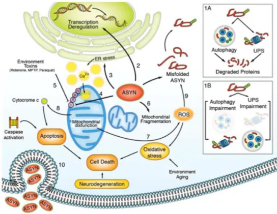
Figure 1.5. Schematic representation of the established αSyn pathological mechanisms in the cell. (1A) Autophagy and
UPS are essential to degrade misfolded protein and maintain cell homeostasis; (1B) when aberrant αSyn accumulates due to a failure of these mechanisms, several organelles are affected, and αSyn will cause (2) transcription deregulation, (3) ER stress, (4) mitochondrial stress, that can also be exacerbated by (5) environmental toxins and (6) mitochondrial fragmentation, which will culminate in the (7) production of ROS and (8) apoptotic activation and, consequently (9) more misfolded proteins. All these events lead to cell death and consequently neurodegeneration [Wales et al, 2013].
16
1.4. PTMs of αSyn
αSyn is prone to several types of PTMs, namely the already identified: ubiquitination [Shimura et al, 2001], C-terminal truncation [Li et al, 2005] and phosphorylation, which are the major disease-associated modifications [Wales et al, 2013], but also SUMO modification (sumoylation) [Dorval and Fraser, 2006], N-terminal acetylation [Anderson et al, 2006], nitration on tyrosine residues [Giasson et al, 2000] and glycosylation [Dorval and Fraser, 2006]. These PTMs are able to alter the conformation and/or biological function of proteins as well as the impact of the protein on a number of pathological processes, such as protein folding and aggregation, LBs formation and neurotoxicity, thereby playing a critical role in PD [Tenreiro et al, 2014b].
1.4.1. Phosphorylation of αSyn
A total of four serine, four tyrosine and ten threonine residues are putative sites of phosphorylation in the αSyn sequence [Tenreiro et al, 2014b]. However, it is suggested that αSyn might be predominantly unphosphorylated under physiological conditions [Okochi et al, 2000], [Fujiwara et al, 2002]. For this reason, it was hypothesized that changes in αSyn phosphorylation could represent a response to biochemical events associated with PD pathogenesis [Tenreiro et al, 2014b]. Phosphorylation is the most common PTM of αSyn and occurs predominantly at serine residues 129 (S129) [Fujiwara et al, 2002] and, to a lesser extent, 87 (S87) [Paleologou et al, 2010] and at tyrosine residues 125, 133 and 136 (Y125, Y133 and Y136, respectively) [Okochi et al, 2000], [Nakamura et al, 2001]. Phosphorylation at the tyrosine residues have been inversely correlated with aggregation and S129 phosphorylation related toxicity, indicating a probable posttranslational cross-talk amongst several sites [Bill et al, 1989], however no correlation was established between increased levels of phosphorylation in these residues and the pathological condition [Chen et al, 2009], [Mahul-Mellier et al, 2014].
Except for the S87 residue, that it is localized in the NAC region of αSyn [Paleologou et al, 2010], these phosphorylation sites are all localized in the C-terminal of the protein and highly conserved across species [Oueslati et al, 2010] (Figure 1.6).
Figure 1.6. Schematic representation of αSyn protein and its phosphorylation sites. The two phosphorytable serines S87
and S129 are localized in the NAC and C-terminal domain of the protein, respectively, while the tyrosine residues Y125, Y133 and Y136 are localized in the C-terminal. S129 residue, the most common phosphorylated residue in LBs (90% of αSyn is phosphorylated, compared to the 4% under physiological conditions) is represented in red. Adapted from [Taymans and Baekelandt, 2014].
Phosphorylation can majorly affect protein conformation, function and fate in many different ways: phosphorylation may be required for proper protein folding, induce conformational changes that can result in lower or higher catalytic activity of the phosphorylated protein, may precede or function as a recognition signal for further modifications, such as ubiquitination, it is able to alter the subcellular
![Figure 1.1. Schematic representation of PD pathology. On the right, light microscopy of a neuronal cell containing LBs (dark stain) [Lees et al, 2009]](https://thumb-eu.123doks.com/thumbv2/123dok_br/18180498.874464/21.892.247.674.99.461/figure-schematic-representation-pathology-right-microscopy-neuronal-containing.webp)