Faculdade de Medicina de Lisboa
B cells at the crossroad of immune responses:
insights from primary B-cell immunodeficiencies
Rita R. Barbosa
Doutoramento em Ciências Biomédicas Especialidade de Imunologia
Faculdade de Medicina de Lisboa
B cells at the crossroad of immune responses:
insights from primary B-cell immunodeficiencies
Rita R. Barbosa
Tese orientada pela Professora Doutora Ana E. Sousa
Doutoramento em Ciências Biomédicas Especialidade de Imunologia
2012
Todas as afirmações efectuadas no presente documento são da exclusiva responsabilidade do seu autor, não cabendo qualquer responsabilidade à Faculdade de Medicina de Lisboa pelos conteúdos nele apresentados.
A impressão desta dissertação foi
aprovada pela Comissão Coordenadora
do Conselho Científico da Faculdade de
Medicina de Lisboa, em reunião de 25
de Setembro de 2012.
Dissertação apresentada à Faculdade de
Medicina da Universidade de Lisboa para
obtenção do grau de Doutor em Ciências
Biomédicas.
A presente dissertação foi realizada na Unidade de Imunologia Clínica do Instituto de Medicina Molecular, Faculdade de Medicina da Universidade de Lisboa.
O trabalho aqui apresentado foi co-financiado pelo POCI 2010 e o FSE.
Bolsa de Doutoramento da Fundação para a Ciência e Tecnologia.
“Para ser grande, sê inteiro: nada Teu exagera ou exclui. Sê todo em cada coisa. Põe quanto és
No mínimo que fazes. Assim em cada lago a lua toda
Brilha, porque alta vive.”
ACKNOWLEDGEMENTS ... i
ABBREVIATIONS ... iii
SUMMARY ... v
SUMÁRIO ... vii
CHAPTER1–INTRODUCTION ... 1
PARTI.B CELLS AT THE CROSSROAD OF IMMUNE RESPONSES ... 3
I.a. Ontogeny, Development and Differentiation of B lymphocytes ... 3
I.b. B cells as a part of the innate immune system ... 9
I.c. The relevance of T-B cell interactions for immune responses ... 10
I.c.i. Organization of germinal centres ... 10
I.c.ii. Co-stimulatory molecules ... 12
I.c.iii. Soluble factors ... 13
PARTII.PRIMARY B-CELL IMMUNODEFICIENCIES ... 15
II.a. Historical perspective, clinical manifestations, aetiology and treatment ... 15
II.b. B-cell disturbances in Common Variable Immunodeficiency ... 24
II.c. How are T cells affected in B-cell immunodeficiencies ... 29
II.d. Other immune compartments affected in B-cell immunodeficiencies ... 32
REFERENCES ... 36
CHAPTER2–AIM AND WORKPLAN ... 61
CHAPTER3–RESULTS ... 69
3.1.MODULATION OF BAFF-R AND TACI EXPRESSION IN COMMON VARIABLE IMMUNODEFICIENCY ... 71
Abstract ... 73
Introduction ... 74
Materials and Methods ... 76
Results ... 79
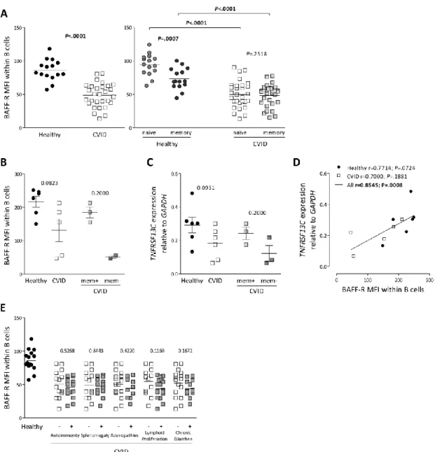
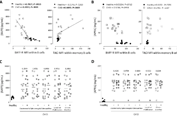
Reduced ex vivo BAFF-R expression and increased TACI expression were associated with high serum levels of BAFF in CVID patients ... 79
BAFF-R protein expression was modulated upon culture in the presence of BAFF ... 82
BAFF-R expression and BAFF serum levels did not change significantly upon starting IgG replacement therapy in CVID patients ... 85
Discussion ... 87
Supplemental Data ... 90
References ... 96
3.2.RELATIONSHIP BETWEEN B- AND T-CELL IMBALANCES IN PRIMARY B-CELL IMMUNODEFICIENCIES ... 101
Abstract ... 103
Decreased frequency of TH17 cells in individuals without B cells ... 111
Direct correlation between the frequency of circulating TH17 cells and switched-memory B cells in healthy individuals ... 112
Discussion ... 115
Supplemental Data ... 119
References ... 121
Supplement A. Evaluation of the frequency of TH17 cells in patients with Selective IgA Deficiency ... 125
Supplement B. Differentiation of TH17 cells from naïve CD4 T cells from Congenital Agammaglobulinemia patients ... 129
Supplement C. Regulatory T cells in Common Variable Immunodeficiency ... 135
3.3.MONOCYTE IMBALANCES IN PRIMARY B-CELL IMMUNODEFICIENCIES AND THEIR POSSIBLE RELATIONSHIP WITH INCREASED MICROBIAL TRANSLOCATION AND CHRONIC IMMUNE ACTIVATION IN COMMON VARIABLE IMMUNODEFICIENCY ... 151
Abstract ... 153
Introduction ... 154
Materials and Methods ... 156
Results ... 159
CVID was associated with monocyte activation ... 159
Monocyte activation was unrelated to plasma LPS levels in CVID ... 161
Monocyte activation was directly associated with T-cell disturbances in CVID patients ... 162
CVID patients grouped according to the EUROclass classification featured distinct monocyte imbalances ... 164
Discussion ... 166
References ... 169
Supplement A. Longitudinal study of CVID patients prior to and after starting IgG replacement therapy ... 173
CHAPTER4–CONCLUSIONS AND FUTURE PERSPECTIVES ... 179
CHAPTER1–INTRODUCTION
Figure 1. B-cell development. 4
Figure 2. Peripheral B-cell differentiation. 6
Figure 3. The 3-signal model for naïve B-cell activation. 7
Figure 4. Simplified schematic representation of BAFF-R-mediated signalling events. 8
Figure 5. Germinal centre. 11
Figure 6. The EUROclass classification scheme for Common Variable Immunodeficiency
patients. 25
Figure 7. Model of pathophysiological background of five different B-cell patterns in Common
Variable Immunodeficiency patients, based on proliferation history and somatic
hypermutation levels. 26
CHAPTER3.1–MODULATION OF BAFF-R AND TACI EXPRESSION IN COMMON VARIABLE
IMMUNODEFICIENCY
Figure 1. B-cell expression of BAFF-R was decreased in CVID patients, irrespective of clinical
phenotype. 80
Figure 2. TACI expression was increased in CVID, particularly in patients with splenomegaly. 81
Figure 3. High serum levels of BAFF were associated with reduced BAFF-R expression and
increased TACI in CVID. 81
Figure 4. BAFF-R protein expression was modulated upon culture in the presence of BAFF. 83
Figure 5. Autologous serum from CVID patients was able to recapitulate the effects of
recombinant BAFF in culture. 84
Figure 6. BAFF-R expression and BAFF serum levels did not significantly change upon starting
IgG replacement therapy. 85
Supplemental Figure 1. BAFF-R expression within peripheral blood populations. 90
Supplemental Figure 2. B-cell populations in CVID patients before and up to 12 months after
starting IgG replacement therapy. 91
Supplemental Figure 3. Down-regulation of BAFF-R expression upon BAFF binding appeared to
occur independently of receptor occupancy. 92
CHAPTER3.2–RELATIONSHIP BETWEEN B- AND T-CELL IMBALANCES IN PRIMARY B-CELL
IMMUNODEFICIENCIES
Figure 1. TH17 cells in patients with CVID. 109
Figure 2. Decreased frequency of TH17 cells in individuals lacking B cells. 111
Figure 3. Direct correlation between the frequencies of circulating TH17 cells and switched-
memory B cells in healthy individuals. 112
Figure 4. Negative correlation between the frequency of TH17 cells and serum BAFF levels in
healthy subjects. 113
Supplemental Figure 1. B-cell disturbances in CVID patients. 119
Supplemental Figure 2. T-cell disturbances in CVID and Congenital Agammaglobulinemia
Figure 1. TH17 differentiation in naïve CD4 T cells from Congenital Agammaglobulinemia
patients. 130
Supplement C
Figure 1. CVID was associated with decreased CD25 expression, while FOXP3 expression
was maintained. 136
Figure 2. Regulatory T cells in relation to naïve and memory T-cell subsets. 137
Figure 3. Expression of the ectoenzyme CD39 in regulatory T cells. 139
Figure 4. Relationship between regulatory T-cell subsets and T-cell activation in CVID. 140
Figure 5. Relationship between naïve and memory regulatory T-cell subsets and T-cell
activation in CVID. 141
Figure 6. The frequency of regulatory T cells was directly associated with the frequency
of naïve T cells in CVID. 141
Figure 7. Direct association between the frequency of IL-17-producing CD4 T cells and
regulatory T cells in CVID. 142
Figure 8. Expansion of T-cell populations producing pro-inflammatory cytokines was
directly associated with loss of regulatory T cells in CVID. 143
Figure 9. Association of regulatory T-cell imbalances with clinical manifestations in CVID
patients. 144
Figure 10. Expansion of the CD21lowCD38low B-cell population correlated with loss of
regulatory T cells in CVID. 146
CHAPTER3.3–MONOCYTE IMBALANCES IN PRIMARY B-CELL IMMUNODEFICIENCIES AND THEIR POSSIBLE RELATIONSHIP WITH INCREASED MICROBIAL TRANSLOCATION AND CHRONIC IMMUNE ACTIVATION IN
COMMON VARIABLE IMMUNODEFICIENCY
Figure 1. Monocyte activation markers in CVID and Congenital Agammaglobulinemia. 159
Figure 2. Plasma LPS levels and related molecules in CVID. 161
Figure 3. Relationship between markers of monocyte and T-cell activation in CVID. 163
Figure 4. Markers of monocyte activation in CVID according to the EUROclass classification. 164
Supplement A
Figure 1. Monocyte populations and HLA-DR expression on monocytes in CVID patients,
before and up to 12 months after starting IgG replacement therapy. 174
Figure 2. T-cell populations in CVID patients, before and up to 12 months after starting
IgG replacement therapy. 176
CHAPTER4–CONCLUSIONS AND FUTURE PERSPECTIVES
Figure 1. Simplified schematic representation of the disturbances of the immune system
CHAPTER1–INTRODUCTION
Table 1. Genetic defects underlying Congenital Agammaglobulinemia. 17
Table 2. Known genetic defects underlying Common Variable Immunodeficiency. 22
CHAPTER3.1–MODULATION OF BAFF-R AND TACI EXPRESSION IN COMMON VARIABLE
IMMUNODEFICIENCY
Supplemental Table 1. Analysis of sequence variants in the BAFF-R gene in the cohort of CVID
patients studied. 93
Supplemental Table 2. Analysis of sequence variants in the TACI gene in the cohort of CVID
patients studied. 94
CHAPTER3.2–RELATIONSHIP BETWEEN B- AND T-CELL IMBALANCES IN PRIMARY B-CELL IMMUNODEFICIENCIES
Table 1. Clinical and epidemiological data of the cohorts studied. 107
CHAPTER3.3–MONOCYTE IMBALANCES IN PRIMARY B-CELL IMMUNODEFICIENCIES AND THEIR POSSIBLE RELATIONSHIP WITH INCREASED MICROBIAL TRANSLOCATION AND CHRONIC IMMUNE ACTIVATION IN COMMON
VARIABLE IMMUNODEFICIENCY
A
CKNOWLEDGEMENTS
Quero começar por agradecer, antes de mais, à minha orientadora, Professora Doutora Ana Espada de Sousa, por todo o tempo dedicado à minha formação durante estes anos. Quero agradecer-lhe por ter investido em mim e me ter permitido fazer doutoramento no seu laboratório, mas principalmente por ter aberto os meus horizontes em relação à Ciência. Deixo igualmente uma palavra de apreço ao Professor Doutor Rui Victorino.
De seguida, não posso deixar de agradecer às pessoas que estiveram directamente envolvidas comigo neste trabalho, nomeadamente Susana Lopes da Silva, Sara Silva, Alcinda Campos Melo e Maria da Conceição Pereira Santos, pessoas sem as quais este não seria possível e que em muito contribuíram para o bom andamento do trabalho. Um grande e especial “muito obrigada”.
Neste sentido, quero também agradecer a todos os doentes e controlos saudáveis que foram incluídos neste trabalho. Sem o seu contributo, não teríamos aqui chegado.
Igualmente importante foi a ajuda que fui tendo ao longo dos anos, por parte de pessoas que, não estando directamente envolvidas no meu trabalho, me ofereceram toda a sua disponibilidade e dedicação. Quero, neste sentido, agradecer à Paula Matoso, Adriana Albuquerque, Ana Isabel Pinheiro, Vânia Silva e Rita Tendeiro, que em momentos de necessidade me deram a contribuição que permitiu que chegasse a bom porto.
Aos restantos colegas da Unidade de Imunologia Clínica, presentes e passados, quero agradecer pela boa disposição e camaradagem, por tornarem a vida no laboratório um pouco mais “familiar”.
Um agradecimento especial tem que ser feito à Íris Caramalho. Pelas longas conversas no meio do trânsito e a aturar vizinhos mal-dispostos, pela preocupação incondicional, por acreditar nas minhas capacidades “científicas” e me fazer desafiá-las para poder ir mais além.
Por um apoio que eu nunca poderia sequer pedir, e por me fazer acreditar que um futuro na Ciência é possível. Muito obrigada, miúda.
Quero também agradecer às pessoas com quem trabalhei no passado, Naomi Taylor, Filipe Santos Silva e Alexandre do Carmo, por terem, também eles, apostado na minha formação e por me terem permitido atingir uma maior maturidade para enfrentar um doutoramento.
Uma palavra para os membros do meu Comité de Tese, Bruno Silva Santos, Henrique Veiga Fernandes e Margarida Correia Neves, pelas discussões proveitosas e críticas pertinentes.
Muito importante neste meu caminho foi a presença de pessoas que, de meras caras anónimas no corredor, se tornaram grandes amigas. Raquel Lourenço, foste uma agradável surpresa. Muito obrigada por todos os gostos partilhados.
Aos meus amigos de sempre, João Manuel, Rita e João Luís, Carine, e Marta, não há propriamente palavras para vos agradecer o facto de fazerem parte da minha vida. Cada um à sua maneira, vocês fazem-me ser mais (e melhor) eu.
Embora o passado não seja o presente, com certeza ajudou a fazê-lo. Rui, agradeço-te a nossa história, porque também ela me conduziu ao dia de hoje.
Quero agradecer às minhas primas lisboetas, Carolina e Cristina, por manterem a minha sanidade mental e cultural. Um grande, grande “muito obrigada” a toda a minha família, por ser, sem dúvida, o meu pilar emocional. Ao meu cunhado, Rui, por ser o irmão mais velho que não tive. À minha sobrinha, Carlota, por tornar os meus dias incomparavelmente mais felizes.
Aos meus pais, Maria de Lurdes e António, e à minha irmã, Sara, tenho que agradecer infinitamente, pois são eles os grandes responsáveis por ter chegado até aqui, por ser a pessoa que sou. Obrigada por me aturarem há já trinta anos, o que é uma tarefa gigantesca (e eu sei bem do que estou a falar!).
Por último, um agradecimento um tanto “imaterial” a Cinfães, por ser a terra onde orgulhosamente cresci, o que em muito abriu os meus horizontes de vida.
A
BBREVIATIONS
ADAM – A Disintegrin and Metalloproteinase AHA – Autoimmune Haemolytic Anaemia AID – Activation-induced Cytidine Deaminase AIDS – Acquired Immunodeficiency Syndrome APC – Antigen-presenting cell
APRIL – A Proliferation Ligand
BAFF – B-cell Activating Factor of the TNF family BAFF-R – BAFF Receptor
Bcl-6 – B-cell Lymphoma 6 BCR – B-cell Receptor
BCMA – B-cell Maturation Antigen
Blimp-1 – B Lymphocyte-induced Maturation
Protein-1
BLNK – B cell Linker Protein Btk – Bruton’s Tyrosine Kinase CSR – Class-switch Recombination
CVID – Common Variable Immunodeficiency DC – Dendritic cell
ELISA – Enzyme Linked Immunosorbent Assay EndoCAb – Endotoxin Core Antibodies FcRH4 – Fc Receptor Homologue 4 FDC – Follicular Dendritic cell FOXP3 – Forkhead Box P3 GC – Germinal Centre HIGM – Hyper IgM
HIV – Human Immunodeficiency Virus HLA – Human Leukocyte Antigen
HLA-DR – Human Leukocyte Antigen D-related HSC – Hematopoietic Stem cell
HSCT – Hematopoietic Stem Cell Transplantation IBD – Inflammatory Bowel Disease
ICOS – Inducible Co-stimulator
ICOSL – Inducible Co-stimulator Ligand IFN – Interferon
Ig – Immunoglobulin IgAD – IgA Deficiency IL – Interleukin
iNKT – Invariant Natural Killer T cell IRAK4 – Interleukin-1 Receptor-associated
Kinase 4
ITP – Autoimmune Thrombocytopenia Purpura IVIg – Intravenous Immunoglobulin Therapy LBP – Lipopolysaccharide-binding Protein LPS – Lipopolysaccharide
mAb – Monoclonal Antibody
Mcl-1 – Myeloid Cell Leukemia Sequence 1 mDC – Myeloid Dendritic cell
mTORC1 – Mammalian Target of Rapamycin
Complex 1
MFI – Mean Fluorescence Intensity MHC – Major Histocompatibility Complex mRNA – Messenger RNA
MyD88 – Myeloid Differentiation Factor 88 MZ – Marginal Zone of the spleen
NF-B – Nuclear Factor B
NIK – NF-B-inducing kinase
NK – Natural Killer
PBL – Peripheral Blood Lymphocyte PBMC – Peripheral Blood Mononuclear Cell PC – Plasma Cell
pDC – Plasmacytoid Dendritic cell PD-1 – Programmed Death-1 molecule PD-L1 – PD-1 Ligand 1
PD-L2 – PD-1 Ligand 2
PID – Primary Immunodeficiency Disease PI3K – Phosphoinositide 3-kinase PMA – Phorbol Myristate Acetate RTE – Recent Thymic Emigrant
SAC – Staphylococcus aureus Cowan I SAP – Signaling Lymphocytic Activation
Molecule-associated Protein
sCD14 – Soluble CD14
SCID – Severe Combined Immunodeficiency SHM – Somatic Hypermutation
sIgAD – Selective IgA Deficiency SLE – Systemic Lupus Erythematosus
TACI – Transmembrane Activator and
Calcium-modulating Cyclophilin Ligand Interactor
TCR – T-cell Receptor
TFH – Follicular Helper CD4 T cell
TGF- - Tumour Growth Factor
TH1 – Helper CD4 T cell Type 1
TH2 – Helper CD4 T cell Type 2
TH17 – Helper CD4 T cell Type 17
TLR – Toll-like Receptor TNF – Tumour Necrosis Factor
TRAF – TNF-receptor associated factor TREC – T-cell Receptor Excision Circle Treg – Regulatory T cell
UNG – Uracil-DNA Glycosylase XLA – X-linked Agammaglobulinemia
S
UMMARY
B lymphocytes are important players in adaptive immunity and the main targets of current vaccination strategies, with the interaction between B and T cells being fundamental to generate long-term immunity. B cells also play a key role in linking innate and adaptive immunity by expressing receptors that recognize both specific antigens and microbial patterns.
The general aim of this work was to investigate the interplay between B cells and other components of the immune system through the study of B-cell immunodeficiencies, namely Common Variable Immunodeficiency (CVID), characterized by impaired antibody production due to defective mature cell differentiation, and Congenital Agammaglobulinemia, where early B-cell development is abrogated, commonly resulting in the absence of peripheral B B-cells.
We found BAFF-R expression to be reduced in CVID, particularly in patients with low memory B cells, and associated with high serum levels of BAFF, while TACI expression was significantly increased. BAFF induced BAFF-R down-regulation in vitro, both in healthy individuals and CVID patients. However, the degree of modulation in CVID was impaired, suggesting that these dynamics are affected, with a possible impact in B-cell homeostasis.
We also observed that CVID was associated with monocyte activation, irrespective of LPS levels, but in direct association with T-cell activation and B-cell imbalances.
To explore the mechanisms underlying chronic immune activation, we studied the role of IL-17, a major pro-inflammatory cytokine implicated in autoimmunity and inflammatory conditions, frequently found in CVID. However, no increase in TH17 cells was found and their
frequency was inversely correlated with markers of germinal centre impairment. TH17 cells were
severely reduced in Congenital Agammaglobulinemia and directly associated with switched-memory B cells in healthy subjects. Our data support a link between B-cell differentiation and TH17 homeostasis, with implications for the understanding of the pathogenesis of
inflammatory/autoimmune diseases and the physiology of B-cell depleting therapies.
Keywords: Primary B-cell immunodeficiencies, CVID, Congenital Agammaglobulinemia, B
S
UMÁRIO
Linfócitos B têm um papel preponderante na imunidade adaptativa e são os principais alvos das estratégias de vacinação, sendo a interacção entre linfócitos B e T fundamental para desenvolver imunidade a longo prazo. Linfócitos B têm também um papel importante no estabelecimento da ponte entre imunidade inata e adaptativa.
O objectivo principal deste trabalho foi investigar o diálogo entre linfócitos B e outros elementos do sistema imunitário, através do estudo da Imunodeficiência Comum Variável (IDCV), caracterizada por produção deficiente de anticorpos resultante de defeitos na maturação de linfócitos B, e da Agamaglobulinémia Congénita, em que ocorre um bloqueio precoce no desenvolvimento de linfócitos B, resultando frequentemente na sua ausência em circulação.
A expressão de BAFF-R encontrou-se diminuída na IDCV, particularmente em doentes que possuíam uma frequência baixa de linfócitos B de memória, associada a níveis séricos de BAFF elevados. BAFF induziu diminuição de expressão de BAFF-R in vitro, no entanto de forma alterada na IDCV, sugerindo que esta dinâmica está afectada, com possível impacto na homeostasia de linfócitos B.
A IDCV associou-se com activação monocitária, não relacionada com níveis de LPS, mas com activaçãode de linfócitos T e alterações de linfócitos B.
Para explorar os mecanismos subjacentes à activação imunitária crónica, estudámos o papel da IL-17, citocina pró-inflamatória envolvida em autoimunidade e manifestações inflamatórias, as quais são frequentemente associadas a IDCV. No entanto, não houve aumento de linfócitos TH17 e a sua frequência associou-se inversamente com evidências de defeitos de
centros germinativos. Linfócitos TH17 estavam reduzidos na Agamaglobulinémia Congénita, e em
indivíduos saudáveis encontravam-se associados com a frequência de linfócitos B de memória com switch. Estes resultados mostram que a homeostasia de linfócitos TH17 se associa à
maturação de linfócitos B, com implicações para a compreensão da patogénese de doenças inflamatórias/autoimunes e da fisiologia de terapêuticas depletivas de linfócitos B.
Palavras-Chave: Imunodeficiências de linfócitos B, IDCV, Agamaglobulinémia Congénita,
Linfócitos B, BAFF-R, BAFF, Activação monocitária, Activação de linfócitos T, Linfócitos TH17,
C
HAPTER
1
This introduction will be divided in two main parts. In the first part will be made an overview of the development of B lymphocytes, as well as of the role they play in the generation of immune responses. In the second part, primary B-cell immunodeficiencies will be addressed, with a brief historical overview followed by a description of the major clinical complications involved, together with a perspective on the aetiology of the different diseases and a short outline of the therapeutic strategies that are currently used in such conditions.
P
ARTI
B
CELLS AT THE CROSSROAD OF IMMUNE RESPONSESa. ONTOGENY,DEVELOPMENT AND DIFFERENTIATION OF B LYMPHOCYTES
B lymphocytes are a population of haematopoietic cells that mainly dedicated to the production of antibodies. Importantly, current vaccination strategies rely on memory B-cell generation and antibody production by B cells. B lymphocytes are thus at the crossroad of immune responses. In evolutionary terms, their origin goes back more than 500 million years to the adaptive immune system of jawed vertebrates1. The discovery of B cells was essentially simultaneous to the discovery of T cells. In the mid-1960s, pioneer work by Max Cooper and Robert Good implied that the adaptive immune system was composed of two different populations: one that was accountable for the production of antibodies and derived from the chicken bursa of Fabricius, and another deriving from the thymus that was required for delayed hypersensitivity and graft versus host reactions2,3. This functional characterization was also at the basis of the nomenclature used then on: T (thymus-derived) and B (bursa-derived) lymphocytes. In mammals, B lymphocytes are bone marrow derived, also suiting the classification.
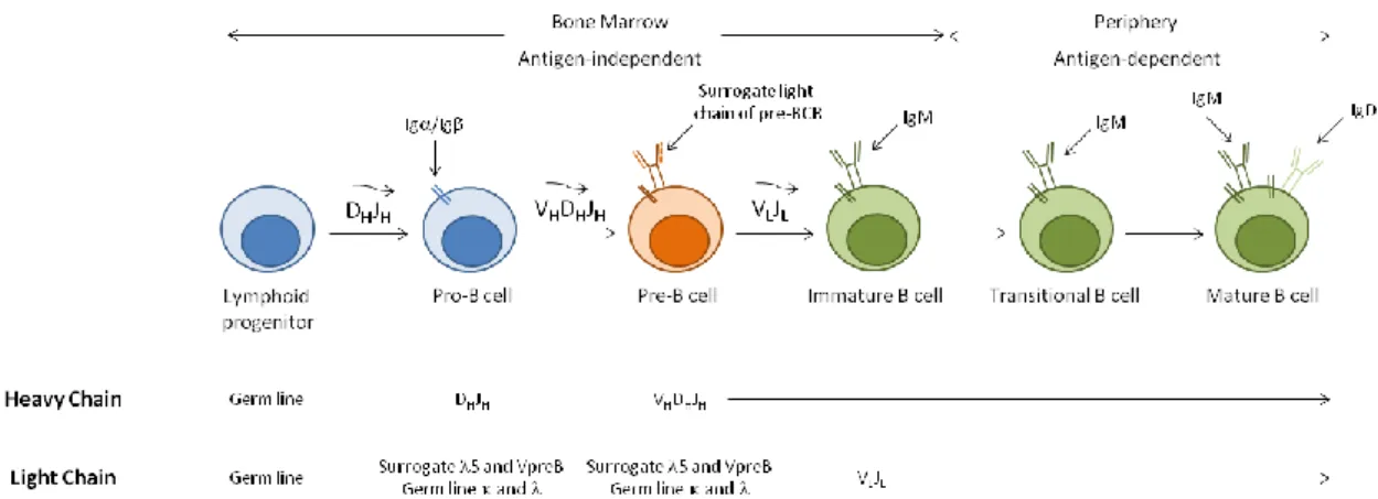
Earlier stages of B-cell development occur in primary lymphoid organs, such as the foetal liver and the bone marrow, and then differentiation proceeds in secondary lymphoid organs, such as the spleen and lymph nodes, where B cells achieve functional maturation (Figure 1). Haematopoietic stem cells (HSCs) generate lymphoid progenitors that give rise to the earliest cell committed to the B-cell lineage, the progenitor B cell or pro-B cell. Proliferation of pro-B cells in the bone marrow is accompanied by the sequential rearrangement of the immunoglobulin (Ig) heavy chain, originating pre-B cells. The rearranged Ig heavy chain forms, together with the surrogate light chains, 5 and VpreB, the pre-B cell receptor (BCR).
Figure 1. B-cell development. Outline of the events involved, depicting the bone marrow, antigen-independent phase,
and the functional maturation in the periphery (adapted from Immunology, by Goldsby et al.4).
The progeny of pre-B cells will then proceed with the rearrangement of the Ig light chain, developing into immature B cells expressing membrane IgM at their surface, in addition to a functional BCR5-7. At this stage, expression of an autoreactive BCR can direct the internalization of the self-antigen-BCR complexes and the activation of a pathway of intracellular signals that arrests differentiation and initiates a secondary rearrangement of the Ig light chain that will replace the original one. This process is called receptor editing and provides the first checkpoint on B-cell development for autoreactivity8,9. In addition to receptor editing, B cells with autoreactivities can be eliminated by clonal deletion, undergoing apoptosis if they recognize self-antigens expressed in the bone marrow10,11. Even though IL-7 has been shown to be critically
involved in murine B-cell development, promoting VDJ rearrangements and inducing proliferation and survival of B-cell precursors12, such role for IL-7 in human B-cell development remains elusive. Patients with severe combined immunodeficiency (SCID) due to mutations in the IL-7 receptor have normal levels of circulating B cells13, arguing against a major role for IL-7 in this process.
Following the bone marrow phase of development, immature B cells migrate into secondary lymphoid organs to integrate an antigen-dependent phase of maturation in the periphery7,14. Immature B cells, also termed transitional B cells, are fundamentally short-lived and migrate to the spleen through the bloodstream, where they develop into mature long-lived B cells. Upon exiting the bone marrow, transitional B cells acquire expression of markers such as IgD or CD21 (also known as complement receptor 2, CR2)14. CD21 is part of the CD19/CD81/CD21/CD225 complex, which is associated with signal transduction from the BCR, thus playing an important role in the bridging of innate and adaptive responses15. Transitional B cells have been mainly characterized in the murine system, where they can be subdivided into
T1 and T2 transitional populations. These two populations can be distinguished according to the expression of phenotypic markers, such as CD21 and IgD, with T1 cells being IgMbrightCD21-IgD -and T2 cells defined IgMbrightCD21brightIgDbright. However, some controversy remains regarding whether they are developmentally related or completely independent populations14. Transitional B cells have also been described in humans and have been proposed to include two populations expressing differential levels of CD21, which would correspond to the T1 and T2 transitional B-cell populations in the mouse16,17. These two populations are thought to represent distinct developmental stages, based on the assessment of B-cell reconstitution following HSC transplantation (HSCT)17. A third, more mature, transitional B-cell subset has also been proposed in humans, possessing a more transient nature and rapidly differentiating into mature naïve B cells18. Importantly, in addition to BCR signalling, transitional T1 and T2 B-cell populations have differential requirements for B-cell Activating Factor of the TNF family (BAFF)-mediated survival signals, since BAFF-deficient mice present impaired B-cell maturation arrested at the transitional T1 stage19, while BAFF-transgenic mice have an accumulation of B cells at the transitional T2 stage20. It is unclear whether this requirement is also observed for human transitional B cells16,21. The transitional B-cell stage is particularly relevant given that it includes a second checkpoint for autoreactivity, in the event of an autoreactive B cell escaping the first checkpoint in the bone marrow. BCR signalling at this stage induces apoptosis and is thus very important to remove autoreactivities22. B cells that recognize autoantigens can also be converted to a state where they can no longer respond to BCR engagement, in a process termed clonal anergy or functional unresponsiveness23,24. Importantly, human transitional B cells can respond to Toll-like receptor (TLR) 9 stimulation by differentiating into IgM-secreting plasma cells (PCs), arguing that transitional B cells can actually play an important role in building a rapid immune response against bacterial antigens25,26.
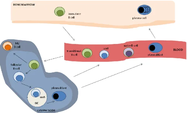
Mature B cells recirculate between the follicles of the spleen and the lymph nodes, playing a crucial role in adaptive immune responses (Figure 2). In humans, five major populations are described in peripheral B-cell development: the abovementioned transitional B cells, naïve B cells that have not yet encountered antigen, germinal centre (GC) B cells that are found in peripheral lymphoid organs and are actively involved in ongoing immune responses, memory B cells, expressing the memory marker CD27, that are generated through GC reactions and survive for long periods of time, and PCs that produce antibodies, being the effector arm of the B-cell immune response7.
Figure 2. Peripheral B-cell differentiation. Simplified schematic outline, with the main B-cell subsets depicted. MZ:
marginal zone; smB: switched-memory B cell.
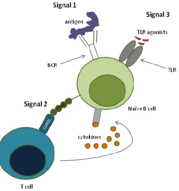
Naïve B cells constitute the major B-cell subset found in the peripheral blood of healthy adult individuals27. Full activation of naïve B cells requires BCR cross-linking by antigen recognition, together with signals coming from CD40 ligation and cytokine stimulation (to be discussed in the next sections) and stimulation by TLR agonists28 (Figure 3). Following activation, naïve B cells can either differentiate into short-lived IgM-producing PCs through extrafollicular reactions or they can enter B-cell follicles and participate in GC reactions, where they differentiate into long-lived memory B cells and PCs29. Memory B cells possess less stringent requisites for activation and rapidly produce specific IgG when they re-encounter the antigen, while persisting once the antigen is cleared. Surface expression of CD27 is currently accepted as the best marker for memory, antigen-experienced B cells27. PCs directly differentiate from plasmablasts that migrate from the secondary lymphoid organs to the bone marrow29. They are normally not found in the peripheral blood of healthy individuals.
The main origin of IgM-expressing human memory B cells is the subject of great controversy. While some argue that they derive from GC reactions30, others actually propose them to be independent of the GC, since they are present in patients with impaired GC formation and class-switch recombination (CSR)31-34. In addition, it has also been suggested that this population includes B cells that are the circulating counterparts of splenic marginal zone (MZ) B cells, which are important in the responses against encapsulated bacteria, and that they would undergo somatic hypermutation (SHM) very early during ontogeny35,36.
Figure 3. The 3-signal model for naïve B-cell activation. Full activation of naïve B cells requires the integration of BCR
stimulation (Signal 1), and help provided by T cells (Signal 2), both in the form of co-stimulation and cytokines. BCR stimulation drives the up-regulation of TLR expression37, which then provides Signal 3 for B-cell activation.
In the mouse, follicular (also termed B2) B-cell differentiation is essentially similar to that described above for humans. In addition, two separate lineages of B cells have also been well characterized, with features that put them closer to the innate arm of the immune system: B1 B cells and the already mentioned MZ B cells38. While MZ B cells share developmental stages with
B2 B cells39, B1 B cells develop from distinct B-cell progenitors in the foetal liver40. B1 B cells, originally identified as a population of CD5+ splenic B cells, are a self-renewing population and express germline-encoded antibodies that form the so-called natural antibody repertoire41. The terminology derives from the fact that these antibodies are generated in the absence of antigen exposure and hence the proximity to a more innate-like function41. B1 B cells are believed to play an important role in the suppression of inflammatory responses41,42. Even though human equivalents to mouse B1 B cells have been reported over time43-45, these cells are not resident in the peritoneal cavity46, unlike mouse B1 B cells. The recent report that the human equivalent to murine B1 B cells expresses CD27 and IgM, in addition to CD43, argues that at least some B cells that have been previously described as IgM-expressing memory B cells are in fact B1 B cells44. MZ B cells are, as the nomenclature indicates, located in the marginal zone of the spleen. They represent a distinct, non-circulating, lineage of murine splenic B cells, critical for the first line of defence against encapsulated bacteria47. Contrary to what is seen in the mouse, human MZ B
cells can recirculate and are thus found both in the spleen and the peripheral blood. In addition and as previously mentioned, they express the memory marker CD27 and have been described to undergo SHM in the absence of typical immune responses36,47,48. Nevertheless, it has been proposed that, similar to what happens in the murine system, MZ B cells are critical for the fast protective, T-independent response against encapsulated bacteria47.
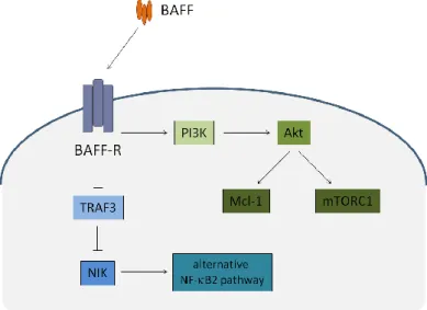
Peripheral B-cell survival and differentiation largely depend on two factors belonging to the TNF family, BAFF and A Proliferation Ligand (APRIL), and on their interaction with the receptors BAFF Receptor (BAFF-R), Transmembrane Activator and Calcium-modulating Cyclophilin Ligand Interactor (TACI), and B-cell Maturation Antigen (BCMA)49. BAFF signalling through BAFF-R is particularly relevant for the survival and maturation of peripheral B cells, while APRIL acting through BCMA appears to be critical for PC survival in the bone marrow50-53. BAFF-R stimulation by BAFF mainly activates the alternative Nuclear Factor-B (NF-B) pathway, mediated by the stabilization of the NF-B-inducing kinase (NIK) induced by TNF-receptor associated factor (TRAF) 3 degradation, thereby resulting in increased B-cell survival49,54-56. In addition, BAFF stimulation of BAFF-R also has an impact in the metabolic fitness of B cells by activating the mammalian target of rapamycin complex 1 (mTORC1), in a pathway that sequentially involves Phosphoinositide 3-kinase (PI3K) and Akt57,58. The activation of Akt in also critically involved in the up-regulation of the anti-apoptotic molecule myeloid cell leukemia sequence 1 (Mcl-1)54,58 (Figure 4). The integration of the signals delivered through BAFF-R stimulation to these signalling pathways thus mediates the effects of BAFF on B-cell survival and metabolic fitness.
b. B CELLS AND THE INNATE IMMUNE SYSTEM
Whenever a pathogen enters the organism, an immune response needs to take place in order to clear it and prevent further damage. Given that failing to control the initial phase of an infection can have serious and even life-threatening outcomes, there is the need for immune defences that are ready-to-go. The innate immune system has the task of initiating a local inflammatory response at the place of pathogen entry, thereby limiting the extent of infection until an adaptive immune response is developed. In addition, the innate immune system also has a crucial role in the priming of the initial adaptive immune response. B cells have an important role in linking cell-intrinsic innate and adaptive immune responses, given the fact that they express both an antigen-specific BCR and TLRs59. TLRs are receptors that recognize microbial antigens and activate the innate immune system. The integration of signals coming from these two pathways thus determines the fine-tuning of the overall B-cell response.
As previously mentioned, two subsets of B cells have characteristics that places them closer to the innate immune system: B1 B cells and MZ B cells. These B-cell subsets are crucially involved in T-independent immune responses, where they generate rapid although low-affinity antibody production60. Moreover, the BCRs expressed by B1 and MZ B cells are of limited diversity and preferentially recognize microbial antigens and self-peptides, and TLR signalling is important for their responses. In fact, B1 and MZ B cells show higher responsiveness to TLR signals than follicular B cells61,62. TLR stimulation induces the up-regulation of Blimp-1 specifically in B1 and MZ B cells, thus driving their differentiation into PCs61. In addition, B1 and MZ B cells are highly responsive to BAFF63,64, which is mainly produced by cells of the innate immune system, such as dendritic cells, monocytes, macrophages, neutrophils and FDCs, and can also be stimulated in response to TLR ligands49. Also, naïve and memory B cells respond differently to TLR stimulation, with memory B cells expressing constitutively higher levels of several TLRs37,65. This renders memory B cells able to differentiate into PCs through TLR stimulation bypassing the need for T-cell help37,66.
Such innate-like immune responses of B cells are particularly important in the context of mucosal tissues. Especially in the intestine, B cells mediate immunity against commensal bacteria through the production of IgA by mucosal follicular and extrafollicular reactions67,68. At these sites, immune responses are highly dependent on TLR stimulation, as well as on factors such as BAFF, APRIL, TGF-, and IL-1069-71.
TLR stimulation of innate cell types can also have an effect in the modulation of B-cell responses. Plasmacytoid dendritic cells (pDCs) were shown to have a better ability to support B-cell differentiation when compared to myeloid dendritic B-cells (mDCs)72,73. Interestingly,
TLR-stimulated mDCs and macrophages may play a suppressive role in controlling the activation of autoreactive B cells, through the secretion of IL-6 and soluble CD40L74,75. This suppressive effect was specific for autoreactive B cells, since the activation of naïve B cells was not affected in this system75. In line with this observation, patients with defects in TLR signalling (i.e., deficiencies in Myeloid Differentiation Factor 88 (MyD88), Interleukin-1 Receptor-associated Kinase 4 (IRAK4), and UNC93B) have an appearance of autoreactive mature naïve B cells in the peripheral blood76.
Understanding how B cells translate the different signals around them, coordinating the innate and the adaptive immune systems, is of paramount importance, both for the development of adequate vaccination protocols and treatments for autoimmune diseases77.
c. THE RELEVANCE OF T-B CELL INTERACTIONS FOR IMMUNE RESPONSES
The interaction between B cells and T cells is fundamental to generate adaptive immune responses. The establishment of long-term humoral immunity, namely in vaccination strategies, relies on B cells receiving appropriate help from CD4 T cells in the context of GC reactions, with the generation of memory B cells and antibody-producing plasma cells78. It should also be appreciated that B cells themselves play a crucial role in the development and maintenance of the immune response, in addition to their effector function of antibody production, by acting as antigen-presenting cells (APCs), producing cytokines, recruiting and co-stimulating CD4 T cells7.
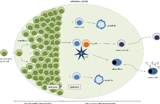
i. ORGANIZATION OF GERMINAL CENTRES
GCs are specific structures within B-cell follicles where antigen-driven SHM and CSR occur79. The GC reaction starts with the recruitment and expansion of antigen-experienced B cells to the B-cell follicles of secondary lymphoid organs, which have as main function bringing together APCs and lymphocytes in order to mount immune responses. Whether B cells differentiate into plasmablasts in extrafollicular reactions or seed the GC seems to be related to BCR primary affinity for the antigen. BCRs with high affinity most likely drive B cells into the extrafollicular reaction, whereas moderate or lower affinity will push B cells into the GC80,81. As the GC reaction develops, a defined anatomical structure comprising light and dark zones becomes apparent (Figure 5). The light zone is mainly occupied by follicular dendritic cell (FDC) processes, while lymphocytes are compacted in the dark zone. The chemokine receptors CXCR4 and CXCR5 are crucial for the positioning of lymphocytes to the dark zone and the light zone. CXCL12, the ligand for CXCR4, is more abundant in the dark zone, while CXCL13, the ligand for CXCR5, is more abundant in the light zone79,82. GC B cells in the dark zone are called centroblasts and express high levels of CXCR4, while GC B cells present in the light zone are termed
centrocytes and express high levels of CXCR5. Different events of the GC reaction take place at different zones of the GC; SHM occurs in proliferating centroblasts in the GC dark zone, while it is in the light zone that interactions between T cells, centrocytes and FDCs happen79,82,83. During the GC reaction, B cells clonally expand to ultimately generate a high affinity BCR to the given antigen, with the locus of the Ig heavy chain undergoing CSR and its variable regions going through SHM. Both of these processes are mainly mediated by an enzyme termed activation-induced cytidine deaminase (AID)84.
Figure 5. Germinal Centre. Schematic representation of the structure of a germinal centre, with dark and light zones
depicted. Somatic hypermutation occurs in the dark zone, while class-switch recombination is limited to the light zone, where B cells interact with follicular helper T cells (TFH) and follicular dendritic cells (FDC) (adapted from Klein and Dalla-Favera84).
T-cell help is known to be essential to the induction and subsequent organization of GCs. This help is attributed to a particular subset of T cells, follicular helper T cells (TFH)85,86, which are
identified by the expression of the chemokine receptor CXCR5, necessary for their specific homing to B-cell follicles and within GCs87-89. In addition to CXCR5 expression, TFH cells are
characterized by the expression of the co-stimulatory molecules CD40L and ICOS85,86, crucial for their function of B-cell help provision (see next section), and the inhibitory receptor Programmed Death-1 molecule (PD-1)90. A major function of TFH cells is the production of
below). After a long standing debate regarding the functional relationship between other helper T-cell populations and TFH cells, they have been described as a separate lineage of helper T
cells91-93, with its generation being governed by the transcription factor B-cell Lymphoma 6 (Bcl-6)94-96. In B cells, Bcl-6 expression is restricted to the GC and it suppresses the differentiation into plasma cells, by inhibiting the expression of the transcription factor B Lymphocyte-induced Maturation Protein-1 (Blimp-1)97,98. In addition, Blimp-1 has also been shown to inhibit TFH cell
differentiation and function96. Thus, Bcl-6 is crucial for GC development and function, both at the B-cell and the T-cell levels95. The lack of adequate T-cell help during the priming of B cells results in apoptosis, rather than their differentiation into GC B cells or plasma cells78,99,100.
Despite the fact that much is known about the attributes of adaptive immune responses, the factors that govern the differentiation of memory B cells and their long-term maintenance remain largely unknown101.
ii. CO-STIMULATORY MOLECULES
The functional interaction between T cells and B cells within GCs rely on the expression of co-stimulatory molecules, such as CD40-CD40L and the Inducible Co-Stimulator (ICOS) molecule102.
CD40 is a member of the tumour necrosis factor (TNF) receptor family and a co-stimulatory molecule expressed on B cells that binds CD40L on the surface of activated T cells. This interaction activates B cells and, in combination with soluble cytokines, induces production of different classes of antibodies103,104. For instance, CD40 stimulation together with IL-4 induces preferential isotype switch to IgE105. Humoral responses are impaired in both mice106,107 and humans108-113 with defective CD40L function, underpinning the important role of CD40-CD40L interactions in GC generation, isotype switching and SHM. Also, blocking CD40-CD40L interactions impairs the formation of the GC structure and the proliferation of B cells114-116. Importantly, CD40 signals are essential to prevent centrocyte apoptosis99,100.
ICOS, a member of the CD28 family of T-cell co-stimulatory molecules117, plays a critical role in the regulation of humoral immunity. ICOS expression is up-regulated on T cells following activation and is expressed at high levels on TFH cells85,86,117. ICOS specifically binds to ICOS ligand
(ICOSL), constitutively expressed on B cells118. ICOS co-stimulation induces IL-4 and, more specifically, IL-10 production by T cells, both of which have an important role in antibody production117 (discussed in the next section). Of note, ICOS co-stimulation also induces up-regulation of CD40L expression117, further enhancing the B-cell helper capacity of the T cell. Both ICOS-deficient mice119-122 and human individuals31,123 exhibit profoundly defective isotype
class-switch, as well as impaired GC formation. Homozygous deletion of the ICOS gene in humans leads to a phenotype of Common Variable Immunodeficiency (CVID), with low numbers of B cells, low serum antibody concentrations and lack of memory B cells31. It is noteworthy to mention that patients with defective ICOS or CD40L show a decreased frequency of circulating CXCR5+ CD4 T cells124, which are suggested to be the counterparts of T
FH cells found in secondary
lymphoid organs124,125. Together with the finding of reduced TFH cells in the spleens and lymph
nodes of ICOS- and ICOSL-deficient mice124,126, this suggests that ICOS is important for the development of TFH cells. In line with these observations, ICOS has recently been shown to
supply a vital signal for the early induction of Bcl6 on T cells, which then induces expression of CXCR5, committing cells to the TFH lineage127.
PD-1 expression by TFH cells also plays a critical role in the GC reaction. GC B cells
up-regulate the expression of the PD-1 ligands Programmed Death-1 molecule Ligand 1 (PD-L1) and Programmed Death-1 molecule Ligand 2 (PD-L2). The study of mice deficient for either of the molecules involved in this pathway showed that PD-1 expression on T cells and PD-L2 expression on B cells is in fact regulating TFH cell and PC numbers128. PD-1 deficiency also resulted in lower
production of IL-4 and IL-21. It has thus been proposed that PD-1 directs the development of long-lived PCs by regulating the survival and selection of B cells in the GC, through its role in the function of TFH cells128.
In addition to the aforementioned molecules, other factors that affect the interaction between T and B cells also have an impact in GC development and function101. One of such cases is the signaling lymphocytic activation molecule-associated protein (SAP), expressed on T cells, whose absence results in defective interaction between TFH and B cells, with a consequent
inability of TFH cells to sustain GC reactions129.
iii. SOLUBLE FACTORS
T-cell derived cytokines present during the events of B-cell activation are crucial in the modulation of the B-cell response, namely by regulating the CSR process and determining the class of antibody that the B cell will produce.
IL-21 is probably the best example of cytokines involved in the functional interaction between T cells and B cells130-132. IL-21 was first described as having a critical role in the regulation of antibody production by B cells131,133,134. In addition, the GC requirement for IL-21 was also found to reflect an intrinsic requisite for TFH maintenance, acting in an autocrine
fashion132. Nevertheless, IL-21 is in fact a central player in the B helper function provided by TFH
expression and thus has a crucial role in the development of GC responses135,136. In the absence of IL-21 signalling, GCs are reduced and the production of high-affinity plasma cells is impaired135,136. IL-21 production is induced by ICOS stimulation, through the regulation of the transcription factor c-Maf137. Paradoxically, IL-21 was also found to promote B-cell apoptosis138. This effect is however dependent on the activation signals provided to B cells and may thus represent an important role of IL-21 in regulating B-cell homeostasis138,139.
IL-4, the master cytokine involved in Th2 responses, was originally identified as a B-cell stimulating factor140,141. IL-4 was shown to be critical for isotype switch to IgE and IgG1105,142, and also to cooperate with IL-21 in regulating isotype switch to different IgG subclasses and IgA131,143. Importantly, TFH cells have been shown to produce IL-4 upon immunization with given antigens,
in particular with helminths144,145.
IL-2 was initially described as promoting B-cell growth146,147. In addition, it has later been demonstrated to participate in the induction of antibody production148-152. However, recent data has challenged this view, with IL-2 administration being described to impair influenza-specific GC formation, long-term IgG responses and limiting the differentiation of TFH cells153. However,
other recent reports have identified T-cell derived IL-2 to be critically involved in human PC differentiation154,155. If these conflicting data represent an essential functional difference between the murine and the human systems is something that needs to be clarified.
IL-6 is another T-cell derived cytokine with a role in the regulation of B-cell responses156, inducing plasma cell differentiation and antibody production152,157,158. Importantly, IL-6 was also shown to promote IL-21 production159,160 and to drive the differentiation of T cells with the ability to provide B-cell help160.
IL-10 has a well recognized role in PC differentiation, especially in humans78,148,157,161,162. Along with Tumour Growth Factor (TGF)-, it mediates isotype switch to IgA163, particularly
important at the mucosal sites. IL-10 seems to be especially relevant in stopping B-cell proliferation in the GC and inducing differentiation into PCs164. IL-10 production is enhanced by ICOS stimulation117,165,166, which is particularly relevant in the context of TFH cells. Accordingly,
human TFH cells were found to produce IL-10 in an ICOS-dependent manner167,168, which adds up
to their function in providing B-cell help. Recent studies also suggest that IL-10 production by B cells, the so-called regulatory B-cell population, can be important to modulate T-cell responses169,170.
PART II
PRIMARY B-CELL IMMUNODEFICIENCIES
a. HISTORICAL PERSPECTIVE, CLINICAL MANIFESTATIONS, AETIOLOGY AND TREATMENT OF PRIMARY B-CELL IMMUNODEFICIENCIES
Primary B-cell immunodeficiencies include several disorders that have as a common feature a defect in antibody. Currently, several classes of primary antibody deficiencies are considered - defective early B-cell development in the bone marrow; impairments in CSR, mainly comprising Hyper IgM (HIGM) syndromes; isotype or light chain deficiency, including selective IgA deficiency; specific antibody deficiency; and CVID. Given the fact that this work will be mainly focused on patients affected by defects in early B-cell development and CVID patients, the other classes will only be briefly mentioned.
CSR defects, also referred to as HIGM syndromes, are observed in patients with recurrent infections that have normal or elevated serum levels of IgM, in the presence of reduced IgG, IgA, and IgE. In addition to the CSR defect, SHM can also be affected171. This group of genetic defects can be B-cell restricted or it can affect other cell types, such as T cells, macrophages or DCs. More than half of the patients with CSR defects present X-linked mutations in the gene encoding CD40L108-110,112,113, expressed by activated T cells. In this case, patients usually suffer from opportunistic infections and neutropenia, presenting a clinical picture that is more severe than the one seen in patients with pure humoral defects171. CD40 deficiency has also been identified, although in very rare cases, with a clinical phenotype identical to the CD40L deficiency172. In terms of CSR defects that are B-cell restricted, autosomal recessive mutations in AID account for the majority of the cases173,174. These patients have a later onset of disease, while frequently presenting adenopathies and a high incidence of autoimmunity175. Autosomal dominant mutations have also been described in a small number of patients176. In addition, autosomal recessive mutations in uracil-DNA glycosylase (UNG), an enzyme responsible for the deglycosylation and removal of uracil residues resulting from the action of AID, have also been described in very few cases of HIGM patients177. The clinical phenotype is similar to the one observed in AID-deficient patients.
X-linked Agammaglobulinemia (XLA) was one of the first immunodeficiencies to be described and is often regarded as a prime example. Back in 1952, Ogden C. Bruton reported a case of an 8-year-old male presenting recurrent pneumococcal sepsis with complete absence of
the serum gamma globulin fraction, with otherwise normal serum proteins178. These laboratory findings led to the use of tailored therapy, through the subcutaneous administration of human immune serum globulin, while completely explaining the clinical symptoms. In 1953, Charles A. Janeway and colleagues documented the same phenomena (agammaglobulinemia and recurrent infections) in an adult individual, in what was the first report of the nowadays termed CVID179.
While having a similar outline, it was since early recognized that differences existed between the two observations. Agammaglobulinemia mainly affected males when presenting during childhood, often with an X-linked mode of inheritance, whereas in adults such pattern was not evident, with both females and males being equally affected179-184. Later on, it was established that patients with XLA presented virtually no circulating B lymphocytes, at the same time as adult CVID patients typically had close to normal numbers of B cells185-188. It was not until more than 40 years after the original description that the molecular defect underlying XLA was unveiled and shown to affect a cytoplasmic tyrosine kinase expressed at all stages of the B-cell lineage and in myeloid cells, later termed Bruton’s Tyrosine Kinase, or Btk189,190. Mutations in the Btk gene arrest early B-cell development in the bone marrow at the pre-B cell stage191-194, frequently, but not always195,196, leading to a complete absence of circulating mature B cells. This block in B-cell development results from defective signal transduction by Btk through the pre-BCR and pre-BCR197,198.
Regarding clinical manifestations, the most common feature of XLA patients is recurrent infections of the respiratory tract that usually start between 3 and 18 months of age199-201. Although some problems can arise from viral infections at early ages, they are well dealt with by XLA patients once T-cell immunity is developed. The most frequent infectious agents are the encapsulated bacteria Streptococcus pneumoniae and Haemophilus influenza, in terms of respiratory tract infections, and the protozoan parasite Giardia, in what concerns gastrointestinal infections199.
In addition to the X-linked form of agammaglobulinemia, autosomal recessive forms have also been identified, accounting for approximately 15% of the cases of patients suffering from early defects in B-cell development202 (Table 1). As happens with patients with XLA, all of these patients are characterized by presenting an onset of recurrent infections in the first 5 years of life, severe hypogammaglobulinemia, remarkably reduced or absent circulating B cells, and a block in B-cell development in the bone marrow.
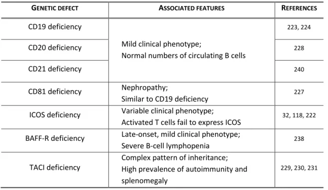
Table 1. Genetic defects underlying Congenital Agammaglobulinemia.
GENETIC DEFECT ASSOCIATED FEATURES REFERENCES
Btk deficiency
Severe bacterial infections; Absence of circulating B cells;
Normal numbers of pro-B cells in the bone marrow
173, 176, 177
heavy chain deficiency 198
5 deficiency 200
Ig deficiency 201
Ig deficiency 202, 203
BLNK deficiency 204
PI3K deficiency
Severe bacterial infections; Absence of circulating B cells;
Markedly reduced numbers of pro-B cells in the bone marrow
205
Autosomal recessive forms of agammaglobulinemia include mutations in the genes encoding for components of the pre-BCR or the BCR, or in the adaptor protein B cell Linker Protein (BLNK), which also plays a role in signal transduction from the BCR. Mutations in the heavy chain, which defines the IgM isotype and is part of the BCR, were identified to underlie a clinical picture similar to XLA203. However, the clinical manifestations found in these patients tend to be biased towards a more severe phenotype, with an earlier onset of disease and more clinical complications throughout life204. A smaller number of patients have been identified with mutations in the genes encoding 5205, Ig206, and Ig207,208, all of which are part of the pre-BCR complex. In addition, mutations in BLNK have also been described to give rise to a phenotype similar to XLA209. The vast part of the patients presents null mutations that abrogate protein function. In clinical terms, these patients are identical to XLA patients, even though they normally present an earlier onset of disease. Very recently, it has been described a case of a young female with agammaglobulinemia in the absence of circulating B cells that presented a mutation in PI3K, resulting in the absence of the p85 subunit210. Interestingly, the absence of p85 in the patient results in an early and striking defect in B-cell development in the bone marrow, but with a clinical picture restricted to colitis. The block in B-cell development was observed at the earliest stage of commitment to the B-cell lineage, earlier to what is seen for XLA or patients with other forms of autosomal recessive agammaglobulinemia210.
CVID is the most frequent primary immunodeficiency with clinical relevance, with an estimated prevalence of 1:25,000 – 1:50,000 in the European population211. The age of disease onset has a bimodal distribution, with some patients being diagnosed during childhood while the majority presents in early to mid adulthood. CVID is clinically defined by reduced serum levels of IgG and at least another Ig isotype, impaired antibody response to vaccines, and exclusion of defined causes of hypogammaglobulinemia212. Nevertheless, CVID patients present highly heterogeneous clinical and immunological profiles. In addition to the high frequency of respiratory and gastrointestinal tract infections, a myriad of non-infectious complications have been reported in more than two-thirds of CVID patients, ranging from chronic lung disease and gastrointestinal inflammatory disease, to autoimmune manifestations, granulomatous disease and neoplasias211,213,214. Autoimmunity is being increasingly recognized as a serious medical issue in CVID patients, affecting more than 20% of the patients215,216. A recent study has reported the immunological parameters, clinical complications and mortality statistics from 473 CVID patients followed for over 4 decades214. In this report, it was found that CVID patients had a significantly shorter survival rate than age- and gender-matched control populations. In addition, this reduced survival was significantly associated with age at diagnosis, lower circulating B cells, and lower baseline serum levels of IgG and higher levels of IgM. Patients affected by non-infectious complications had a significantly higher mortality risk, which was associated with lymphoma, liver or lung disease, and gastrointestinal manifestations, but not with other complications214. Moreover, an Italian study has concluded that malignancies are the major cause of death in adult-onset CVID217. Another study has analysed and divided 334 CVID patients into distinct clinical phenotypes, but failed to identify useful predictors213.
Even though the vast majority of CVID cases are sporadic, familial patterns of inheritance are observed in 10 – 20% of the patients, with a family history of autoimmunity or disorders of antibody production218,219. In some families, CVID co-exists with IgA deficiency (IgAD). This primary immunodeficiency can be selective for IgA or be associated with deficiency in subclasses of IgG, and is the most common primary immunodeficiency. However, the majority of IgAD patients are asymptomatic and thus the condition is clinically not very relevant220,221. Some IgAD patients progress to CVID, suggesting a degree of common genetic aetiology. In this sense, early studies identified certain Human Leukocyte Antigen (HLA) haplotypes to be more common in both conditions219,222-226.
According to the latest update on the classification of Primary Immunodeficiency Diseases (PIDs)13, CVID, now standing for Common Variable Immunodeficiency Disorders, groups all patients with a severe reduction of at least two Ig isotypes with normal or low B-cell numbers
of unknown aetiology. In this recent classification, the molecular defects that give rise to a CVID-like phenotype are now in separate subgroups, namely mutations in ICOS, CD19, CD81, CD20, TACI, and BAFF-R13.
ICOS deficiency was the first genetic defect to be identified in four patients with adult-onset CVID, in 200331. Patients failed to express ICOS at the surface of activated T cells, due to a
partial homozygous deletion in the ICOS gene. Five additional patients have been later described to present the same homozygous deletion, indicating that all nine patients probably share a common founder227. It is interesting to note that this first description of an autosomal recessive disorder underlying CVID was related to a T-cell intrinsic defect that affects the cross-talk between T and B cells in GCs. In the original report of the first four patients, ICOS-deficient patients were described as lacking major non-infectious complications, such as splenomegaly or autoimmunity31. However, the subsequent identification of five additional patients, who included patients with early-onset disease, showed that ICOS deficiency can present with the full clinical spectrum of CVID123.
Mutations in CD19 were described in 2006, resulting in a phenotype of hypogammaglobulinemia compatible with CVID228. There are currently six cases identified, all of which present normal numbers of circulating B cells with minimal or no expression of CD19 at their surface199,202,228,229. These include both early- and adult-onset CVID-like phenotypes. Given that CD19 is part of a complex that is associated with signal transduction from the BCR230,231, mutations in CD19 likely hinder antigenic stimulation and hence result in defective humoral responses. Accordingly, B cells from CD19-deficient patients showed defective calcium fluxes following BCR stimulation. Vaccination responses were also impaired228. Patients with CD19 deficiency did not present signs of non-infectious complications, including autoimmunity, lymphoid proliferation or neoplasias228.
CD81 deficiency was reported in 2010 in one patient with severe nephropathy and hypogammaglobulinemia232. B cells from the patient did not express CD19, but had normal CD19 alleles. Instead, a homozygous mutation in CD81 was found, leading to a complete absence of CD81 expression. Transduction experiments showed that CD81 expression was crucial for CD19 membrane expression. So, CD81 deficiency leads to a functional phenotype that is similar to CD19 deficiency, with impaired responses through BCR stimulation232.
Also in 2010, CD20 deficiency was described in a young patient with persistent hypogammaglobulinemia233. The patient presented normal peripheral B-cell numbers, but a severe decrease in the frequency of memory B cells. SHM was defective and even though the patient mounted adequate antibody responses after vaccination with recall antigens, responses