This Accepted Author Manuscript is copyrighted and published by Elsevier. It is posted here by agreement between Elsevier and University of Brasilia. Changes resulting from the publishing process - such as editing, corrections, structural formatting, and other quality control
mechanisms - may not be reflected in this version of the text. The definitive version of the text was subsequently published in [Small Ruminant Research, Volume 107, Issue 2-3, October 2012, Pages 121–130, doi:10.1016/j.smallrumres.2012.04.009].You may download, copy and
otherwise use the AAM for non-commercial purposes provided that your license is limited by the following restrictions:
(1) You may use this AAM for non-commercial purposes only under the terms of the CC-BY-NC-ND license.
(2) The integrity of the work and identification of the author, copyright owner, and publisher must be preserved in any copy.
(3) You must attribute this AAM in the following format: [agreed attribution language, including link to CC BY-NC-ND license + Digital Object Identifier link to the published journal article on Elsevier’s ScienceDirect® platform].
________________________________________________________________________
Este Manuscrito do Autor Aceito para Publicação (AAM) é protegido por direitos autorais e publicado pela Elsevier. Ele esta disponível neste Repositório, por acordo entre a Elsevier e a Universidade de Brasília. As alterações decorrentes do processo de publicação - como a edição, correção, formatação estrutural, e outros mecanismos de controle de qualidade - não estão refletidas nesta versão do texto. A versão definitiva do texto foi posteriormente publicado em [Small Ruminant Research, Volume 107, Número 2-3, Outubro 2012, Pages 121–130 ,
doi:10.1016/j.smallrumres.2012.04.009]. Você pode baixar, copiar e utilizar de outra forma o AAM para fins não comerciais , desde que sua licença seja limitada pelas seguintes restrições:
(1) Você pode usar este AAM para fins não comerciais apenas sob os termos da licença CC- BY- NC-ND.
(2) A integridade do trabalho e identificação do autor, detentor dos direitos autorais e editor deve ser preservado em qualquer cópia.
Morphologic, viability and ultrastructural analysis of vitrified sheep
preantral follicles enclosed in ovarian tissue
Franciele Osmarini Lunardi
Valdevane Rocha Araújo
Luciana Rocha Faustino
Adeline de Andrade Carvalho
Raphael Fernando Braga Gonçalves
Casie Shantel Bassa
Sônia Nair Báo
Khesller Patrícia Olázia Name
Cláudio Cabral Campello
José Ricardo de Figueiredo
Ana Paula Ribeiro Rodrigues
Abstract
The main objective was to compare the efficiency of vitrification techniques and solutions on the preservation of morphology, ultrastructure and viability of sheep preantral follicles enclosed in ovarian tissue fragments. The fragments were cryopreserved by using macrotube vitrification (MTV), solid-surface vitrification (SSV) or conventional vitrification (CV). These techniques were combined with one of the six solutions containing 6 M ethylene glycol (EG) and with or without sucrose (SUC) (0.25 or 0.50 M) and with or without fetal calf serum (FCS) (10%). After one week, samples were warmed and histological analysis was performed, showing that the percentage of normal follicles after CV (66.20 ± 8.87%) using a solution containing 6 M EG, 0.25 M SUC and 10% FCS (vitrification solution 4 – VS4) was similar to fresh control (79.40 ± 7.83%), MTV (53.40 ± 10.60%) and SSV (56.75 ± 15.33%), all of them with the same vitrification solution (P < 0.05). For follicular viability evaluation, ovarian fragments were
vitrified as described above. After warming, follicles were assessed by trypan blue dye. Controversially, the highest percentage of viable follicles was observed in MTV (97.06%) and was similar to fresh control (92.62%) (P < 0.05), but was significantly different from SSV
(81.08%) and CV (83.81%) (P < 0.05). These results were validated by transmission electron
microscopy that showed normal follicles observed in MTV and in fresh control. In addition, to verify the MTV with VS4 (a combination of the best technique plus the best solution), follicle viability was evaluated after 48 h in vitro culture. The viability assay was performed by
fluorescence microscopy (calcein-AM and ethidium homodimer-1) analysis as follows: follicles isolated from fresh tissue were forthwith analyzed or underwent 48 h in vitro culture before
analysis, whereas others fragments were vitrified/warmed and immediately analyzed or underwent 48 h in vitro culture before analysis. These results showed that, although follicular
viability after MTV/VS4 (65%) was reduced when compared to the non-vitrified follicles at day 0 (100%), follicular viability after MTV/VS4 at day 2 (36.5%) was similar to follicles vitrified at day 0 (65%) and similar to non-vitrified follicles at day 2 (62.5%) (P > 0.05). As the decrease of
experimental conditions of the present study, an efficient solution (VS4: 6 M EG, 0.25 M SUC and 10% FCS) and technique (MTV) were successfully used to vitrify ovine ovarian tissue.
Keywords
Short-term culture; Ovarian fragments; Cryopreservation; Cryoprotectant; Ovary; Ewes
1.
Introduction
Advances in cryopreservation techniques and protocols for germinal tissue over the past
decades have contributed greatly to the establishment of germplasm banks. These genetic
banks are crucial for the preservation of genetic material with potentially high economic value
or for use with endangered species populations (Liu et al., 2008 and Santos et al., 2010). In
addition, the association between cryotechnology and assisted reproduction techniques (ART)
has important clinical relevance, as it permits the development of alternative strategies for
restoring fertility in women at risk of premature ovarian failure, especially those undergoing
cancer therapies. Admittedly, high dose chemotherapy and radiotherapy destroy a significant
portion of ovarian follicular population, often times leading to permanent infertility in women
(Meirow and Nugent, 2001 and Chemaitilly et al., 2006).
The main alternatives for fertility preservation in routine clinical use are limited to the
protection of the ovaries (oophoropexy) against radiation, or emergency in vitro fertilization
(IVF) ( Sonmezer and Oktay, 2004). Although oophoropexy may offer some protection to germ
cells, this technique can greatly reduce the success of future pregnancies ( Wallace et al.,
2005). There are also serious limitations in the emergency use of IVF in patients with cancer, as
hormonal stimulation is required to obtain mature oocytes. The possibility of utilizing these
hormones in patients with hormone-sensitive cancers, as well as in prepubertal patients (
Sonmezer and Oktay, 2004), is immensely restricted. Currently, cryopreservation of ovarian
tissue is a possible fertility preservation alternative for patients in need of treatment for
malignant diseases and is recommended by the American Society of Clinical Oncology (ASCO) (
Lee et al., 2006).
In veterinarian medicine, embryo cryobiology has been emphasized when regarding
conservation of endangered species or pets. However, this practice is not feasible in cases of
accidental or sudden loss of valuable females and, therefore, cryopreservation of ovarian
tissue is indicated as a better alternative in these situations (Takahashi et al., 2001). With
regard to the ovarian tissue cryopreservation of livestock animals, such as sheep, several
et al., 2002, Salle et al., 2003 and Imhof et al., 2006) and vitrification (Bordes et al., 2005 and
Lornage et al., 2006) methods through the birth of healthy offspring after transplantation of
ovarian tissue.
Itrification is a fairly recent alternative method of cryopreservation and, when compared
to slow freezing, is quicker and cheaper. However, the vitrification method is characterized by
using high concentrations of cryoprotectants (Vajta et al., 1998), which can increase the toxic
effect caused by these substances on preantral follicles. Moreover, it is known that factors
such as high concentrations of cryoprotectant agents, osmotic stress and the techniques used
for vitrification loading may contribute to the reduction of normal preantral follicles after
warming (Huang et al., 2008).
In the last decade, studies have been completed using ethylene glycol (EG) with vitrified
ovarian tissue or isolated preantral follicles in rat (Sugimoto et al., 2000), mouse (Kagabu and
Umezu, 2000 and Kim et al., 2010), goat (Santos et al., 2007 and Carvalho et al., 2011), cow
(Gandolfi et al., 2006 and Kagawa et al., 2009), pig (Moniruzzaman et al., 2009) and human
(Isachenko et al., 2003 and Silber et al., 2010). However, very few investigators have tested EG
in vitrification solution with ovine preantral follicles enclosed in fragments of ovarian tissue
(Amorim et al., 2003 and Melo et al., 2011). Developments in sheep ovarian vitrification may
have relevance as ewe ovaries are similar to the human ovary in its anatomy and physiology
(Gosden et al., 1994, Oktay et al., 2000 and Salle et al., 2002). While positive results have
recently been obtained with the vitrification of mouse ovaries (Wang et al., 2011), these
methods cannot be easily transferred to human tissue. This is, in part, due to the vast
morphological and physiological differences between mouse and human ovaries. Despite
having larger ovaries, neither bovine nor porcine can be considered a relevant model for
human tissue vitrification (Gandolfi et al., 2006). In addition, researchers have published
promising results regarding ovarian tissue cryopreservation in the presence of an extra-cellular
cryoprotectant, like sucrose (SUC) (Santos et al., 2006a) or fetal calf serum (FCS) (Chen et al.,
2006). Information detailing whether the addition of sucrose at concentrations of 0.25 or 0.5
M with or without 10% FCS may be essential for ovarian tissue vitrification, despite being
important, is limited in sheep.
The current study aimed (1) to compare different vitrification techniques in ovine ovarian
tissue and (2) to test the effects of varying concentrations of SUC, FCS or both combined with 6
M EG as a vitrification solution (VS). Morphology, by classical histology and transmission
electron microscopy, and viability, by trypan blue stain and fluorescent markers, were assessed
in fresh ovarian fragments, vitrified/thawed fragments, and vitrified/thawed samples after in
2.
Materials and methods
2.1.
Source and preparation of ovarian tissue
Ovaries (n = 30) were collected at a local abattoir from 15 adult non-pregnant mixed-breed
ewes. Immediately after postmortem, under aseptic conditions, the ovaries were washed in
70% alcohol for 10 s, followed by two washes in HEPES buffered minimum essential medium
(MEM) (Sigma Chemical Co., St. Louis, MO, USA) supplemented with 100 g/mL penicillin and
100 g/mL streptomycin. The ovaries were transported into tubes containing 20 mL of MEM
within thermos flasks maintained at 20 °C to the laboratory within 1 h after they were
recovered.
2.2.
Experiment I: morphology, viability and ultrastructure of preantral
follicles in vitrified ovarian cortex
2.2.1.
Ovarian tissue vitrification: solution composition and technique
At the laboratory, ovarian pairs (n = 5) were stripped of adhering tissue and fat, and cut
with a scalpel into approximately 3 mm × 3 mm × 1 mm (9 mm3) or 1 mm × 1 mm × 1 mm (1
mm3) fragments, according to the vitrification technique used, macrotube vitrification (MTV),
solid-surface vitrification (SSV) or conventional vitrification (CV) and were randomly assigned
to each treatment. One fragment (9 mm3) from each pair of ovaries was immediately fixed in
Carnoy's solution for 12 h for histological analysis (fresh control). Twelve 9 mm3 fragments (for
MTV or for SSV) and six 1 mm3 fragments (for CV) were exposed to one of the six vitrification
solutions (VS): (VS1–6, description to follow) for 5 min at 20 °C. After this duration, the
fragments underwent MTV, SSV or CV. The base medium (BM), composed of 6 M ethylene
glycol (EG) in MEM, was supplemented with SUC with or without 10% FCS to produce the six
VS and are referred to as: VS1: (BM); VS2: (BM + FCS); VS3: (BM + 0.25 M SUC); VS4: (BM +
0.25 M SUC + FCS); VS5: (BM + 0.50 M SUC) and VS6: (BM + 0.50 M SUC + FCS). For
Table 1.
Composition of six vitrification solutions and their arrangement with vitrification techniques.
BM, base medium; FCS, fetal calf serum; SUC, sucrose; VS, vitrification solution; MTV, macrotube vitrification; SSV, solid-surface vitrification; CV, conventional vitrification.
For the MTV, each fragment was inserted into a macrotube (Minitub do Brasil Ltda., Porto
Alegre, RS, Brazil) containing 1.8 mL of VS (VS1–6) (20 °C), which was then immersed in liquid
nitrogen (LN2) after 5 min (Carvalho et al., 2011). The SSV procedure was performed as
previously reported by Santos et al. (2007) with slight modifications. A total of six fragments
were exposed to 1.8 mL VS (VS1–6) (20 °C) for 5 min, removed from the solutions, and dried
(using an absorbent paper). These samples were then individually placed on the surface of a
metal cube floating in LN2. After this, vitrified fragments were transferred (with LN2 cooled
forceps) into cryovials for storage in LN2. The CV procedure was conducted according to the
methodology previously described by Chen et al. (2006). Briefly, six fragments were loaded
individually into 0.5 mL French straws that were partially filled with a column of VS (VS1–6)
(∼2 cm) (20 °C), followed by an air space (∼1 cm), and another VS column. After insertion of a
tissue sample, another air space, and a final column of VS were drawn into the straws. The
straws were then heat-sealed and after 5 min, plunged into LN2.
After one week of cryostorage, all treatment fragments were removed from the LN2, kept
at room temperature (RT) (∼25 °C) for 1 min, and then immersed in a water bath at 37 °C until
the VS was completely melted (∼1–2 min). The cryoprotectant was removed from ovarian
cortex fragments in three step washes containing MEM supplemented with 10% FCS and
decreasing concentrations of SUC (0.50 M, 0.25 M, and no SUC, respectively) for 5 min each.
The efficiency of these VS for the preservation of preantral follicles was evaluated
histologically.
2.2.2.
Histological analysis
After 12 h of fixation in Carnoy's fluid, fresh and vitrified ovarian tissue fragments were
dehydrated in a graded series of ethanol, clarified with xylene and embedded in paraffin wax.
Serial sections 7 μm thick were cut and every fifth section was mounted on glass slides and
(Nikon, Tokyo, Japan) at 400× magnification. In each treatment, a total of 150 preantral
follicles were examined and were defined according to Hulshof et al. (1994) as follows: (1)
primordial follicle: had an oocyte surrounded by one layer of flattened pre-granulosa cells; (2)
primary follicle: had an oocyte surrounded by a single layer of cuboidal granulosa cells; or (3)
secondary follicle: had an oocyte surrounded by two or more layers of cuboidal granulosa cells
without antrum formation. Antral follicles were not counted in this study. Normal morphology
was defined as a follicle containing a spherical oocyte with uniform cytoplasm and
well-organized granulosa cell layers. Degenerated follicles were those containing a retracted oocyte
with or without a pyknotic nucleus or degeneration of either oocyte or granulosa cells. To
avoid evaluating and counting the same follicle more than once, preantral follicles were
analyzed only in the sections in which an oocyte nucleus was observed.
2.2.3.
Follicular isolation, trypan blue staining and assessment of follicular
viability
To evaluate the effect of vitrification on preantral follicle viability in ovarian tissue,
samples underwent the three tested techniques: MTV, SSV and CV. The only VS utilized was
VS4, as it yielded the highest percentage of morphologically normal follicles shown by
histological analysis from the MTV/VS4, SSV/VS4 and CV/VS4 treatments.
Four fragments were retrieved from five pairs of ovaries (n = 5), with one fragment
immediately undergoing follicle isolation (fresh control) and viability testing. The other three
fragments were first vitrified in MTV/VS4, SSV/VS4 and CS/VS4 and stored for one week. After
this time, fragments were warmed and follicle isolation was completed. Preantral follicles were
isolated from ovarian tissue by using a mechanical procedure as described by Amorim et al.
(2000). Briefly, samples were cut into small pieces with a tissue chopper (The Mickle
Laboratory Engineering Co., Gomshal, Surrey, UK) adjusted to a sectioning interval of 87.5 μm.
Samples were then placed in 2 mL of MEM supplemented with 3 mg/mL bovine serum albumin
(BSA), and suspended 100 times with a large Pasteur pipette (inner diameter ∼ 1600 μm),
followed by 100 times with a smaller Pasteur pipette (inner diameter ∼600 μm) to dissociate
preantral follicles from stroma. The suspension was successively filtered through 500 and 100
μm nylon-mesh filters. This procedure was performed within approximately 10 min at RT. After follicular isolation, preantral follicle viability was assessed by trypan blue dye. Briefly,
300 μL of the suspension plus 15 μL of 0.4% trypan blue (Sigma Chemical Co., St. Louis, MO,
USA) were incubated for 1 min at RT (Celestino et al., 2008). Afterwards, follicles were viewed
unstained or non-viable if stained with trypan blue. An average of 93 preantral follicles was
analyzed in each treatment (total of 372 follicles) and the percentages of viable follicles were
calculated.
2.2.4. Ultrastructure evaluation
The procedures for ultrastructure evaluation were conducted according to Santos et al.
(2006b). Briefly, ultrastructural studies were carried out using fresh control fragments and
from fragments that underwent MTV/VS4, SSV/VS4 and CV/VS4 treatments. For these, tissue
fragments with a maximum dimension of 1 mm3 were fixed in 2% paraformaldehyde and 2.5%
glutaraldehyde in 0.1 M sodium cacodylate buffer (pH 7.2) for 4 h. After fixation and five
washes, specimens were post-fixed in 1% osmium tetroxide, 0.8% potassium ferricyanide and
5 mM calcium chloride in 0.1 M sodium cacodylate buffer for 1 h at RT. Subsequently, the
samples were dehydrated through a gradient of acetone solutions (31–100%) and the tissues
were embedded in Spurr. Semi-thin sections (3 μm) were stained with Toluidine blue. The
ultra-thin sections (60–70 nm) were contrasted with uranyl acetate and lead citrate, and
examined under a Jeol JEM 1011 transmission electron microscope (Jeol, Tokyo, Japan).
2.3. Experiment II: in vitro culture of preantral follicles after vitrification
Ewe ovarian tissue was cultured in vitro after vitrification with the MTV/VS4 treatment,
which yielded the highest percentage of viable preantral follicles after being dyed with trypan
blue in experiment I.
In each replicate (n = 5), four cortex fragments were prepared; one was immediately
analyzed as a fresh control for viability using fluorescence microscopy. Of the remaining three
fragments, two were vitrified in MTV/VS4 and the remaining one was cultured in vitro for 48 h.
One week after vitrification, both fragments were warmed. One of the fragments was then
assessed for viability using fluorescence markers, whereas the other one was further cultured
in vitro for 48 h. After culture, fragments were processed for viability assessment, as described
below.
Preantral follicles were first isolated and incubated at 37 °C for 15 min in 4 μM calcein-AM
and 2 μM ethidium homodimer-1 (Molecular Probes, Invitrogen, Karlsruhe, Germany) (Santos et al., 2007). Afterwards, follicles were examined using an epifluorescence microscope (Nikon,
Tokyo, Japan) at magnification 400×. The emitted fluorescent signals of calcein-AM and
detected intracellular esterase activity of viable cells, whereas the latter labeled nucleic acids
of non-viable cells with plasma membrane disruption. Oocytes and granulosa cells were
considered live if the cytoplasm was stained positively with calcein-AM (green) and chromatin
was not labeled with ethidium homodimer (red). While trypan blue only detects membrane
intactness, fluorescent markers give information regarding DNA integrity. Examples of
follicular viability using these probes have been published by our team (Carvalho et al., 2011).
The culture medium comprised α-MEM (pH 7.2–7.4) supplemented with ITS (10 μg/mL
insulin, 5.5 μg/mL transferrin, 5 ng/mL selenium), 2 mM glutamine, 2 mM hypoxanthine, 3
mg/mL bovine serum albumin, 50 μg/mL ascorbic acid, 50 μg/mL recombinant follicle stimulating hormone (rFSH), and 100 μg/mL penicillin–streptomycin. Fresh and vitrified/warmed ovarian tissues were transferred to 24-well culture dishes containing 1 mL of
culture media per well and were cultured at 39 °C in 5% CO2 in a humidified incubator for 48 h.
An average of twenty preantral follicles was analyzed in each treatment (fresh control, in
vitro culture with or without prior vitrification), which resulted in a total of 60 examined
preantral follicles.
2.4. Statistical analysis
For follicle morphology data, Kolmogorov–Smirnov and Bartlett tests were used to confirm
normal distribution and homogeneity of variances, respectively. Two-way ANOVA was then
carried out using PROC GLM procedure of SAS (SAS Institute Inc., Cary, NC, USA) according to a
3 × 6 factorial arrangement of treatments with vitrification technique (MTV, SSV and CV) and
VS (EG with or without SUC and with or without FCS – VS1–6) as the main effects. Ovarian
fragments were considered as the experimental unit and the following model was used:
Y
i j=
μ
+ VTi+ VS j + ( VTi×VS j ) + e
i jwhere Yij, dependent variable (percentage of morphologically normal preantral follicles); VTi,
vitrification technique; VSj, vitrification solution; VTi × VSj, interaction between vitrification
technique and vitrification solution; and eij, residual error.
When main effects or the interaction was significant, means were compared by Student–
Newman–Keuls (SNK) test, whereas Dunnett's test was applied for comparisons of each
treatment to fresh control group. Percentages of viable follicles assessed by trypan blue or
fluorescent markers were compared by Chi-square test or by Fisher's exact test (when the
number of replicates was smaller than 30 units). For all statistical analyses, P < 0.05 was
3. Results
3.1. Percentages of morphologically normal sheep preantral follicles
A total of 2850 preantral follicles (150 follicles each treatment) were examined by classic
histology. They were evaluated according to oocyte, granulosa cell and membrane integrity.
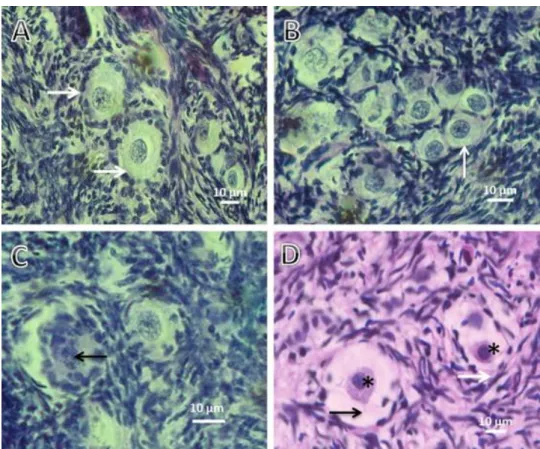
Morphologically normal (Fig. 1A and B) and atretic preantral follicles (Fig. 1C and D) were
found after all treatments as well as in fresh control. The predominant degenerative
characteristics were shrunken oocytes, pyknosis of oocyte nucleus, oocyte cytoplasm
vacuolization and disorganized granulosa cells.
Fig. 1. Photomicrographs of ovarian cortical histological sections showing preantral follicles before (A) and after vitrification (B–D). (A) Normal follicles with one to two layers of cuboidal granulosa cells (white arrows). Normal follicles (white arrow) after vitrification through MTV/VS4 (B). (C) Nucleus of degenerated follicle (black arrow) after vitrification through SSV/VS4. (D) Degenerated follicles displaying oocyte nuclear pyknosis (asterisks), slight cytoplasm retraction (black arrow) and disorganization of granulosa cells layers (white arrow) after vitrification through CV/VS4.
The percentage of morphologically normal preantral follicles in ovarian fragments from
fresh control (79.40 ± 7.83%) and after all vitrification treatments is shown in Table 2. All
treatments had significantly reduced percentages of morphologically normal follicles when
Vitrification techniques only presented significant differences, between them, when using VS6,
with a higher percentage of morphological normal follicles with SSV (59.00 ± 13.09).
Concerning the vitrification solution, no differences were observed when performing MTV but
the CV using the VS4 allowed attaining the higher percentage of morphological normal
follicles. When using SSV, both VS6 and VS4 allowed the observation of a higher percentage of
morphological normal follicles.
Table 2.
Percentages of morphologically normal ovine preantral follicles in fresh (control) and in
vitrified ovarian tissue by using macrotube vitrification (MTV), solid-surface vitrification
(SSV) or conventional vitrification (CV) and six solutions containing 6 M ethylene glycol
(EG) and with or without sucrose (SUC) (0.25 or 0.50 M) and with or without fetal calf
serum (FCS) (10%).
Data are displayed as the mean ± SD. A,BDiffering capital letters within a row illustrate significantly differing percentages among vitrification techniques (P < 0.05). a,bDiffering lowercase letters within a column illustrate significantly differing percentages among vitrification solutions (P < 0.05).
MTV, macrotube vitrification; SSV, solid-surface vitrification; CV, conventional vitrification; VS, vitrification solution; BM, base medium; FCS, fetal calf serum; SUC, sucrose.
*Percentage significantly different (P < 0.05) from the non-vitrified ovarian cortex (control).
The percentage of normal follicles was significantly greater in VS4 compared to VS1
and VS6 (when using the SSV or CV techniques). Therefore, VS4 was selected for assessment of
viability and ultrastructure.
3.2. Preantral follicle viability using trypan blue after vitrification of ovine ovarian tissue
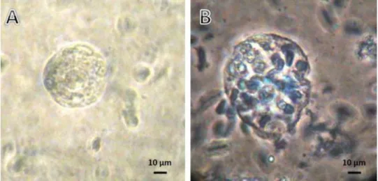
Viable and nonviable follicles obtained from vitrified ovarian cortex fragments are shown
in Fig. 2. The percentage of viable preantral follicles was similar to that in the fresh control
(92.62%) only after MTV/VS4 (97.06%) (P < 0.05). Furthermore, MTV/VS4 preserved the
follicular viability better than SSV/VS4 (81.08%) or CV/VS4 (83.81%) (P < 0.05). These results
Fig. 2. Photomicrographs of trypan blue dye-treated preantral follicles that were mechanically isolated from vitrified ovarian tissue showing (A) viable follicle (not stained) and a (B) nonviable follicle (stained). (For interpretation of the references to color in this figure legend, the reader is referred to the web version of the article.)
3.3. Ultrastructural analysis of vitrified preantral follicles
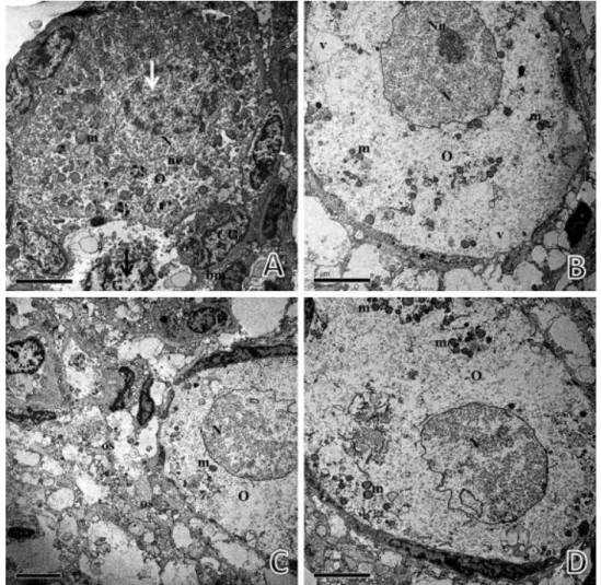
The ultrastructural analysis showed that follicles from the fresh control group (Fig. 3A) and
follicles from MTV/VS4 (Fig. 3B) treatment were similar, presenting oocytes with a large
central nucleus well-defined by a nuclear envelope. Organelles were uniformly distributed
throughout the homogeneous cytoplasm, with mitochondria being the most evident organelle,
and granulosa cells were normal in appearance. In the fresh control group (Fig. 3A), it is
possible to see some vesicles. Although MTV/VS4 (Fig. 3B) follicular ultrastructure was similar
to fresh control follicles, some discreet changes could be observed, like a slight shrinkage in
the nuclear envelope.
Conversely, more drastic alterations could be observed during ultrastructure analysis
of SSV/VS4 (Fig. 4A) and CV/VS4 (Fig. 4B–D) follicles. For example, oocytes exhibited cytoplasm
vacuolization with a granulated appearance and contained empty spaces. In the SSV/VS4
follicles, granulosa cells had a loss of cytoplasmic content. CV/VS4-treated follicles resulted in
the greatest ultrastructural abnormalities with vacuolated follicles and the majority of
organelles lost in the oocyte cytoplasm (Fig. 4B), which presumably left empty spaces in the
oocyte cytoplasm content. The ovarian stromal area also had signs of degeneration, with
largely vacated areas (Fig. 4C).
3.4.
In vitro culture of preantral follicles after vitrification
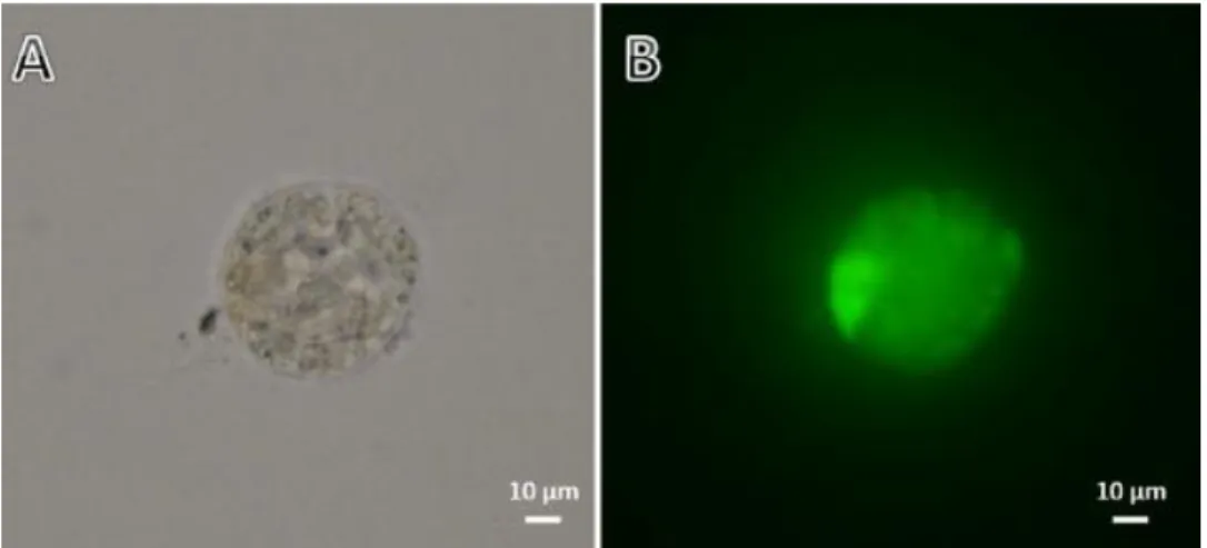
Follicular viability, based on fluorescent markers (Fig. 5), immediately after MTV/VS4 (65%)
was reduced when compared to the non-vitrified follicles at day 0 (100%) (P < 0.05) ( Table 3).
However follicular viability after MTV/VS4 at day 2 (36.5%) was similar to follicles vitrified at
day 0 (65%) and similar to non-vitrified follicles at day 2 (62.5) (P > 0.05). As the decrease of
viability in non-vitrified follicles at day 2 was similar to the decrease of MTV/VS4 in the same
time, follicle viability at day 2 is not affected by MTV/VS4.
Fig. 5. View of viability evaluation of isolated preantral follicles. (A) Direct observation of isolated ovarian follicles. (B) Fluorescent staining with calcein-AM and ethidium homodimer-1. No dead follicle is shown in this view.
Table 3.
Percentages of viable ovine preantral follicles non-vitrified and vitrified at day 0 and day 2 of in vitro culture.
A,B Differing capital letters within a row illustrate significantly differing percentages between follicles before (at day 0) and after (at day 2) 48 h in vitro culture (P < 0.05). a,bDiffering lowercase letters within a column illustrate significantly differing percentages between follicles before (non-vitrified) and after MTV/VS4 (P < 0.05)
4.
Discussion
In this study were evaluated the effects of three different techniques for vitrification of
sheep ovarian tissue: the vitrification in macrotubes (MTV), solid-surface vitrification (SSV) and
conventional vitrification (CV), combined with six different vitrification solutions, on the
follicular morphology and viability preservation. Based on our previous experimental data
(Carvalho et al., 2011), the solutions tested were composed of minimum essential medium
calf serum (FCS) and with or without sucrose (SUC) in two different concentrations (0.25 or
0.50 M).
In the first experiment, we found higher percentage of morphologically normal preantral
follicles cryopreserved when used the VS4 (EG + SUC 0.25 M + 10% FCS) in all the vitrification
techniques. In addition, this solution was the only that presented percentage of normal
follicles similar to fresh control (CV/VS4). This result is probably due to the optimized
combination of low SUC concentration (0.25 M) with FCS (10%). The absence of SUC in VS
probably reduced its dehydration potential, allowing higher intracellular water and,
consequently, promoting more intracellular water crystallization (Bao et al., 2010), thereby
demonstrating that a delicate balance between SUC and FCS is essential for preserving normal
preantral follicle morphology after cryopreservation process. On the other hand, VS with
higher concentration of SUC could have promoted deleterious dehydration to the tissue,
causing evaluable damage. Both, the SUC and the FCS, acted synergistically permitting to influx
and efflux of substances through the plasma membranes of preantral follicles (Jain and
Paulson, 2006). This effect results in a favorable transport of EG and water in this cellular type.
Moreover, the combination between 0.25 M of SUC and FCS in the basic medium as observed
above was beneficial for the preservation of follicular morphology in all vitrification techniques
(CV/VS4, MTV/VS4 and SSV/VS4). And at the same time, high SUC concentration (0.5 M)
associated with FCS (10%) also permitted the preservation of percentage of morphologically
normal follicles in SSV/VS6. Probably, the fragments were better preserved in this association
because in SSV the thawing procedure occurred without contact with the vitrification solution,
whereas the thawing procedure in CV and MTV keeps the fragments in contact with the
vitrification solution, causing damage due to an additional exposure to this solution (Jain and
Paulson, 2006).
The results were similar to those previously reported by Bao et al. (2010) that in vitrified
bovine follicles in a solution containing, among others, EG and FCS, the addition of SUC was
essential to maintain the follicle morphology and the potential for further development to
advanced stages. One study performed by Santos et al. (2007) demonstrated that the
combination of EG and SUC was the most suitable for vitrification of goat ovarian tissue,
obtaining higher rates of morphologically viable follicles. Despite the use of a slow freezing
technique, recently, Santos et al. (2011) also showed that sheep preantral follicles are best
preserved (morphology and viability) when SUC was associated with EG in the freezing
solution.
The cryoprotectant EG, a basic component of vitrification solution in this study, was
(Massip, 2001 and Amorim et al., 2003) and low toxicity (Zhang et al., 2010). The
cryoprotectants reduce cell damage caused by cryopreservation, since they act by partially
replacing the water inside the cell and binding to hydrogen molecules in the intracellular
water, increasing the viscosity of the cryopreservation solution and thus reducing the freezing
point (Fuller and Paynter, 2004).
Unlike intracellular cryoprotectants, the extracellular cryoprotectants, as SUC, remain
outside the cell and interact with the water-free present in the solution, influencing indirectly
the processes of osmotic cellular dehydration. The SUC acts as a buffer against osmotic stress
during the addition and the removal of intracellular cryoprotectant and has the potential to
stabilize the cell membrane, minimizing cell damage (Fabbri et al., 2001). Despite the process
of cellular dehydration prevents intracellular ice formation (IIF), dehydration can also cause
excessive osmotic stress (Vajta et al., 1998). Therefore, we believed this is the reason why the
low SUC concentration used in this experiment (0.25 M) had supported better results.
The FCS, which composition is not defined, or in some cases semi-defined, also participates
in the controlling of the water flow through cell membranes. Its action occurs through proteins
and large macromolecules, which bind to the lipid membranes, protecting these structures
(Wang et al., 1997). In our study we found that the FCS is related to the preservation of the
morphology of sheep preantral follicles after vitrification, similarly as shown in goat ovarian
tissue by Carvalho et al. (2011). We believe that this substance promotes the exchange of
fluids between the vitrification solution and cellular environment, an effect that apparently
was enhanced by the association with low SUC concentration (0.25 M). In addition to the
osmotic equilibrium maintenance, the FCS can act as a source of nutrients (protein,
carbohydrates, lipids and vitamins), possibly aiding in the recovery of cellular metabolism after
the cryopreservation procedures (Wang et al., 1997).
Unlike the observed in the morphological analysis, whose best results were obtained with
the CV technique, when concerning follicular viability it was observed that follicles were better
preserved using MTV. Furthermore, this same treatment MTV/VS4 was the only that showed
follicles with ultrastructural characteristics similar to those from fresh control. This can be
explained by the fact that histological studies identify only a few signs of follicular atresia, such
as nuclear pyknosis, detachment of the granulosa cells of the oocyte and the basement
membrane abnormalities.
Thus, the visual observation of follicular morphology alone is insufficient to assess the
efficacy of the cryopreservation process by itself (Martinez-Madrid et al., 2004).
In this regard, the viability and ultrastructure assessments determined that MTV was the
vitrification of larger fragments than CV technique, because the French straws are narrow and
do not allow proper handling of 9 mm3 fragments. Similar to MTV, the SSV technique,
described by Begin et al. (2003) and still used by many researchers (Santos et al., 2007 and
Carvalho et al., 2011), permits the vitrification of larger fragments. However, to perform this
technique the tissue must to be in contact with a cooled aluminum metal partly immersed into
liquid nitrogen (Begin et al., 2003). Such tissue exposure to liquid nitrogen is discouraged, since
there is the possibility of cellular contamination by nitrogen contact (Criado et al., 2011 and
Isachenko et al., 2009). While some authors indicate techniques such as ultraviolet sterilization
or filtration of liquid nitrogen, these procedures would not prevent cross-contamination
between cells of different individuals in the same cryogenic tank (Parmegiani et al., 2010).
Studies have shown that short-term culture can be used as a valuable tool to verify the
developmental ability of cryopreserved preantral follicles (Choi et al., 2008). Based on these
previous studies, we conducted a second experiment including the short-term in vitro culture
as an additional tool to precisely evaluate the follicular viability after MTV/VS4. Also, it is
known that this type of culture has no intention of promoting follicular growth, but allows the
tissue to return to its normal metabolic conditions, recovering from possible cryoinjuries, or
even allowing the cells to express the molecular damage that may have occurred during
cryopreservation, undetectable at the post-thaw ( Paynter et al., 1999).
To assess the percentages of viable non-vitrified and vitrified ovine preantral follicles at
day 0 and day 2 of in vitro culture, we used the fluorescent markers ethidium homodimer-1,
which enters into cells with disrupted plasmatic membrane and binds to DNA, and calcein-AM,
which assesses esterase activity in viable cells. This method is more sensitive than histology for
the assessment of follicular quality and more accurate than the trypan blue previously used in
the first experiment of this study, by evaluating both, morphological and functional structures
simultaneously. With these markers, it was found that follicular viability immediately after
MTV/VS4 (65%) was reduced when compared to the non-vitrified follicles at day 0 (100%) (P <
0.05). Similarly, other studies (Gonçalves et al., unpublished data; Oskam et al., 2011) did not
obtain satisfactory results using similar conditions of in vitro culture even after 2 and 4 h after
freezing sheep ovarian tissue compared to non-vitrified follicles at day 0. However, in the
present study the significant reduction in viability from day 0 to day 2 was observed only in
follicles from non-vitrified tissue. Thus, follicular viability after MTV/VS4 at day 2 (36.5%) was
similar to follicles vitrified at day 0 (65%) and similar to non-vitrified follicles at day 2 (62.5) (P >
0.05). Due to the decrease of viability in non-vitrified follicles at day 2, the viability at day 2
MTV/VS4 is suitable for the preantral follicles, because they are able to survive after
short-term in vitro culture as successfully as fresh follicles were.
In conclusion, when using the experimental conditions of the present study, an efficient
solution (VS4: 6 M EG, 0.25 M SUC and 10% FCS) and technique (MTV) were successfully used
for the vitrification of ovine ovarian tissue.
Acknowledgements
This work was supported by CNPq, Brazil. F.O. Lunardi is supported by a grant from Fundação
Cearense de Apoio ao Desenvolvimento Científico e Tecnológico (FUNCAP). J.R. Figueiredo,
A.P.R Rodrigues and S.N. Báo are supported by a grant from CNPq.
REFERENCES
Amorim, C.A., Rodrigues, A.P.R., Lucci, C.M., Figueiredo, J.R., Gonçalves, P.B.D., 2000. Effect of
sectioning on the number of isolated ovine preantral follicles. Small Rumin. Res. 37,269-277.
Amorim, C.A, Rondina, D„ Rodrigues, A.P.R., Costa, S.H.F., Gonçalves, P.B.D., de Figueiredo, J.R., Giorgetti, A, 2003. Isolated ovine primordial follicles cryopreserved in different
concentrations of ethylene glycol. Theriogenology 60,735-742.
Bao, R.M., Yamasaka, E., Moniruzzaman, M., Hamawaki, A., Yoshikawa, M„ Miyano, T„ 2010.
Development of vitrified bovine secondary and primordial follicles in xenografts.
Theriogenology 74,817-827.
Begin, I., Bhatia, B., Baldassarre, H., Dinnyes, A., Keefer, C.L, 2003. Cryopreservation
of goat oocytes and in vivo derived 2-to 4-cell embryos using the cryoloop (CLV) and
solid-surface vitrification (SSV) methods. Theriogenology 59,1839-1850.
Bordes, A., Lomage, J., Demirci, B., Franck, M., Courbiere, B., Guerin, J.F., Salle, B„ 2005.
Normal gestations and live births after orthotopic autograft of vitrifled-warmed hemi-ovaries
Carvalho, AA, Faustino, L.R., Silva, C.M.G., Castro, S.V., Luz, H.K.M., Rossetto, R., Lopes, C.A.P.,
Campello, C.C., Figueiredo, J.R., Rodrigues, A.P.R., Costa, A.P.R., 2011. Influence of vitrification
techniques and solutions on the morphology and survival of preantral follicles after in vitro
culture of caprine ovarian tissue. Theriogenology 76,933-941.
Celestino, dos Santos, R.R., Lopes, CAP., Martins, F.S., Matos, M.H.T., Melo, MAP., Bao, S.N.,
Rodrigues, AP.R., Silva, J.R.V., de Figueiredo, J.R., 2008. Preservation of bovine preantral follicle
viability and ultrastructure after cooling and freezing of ovarian tissue. Anim. Reprod.
Sci. 108,309-318.
Chemaitilly, W., Mertens, AC., Mitby, P., Whitton, J., Stovall, M., Yasui, Y., Robison, LL, Sklar,
CA, 2006. Acute ovarian failure in the childhood cancer survivor study. J. Clin. Endocrinol.
Metab. 91,1723-1728.
Chen, S.U., Chien, C.L, Wu, M.Y., Chen, T.H., Lai, S.M., Lin, C.W., Yang, Y.S., 2006. Novel direct
cover vitrification for cryopreservation of ovarian tissues increases follicle viability and
pregnancy capability in mice. Hum. Reprod. 21,2794-2800.
Choi, J„ Lee, B„ Lee, E„ Yoon, B.-K., Bae, D„ Choi, D„ 2008. Cryopreservation of ovarian tissues temporarily suppresses the proliferation of granulosa cells in mouse preantral follicles.
Cryobiology 56,36-42.
Criado, E., Moalli, F., Polentarutti, N., Albani, E., Morreale, G., Menduni, F., Levi-Setti, P.E.,
2011. Experimental contamination assessment of a novel closed ultravitrification device. Fertil.
Steril. 95,1777-1779.
Fabbri, R., Porcu, E., Marsella, T., Rocchetta, G., Venturoli, S., Flamigni, C., 2001. Human oocyte
cryopreservation: new perspectives regarding oocyte survival. Hum. Reprod. 16,411-416.
Fuller, B„ Paynter, S., 2004. Fundamentals of cryobiology in reproductive medicine. Reprod. Biomed. Online 9,680-691.
Gandolfi, F., Paffoni, A., Brambilla, E.P., Bonetti, S., Brevini, T.A.L., Ragni, G„ 2006. Efficiency of
equilibrium cooling and vitrification procedures for the cryopreservation of ovarian tissue:
R.G., Baird, D.T., Wade, J.C., Webb, R., 1994. Restoration of fertility to oophorectomized sheep
by ovarian autografts stored at -1 9 6 ° C. Hum. Reprod. 9,597-603.
Huang, L.L, Mo, Y.Q,, Wang, W.J., Li, Y„ Zhang, Q.X., Yang, D.Z., 2008. Cryopreservation of
human ovarian tissue by solid-surface vitrification.
Eur. J. Obstet. Gynecol. Reprod. Biol. 139,193-198.
Hulshof, S.C.J., Figueiredo, J.R., Beckers, J.F., Bevers, M.M., Vandenhurk, R., 1994. Isolation and
characterization of preantral follicles from fetal bovine ovaries. Vet. Quart. 16,78-80.
Imhof, M., Bergmeister, H., Lipovac, M., Rudas, M., Hofstetter, G., Huber, J., 2006. Orthotopic
microvascular reanastomosis of whole cryopreserved ovine ovaries resulting in pregnancy and
live birth. Fertil. Steril. 85,1208-1215.
Isachenko, E„ Isachenko, V., Rahimi, G., Nawroth, F., 2003. Cryopreservation of human ovarian tissue by direct plunging into liquid nitrogen. Eur. J. Obstet. Gynecol. Reprod. Biol.
108,186-193.
Isachenko, V., Isachenko, E., Weiss, J.M., Todorov, P., Kreienberg, R., 2009. Cryobanking of
human ovarian tissue for anti-cancer treatment: comparison of vitrification and conventional
freezing. Cryoletters 30, 449-454.
Jain, J.K., Paulson, R.J., 2006. Oocyte cryopreservation. Fertil. Steril. 86, 1037-1046.
Kagabu, S., Umezu, M., 2000. Transplantation of cryopreserved mouse, Chinese hamster,
rabbit, Japanese monkey and rat ovaries into rat recipients. Exp. Anim. (Tokyo) 49,17-21.
Kagawa, N„ Silber, S., Kuwayama, M„ 2009. Successful vitrification of bovine and human ovarian tissue. Reprod. Biomed. Online 18, 568-577.
Kim, GA, Kim, H.Y., Kim, J.W., Lee, G., Lee, E., Um, J.M., 2010. Ultrastructural deformity of
Lee, S.J., Schover, L.R., Partridge, A.H., Patrizio, P., Wallace, W.H., Hagerty, K., Beck, L.N.,
Brennan, L.V., Oktay, K„ 2006. American Society of Clinical Oncology recommendations on fertility preservation in cancer patients. J. Clin. Oncol. 24,2917-2931.
Liu, LJ., Xie, X.Y., Zhang, R.Z., Xu, P., Bujard, H., Jun, M., 2008. Reproduction and fertility in
wild-type and transgenic mice after orthotopic transplantation of cryopreserved ovaries from
10-d-old mice. Lab Anim. 37, 353-357.
Lomage, J., Courbière, B., Mazoyer, C., Odagescu, V., Baudot, A., Bordes, A, Poirel, M.T.,
Franck, M„ Salle, B„ 2006. Ovarian tissue vitrification: cortex and whole ovary in sheep. Gynecol. Obstet. Fertil. 34, 746-753.
Martinez-Madrid, B„ Dolmans, M.M., Van Langendonckt, A, Defrere, S., Donnez, J., 2004.
Freeze-thawing intact human ovary with its vascular pedicle with device a passive cooling.
Fertil. Steril. 82, 1390-1394.
Massip, A, 2001. Cryopreservation of embryos of farm animals. Reprod. Domest. Anim.
36,49-55.
Meirow, D„ Nugent, D„ 2001. The effects of radiotherapy and chemotherapy on female reproduction. Hum. Reprod. Update 7,535-543.
Melo, MAP., Oskam, I.C., Celestino, J.J.H., Carvalho, AA, Castro, S.V., Figueiredo, J.R.,
Rodrigues, A.P.R., Santos, R.R., 2011. Adding ascorbic acid to vitrification and IVC medium
influences preantral follicle morphology, but not viability. Reprod. Domest. Anim. 46,742-745.
Moniruzzaman, M., Bao, R.M., Taketsuru, H„ Miyano, T., 2009. Development of vitrified
porcine primordial follicles in xenografts. Theriogenology 72,280-288.
Oktay, K., Karlikaya, G.G., Aydin, B A, 2000. Ovarian cryopreservation and transplantation:
basic aspects. Mol. Cell. Endocrinol. 169,105-108.
Oskam, I.C., Lund, T„ Santos, R.R., 2011. Irreversible damage in ovine ovarian tissue after
cryopreservation in propanediol: analyses after in vitro culture and xenotransplantation.
Parmegiani, L, Accorsi, A., Cognigni, G.E., Bernardi, S., Troilo, E„ Filicori,M„ 2010. Sterilization
of liquid nitrogen with ultraviolet irradiation for safe vitrification of human oocytes or
embryos. Fertil. Steril. 94, 1525-1528.
Paynter, S.J., Cooper, A., Fuller, B.J., Shaw, R.W., 1999. Cryopreservation of bovine ovarian
tissue: structural normality of follicles after thawing and culture in vitro. Cryobiology
38,301-309.
Salle, B„ Demirci, B„ Franck, M„ Berthollet, C„ Lornage, J„ 2003. Long-term follow-up of cryopreserved hemi-ovary autografts in ewes: pregnancies, births, and histologic assessment.
Fertil. Steril. 80,172-177.
Salle, B., Demirci, B., Franck, M., Rudigoz, R.C., Guerin, J.F., Lornage, J., 2002. Normal
pregnancies and live births after autograft of frozen-thawed hemi-ovaries into ewes. Fertil.
Steril. 77,403-408.
Santos, R.R., Amorim, C„ Cecconi, S., Fassbender, M„ Imhof, M„ Lornage, J., Paris, M.,
Schoenfeldt, V., Martinez-Madrid, B., 2010. Cryopreservation of ovarian tissue: an emerging
technology for female germline preservation of endangered species and breeds. Anim. Reprod.
Sci. 122,151-163.
Santos, R.R., Tharasanit, T„ Figueiredo, J.R., van Haeften, T„ van den Hurk, R., 2006a.
Preservation of caprine preantral follicle viability after cryopreservation in sucrose and
ethylene glycol. Cell. Tissue Res. 325, 523-531.
Santos, R.R., Rodrigues, A.P.R., Costa, S.H.F., Silva, J.R.V., Matos, M.H.T., Lucci, C.M., Bao, S.N.,
van den Hurk, R., Figueiredo, J.R., 2006b. Histological and ultrastructural analysis of
cryopreserved sheep preantral follicles. Anim. Reprod. Sci. 91,249-263.
Santos, R.R., Tharasanit, T., Van Haeften, T., Figueiredo, J.R., Silva, J.R.V., Van den Hurk, R.,
2007. Vitrification of goat preantral follicles enclosed in ovarian tissue by using conventional
Santos, R.R., Van den Hurk, R., Rodrigues, A.P.R., Figueiredo, J.R., 2011. Viability of oocytes and
granulosa cells from cryopreserved ovine ovarian primordial, primary and secondary follicles.
Small Rumin. Res. 99, 203-207.
Silber, S., Kagawa, N., Kuwayama, M., Gosden, R., 2010. Duration of fertility after fresh and
frozen ovary transplantation. Fertil. Steril. 94, 2191-2196.
Sonmezer, M„ Oktay, K„ 2004. Fertility preservation in female patients. Hum. Reprod. Update
10,251-266.
Sugimoto, M., Maeda, S., Manabe, N., Miyamoto, H., 2000. Development of infantile rat
ovaries autotransplanted after cryopreservation by vitrification. Theriogenology 53,1093-1103.
Takahashi, E„ Miyoshi, I., Nagasu, T., 2001. Rescue of a transgenic mouse line by transplantation of a frozen-thawed ovary obtained postmortem. Contemp. Top. Lab. Anim. Sci.
40,28-31.
Vajta, G., Holm, P., Kuwayama, M., Booth, P.J., Jacobsen, H., Greve, T., Callesen, H., 1998. Open
pulled straw (OPS) vitrification: a new way to reduce cryoinjuries of bovine ova and embryos.
Mol. Reprod. Dev. 51, 53-58.
Wallace, W.H.B., Anderson, R.A., Irvine, D.S., 2005. Fertility preservation for young patients
with cancer: who is at risk and what can be offered? Lancet Oncol. 6,209-218.
Wang, S., Liu, Y., Holyoak, G.R., Bunch, T.D., 1997. The effects of bovine serum albumin and
fetal bovine serum on the development of preand postcleavage-stage bovine embryos
cultured in modified CR2 and Ml 99 media. Anim. Reprod. Sci. 48,37-45.
Wang, X.Q., Catt, S., Pangestu, M., Temple-Smith, P., 2011. Successful in vitro culture of
pre-antral follicles derived from vitrified murine ovarian tissue: oocyte maturation, fertilization,
and live births. Reproduction 141,183-191.
Zhang, J.M., Liu, X.L, Yang, Y.X., Wan, X.P., 2010. Comparisons of different protocols for