ENZYME-LINKED IMMONOSORBENT ASSAY (ELISA)
The
development of a Direct Competitive ELISA for the determination
of histamine in wine
Haline Garcia Victório
Dissertação para obtenção do Grau de Mestre em Engenharia Alimentar
Orientador: Doutora Maria Adélia Ferreira
Co-orientadores: Doutor Abel Oliva e Dra. Vitoria San Romão
Júri:Presidente: Doutor Ricardo Manuel de Seixas Boavida Ferreira, Professor Catedrático do Instituto Superior de Agronomia da Universidade Técnica de Lisboa.
Vogais: Doutora Maria Adélia da Silva Santos Ferreira, Professora Auxiliar do Instituto Superior de Agronomia da Universidade Técnica de Lisboa;
Doutor Abel Martin González Oliva, Investigador Auxiliar do Laboratório de Diagnóstico Biomolecular – Instituto de Tecnologia Química e Biológica da Universidade Nova de Lisboa; Doutora Ana Isabel Amaro Gonçalves Domingos, Investigadora Auxiliar da Unidade de Tecnologia de Proteínas e Anticorpos Monoclonais do Instituto de Higiene e Medicina Tropical.
i Acknowledgements
I am grateful to Dr. Maria Adélia Ferreira, Dr. Abel Oliva and Dr. Vitoria San Romão for the technical support and guidance over the development of the research and evaluation of this work.
I also thank Dr. Claudia Sánchez Lara and Elisa Campos from ITQB for excellent technical assistance.
Finally, I thank Dr. Ana Domingos, Dr. Fernando Cardoso and Dr. Carlos Novo for permitting the execution of the assays in the facilities of UTPAM/IHMT.
ii Abstract .
The presence of biogenic amines in wines, specifically the histamine (0,1 mg/L to 20 mg/L) has been a constant source of concern for wine producers, since this substance presents toxic effects for human beings.
The goal of this work was the development of a fast, efficient and low cost technique for the determination and quantification of histamine in wine. For that end, the strategy was the use of a competitive ELISA method.
The first step for the development of this method was the acquisition of the antibody for histamine. Among the available options, it was decided to purchase the commercial polyclonal antibody for histamine.
The second step was the conjugation of the histamine with the peroxidase. Three methods of conjugation were performed, followed by their subsequent analysis.
Initially, it was necessary to perform assays for the optimization of the antibody and of the conjugate, as well as to test the blocking buffers (1,0% (w/v) gelatin and 0,5% (w/v) sodium caseinate).
Before the application of the ELISA competitive method, auxiliary tests such as indirect ELISA, ELISA inhibition test and ELISA binding test were performed. However, continuously unsatisfactory results were obtained, leading to the perception that there could be a low efficiency of the conjugation processes and/or a lack of specificity between the histamine used and the polyclonal antibody acquired for the assays.
iii Resumo em Português
A presença de aminas biogénicas em vinhos, especificamente a histamina (0,1 mg/L a 20 mg/L) tem sido uma constante preocupação para o mercado vinícola, já que esta substância apresenta efeitos tóxicos para seres humanos.
O objectivo do presente trabalho foi desenvolver uma técnica rápida, eficaz e de baixo custo para a determinação e quantificação de histamina em vinhos. Para esse fim, foi proposto o método ELISA competitivo.
O primeiro passo para o desenvolvimento do método foi a obtenção do anticorpo anti- histamina. De entre as alternativas existentes, optou-se pela compra de um anticorpo policlonal.
O segundo passo foi a conjugação da histamina com a peroxidase. Foram feitos três processos de conjugação, seguidos de suas respectivas análises.
Inicialmente, foram realizados ensaios para a optimização do anticorpo e do conjugado, assim como foram testados dois tampões de bloqueio (1,0% (p/v) gelatina e 0,5% (p/v) caseinato de sódio).
Anteriormente à aplicação do método ELISA competitivo, foram realizados ensaios como ELISA indirecto, ELISA inhibition test e o ELISA binding test. Porém, os resultados foram pouco satisfatórios indicando uma possível falha no processo de conjugação e/ou baixa especificidade da histamina com o anticorpo utilizado.
iv Resumo alargado em Português
A presença de aminas biogénicas em vinhos, especificamente a histamina (0,1 mg/L a 20 mg/L) tem sido uma constante preocupação para seus produtores e consequentemente para o mercado vinícola português, já que esta substância apresenta efeitos tóxicos para seres humanos. Uma das técnicas mais utilizadas para detecção dessa substância em vinhos é o HPLC (High-Performance Liquid Chromatography) porém, este método apresenta um custo elevado de equipamentos e manutenção, além de exigir técnicos treinados para o seu desenvolvimento.
O objectivo deste presente trabalho foi desenvolver uma técnica rápida, eficaz e de baixo custo. Para esse fim, foi desenvolvido um método imunoenzimático que se baseia na utilização de antigénios ou anticorpos marcados com enzimas que permitem a detecção e quantificação de substâncias de interesse biológico. Foi proposto portanto, o método ELISA competitivo, cujo princípio se baseia nas seguintes etapas: (i) o revestimento dos poços de uma placa ELISA com anticorpo específico contra a histamina; (ii) adição simultânea da histamina livre e da histamina marcada com a enzima peroxidase, com o objectivo de provocar uma competição pelo local de ligação do anticorpo; (iii) adição de um substrato cromogénico específico (TMB- tetrametilbenzidine); (iv) adição de ácido sulfúrico 1M com o objectivo de parar a reacção e medir fotometricamente a absorvância a 450 nm. Os resultados dos valores de absorvância esperados deveriam ser inversamente proporcionais à concentração de histamina da amostra.
Embora haja no mercado kits ELISA comercias para determinação de histamina em vinhos, todos esses métodos fazem um pré-tratamento (acilação ou derivatização) da histamina. O propósito do presente trabalho, foi avaliar o comportamento da histamina sem a etapa de pré-tratamento a fim de tornar o método mais fácil e rápido.
O primeiro passo para o desenvolvimento do método foi a obtenção do anticorpo anti-histamina. De entre as opções existentes, optou-se pela compra de um anticorpo policlonal anti-histamina.
O segundo passo foi a conjugação da histamina com a peroxidase. Este processo de conjugação é necessário para que seja possível quantificar através de um
v
espectrofotómetro a histamina marcada (neste caso com a enzima peroxidase), e diferenciá-la da histamina livre presente na amostra. Três métodos de conjugação foram realizados, um via periodato e os outros dois utilizando glutaraldeído. Foram realizadas análises para determinação da eficiência de conjugação e concluiu-se que o terceiro método de conjugação, usando glutaraldeído, era o que apresentava melhores condições para utilização nos ensaios subsequentes.
Inicialmente, foram realizados ensaios para a optimização do anticorpo anti-histamina e do conjugado (histamina + peroxidase), assim como foram testados dois tampões de bloqueio (1,0% (p/v) gelatina e 0,5% (p/v) caseinato de sódio); concluiu-se, portanto, que diluições na ordem de 1/1000 no caso do anticorpo e de 1/400 no caso do conjugado seriam apropriadas.
Após optimização da reacção entre anticorpo e conjugado, optou-se por iniciar os ensaios pelo método ELISA indirecto, utilizando ainda dois tampões de bloqueio, 1,0% (p/v) gelatina e 0,5% (p/v) caseinato de sódio. Porém, os resultados foram pouco satisfatórios indicando uma possível falha no processo de conjugação.
Outros ensaios foram realizados, como o ELISA inhibition test e o ELISA binding test. No primeiro ensaio, os resultados indicaram que este pode ser um possível método para determinação de histamina em alimentos que possuam maiores concentrações deste composto, já que somente a partir de 100 ug/µL de histamina foi possível diferenciar os valores de densidade óptica; no segundo ensaio, embora o conjugado não tenha sido utilizado e sim a histamina livre fixada na placa através da utilização da poly-l-lysine, resultados obtidos continuavam a ser muito aquém do esperado, o que poderia reforçar a ideia da falta de especificidade da histamina com o anticorpo em questão.
A fim de verificar a diferença entre trabalhar com a histamina na forma livre como foi proposto neste trabalho e a histamina derivatizada como é proposto pela maioria dos kits ELISA comerciais, considerou-se adequado realizar um ensaio com o kit comercial Histamine ELISA kit (Ref: IB 89128, Immuno Biological Laboratories, Inc. Minneapolis, USA).
De entre as perspectivas apresentadas, outras hipóteses para o não funcionamento do método poderiam ser sugeridas. Uma delas seria a pouca especificidade entre a histamina utilizada nos ensaios e o anticorpo. Outra hipótese seria a de que o processo
vi
de derivatização da histamina, como ocorre na maioria dos kits comerciais, seja fundamental para o sucesso dos ensaios – porém, para que isso ocorra, é fundamental que o processo de derivatização da histamina seja realizado, seguido da conjugação desta com moléculas como BSA (Bovine Serum Albumin), KLH (Keyhole Limpet Hemocyanin), HSA (Human Serum Albumin) e a imunização dos animais para que o anticorpo produzido seja específico para a histamina a ser utilizada.
vii Contents
Abstract . ...ii
Resumo em Português ... iii
Resumo alargado em Português ... iv
1. Introduction ... 1
2. Definition – Biogenic amines ... 2
2.1. Biogenic amines in food. ... 3
2.2. Toxic action ... 5
2.2.1. Histamine and histamine metabolism ... 7
2.2.2. Histamine intolerance... 9
2.2.3. Histamine and headache ... 9
2.2.4. Histamine and gastrointestinal ... 9
2.3. Origin of biogenic amines in wine ... 10
2.4. Factors that influence the concentration of amines in wine ... 10
2.4.1. Raw materials: grapes and must... 11
2.4.2. Influence of pH... 11
2.4.3. Amino acid concentration ... 12
2.4.4. Influence of winemaking ... 12
2.4.5. Influence of vinification conditions ... 13
2.4.6. Influence of malolactic fermentation and storage on biogenic amines concentration. ... 14
2.4.7. Formation of amines during alcoholic and malolactic fermentation ... 14
2.5. Immunochemical methods ... 15
2.5.1. Definition of immunochemical methods ... 15
2.5.2. Immunoenzyme methods ... 16
2.5.2.1. Heterogeneous EIA – ELISA techniques ... 16
i. Competitive ELISA for antigens and haptens ... 17
ii. Sandwich ELISA for antigens ... 18
iii. Indirect ELISA for antigens ... 18
iv. Sandwich ELISA for antibodies ... 19
viii
2.5.3.1. Principles of ELISA ... 20
2.5.3.2. Steps in ELISA ... 21
2.5.3.2.1. Solid-Phase ... 22
2.5.3.2.2. Blocking conditions and nonspecific reactions ... 23
2.5.3.2.3. Washing ... 23 2.5.3.2.4. Addition of sample ... 24 2.5.3.2.5. Incubation ... 24 2.5.3.2.6. Enzyme conjugates ... 24 2.5.3.2.7. Development of color ... 24 2.5.3.2.8. Stopping reactions ... 25 2.5.3.2.9. Spectrophotometric reading ... 25 2.5.3.3. Advantages in ELISA ... 25
3. Materials and Methods ... 27
3.1. Chemicals ... 27
3.2. Preparation of a histamine-HRP conjugate I via periodate method ... 27
3.3. Determination of the percentage of peroxidase in the conjugate I ... 27
3.4. Determination of the effect of HRP enzyme - TMB substrate reaction time on absorbance ... 28
3.5. Preparation of a histamine-HRP conjugate II... 28
3.6. Synthesis of histamine-enzyme conjugate III... 28
3.7. Chessboard titration (titration of antigen and antibody) ... 29
3.7.1. Detailed procedures of the chessboard titration ... 29
3.8. Indirect ELISA ... 30
3.9. ELISA – Inhibition test ... 32
3.10. ELISA binding test (poly-l-lysine) ... 32
3.11. Direct competitive enzyme immunoassay ... 33
3.12. Histamine ELISA Kit (Immuno Biological Laboratories,Inc. Minneapolis, USA) 34 4. Results and Discussion ... 35
4.1. Determination of the percentage of peroxidase in the conjugate ... 38
4.1.1. HRP optical density ... 38
ix
4.2. Determination of the effect of HRP enzyme - TMB substrate reaction time on
absorbance. ... 40
4.2.1. Determination of the effect of conjugate I - TMB substrate reaction time on absorbance ... 41
4.2.2. Determination of the effect of conjugate II - TMB substrate reaction time on absorbance ... 42
4.2.3. Determination of the effect of conjugate III - TMB substrate reaction time on absorbance ... 43
4.3. Chessboard titration ... 44
4.4. Indirect ELISA ... 47
4.5. ELISA inhibition test ... 48
4.6. ELISA binding test ... 51
4.7. ELISA competitive ... 53
4.8. Histamine ELISA Kit (Immuno Biological Laboratories, Inc. Minneapolis, USA) 54 5. Conclusion ... 57
6. Possible steps for further studies on this subject ... 59
7. References ... 60
8. Annexes. ... 65
8.1. Preparation of solutions used in this work ... 65
8.2. Problems and Solutions in ELISA ... 67
x Contents of figures
Figure 1. Precursor amino acids of biogenic amines. ... 3
Figure 2. Summary of histamine-mediated symptoms. ... 7
Figure 3. Summary of the histamine metabolism. ... 8
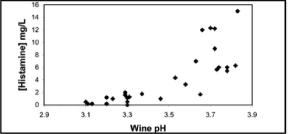
Figure 4. pH values versus histamine concentration from 28 different red, rosé, and white wines analyzed with malolactic fermentation accomplished. ... 11
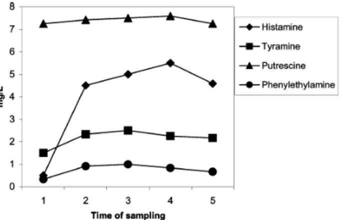
Figure 5. Evolution of histamine, tyramine, phenylethylamine, and putrescine concentration of wines from Utiel-Requena, before (1) and after (2) malolactic and 3 (3), 6 (4), and 12 (5) months of storage. ... 14
Figure 6. Competitive ELISA of haptens and antigens ... 17
Figure 7. Sandwich ELISA for antigens ... 18
Figure 8. Indirect ELISA for antigens ... 19
Figure 9. Sandwich ELISA for antibodies ... 19
Figure 10. Chessboard titration of antigen and antibody. ... 29
Figure 11. Diagrams of ELISA ... 31
Figure 12. Indirect ELISA ... 31
Figure 13. ELISA Inhibition test ... 32
Figure 14. ELISA binding test ... 33
Figure 15. Competitive ELISA ... 34
Figure 16. Determination of optical spectrum of HRP... 38
xi
Figure 18. Calibration curve of HRP ... 41
Figure 19. Calibration curve conjugate I HRP-HA via periodate method ... 42
Figure 20. Calibration curve for the conjugate II HRP-GA-HA. ... 43
Figure 21. Calibration curve for the conjugate III HRP-GA-HA ... 44
Figure 22. Chessboard titration conjugate II (HA-GA-HRP) – 1,0% (w/v) gelatin ... 45
Figure 23. Chessboard titration Conjugate II (HA-GA-HRP) – 1,0% (w/v) sodium caseinate ... 46
Figure 24. Chessboard titration Conjugate III (HA-GA-HRP) – 0,5% (w/v)sodium caseinate ... 46
Figure 25. Indirect ELISA – blocking buffer 1,0% (w/v) gelatin ... 47
Figure 26. Indirect ELISA – blocking buffer 0,5% (w/v)sodium caseinate ... 48
Figure 27. ELISA Inhibition test – HA-GA-BSA ... 49
Figure 28. ELISA Inhibition test – HA-GA-BSA. Higher concentration of histamine ... 49
Figure 29. ELISA Inhibition test – HA-GA-HRP. ... 50
Figure 30. ELISA binding test – poly-l-lysine ... 51
Figure 31. ELISA binding test – poly-l-lysine. Higher concentration of histamine ... 52
xii Contents of tables
Table 1. Species of lactic acid bacteria able to produce biogenic amines ... 5
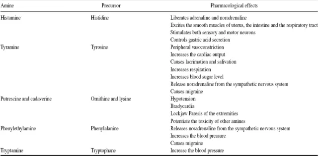
Table 2. Biogenic amines and their pharmacological effects ... 6
Table 3. Determination of the percentage of peroxidase in the conjugate I ... 40
xiii Abbreviations
BA: Biogenic Amines HA: Histamine GA: Glutaraldehyde
HRP: Horseradish Peroxidase BSA: Bovine Serum Albumin
TMB: 3,3’, 5’5 Tetramethylbenzidine Ab: Antibody
Ag: Antigen
Conjugate I (HA-HRP): conjugation of histamine with the enzyme via periodate method according to the Schneider protocol (Schneider E. et al, 1997).
Conjugate II (HA-GA-HRP): conjugation of histamine with the enzyme according to the Rice protocol (Rice T.K. et al, 1982).
Conjugate III (HA-GA-HRP): conjugation of histamine with the enzyme according to the Fujiwara protocol (Fujiwara et al., 1997)
HA-GA-BSA: conjugation of histamine with BSA according to the Fujiwara protocol (Fujiwara et al., 1997)
KLH: Keyhole Limpet Hemocyanin HSA: Human Serum Albumin PBS: Phosphate Buffered Saline TBS: Tris Buffered Saline
1 1. Introduction
The presence of Biogenic Amines (“BA”) in fermented foods, such as beer and wine, cheese and other dairy products, fish, sausages and meat has been receiving increasing attention due to its potential undesirable physiological effects in sensitive humans.
BA have been referred as being involved in relevant pathologies like immunological, neurological, and gastrointestinal diseases like gastric acid secretion (Marcobal et al., 2005, Aygun et al., 1999).
In wines, BA may be formed by lactic acid bacteria mostly after malolactic fermentation and/or during wine aging (Lonvaud-Funel, 2001). The main BA in wine is histamine, tyramine, putrescine, cadaverine and phenylethylamine. The presence of BA in wine is well documented and a wide range of biogenic amine concentrations are reported among different types of wine (from various origins and different winemaking processes), as reported by Lonvaud-Funel and Joyeux in a review issued in 1994.
There are two main reasons for the increasing interest in the study of amines in wines: (a) the toxicological risks associated with the ingestion of elevated amounts of these substances; and (b) the possible relationship between (i) high amine content and (ii) the quality of the grapes used in the production of the wine as well as the hygienic-sanitary conditions prevalent during processing (Souza et al., 2005).
The rising consumer demand for higher quality and safe food has led to a renewed interest in studies of biogenic amines. Nowadays, the analytical methods used for separation and quantification of BA in foods and beverage in general are mainly based on chromatography methods: gas chromatography (GC), thin-layer chromatography (TLC), and high-performance liquid chromatography (“HPLC”) with precolumn or postcolumn derivatization techniques (Karovicova et al., 2003). HPLC methods are reported to be the most efficient, sensitive and reproducible ones. However, HPLC techniques require costly equipment, careful maintenance, expensive solvents and accessories, and specially trained personnel (Marcobal et al., 2006; Vidal-Carou et al., 2003). In order to enhance food control measurements, and possibly clarify the role of dietary histamine for human
2
health, it would be important the availability of easier, cheaper and faster methods for quantification of BA in food and beverage (Ayun et al., 1999).
Most immunochemical methods (enzyme immunoassays) for the detection of histamine in human serum, biological fluids, and food are based on antibodies against N-amino derivatives of histamine synthesized by reaction with, for example, p-benzoquinone or propionic acid esters. More recently, Claeys-Bruno et al., 2006, suggested methodological approaches for histamine quantification using derivatization by choroethylnitrosourea. However, since the antibodies used in these tests are not reactive with the natural histamine compound but only with the modified histamine, derivatization of histamine is required before analysis, which either implicates in a time-consuming process (propionic acid esters) or requires toxic reagents (p-benzoquinone, choroethylnitrosourea) (Ayun et al., 1999).
Among the several options in food and beverage products, it seemed appropriated to focus on wine, due to its relevance to the Portuguese economy as well as the rising interest on the subject worldwide.
Therefore, the goal of the study conducted was the pursuit of a simpler, more practical, faster and less costly method to quantify free histamine without previous derivatization by immunological methods (e.g. a direct competitive ELISA) in wine.
2. Definition – Biogenic amines
Biogenic amines are basic nitrogenous compounds formed mainly by decarboxilation of amino acids or by amination and transamination of aldehydes and ketones. They are organic bases with low molecular weight and are synthesized by microbial, vegetable and animal metabolisms. Biogenic amines in food and beverage are formed by the enzymes of raw material or are generated by microbial decarboxylation of amino acids, although it has been found that some of the aliphatic amines can be formed “in vivo” by amination from corresponding aldehydes (Silla Santos, 1996).
3
The chemical structure of biogenic amines can either be: aliphatic (putrescine, cadaverine, spermine, spermidine), aromatic (tyramine, phenylethylamine) or heterocyclic (histamine, tryptamine). Several authors have classified cadaverine, putrescine, spermine, and spermidine among polyamines (Karovicova et al., 2003). The precursors of the main biogenic amines involved in food poisoning are the describe below:
Figure 1. Precursor amino acids of biogenic amines. Source: Ancín-Azpilicueta, et al, 2008
The prerequisites for biogenic amine formation by microorganisms are: (i) availability of free amino acids, even if not always leading to amine production; (ii) presence of decarboxylase-positive microorganisms; and, (iii) conditions that allow bacterial growth, decarboxylase synthesis and decarboxylase activity.
2.1. Biogenic amines in food.
Biogenic amines are usually found in virtually all food that contain proteins or free amino acids and are subject to conditions enabling microbial or biochemical activity. The total amount of the different amines formed strongly depends on the nature of the food and the microorganisms present. Biogenic amines are present in a wide range of food products including fish products, meat products, dairy products, wine, beer, vegetables, fruits, nuts and chocolate (Silla Santos, 1996).
4
In non-fermented food, the presence of biogenic amines above a certain level is considered as indicative of undesired microbial activity. However, the presence of biogenic amines in food does not necessarily correlate with the growth of spoilage organisms, because they are not all decarboxylase-positive.
Scombroid fish have most commonly been associated with incidents of histamine intoxication (scombrotoxicosis). The formation of histamine in scombroid and other marine fish containing abundant endogenous histidine has been attributed to microbial action rather than endogenous histidine decarboxilase activity (Karovicova et al., 2003; Silla Santos, 1996).
In fermented foods, it is expected that many kinds of microorganisms are present, some of them being capable of producing biogenic amines. Table 1 shows the lactic acid bacteria species capable of producing biogenic amines like histamine, tyramine, phenylethyllamine and putrescine.
5
Table 1. Species of lactic acid bacteria able to produce biogenic amines
Source: Ancín-Azpilicueta et al, 2008
2.2. Toxic action
Histamine is the most studied biogenic amine and its quantity in food products is usually taken as an indicator of freshness and quality. The effects produced by histamine are the well-known as they are associated with most cases of food poisoning. The intake of 5 mg/L of histamine in wine is considered a tolerable level; the amount of 10 mg/L is considered the maximum level admitted and a proportion higher than 20 mg/L induces a high toxicity (Rauscher-Gabernig et al., 2009).The toxicological levels of biogenic amines have still not been established and depend on individual characteristics and the presence
6
of other amines. Legal upper limits of 100 mg/kg of histamine in food and 2 mg/L of alcoholic beverage have been suggested (Silla Santos, 1996).
The definition of wine quality should include a threshold quantity for biogenic amines related to the toxicological effects directly caused by their ingestion; however this limit is still under discussion, with a myriad of opinions being expressed by different countries. Thus, upper limits for histamine in wine have been recommended in Germany (2 mg/L), Belgium (5-6 mg/L), France (8 mg/L) and Switzerland (10 mg/L) (Landete et al., 2005).
Among the amines found in wine, not all of them present toxic effects for consumers. Table 2 shows the main effects of amines that provoke toxic effects produce in human beings.
Table 2. Biogenic amines and their pharmacological effects
Source: Ancín-Azpilicueta et al, 2008
It must be said, however, that Kanny et al. (2001), using histamine-poor or histamine-rich wines claims that there is no correlation between the histamine content of wine and human wine tolerance, based on the argument that the presence of others compounds in wine, such as acetaldehyde, could explain the fact that some people are intolerant to this beverage as these compounds could provoke the liberation of endogenous histamine thus provoking such intolerance (Ancín-Azplilicueta et al, 2008).
7 2.2.1. Histamine and histamine metabolism
Histamine is synthesized by mast cells, basophils, platelets, histaminergic neurons, and enterochromaffine cells and exerts its effects by binding to its 4 receptors [histamine 1 receptor (H1R), H2R, H3R, and H4R] on target cells in various tissues (figure 2). It causes smooth muscle cell contraction, vasodilatation, increased vascular permeability and mucus secretion, tachycardia, alterations of blood pressure, and arrhythmias, and it stimulates gastric acid secretion and nociceptive nerve fibers. In addition, histamine has been known to play various roles in neurotransmission, immunomodulation, hematopoieses, wound healing, day-night rhythm, and the regulation of histamine and polyamine induced cell proliferation and angiogenesis in tumor models and intestinal ischemia (Maintz and Novak, 2007).
Figure 2. Summary of histamine-mediated symptoms. Adapted with permission from Maintz L et al. Dtsch Artzebl 2006;103:A3477-83. Source: Maintz and Novak, 2007
8
Histamine can be metabolized in 2 ways: by oxidative deamination by DAO - Diamine Oxidase - (former name: histaminases) or by ring methylation by histamine-N-methyltransferase (HNMT) (fig 3). Whether histamine is catabolized by DAO or HNMT is supposed to depend on the localization of histamine. The DAO protein is stored in plasma membrane-associated vesicular structures in epithelial cells and is secreted into the circulation on stimulation. Therefore, it has been proposed that DAO may be responsible for scavenging extracellular histamine (e.g. after ingestion of histamine-rich food) (Maintz and Novak, 2007).
Figure 3. Summary of the histamine metabolism. The biogenic amine histamine is synthesized by decarboxylation of the amino acid histidine catalyzed by l-histidine decarboxylase (HDC) (1). Histamine can be metabolized by extracellular oxidative deamination of the primary amino group by diamine oxidase (DAO) (2) or intracellular methylation of the imidazole ring by histamine-N-methyltransferase (HNMT) (3). Therefore, insufficient enzyme activity caused by enzyme deficiency or inhibition may lead to accumulation of histamine. Both enzymes can be inhibited by their respective reaction products in a negative feedbackloop (4), N-Methylhistamine is oxidatively deaminated to N-methyl-imidazole acetaldehyde by monoamine oxidase B (MAO B) (5) or by DAO (6). Because the methylation pathway takes place in the cytosolic compartment of cells, MAO B (5) has been suggested to catalyze this reaction in vivo (35). Source: Maintz and Novak, 2007
9 2.2.2. Histamine intolerance
Histamine intolerance results from a disequilibrium of accumulated histamine and capacity for histamine degradation. The main enzyme for metabolism of ingested histamine is diamine oxidase (DAO). An impaired histamine degradation based on a reduced DAO activity and the resulting excess of histamine may cause numerous symptoms mimicking an allergic reaction. Ingestion of histamine-rich food, alcohol, or drugs that release histamine or block DAO may provoke diarrhea, headache, congestion of the nose, asthmatoid wheezing, hypotension, arrhythmia, urticaria, pruritus, flushing, and other conditions in these patients (Maintz and Novak, 2007).
2.2.3. Histamine and headache
Headache can be induced dose-dependently by histamine in healthy persons as well as in patients with migraine. Histamine-induced headache is a vascular headache caused mainly by nitrate monoxide. Histamine releases endothelial nitrate monoxide upon stimulation of H1R, which is also expressed in the large intracranial arteries. In migraine patients, plasma histamine concentrations have been shown to be elevated both during headache attacks and during symptom-free periods. An increase in the number of brain mast cells is associated with pathologic conditions such as migraine, cluster headache, and multiple sclerosis. Many migraine patients have histamine intolerance evidenced by reduced DAO activity, triggering of headache by food rich in histamine (e.g. wine), and the alleviation of headache under a histamine-free diet and therapy with antihistamines (Maintz and Novak, 2007).
2.2.4. Histamine and gastrointestinal
Besides headache, gastrointestinal ailments including diffuse stomachache, colic, flatulence, and diarrhea are leading symptoms of histamine intolerance. Elevated histamine concentrations and diminished DAO activities have been shown for various inflammatory and neoplastic diseases such as Crohn disease, ulcerative colitis, allergic enteropathy, food allergy, and colorectal neoplasmas. In the colonic mucosa of patients with food allergy, a concomitant reduced HNMT and an impaired total histamine
10
degradation capacity (THDC) have been found, so that the enzymes cannot compensate each other (Maintz and Novak, 2007).
2.3. Origin of biogenic amines in wine
The first report on the levels of histamine in wines was published in 1965, due to histamine poisoning associated with wine samples. In the 1980s interest was extended to other amines, among which tyramine, putrescine and cadaverine, due to technological and toxicological aspects. From a technological point of view, high levels of these amines are associated with low quality products or with defective winemaking practices, indicating poor hygienic conditions during processing. Furthermore, putrescine and cadaverine were observed to potentiate the toxic effect of histamine and tyramine. Some amines are also significant to wines in terms of aroma and flavor. In general, a weakening of the flavor impression is attributed to amines, whereby an unpleasant bitter aftertaste has been described in wines with high amine levels. Furthermore, putrescine and cadaverine can negatively affect the sensory quality of wines (Manfroi et al., 2009).
Beside the amines already present in grapes, several amines can be formed and accumulated during winemaking. The formation of amines depends on the pH of the wine, addition of sulphur dioxide, use of clarification agents, ageing with or without lees and, length of barrel aging (Manfroi et al., 2009).
2.4. Factors that influence the concentration of amines in wine
The total content of amines in wine is variable. This variability is due to the fact that numerous factors could influence the formation of biogenic amines. Some of these factors could have an indirect influence due to a modification in the concentration of precursor amino acids of the biogenic amines in the medium. Other factors could modify the development of microorganisms with amino-genic capacity. The factors which can influence the concentration of amines in wine, are described below (Ancín-Azpilicueta et al., 2008).
11 2.4.1. Raw materials: grapes and must
In general, low concentrations of amines are present in both the grape and the must. However, different concentrations of these compounds have been found in raw materials because amines, especially polyamines, are indispensable components of all living cells (Ancín-Azpilicueta et al., 2008).
Some amines are normal constituents of grapes, being in variable amounts in different types of grapes. Among biogenic amines detected in grapes, putrescine and spermidine are usually abundant (20 and 45 mg/kg of fresh fruit, respectively), whereas ethanolamine, agmatine, cadaverine, spermidine, histamine and tyramine have been found in lower amounts (Landete et al., 2005).
2.4.2. Influence of pH.
The wines that have higher pH generally have higher amine concentrations. Wines that exhibited pH values higher than 3.6 set a threshold for high histamine content (Fig.4). This is consistent with the fact that white wines (which by rule have lower pH values, i.e. around 3.11) show lower histamine concentration. The relationship between pH and amines is explained by the fact that the higher the pH, the greater the amount of bacteria that can develop, thus increasing the probability of having strains able to form amines (Landete et al., 2005).
Figure 4. pH values versus histamine concentration from 28 different red, rosé, and white wines analyzed with malolactic fermentation accomplished. Source: Landete et al, 2005
12 2.4.3. Amino acid concentration
The content of free amino acids in the must could be related to the quantity of amines in wine. The amino acids present in grapes constitute a major source of yeast assimilable nitrogen and they are used by yeast as sources of nitrogen during alcoholic fermentation. Furthermore, amino acids also act as nutrients for the bacteria during the secondary fermentations. The concentration of amino acids in must depends on many factors such as grape variety, geographical origin, nitrogenous fertilization, degree of grape maturity, vintage and climatic conditions. In addition, different winemaking technologies like prefermentation, clarification, crushing and duration of maceration process have an effect on the amino acid fraction in must.
Therefore, there is a correlation between higher amounts of amino acids in must and higher amounts of biogenic amines after malolactic fermentation. However, in real conditions, it is difficult to establish a correlation between the concentration of biogenic amines and the consumption of their precursors amino acids during alcoholic fermentation. This could be due to the fact that during the alcoholic fermentation, yeasts mainly use amino acids directly in biosynthesis or as a nitrogen source and to a lesser extent for decarboxylation reaction where biogenic amines are formed (Ancín-Azpicueta et al., 2008).
2.4.4. Influence of winemaking
Some technological practices can influence on biogenic amines content in red wines. Studies showed that the longer time period of skin maceration increased the formation of histamine, tyramine, and putrescine and that the wine aging on lees mainly increased the concentration of putrescine and methylamine. It would seem that the fungi that attack the grape in certain vintages could also influence the amino acid concentration in wine (Ancín-Azpicueta et al., 2008).
Some techniques used in winemaking can reduce the biogenic amine content in wine, such as the use of bentonite (clarififying agent). Another technique proposed for elimination of histamine in wine is a thermal treatment of the grape after pressing. In
13
addition, there is the possibility of making refermentations with histaminolytics, but it does not seem to have been any positive results (Ancín-Azpicueta et al., 2008).
2.4.5. Influence of vinification conditions
The type of vinification conditions can influence the amine contents in wine; thus, white and rosé wines present lower amine concentrations than red wines. This may be attributed to various causes: (i) lower amino acids content in berries because they are harvested earlier than red grapes; (ii) short or no maceration with skins; (iii) short contact time with lees; and (iv) no malolactic fermentation (Landete et al., 2005).
Landete et al. (2005) observed that malolactic fermentation increases histamine concentration, which is probably due to the combination of two factors: the increase of amino acids in wine as a consequence of yeast lysis after alcoholic fermentation and the proliferation of lactic acid bacteria with decarboxylase activity. In a medium poor in nutrients such as the wine, lactic acid bacteria can obtain energy to grow from decarboxylation of amino acid precursors.
Accordingly, Aerny (1990) found that high pH values favored the proliferation of bacteria strains, which can be responsible for the formation of biogenic amines in wines. In such study, it was also argued that the addition of SO2 after alcoholic fermentation, even in small quantities, slowed down the malolactic fermentation and favored the development of Pediococcus, which are bacteria with a high aminogenic capacity (Ancín-Azpilicueta et al., 2008).
Vidal-Carou et al (1990) studied the relationship between (i) the concentration of histamine and tyramine in wine and (ii) the level of SO2 and volatile acidity. These authors found that there existed a correlation (99.9%) between total sulfur dioxide level and biogenic amines in red wines. The highest amine contents were found in wines with low total sulfur dioxide level.
The influence of other wine compounds such as malic acid, citric acid, ethanol and sugar has also been studied. Rollan et al. (1995) found that high ethanol (12%v/v), l-lactic acid,
14
and citric concentrations reduced the histidine decarboxylase activity of cell suspensions of a strain of Oenococcus oeni (Leuconostos oenos 9204) (Ancín-Azpilicueta, et al., 2008).
In conclusion, the factors which favor the formation of biogenic amines during red wine vinification are high fermentation temperatures, maceration of the solid parts of the grape, excessive non-acid pHs, high yeasts biomass, development malolactic fermentation, and low levels of free SO2 (Ancín-Azpilicueta, et al., 2008).
2.4.6. Influence of malolactic fermentation and storage on biogenic amines concentration.
According to figure 5, it is possible to observe the increase of histamine after malolactic fermentation; the others amines also increased, but to a lesser extent. During the first six months of storage in bottles, the histamine showed another increase, although smaller than that observed after malolactic fermentation.
Figure 5. Evolution of histamine, tyramine, phenylethylamine, and putrescine concentration of wines from Utiel-Requena, before (1) and after (2) malolactic and 3 (3), 6 (4), and 12 (5) months of storage. Source: Landete et al, 2005
2.4.7. Formation of amines during alcoholic and malolactic fermentation
Among other possibilities, BA are formed during alcoholic fermentation, even from the very beginning, since it has been observed that the apiculate yeasts posses the capacity for the formation of these substances. During alcoholic fermentation, the formation of amines will depend, among others factors which have been previously outlined, on the strain of
15
Saccharomyces cerevisiae that predominates during fermentation (Ancín-Azpilicueta et al., 2008).
The control of malolactic fermentation is one of the most important measures to take into account in order to avoid accumulation of biogenic amines in wine.
2.5. Immunochemical methods
2.5.1. Definition of immunochemical methods
All immunochemical methods are based on a biospecific linkage between an antigen (hapten) and an antibody, or occasionally, on the non-specific interactions of free or bound antibodies with certain compounds (complement, protein A) or cell receptors (Fc-receptors). Immunochemical methods are used to demonstrate the presence of an antigen or to determine its quantity (when specific antibody is available) or the quantity of an antibody (when a specific antigen or hapten is available). The course of the antigen-antibody reaction is measured by a decrease of one of the pair of reacting components or, more frequently, by the formation of a reaction product, the antigen-antibody complex.
In the formation of the antigen- antibody complex, four basic factors should be considered – type of antigen, type of antibody, environment in which the reaction takes place and method of identifying complexes.
The immune complex produced can be detected by the naked eye (precipitate, agglutinate), after staining with common histochemical stains (usually reacting with proteins), by different optical methods (turbidimetry, conventional and laser nephelometry, spectrofluorimetry) or after labeling one of reacting components (antigen, hapten or antibody) with a radioactive isotope, or, occasionally, with another compound functioning as a label. The antigen or antibody must be labeled before their mutual interaction. Radioactive isotopes such as 14C, 3H, 131I and 75Se (radioimmunoassay – RIA), enzymes (enzyme-immunoassay – EIA), etc. may be used as labels.
Some authors distinguish several generations of immunochemical methods. In this work we will concentrate in the third generation that includes highly sensitive methods to
16
quantify antigens, antibodies and haptens. This group of methods includes EIA, method used in this work.
2.5.2. Immunoenzyme methods
Immunoenzyme methods (EIA) can be classified according to the type of compound to be determined (antigen, antibody), according to (i) which component is labeled; (ii) reaction type (competitive or non-competitive); and (iii) whether the free labeled component must be separated from the bound component. For any type of EIA, the most advantageous enzyme and most suitable procedure for conjugating it to the antigen or antibody must be chosen. All immunospecifically reacting sites, i.e. antigenic determinants or binding sites on the antibody, in the resultant conjugate must be preserved. Damage to immunospecifically reacting sites would reduce reaction specificity. Depending on the catalytic activity of the enzyme, the enzyme activity of the conjugate may change or remain unchanged. According to these criteria, EIA methods can be divided into two main groups: (i) Homogeneous EIA methods – the catalytic activity of the enzyme in the conjugate decreases or increases; (ii) heterogeneous EIA methods – the catalytic activity of the enzyme in the conjugate does not change.
In this work we will concentrate in heterogeneous EIA methods.
2.5.2.1. Heterogeneous EIA – ELISA techniques
Compared to homogeneous EIA, heterogeneous EIA methods require that the free labeled reacting component is separated from the labeled reacting component bound in complex with the antibody. The aim of this method is the immobilization of one of the pair of reactants by binding to a solid carrier (usually the wall of the test tube or well of the microplate). Separation of the free and bound components is readily achieved by washing. As the procedure is based on immunoadsorption the technique is known as the enzyme-linked immonosorbent assay (ELISA).
Enzymes used in these techniques should have a low relative molecular weight, high stability and enzyme activity; also, they should bind covalently to antibodies and to various functional groups of antigens; finally, the product of the enzyme reaction should be colored
17
or otherwise readily detectable and the enzymes must be readily available and inexpensive. Among the enzymes fulfilling these conditions, horseradish peroxidase (EC 1.11.1.7) is one of the most widely used, although alkaline phosphatase (EC 3.1.3.1) is also used.
There are several types of ELISA methods which differ in the analytical procedure (competitive or non-competitive) and the type of labeled reactant (antigen or antibody).
i. Competitive ELISA for antigens and haptens
The antibody specific for the test antigen is bound (immobilized) to the solid phase (Fig.6). A known amount of the enzyme-labeled antigen and either a known (standard) or unknown (test) amount of the unlabeled antigen are then added. During the incubation, the labeled antigen competes with the standard or test unlabeled antigen. After incubation, the reaction vessel is washed with an appropriate buffer to remove free reactants. A substrate solution is then added, which, following degradation by the enzyme present in the immune complexes, yields a colored product. The color intensity (absorbance) of the reaction mixture is assayed spectrophotometrically.
Figure 6. Competitive ELISA of haptens and antigens (source: Frenencík Miroslav (1993). Handbook of Immunochemistry)
18 ii. Sandwich ELISA for antigens
In this type of assay, the antibody is also first bound to the solid phase (fig.7). Known (standard) or unknown (test) amounts of the antigen are the bound to it. After washing, a second, enzyme-labeled antibody, which reacts with the remaining free determinants of the antigen bound to the immobilized antibody, is then added. The reaction vessel is washed again and the substrate to detect the enzyme activity is added.
Figure 7. Sandwich ELISA for antigens (source: Frenencík Miroslav (1993). Handbook of Immunochemistry)
iii. Indirect ELISA for antigens
The antigen is bound in the solid phase (after its saturation) and the reaction vessel is washed (Fig.8) When a small antigen is used, it must first be bound to a larger carrier, e.g. bovine serum albumin. The test sample containing the antigen to be measured is then mixed with a specific labeled antibody and the mixture incubated with the immobilized antigen. After incubation, the reaction wells are washed, the substrate is added and the color reaction develops. Indirect ELISA for antigens can also be modified so that an unlabeled antibody (e.g. rabbit IgG) is used instead of the labeled antibody. The immunocomplexes are detected by a labeled anti-immunoglobulin as the second antibody (e.g. pig anti-rabbit IgG).
19
Figure 8. Indirect ELISA for antigens (source: Frenencík Miroslav (1993). Handbook of Immunochemistry)
iv. Sandwich ELISA for antibodies
This modification is also known as indirect ELISA for antibody detection and may be used to demonstrate or titrate antibodies specific for a certain antigen. Its principle is shown in fig.9. An antigen is bound to the solid phase and washed; the diluted antiserum, in which the presence of antibodies is to be determined, is then added. The mixture is incubated to allow the antibodies present to react with the immobilized antigen. The wells are washed and the enzyme-labeled second antibody is added. After another washing, the enzyme substrate is added and the intensity of the color reaction is measured.
Figure 9. Sandwich ELISA for antibodies (source: Frenencík Miroslav (1993). Handbook of Immunochemistry)
20
2.5.3. ELISA (Enzyme Linked Immunosorbent Assay)
ELISAs by definition exploit the use of an enzyme attached to one of the reagents utilized in the test. Subsequent addition of relevant enzyme substrates/chromogens causes a color change; the results can be read both by eye and quantified using specially designed spectrophotometers. The fact that proteins (including antibodies) can be passively attached to plastics has been exploited in most applications of ELISAs. Since one of the components is attached to a solid phase by passive adsorption, subsequent reagents can be added, and after a period of incubation, unreacted material can be simply washed way.
ELISAs provide highly sensitive and precise methods for the estimation of biological parameters, with the added advantage that they can handle large numbers of samples that may then be analyzed rapidly.
These assays are versatile, having been applied to a wide range of biological fields, e.g., viruses, bacteria, fungi, and protozoan and metazoan parasites.
2.5.3.1. Principles of ELISA
The performance of ELISA and related solid phase assays depend on four major principles:
i. A variety of enzymes, including horseradish peroxidase (HRP) and alkaline phosphatase (AP) – the most popular – can be chemically coupled to either antibody or antigen under conditions which retain the biological properties (i.e. substrate interaction, antigen binding, antigenicity) of both components of the conjugate;
ii. Most antigens, for example proteins, peptides, polissaccharides and bacterial lipopolysaccharides, bind spontaneously to plastic surfaces such as the wells of polystyrene microtitre plates. Antibodies, as proteins, also attach whilst retaining their antigen-binding activity. Thus antigen or antibody-coated plates can be prepared as the initial step. Once antigens or antibodies applied to coat or “sensitize” the solid phase are bound, they become resistant to vigorous washing in detergent buffer whilst excess unbound reagent is simply removed by this process;
21
iii. In subsequent steps one or more layers of a solid phase captured immune complex are formed, with unbound entities again efficiently washed away. This affordsthe basis for high specific to non-specific signal ratio when captured enzyme reacts with the substrate; and,
iv. An enzyme conjugated antibody or antigen when bound in the immune complex leaves the enzyme component available for substrate interaction. Addition of substrate, in the usual form of the assay, results in a progressive substrate solution color change. The reaction can be stopped at an appropriate stage and the color signal determined by visual comparison with standards or by optical density measurement.
Examples of specific areas to which ELISAs have been applied in research and diagnosis are:
i. Detection and identification of whole or parts of disease agents, e.g., typing; ii. Discrimination of disease agents, e.g., subtyping;
iii. Quantification of whole or part of the agent, e.g., estimation of parasite load;
iv. Identification of specific antibodies, e.g., serodiagnostics for epidemiological studies; and,
v. Quantification of specific antibody isotypes, e.g., diagnostic usefulness of IgM/IgG ratio, usefulness of IgE in parasitic disease diagnostics.
2.5.3.2. Steps in ELISA
There are some important steps to be considered in an ELISA assay:
i. Coating step: ELISA plates are prepared by coating either with antigen or antibody and are then exposed to a potentially complementary antibody or antigen;
ii. Blocking step: blocking non-specific sites of the solid phase;
iii. Washing steps: remove excess unbound antigens/antibodies, but antigens/antibodies coated to the solid phase become “resistant” to vigorous washing;
22
iv. Detection step: consists of the addition of a substrate resulting in a progressive substrate solution color exchange which absorbance values can be measured, for instance, through an ELISA reader; and,
v. Controls: in an ELISA assay the positive and negative controls, as well as, the controls of the blocking steps and cross-reactions must always be present.
2.5.3.2.1. Solid-Phase
The most widely used solid-phase process, by far, is the 96-well microtiter plate manufactured from polyvinyl chloride (PVC, flexible plates) or polystyrene (inflexible “rigid” plates).
i. Immobilization of antigen/antibody on solid-phase-coating.
A major feature of the solid-phase ELISA is that antigens or antibodies can be attached to surfaces easily by passive adsorption. This process is commonly called coating. Most proteins adsorb to plastic surfaces, probably as a result of hydrophobic interactions between non polar protein substructures and the plastic matrix. The interactions are independent of the net charge of the protein, and thus, each protein has a different binding constant. The rate and extent of the coating depend on:
• The diffusion coefficient of the attaching molecule;
• The ratio of the surface area being coated to the volume of the coating solution; • The concentration of the substance being adsorbed;
• The temperature; and, • The time of adsorption.
These factors are linked and it is very important to determine the optimal antigen/antibody concentration for coating in each system by suitable titrations.
ii. Coating time and temperature
The rate of the hydrophobic interactions depends on the temperature. The higher the temperature, the greater the rate. There are many variations on incubation conditions. The most usual regimens involve incubation at 37°C for 1-3h, overnight at 4oC, or a combination of the two.
23 iii. Covalent Antigen Attachment
Precoating of plates with polymers, such as polyglutaraldehyde and polylysine, has been used to prevent desorption, the antigen being covalently bound. These polymers bind to plates with a high efficiency and act as nonspecific adhesive molecules. This method is particularly useful for antigens with high carbohydrate content, since these normally bind poorly to plastic.
2.5.3.2.2. Blocking conditions and nonspecific reactions
In general, measures have to be taken to prevent nonspecific adsorption of proteins to wells from samples. Nonspecific adsorption of protein can take place with any available plastic sites not occupied by the solid-phase reagent.
There are two methods used to eliminate such binding. One is the addition of high concentrations of immunologically inert substances to the dilution buffer of the added reagent. Commonly used blocking agents are: Human serum albumin (HAS); Bovine serum albumin (BSA); Casein; Gelatin, etc, and they act by competing with nonspecific factors in the test sample for available plastic sites. Such blocking agents can also be added as a separate step before the addition of the sample. This increases the competing ability of the blocker. Nonionic detergents (i.e., Tween 20, Tween 80, Triton X-100, SDS) have also been used to prevent nonspecific adsorption. These are used at low concentration, so as to allow interaction of antigen and antibody. Occasionally, both detergents and blocking substances are added together.
2.5.3.2.3. Washing
The purpose of washing is to separate bound and unbound (free) reagents. The liquid used to wash wells is usually buffered, typically PBS (0,1M, pH 7.4), in order to maintain isotonicity, since most antigen-antibody reactions are optimal under such conditions. Sometimes detergents, notably Tween 20 (0,05%), are added to washing buffers. This can cause problems where excessive frothing takes place producing poor washing condition, since air is trapped and prevents the washing solution from contacting the well surface.
24
When using detergents, it is necessary to carefully avoid adversely affecting the reagents (denature antigen), and greater care is needed to prevent frothing in the wells.
2.5.3.2.4. Addition of sample
Immunoassays involve the accurate dispensing of samples in relatively small volumes. The sample, before addition would be diluted in appropriated buffer generally PBST, TBS and TBS with 0,1,0% (w/v) BSA.
2.5.3.2.5. Incubation
The reaction between antigens and antibodies depends on their distribution, time, temperature, and pH (buffering conditions) at which the incubation step takes place. Two types of incubation conditions are common: (i) incubation of stationary plates and (ii) incubation of rotating plates. These conditions affect the times and temperatures required for successful ELISAs.
2.5.3.2.6. Enzyme conjugates
Intrinsic to the ELISA is the addition of reagents conjugated to enzymes. Enzyme is the substance that can react at low concentration as a catalyst to promote a specific reaction. Assays are then quantified by the build-up of colored product after the addition of substrate. In this study, the antigen (histamine) was conjugated with HRP and the substrate used was TMB.
2.5.3.2.7. Development of color
The substrate is a chemical compound with which an enzyme reacts specifically, usually chosen to yield a colored product. The rate of color development will be proportional, over a certain range, to the amount of enzyme conjugate present. On a kinetic level, reactions are distinguished by their kinetic order, which specifies the dependence of reaction rate on the concentrations of reactants. Under the conditions generally employed in ELISA, the reaction exhibits zero order with respect to the substrate. It can be seen that too little substrate will limit the rate of product production. Thus, sufficient substrate must be
25
present to prevent the substrate and/or cofactors from being rate-limiting. There are some physicochemical parameters that affect the development of color including (i) buffer composition and pH, (ii) reaction temperature, (iii) substrate and/or cofactor concentration and stability, (iv) product stability, (v) enzyme stability, and (vi) substrate and product stability. For the present work, the chosen enzyme was horseradish peroxidase (HRP).
2.5.3.2.8. Stopping reactions
Reagents are added to prevent further enzymatic reaction in ELISA. This is performed at a time as determined in the specific assay. This process is usually called “stopping”, and the reagent used, the “stopping reagent”. The stopping is usually made at a time when the relationship among the enzyme-substrate-product is in the linear phase. Molar concentrations of strong acids or strong bases stop enzymatic activity by quickly denaturing enzymes. For this work, the chosen stopping reagent was H2SO4 1M.
2.5.3.2.9. Spectrophotometric reading
The product of the substrate catalysis by enzyme is measured by transmitting light of a specific wavelength through the product and measuring the amount of adsorption of that light, if any, by an equipment (spectrophotometer, e.g. SPECTRAmax tm 340 Microplate Reader, Sunnyvale, California).
2.5.3.3. Advantages in ELISA
There are some advantages to use ELISA as compared to the use of others analytical methods such as HPLC. The advantages are listed below:
i. Simplicity, because (a) reagents are added in small volumes; (b) separation of bound and free reactants is made by simple washing procedures; (c) passive adsorption of proteins to plastic is easy; (d) specialized equipment is usually readily available;
ii. Facilitated reading, because (a) colored end-product can be read by eye to assess whether tests have worked (avoiding waiting for results with machine reading as in
26
RIA); (b) multichannel spectrophotometers quantify results that can be examined statistically;
iii. Rapidity, because (a) tests can be performed in a few hours (depending on incubation time); (b) Spectrophotometric reading of results is fast (96 wells read in 5 s);
iv. Sensitivity detection levels of 0.01 to 1 μg/mL are easily and consistently achievable. These levels are ideal for most diagnostic purposes;
v. Commercially available reagents offer great flexibility in ELISA design and achievement of specific assays;
vi. Adaptability, as different configurations allow different methods to be examined to solve problems. This is useful in developing tests and research science;
vii. Cost as startup costs are low and reagent costs are low;
viii. Acceptability, as fully standardized ELISAs in many fields are now accepted as "gold-standard" assays;
ix. Safety, because safe non-mutagenic reagents are available. Disposal of waste poses no problem (unlike radioactivity);
x. Availability, because ELISAs can be performed anywhere, even in laboratories where facilities are less than state of the art;
xi. ELISA kits are widespread and often successful; and,
27 3. Materials and Methods
3.1. Chemicals
For this work, histamine (2-[4-Imidazolyl]ethylamine), Sodium periodate, Sodium borohydrate, 3,3’, 5’5 Tetramethylbenzidine (TMB), Peroxidase type VI-A from Horseradish, Alkaline phosphatase Yellow (pNPP) liquid substrate system for ELISA and Anti-Rabbit IgG (whole molecule)–Alkaline Phosphatase antibody produced in goat were purchased from Sigma-Aldrich, Sintra, Portugal. Rabbit Anti - histamine unconjugated was purchased from AbD serotec, Oxford, UK. Histamine ELISA Kit was purchased from Immuno Biological Laboratories,Inc. Minneapolis, USA.
3.2. Preparation of a histamine-HRP conjugate I via periodate method
With regard to the enzyme-labeled antigen in direct competitive EIA, histamine was coupled to horseradish peroxidase (HRP) via the periodate method (Schneider et al, 1996). HRP (27 mg), dissolved in 2 mL distilled water, was stirred with 0,4 mL of an aqueous sodium periodate solution (21,4 mg/mL) for 20 min. The active HRP was dialyzed (dialysis cassettes - Slide-A-lyser 3,5K - 3,500MWCO 0,5-3,0 mL sample volume from Pierce Protein Research Products, Rockford, USA) against 1 mM acetate buffer (pH 4,4); then, 2mg histamine (dissolved in 1 mL distilled water) was added. The pH of this mixture was adjusted to 9,5 with 0,1M NaOH and incubated for 2h at room temperature. Then 0,1 mL sodium borohydride solution (4mg/mL distilled water) was added and incubated at 4°C for one hour. The conjugate was dialyzed against PBS, and stored lyophilized at -18°C.
3.3. Determination of the percentage of peroxidase in the conjugate I
For the determination of the percentage of peroxidase in the conjugate, Laurenti protocol (Laurenti et al., 2000) was followed. Spectrophometric measurements were performed by using a SPECTRAmax tm 340 Microplate Reader, Sunnyvale, California. Also, HRP (6mg/mL) was dissolved in bidestilled water at pH 6,3. The optical density of HRP was characterized at 403 nm.
28
3.4. Determination of the effect of HRP enzyme - TMB substrate reaction time on absorbance
The HRP enzyme induced TMB substrate oxidation reaction is limited by either unreacted substrate or hydrogen peroxidase. For this study, the Theegala and Suleiman protocol (2000) was adapted.
For the calibration curves, 3µL of different dilutions of HRP and labeled antigen were added to 100µL TMB. After specific reaction time, the reaction was stopped with 100µL H2SO4 1M and the absorbance was measured at 450nm.
3.5. Preparation of a histamine-HRP conjugate II
A solution of 5mg histamine and 12mg horseradish peroxidase in 1mL of 0,1M aqueous phosphate buffer (pH 6,8) was prepared. A dropwise 0,05mL of 1,0% (v/v) aqueous glutaraldehyde solution was added to this stirred solution, and then stirred for two hours. The resulting solution was dialyzed (dialysis cassettes - Slide-A-Lyser 3,5K - 3,500MWCO 0,5-3,0 mL sample volume from Pierce Protein Research Products, Rockford, USA ) for 16 hours at 4°C with 2L of phosphate-buffered saline solution. The dialysate was centrifuged for 30 min at 4°C at 20000 rpm to remove particulate matter and to produce the horseradish peroxidase-histamine conjugate.
3.6. Synthesis of histamine-enzyme conjugate III
Histamine (12mg) in 1 mL of a solution containing 1 M sodium acetate was incubated with 1 mL of 30mM glutaraldehyde for 30 s with stirring. After that, 10 mg of a carrier protein (BSA) in 1 mL of sodium acetate was added to this mixture, and incubated for 10 min with slow stirring at room temperature. Then, 5mg of NaBH4 was added so that the double bonds were saturated. The coupling mixture was dialyzed (dialysis cassettes - Slide-A-Lyser 3,5K - 3,500MWCO 0,5-3,0 mL sample volume from Pierce Protein Research Products, Rockford, USA) against several changes of 10 mM sodium acetate overnight at 4°C.
29
3.7. Chessboard titration (titration of antigen and antibody)
Figure 10. Chessboard titration of antigen and antibody. Source: adapted from www.piercenet.com
The strategy of this assay is to dilute the Ab across the plate one way (in a buffer that allows passive adsorption), incubate the plate at 37°C for 1 hour or 4°C overnight, wash the plate, and then dilute the conjugate (HA-GA-HRP) across the plate, the opposite way to the Ab, obtaining a chessboard titration of Ab against conjugate. The conjugate is diluted in a buffer to prevent nonspecific adsorption of the conjugate to any free protein binding sites on the wells. After washing, all the wells receive a solution containing the substrate. Thus, the color developing in each well depends on: (i) the amount of antibody and (ii) the amount of conjugate that has remained bound to that antibody.
3.7.1. Detailed procedures of the chessboard titration
The following steps were performed in this assay:
i. Add 50µL of carbonate buffer to each well of plate;
ii. Dilute of the antibody (5 mg/mL) in carbonate/bicarbonate buffer (1:100); iii. Add 50µL of the diluted antibody to all the wells of column 1;
30
v. Transfer 50 µL to column 2, and so on, until column 11; vi. Incubate at 4°C overnight;
vii. Wash the plate four times with PBS;
viii. Addition of dilutions of conjugate: make up 1 mL of a 1/200 dilution of the conjugate in a 5-mL bottle; add 5 µL of conjugate to 1 mL of blocking buffer;
ix. Add 50µL of blocking buffer to every well of the microplate;
x. Add 50µL of the conjugation dilution into the first row (A 1-12) of the plate. Thus, we have 100 µL of a 1/400 dilution of conjugate in this row.
xi. Mix and transfer 50µL of the conjugate from row A to row B. Repeat the transfer of dilutions to the end of the plate. There should now be 50µL of conjugate dilutions in each wells, in a dilution range from 1/400 in row A to 1/51200 in row H;
xii. Leave at room temperature for 1 h; xiii. Wash the plate;
xiv. Add 50µL/well of TMB;
xv. Examine the color changes at 1, 3, 5, 8 e 10 min;
xvi. Add 50 µL of a 1M H2SO4 to each well after 10 min to stop color development; xvii. Read the plate using a spectrophotometer at 450nm;
3.8. Indirect ELISA
Wells in a microtiter plate were coated with 100µL of a labeled-histamine diluted in 0,5M of bicarbonate buffer pH 9,5 and incubated overnight at 4°C. The wells were further blocked with the blocking buffer (1,0% (w/v) gelatin or 0,5% (w/v) sodium caseinate) specific for each of the assays. The wells were incubated with 100µL of a fixed concentration of anti-histamine antibody (dilution 1/1000 in 0,1% (w/v) specific blocking buffer) and incubated at 37°C. The wells were then incubated with 100 µL goat anti-rabbit IgG labeled with alkaline phosphatase (dilution 1/10000 in TBS/T plus 1% (w/v) BSA) for 1 h at room temperature. The bound enzyme activity was measured using 100 µL of alkaline phosphatase Yellow (pNPP) liquid substrate system for ELISA, incubated for 30 minutes at room temperature and the absorbance was measured at 405 nm.
31
ELISA Diagrams
ENZ
Rabbit Anti Histamine
Anti-Rabbit IgG – Alkaline phosphatase conjugate
Histamine
HA-GA-HRP/ HA-GA-BSA
Substrate - chromogen
Poly-l-lysine
Figure 11. Diagrams of ELISA
Figure 12. Indirect ELISA Indirect ELISA
1. Solid phase coated with labelled antigen (HA-GA-HRP)
2. Add Rabbit Anti – histamine
3. Add Anti-Rabbit IgG (whole molecule)–Alkaline Phosphatase
4. Add Alkaline Phosphatase Yellow (pNPP) liquid substrate