UNIVERSIDADE DE LISBOA
FACULDADE DE CIÊNCIAS
DEPARTAMENTO DE QUÍMICA E BIOQUÍMICA
Diagnosis, Prognosis and Personalized Treatment of
Cystic Fibrosis
Raquel Martins Centeio
Mestrado em Bioquímica
Especialização em Bioquímica Médica
Dissertação orientada por:
Professora Drª. Margarida Amaral,
Acknowledgements and Dedication
First, I would like to thank my supervisors. Prof. Margarida Amaral, for the opportunity of joining her lab and starting my career in science in such a demanding work environment that allowed me to contact head on with innovative techniques in the translational medicine field. For the knowledge shared and for corrections to this thesis. Prof. Karl Kunzelmann, for receiving me in his lab, for acknowledging my hard work and being supportive, for the knowledge shared, for constructive criticism and for teaching me to “work smart”. And Iris Silva, for all the guidance throughout this year, for shared early mornings and late nights of work, for showing me the good and the bad of working in a scientific research environment, for teaching me so so much and always being available to answer my questions, for the fruitful discussions, for the support and help and revision of all my progress reports, for so very thorough corrections to this thesis and for being a driving force for its improvement!
A special thanks goes to my lab colleagues (past and present) who have shared this experience with me and in some way, shape, or form helped build this thesis and the professional I am today.
From Portugal, to Miquéias for being supportive of my struggle, for all the reality checks, for sharing your experience with me and for saving me a position in your future lab (I will not forget this!). To Madalena, for all the conversations about our cloned families, for all the advices and suggestions and for having a work ethic I admire. To Margarida for always carefully but surely speaking her mind, for the readiness to teach and for having a hilarious twisted humour. To João for all the “it’s none of my business but”s, for sharing your work experience and advices, for being another example of work ethics and for some laughs! To Aires for all the pivot tables, keyboard shortcuts, pipelines, codes, macros and micros you taught me about that helped make this thesis so much easier to put together. Also for all the chilled and open-minded talks! To Joana P for having such a mood-upbringing fashion style and for career talks. To Luís M for knowing and sharing so much about microscopy and always being available to help. To Luís S for the never-ending goofiness that make the heavy work lighter. To Filipa for always offering to lend a hand. To Tiago for being my first fellow Carnaxidense in science and for sharing some like-minded ideas. To André, Violeta and Ricardo for joining our team (although one year too late!) and for the thoughts and laughs we’ve shared already. Finally, to Sofia C, Sofia R, Susana, Hugo, Ines, Joana L, Sara, Arsénia, Prof. Carlos Farinha, Prof. Margarida Ramos and Prof. Luka Clarke for the knowledge and help shared. And to Tereza (from Prague) for trusting a newbie with her patients’ samples. Ay caramba!
From Germany, I would like to thank Ji and Prof. Rainer for answering all my “but why”s and always being available to share their knowledge and experience. To Tini for all the 20 L batches of Ringer made last minute for me, for so many help offers and for being so interested in my work and my results. To Patricia for all the times I started my day off with a warming “You smell so good!”. To Silvia for always being available to help me with lab bureaucracies. A very very special thanks goes out to Inês and Robi for taking me in so kindly, for being my German mommas and teaching me general home and life hacks, for forcing me out of my comfort zone and for all the conversations, picnics, parties and trips. Also to Filipe for showing me what true and genuine teamwork is, for all the help and for having my back!
I would also like to thank FCUL in general for becoming my second home, for the great professionals that guided me throughout these five amazing years spent in these facilities, for the friends made and the experience gained, for making a professional out of an unsure teen that thought that five years would be a hell to go through, and now they have flown by, unnoticed. Thank you sincerely.
Aos meus amigos!!! À Cláu e à Jana por participarem nesta jornada de cinco anos comigo, pelas ambições e sonhos partilhados, pela torcida mútua, pelos almoços, jantares e risadas e por tornarem esta experiência e os seus momentos mais difíceis, tão mais fáceis. À Gata por ser o meu maior exemplo de ambição, persistência e organização e por mostrar-me que há sempre tempo para tudo, se as prioridades estiverem bem ordenadas. À Valente por percorrer comigo todas as Padarias Portuguesas de Lisboa e tomar em cada uma delas aquele pequeno-almoço longo onde os desabafos são muitos e a paciência e os ouvidos incansáveis. Ao Sebas por lembrar-me constantemente que aconteça o que acontecer, podia sempre ser pior! À Baltas por ser o yang para o meu yin, por saber ler-me melhor que ninguém, por estar ao meu lado há já 11 anos e ainda não se ter atirado de um penhasco. Ao B por estar sempre pronto a ir, seja onde for! Por todas as viagens planeadas e não concretizadas (é desta!). Ao Du pela alegria e animação constantes, irritantes e contagiantes, e por conseguir sempre tirar-me a cara feia. Ao Gui pela amizade leal, sincera e companheira que um dia serei capaz de retribuir. A todos os meus amigos pelos almoços, cafés e jantares que falhei porque estava a trabalhar ou porque estava a trabalhar, ou porque, quando não estava a trabalhar, estava a trabalhar. Por me conhecerem, por me compreenderem, por me respeitarem e por continuarem sempre aqui, a aguentar-me de pé!
Ao Miguel por ser o meu amuleto da sorte, pelo carinho e apoio incondicionais, pela paciência inumana, pelo verdadeiro companheirismo nos bons e nos maus momentos e por ser feliz com a minha felicidade.
À minha família!!! À minha prima Vânia por ser um exemplo de determinação, luta e conquista, pelos conselhos, pelo apoio e pela motivação. “Yes we can”! E ao Tio Neto, à Tia Alice, à Vanessa e ao Valter (e novos membros!), ao Tio João e família, por torcerem sempre por mim e celebrarem todos os meus pequenos sucessos. Ao meu irmão por puxar-me e empurrar-me sempre para fora da minha zona de conforto, por ser um exemplo de um espírito independente e aventureiro e por pôr sempre a família em primeiro lugar. À minha mãe por desejar o melhor para mim, por fazer-se sentir nas alturas mais difíceis e por estar sempre orgulhosa dos meus pequenos passos para um futuro maior. À minha maninha por assistir ao treino de todas as minhas apresentações “super técnicas”, por aguentá-las até ao fim e criticá-las sempre construtivamente, apesar de não ter uma veia científica no corpo! Pelo carinho e apoio, por ser uma pequena mulher e por ser uma artista inspiradora!!!
Por fim, ao meu pai. À pessoa que sempre acreditou em mim mais do que eu alguma vez acreditei em mim mesma, pelo amor e apoio incondicionais, inquestionáveis e incansáveis nos bons e nos maus momentos, por estar sempre aqui. Por ensinar-me a conhecer o meu valor, a lutar pelo que quero e pelo que mereço, a celebrar humildemente as minhas vitórias, a não gozar louros do que não ganhei pelo meu próprio mérito e a reconhecer e agradecer sempre a contribuição de outros para o meu sucesso. Por ser a matriz do meu carácter, das minhas ambições e do meu empenho. Por querer mais do que tudo a minha felicidade. Espero fazer-te eternamente orgulhoso!
A todos, o meu mais sincero obrigada.
Summary
Cystic Fibrosis (CF) is the most common lethal autosomal recessive genetic disorder in Caucasian populations. CF is caused by over 2000 mutations in the Cystic Fibrosis Transmembrane conductance Regulator (CFTR) gene, that encodes a cAMP-activated and phosphorylation-regulated chloride (Cl-)
and bicarbonate (HCO3-) channel expressed at the apical membrane of epithelial cells. Malfunction of
mutant CFTR leads to dysregulation of ion and fluid homeostasis across several epithelia, resulting in dehydration and obstruction of ducts, atrophic epithelia, gland hypertrophy, inflammation and tissue fibrosis. The airways disease, characterized by an impaired mucociliary clearance and recurrent cycles of infection, inflammation and scarring, that cause progressive loss of lung function, remains the major cause of morbidity and mortality among CF patients. A classification system of CFTR mutations according to the respective functional defect elicited on the CFTR protein has been proposed and these classes (I-VII) have been evolving as theratypes, i.e., groups of mutations that are expected to be rescued by the same therapeutic strategy. Small-molecule drugs have recently been approved for treatment of CF patients carrying selected mutations, namely VX-770 (or ivacaftor, marketed as Kalydeco®), a
potentiator of the CFTR channel, and combinations of this potentiator with correctors of the folding/ trafficking defects of the most frequent mutant (F508del-CFTR), namely VX-809 (lumacaftor, which in combination with ivacaftor is known as Orkambi®) and VX-661 (tezacaftor, known as Symdeco® when
combined with ivacaftor). However, for patients carrying orphan mutations – very rare and un- or mischaracterized variants – it is unknown whether they can benefit from already approved therapies and it is unlikely that further therapeutic efforts will be made towards correction of these mutants. Against this background and given the fact that CF severity is also partially dependent on other non-CFTR related genetic modifiers and non-genetic factors such as environmental determinants, patient-personalized approaches are becoming the gold standard for CF diagnosis, prognosis and therapeutic advice.
In this MSc project, we first aimed to assess CFTR function in native tissues and primary cultures from CF/CF-suspicion patients carrying different (rare) mutations through (i) Ussing chamber measurements of CFTR-mediated Cl- secretion in rectal biopsies, (ii) Forskolin(Fsk)-induced swelling
(FIS) assays of 3D-intestinal organoids derived from these biopsies and (iii) Ussing chamber measurements in isolated human nasal epithelial cells (HNECs) to establish a CF diagnosis and possible prognosis for the studied patients. Our second goal was to repurpose the approved CFTR modulator drugs by testing them in the patient-derived primary cultures and using the above approaches to assess CFTR rescue after incubation with these drugs. Finally, we aimed to identify which among the CFTR function parameters used provide the best correlations with CF-relevant outcomes from patients, so as to further establish them as robust biomarkers for CF diagnosis, prognosis and personalized treatment.
We were able to support a diagnosis for 25/26 studied patients, with 14 being CF (mild) (i.e., with residual CFTR function) and 8 being CF (severe) (i.e., no residual CFTR function). For the remainder 3 patients, a non-CF diagnosis was proposed due to detection of high levels of Cl- secretion and
presentation of a normal or inconclusive clinical phenotype.We were also able to detect rescue of CFTR function by at least one of the current CF-approved drugs, with potential for clinical benefit, for 7/13 patients’ samples assessed, some of which carry mutations outside of the approved drug labels (e.g. S955P). In the end, the cAMP and cholinergic co-activated CFTR-mediated current measured in rectal biopsies correlated best with donor-matched CFTR-dependent organoid swelling at 0.8 µM Fsk, and both these parameters also correlated strongly with more established CF biomarkers, namely the sweat chloride and fecal elastase E1 values. Overall, our results further validate the use of CFTR function measurements in rectal biopsies and 3D-intestinal organoids as personalized predictive tools for CF.
Resumo
A Fibrose Quística (FQ) é a doença autossómica recessiva letal mais comum em populações de origem Caucasiana, e afeta cerca de 85.000 pessoas em todo o mundo. Esta doença é causada por mais de 2000 mutações no gene CFTR (Cystic Fibrosis Transmembrane conductance Regulator), que codifica para um canal de cloreto (Cl-) e bicarbonato (HCO
3-) expresso na membrana apical das células
epiteliais e que é ativado pelo cAMP e regulado por fosforilação. A CFTR atua também na regulação de outros canais epiteliais como o canal epitelial de sódio (Na+) ENaC (Epithelial sodium channel). A
disfunção da proteína CFTR leva à desregulação da homeostase iónica e hídrica em vários tecidos epiteliais, resultando na desidratação e obstrução de ductos, atrofia epitelial, hipertrofia de glândulas, inflamação crónica e fibrose dos tecidos. No caso particular das vias respiratórias, a deficiente secreção de Cl- pela CFTR e a hiperabsorção de Na+ pela ENaC leva à desidratação do líquido que reveste as
superfícies respiratórias, aumentando a viscosidade das secreções e comprometendo o mecanismo de limpeza mucociliar, levando à mucoestase. Isto resulta em colonizações bacterianas recorrentes, obstrução das vias áereas, inflamação crónica e deterioração do tecido, que levam à perda progressiva da função pulmonar, que continua a ser a maior causa de morbilidade e mortalidade para os pacientes com FQ. O elevado número de mutações descritas no gene CFTR tem sido categorizado com base no respetivo defeito funcional causado ao nível da proteína em sete classes, numeradas de I-VII, que têm evoluído para teratipos, i.e., conjuntos de mutações que poderão ser corrigidas pela mesma estratégia terapêutica. Mutações de classe I originam tipicamente um codão stop prematuro no mRNA do CFTR, levando à sua degradação e à redução da expressão duma variante truncada da proteína CFTR; mutações de classe II resultam num defeito do enrolamento tridimensional da proteína (misfolding), levando à sua retenção e degradação no retículo endoplasmático, resultando na sua ausência na membrana apical; mutações de classe III interferem com a regulação do canal da CFTR, reduzindo drasticamente a sua probabilidade de abertura e transporte de iões; mutações de classe IV levam a constrangimentos no poro do canal com a consequente redução da respetiva condutância; mutações de classe V são tipicamente associadas a mecanismos de splicing alternativo que levam à produção de transcritos normais e aberrantes do gene CFTR em diferentes rácios, levando a uma significativa redução dos níveis de proteína normal; mutações de classe VI desestabilizam a CFTR na membrana plasmática, reduzindo assim sua função; mutações de classe VII são chamadas de “irrecuperáveis”, uma vez que causam defeitos tão significativos no CFTR que não podem ser farmacologicamente corrigidos, como, por exemplo, grandes deleções do gene. Estas últimas resultam na ausência de transcritos do CFTR e consequentemente não existe qualquer expressão da proteína. Recentemente, foram desenvolvidas e aprovadas pequenas moléculas para o tratamento de pacientes com FQ que possuem mutações específicas no CFTR, nomeadamente o VX-770 (ou ivacaftor), um potenciador da CFTR que leva ao aumento da probabilidade de abertura do canal, e tratamentos combinatórios deste potenciador com as moléculas VX-809 (ou lumacaftor) ou VX-661 (ou tezacaftor), corretoras de defeitos do tráfego do canal. Estes medicamentos estão comercializados como Kalydeco® (VX-770), Orkambi® (VX-809/770)
e Symdeco® (VX-661/770), respetivamente. No entanto, para pacientes que possuem mutações órfãs –
mutações raras cujo defeito funcional causado na CFTR é indefinido ou está mal caracterizado – ainda não se sabe se poderão beneficiar das terapias já aprovadas. Além disso, não é expectável que venham a ser desenvolvidos esforços direcionados para o desenvolvimento de terapias específicas para estas mutações, devido ao baixo número de pacientes que as expressam, associado à sua elevada distribuição geográfica. Dado isto, e o facto de que a gravidade da FQ é também parcialmente dependente doutros fatores genéticos não associados ao CFTR (p.ex., genes modificadores) e fatores não-genéticos (p.ex., ambientais), as abordagens personalizadas ao paciente individual, que têm em conta além do genótipo do CFTR e do defeito funcional que é causado na proteína, têm-se tornado no padrão-ouro para o
diagnóstico, prognóstico e aconselhamento terapêutico da FQ. Um conjunto de metodologias inovadoras têm vindo a ser desenvolvidas na área da FQ, permitindo a análise laboratorial de células/tecidos derivados do próprio paciente para seleção pré-clínica de pacientes que respondam positivamente a tratamentos já aprovados.
O primeiro objetivo deste projeto de Mestrado foi avaliar a função da CFTR em tecidos nativos e culturas primárias de pacientes com FQ ou suspeita de FQ, apresentando diferentes mutações no gene CFTR, com principal foco em pacientes com mutações raras ou ultra-raras. Isto foi realizado através de (i) medições electrofisiológicas da secreção de Cl- mediada pela CFTR em biópsias rectais de pacientes
usando a câmara de Ussing, (ii) ensaios do inchamento induzido por Forscolina (FIS – Forskolin(Fsk)-induced swelling), um ativador da CFTR que atua no aumento dos níveis intracelulares de cAMP, em culturas primárias de organóides intestinais 3D derivados destas biopsias rectais e (iii) medições electrofisiológicas da atividade da CFTR em culturas primárias de células epiteliais nasais isoladas dos pacientes, usando também a câmara de Ussing, a fim de estabelecermos um diagnóstico e possível prognóstico de FQ para os pacientes estudados. O nosso segundo objetivo foi reaproveitar os fármacos já aprovados para a correção de defeitos na CFTR, testando-os nas culturas primárias dos pacientes e usando novamente as metodologias supramencionadas para avaliar o resgate da função da CFTR após incubação com estas moléculas. Através disto, pretendíamos determinar qual a melhor terapia para cada paciente, de uma forma personalizada, bem como identificar mutações órfãs que respondessem positivamente aos tratamentos já aprovados. Finalmente, o nosso último objetivo foi identificar qual/quais de entre os parâmetros de avaliação da função da CFTR usados correlacionaria melhor com os resultados clínicos dos pacientes, permitindo validá-lo(s) como biomarcadores robustos para o diagnóstico, prognóstico e tratamento personalizado da FQ.
Através da nossa análise pudemos apoiar um diagnóstico para 25/26 pacientes estudados, tendo classificado 14 como pacientes com FQ não-clássica (i.e., com função residual da CFTR) e 8 com FQ clássica/severa (i.e., sem função da CFTR). Para os restantes 3 pacientes, propusemos a exclusão de um diagnóstico de FQ, devido à detecção de níveis elevados de secreção de Cl- mediada pela CFTR nas
suas biópsias rectais (i.e., função normal), a par de uma apresentação clínica não sugestiva de FQ. A análise por FIS dos organóides intestinais 3D dos pacientes, após incubação com os fármacos aprovados para a correção de disfunções da CFTR mutada, permitiu determinar a ocorrência do resgate da função da CFTR por pelo menos um desses fármacos (VX-770, VX-809/770 ou VX-661/770), com baixo, médio ou elevado potencial para benefício clínico, para 7/13 pacientes analisados, permitindo o reaproveitamento destas terapias para estes pacientes, alguns dos quais possuem mutações para as quais os fármacos não estão (ainda) aprovados (p.ex., S955P). Por fim, os nossos resultados da análise de correlações entre os parâmetros da função da CFTR usados neste projecto, mostraram ser a corrente dependente da CFTR, co-activada pelo cAMP e estímulos colinérgicos, detetada na análise de biópsias rectais em câmara de Ussing, o parâmetro que melhor se correlaciona com o inchamento dependente da CFTR dos organóides intestinais 3D, induzido por uma concentração de Fsk de 0.8 µM. Além disso, estes dois parâmetros funcionais também correlacionaram mais significativamente com outros biomarcadores de FQ melhor estabelecidos na área, nomeadamente as concentrações de Cl- no suor e os
valores de elastase fecal E1. Globalmente, os nossos resultados validam o uso das medições da função da CFTR em biópsias rectais e organóides intestinais 3D como ferramentas personalizadas preditivas para a FQ.
Palavras-chave: Fibrose Quística; CFTR; Mutações órfãs, Medicina personalizada; Moduladores da
Table of Contents
Acknowledgements and Dedication iii
Summary v
Resumo vi
Index of Figures and Tables x
List of Acronyms and Abbreviations xiii
1. Introduction 1
1.1. Cystic Fibrosis – An Overview 1
1.2. CFTR 2
1.2.1. Functional classification of CFTR mutations and theratypes 4
1.2.1.1. Orphan mutations 6
1.3. Current Diagnosis Methods 6
1.3.1. Sweat Cl- and Nasal Potential Difference measurements 7
1.3.2. Intestinal current measurements in rectal biopsies 7
1.3.3. Newborn Screening 9
1.3.4. CFTR genotyping 9
1.4. Towards personalized therapies 10
1.4.1. 3D-intestinal organoids 10
1.4.2. Primary human nasal/bronchial epithelial cells (HNECs/HBECs) 10
1.4.3. Cystic Fibrosis Bronchial Epithelial (CFBE) 41o- cell line 11
2. Objectives 12
3. Materials and Methods 13
3.1. Cell Culture 13
3.1.1. Culture conditions of human intestinal organoids 13
3.1.2. Culture conditions of primary HNECs 14
3.1.3. Culture conditions of the human cell-line NCI-H441 15
3.2. Functional analyses 15
3.2.1. Measurement of CFTR-mediated Cl- secretion in rectal biopsies 15
3.2.2. Measurement of CFTR-mediated Cl- secretion in HNEC-monolayers 16
3.2.3. Measurement of non-CFTR epithelial channels’ activity in NCI-H441-monolayers 16
3.2.4. Forskolin-induced swelling (FIS) assay 17
3.3. Statistical Analyses 18
4. Results and Discussion 19
4.1. Patients’ samples 19 4.2. Mild CFTR genotypes 19 4.2.1. D1152H/F508del 19 4.2.2. D1152H/N1303K 23 4.2.3. F508del/S955P 24 4.2.4. P205S/Y1092X 26 4.2.5. D614G/F508del 30 4.2.6. 3849+10kbC>T/F508del 33 4.2.7. 3849+10kbC>T/other 35 4.2.8. F508del/R347P 36
4.2.9. F508del/R334W 37 4.2.10. 2789+5G>A/other 38 4.2.11. 3272-26A>G/other 39 4.3. Severe CFTR genotypes 40 4.3.1. 1717-1G>A/G85E 40 4.3.2. F508del/F508del 43 4.3.3. F508del/G542X 45 4.3.4. 711+1G>T/F508del 47 4.3.5. 711+5G>A/F508del 48
4.4. Summary of results from analyses of patient materials 50 4.5. Functional correlations between different CFTR function parameters, established CF
biomarkers and clinical data 51
4.5.1. Correlations among different CFTR function parameters 51
4.5.1. Correlations between different CFTR function parameters, established CF biomarkers
and clinical data 52
4.6. Effect of CFTR correctors in the biosynthesis of other epithelial channels 56
5. Conclusions and Future Perspectives 58
6. References 60
7. Appendices 64
Appendix I – CFTR structure 64
Appendix II – Results from patients’ analyses: mild CFTR genotypes 65 Appendix III – Results from patients’ analyses: severe CFTR genotypes 70 Appendix IV – Information on CFTR mutations found in patients under study 72 Appendix V – Patient data used for correlation analyses between different CFTR function
parameters, established CF biomarkers and clinical data 73
Index of Figures and Tables
(put in 11 with 1.5 separation)Figure 1.1 – Cystic Fibrosis pathophysiology. ... 1
Figure 1.2 – CFTR structure determined by electron cryomicroscopy (cryo-EM). ... 3
Figure 1.3 – Functional classification of CFTR mutations and theratype-specific CFTR modulators. ... 4
Figure 1.4 – Ussing chamber analysis of rectal biopsies. ... 7
Figure 1.5 – Ion transport processes in human colonic epitelhial cells. ... 8
Figure 1.6 – Representation of Ussing chamber measurements in rectal biopsies using the Cystic Fibrosis diagnosis protocol. ... 8
Figure 1.7 – Key features of Cystic Fibrosis and relative contribution of genetic modifiers and other factors to phenotype variability in CF. ... 9
Figure 1.8 – Representative images of the CFTR-dependent FIS assay. ... 10
Figure 3.1 – Schematic representation of organoid generation from isolated crypts from rectal biopsies. ... 13
Figure 3.2 – Schematic representation of HNECs’ differentiation protocol. ... 14
Figure 3.3 – Rectal biopsies procedure (tissue mounting). ... 15
Figure 3.4 – Organoid surface area increase. ... 18
Figure 4.1 – Results from Ussing chamber measurements in rectal biopsies from patient CFL26 (D1152H/F508del genotype). ... 21
Figure 4.2 – Results from the forskolin-induced swelling (FIS) assay on intestinal organoids from patient CFL26 (D1152H/F508del genotype). ... 22
Figure 4.3 – Results from Ussing chamber measurements in rectal biopsies from patient CFL51 (D1152H/N1303K genotype). ... 24
Figure 4.4 – Results from Ussing chamber measurements in rectal biopsies from patient CFL59 (F508del/S955P genotype). ... 25
Figure 4.5 – Results from the forskolin-induced swelling (FIS) assay on intestinal organoids from patient CFL59 (F508del/S955P genotype). ... 26
Figure 4.6 – Results from Ussing chamber measurements in rectal biopsies from patients CFL05/06 (P205S/Y1092X genotype). ... 27
Figure 4.7 – Results from the forskolin-induced swelling (FIS) assay on intestinal organoids from patient CFL05 (P205S/Y1092X genotype). ... 28
Figure 4.8 – Results from Ussing chamber measurements in human nasal epithelial cells (HNECs) from patient CFL05 (P205S/Y1092X genotype). ... 29
Figure 4.9 – Results from Ussing chamber measurements in rectal biopsies from patient CFL44 (D614G/F508del genotype). ... 30
Figure 4.10 – Results from the forskolin-induced swelling (FIS) assay on intestinal organoids from patient CFL44 (D614G/F508del genotype). ... 31
Figure 4.11 – Results from Ussing chamber measurements in human nasal epithelial cells (HNECs) from patient CFL44 (D614G/F508del genotype). ... 32
Figure 4.12 – Results from Ussing chamber measurements in rectal biopsies from patient CFL57 (3849+10kbC>T/F508del genotype). ... 33
Figure 4.13 – Results from the forskolin-induced swelling (FIS) assay on intestinal organoids from patient CFL57 (3849+10kbC>T/F508del genotype). ... 34
Figure 4.14 – Results from Ussing chamber measurements in rectal biopsies from patient CFL49 (3849+10kbC>T/dele2,3(21kb) genotype). ... 36
Figure 4.15 – Results from Ussing chamber measurements in rectal biopsies from patient CFL54 (F508del/R347P genotype). ... 37
Figure 4.16 – Results from the forskolin-induced swelling (FIS) assay on intestinal organoids from patient CFL69 (F508del/R334W genotype). ... 38
Figure 4.17 – Results from Ussing chamber measurements in rectal biopsies from patient CFL62 (2789+5G>A/dele2,3(21kb) genotype). ... 39
Figure 4.18 – Results from Ussing chamber measurements in rectal biopsies from patient CFL63 (3272-26A>G/W57G genotype). ... 40
Figure 4.19 – Results from Ussing chamber measurements in a rectal biopsy from patient CFL41
(1717-1G>A/G85E genotype). ... 41
Figure 4.20 – Results from the forskolin-induced swelling (FIS) assay on intestinal organoids from patient CFL41
(1717-1G>A/G85E genotype). ... 42
Figure 4.21 – Results from Ussing chamber measurements in rectal biopsies from patient CFL66
(F508del/F508del genotype). ... 43
Figure 4.22 – Results from the forskolin-induced swelling (FIS) assay on intestinal organoids from patient CFL66
(F508del/F508del genotype). ... 44
Figure 4.23 – Results from Ussing chamber measurements in rectal biopsies from patient CFL52 (F508del/G542X
genotype). ... 45
Figure 4.24 – Results from the forskolin-induced swelling (FIS) assay on intestinal organoids from patient CFL52
(F508del/G542X genotype). ... 46
Figure 4.25 – Results from the forskolin-induced swelling (FIS) assay on intestinal organoids from patient CFL68
(711+1G>T/F508del genotype). ... 48
Figure 4.26 – Results from Ussing chamber measurements in rectal biopsies from patient CFL60
(711+5G>A/F508del genotype). ... 49
Figure 4.27 – Correlation analysis between CCH-induced equivalent short circuit currents following IBMX/Fsk
stimulation of rectal biopsies and organoid swelling for [Fsk]=0.8 µM. ... 52
Figure 4.28 – Correlation analyses between CCH-induced equivalent short circuit currents following IBMX/Fsk
stimulation of rectal biopsies and established CF biomarkers. ... 54
Figure 4.29 – Correlation analyses between organoid swelling for [Fsk]=0.8 µM and established CF biomarkers55 Figure 4.30 – Quantification of Ussing chamber measurements in NCI-H441 cells. ... 57 Figure 7.1 – Schematic structure of CFTR. ... 64 Figure 7.2 – Results from Ussing chamber measurements in rectal biopsies from patient CFL50 (D1152H/N1303K
genotype). ... 65
Figure 7.3 – Results from the forskolin-induced swelling (FIS) assay on intestinal organoids from patient CFL06
(P205S/Y1092X genotype). ... 65
Figure 7.4 – Results from Ussing chamber measurements in human nasal epithelial cells (HNECs) from patient
CFL06 (P205S/Y1092X genotype). ... 66
Figure 7.5 – Results from Ussing chamber measurements in rectal biopsies from patient CFL58
(3849+10kbC>T/F508del genotype). ... 66
Figure 7.6 – Results from the forskolin-induced swelling (FIS) assay on intestinal organoids from patient CFL58
(3849+10kbC>T/F508del genotype). ... 67
Figure 7.7 – Results from Ussing chamber measurements in rectal biopsies from patient CFL56
(3849+10kbC>T/621+1G>T genotype). ... 67
Figure 7.8 – Results from Ussing chamber measurements in rectal biopsies from patient CFL55
(2789+5G>A/F508del genotype). ... 68
Figure 7.9 – Results from Ussing chamber measurements in rectal biopsies from patient CFL27
(2789+5G>A/F508del genotype). ... 68
Figure 7.10 – Results from Ussing chamber measurements in rectal biopsies from patient CFL64
(3272-26A>G/F508del genotype). ... 69
Figure 7.11 – Results from Ussing chamber measurements in rectal biopsies from patient CFL65
(F508del/F508del genotype). ... 70
Figure 7.12 – Results from Ussing chamber measurements in rectal biopsies from patient CFL53 (F508del/G542X
genotype). ... 70
Figure 7.13 – Results from Ussing chamber measurements in rectal biopsies from patient CFL61 (F508del/G542X
genotype). ... 71
Figure 7.14 – Results from the forskolin-induced swelling (FIS) assay on intestinal organoids from patient CFL53
(F508del/G542X genotype). ... 71
Figure 7.15 – Results from Ussing chamber measurements of Amiloride responses in human nasal epithelial cells
(HNECs) from patient CFL05 (P205S/Y1092X genotype). ... 75
Table 4.1 – Patients’ data overview ... 20 Table 4.2 – Summary of rectal biopsy and intestinal organoid analyses results for all patients studied ... 50 Table 4.3 – Summary of human nasal epithelial cells (HNECs) analyses results for all patients studied ... 50 Table 4.4 – Correlation analyses between cAMP- and cholinergic-mediated activated equivalent short circuit
currents in rectal biopsies and forskolin-induced organoid swelling. ... 51
Table 4.5 – Correlation analyses between cAMP- and cholinergic-mediated activated equivalent short circuit
currents in rectal biopsies and forskolin-induced organoid swelling with CF outcome parameters. ... 53
Table 7.1 – Information on CFTR mutations found in patients under study ... 72 Table 7.2 – Summary of data from all patients studied used for correlation analyses ... 73
List of Acronyms and Abbreviations
# 2D 3D Two-dimensional Three-dimensional A ABC AC ALI Amil ATP-binding cassette Adenyl cyclase Air-liquid interface Amiloride AMP ANOVA Adenosine monophosphate Analysis of varianceASL Airway surface liquid
ATP AUC
Adenoside triphosphate Area under the curve
C
Ca2+
cAMP CCH
Calcium ion
Cyclic adenosine monophosphate Carbachol
CF CFBE
Cystic Fibrosis
Cystic Fibrosis Bronchial Epithelia CFTR
CFTRinh-172
CFTR-RD
Cystic Fibrosis Transmembrane conductance Regulator CFTR-inhibitor 172
CFTR-related disorders
Cl- Chloride ion
D
DMSO Dimethyl sulfoxide
E
EMA European Medicines Agency
ENaC ER ERQC
Epithelial sodium channel Endoplasmic reticulum
Endoplasmic reticulum quality control
F F12ghp FBS FEE FEV1 FDA FIS FRT Fsk
Advanced DMEM/F12 + 1% Pen/Strep + 1% GlutaMax + 1% HEPES Fetal bovine serum
Fecal elastase E1
Forced expiratory volume in 1 s Food and Drug Administration Forskolin-induced swelling Fischer Rat Thyroid Forskolin
G Gen Genistein H HBECs HNECs HCO3-
Human bronchial epithelial cells Human nasal epithelial cells Bicarbonate I I/F IBMX Ieq-sc ICL ICM IF Indo IBMX/Fsk 3-isobutyl-1-methylxanthine Equivalent short circuit current Intracytoplasmic loop
Intestinal current measurements Immunofluorescence Indomethacin IRT K K+ Immunoreactive trypsinogen Potassium ion N Na+ Sodium ion
NBD Nucleotide binding domain
NBS NMD Newborn screening Nonsense-mediated decay NPD N-term
Nasal potential difference Amino-terminal P PBS Pen/Strep PI Phosphate-buffered saline Penicilline/Streptomycin Pancreatic insufficiency/insufficient PKA PM PS Protein kinase A Plasma membrane Pancreatic sufficiency/sufficient
PTC Premature stop codon
R RD rF508del RT Rte Regulatory domain Rescued F508del Room temperature Transepithelial resistance S
T
t TEER TMD
Time
Transepithelial electrical resistance Transmembrane domain
V
Vte Transepithelial voltage
W
1. Introduction
1.1.
Cystic Fibrosis – An Overview
Cystic Fibrosis (CF) is the most common life-limiting rare disease, affecting around 85,000 people worldwide with prevalence rates significantly higher among Caucasian populations. This genetic condition has an autosomal recessive inheritance and it is caused by mutations in a single gene, the Cystic Fibrosis Transmembrane conductance Regulator (CFTR). The CFTR gene encodes for a cyclic AMP (cAMP)-dependent and phosphorylation-regulated chloride (Cl-) and bicarbonate (HCO
3-)
channel, mostly expressed at the apical membrane of epithelial cells, that is responsible for the regulation of ion and fluid homeostasis across epithelia1–3. Besides functioning mainly as an ion channel, CFTR
also plays a major role in the regulation of other epithelial channels, such as the epithelial sodium (Na+)
channel (ENaC)4.
CF is a multisystem disease that is manifested in the epithelial-lined organs and tissues where CFTR is expressed, such as in the respiratory, reproductive and gastrointestinal tracts, including the biliary system, and the sweat glands. In these tissues, the malfunction of mutant CFTR leads to electrolyte and fluid transport disorders that result in obstructed ducts, atrophic epithelia, gland hypertrophy and ultimately inflammation and fibrosis2,5.
The major cause of morbidity and mortality in CF is pulmonary disease, due to deficient CFTR-mediated Cl- secretion and Na+ hyperabsorption by ENaC. This leads to airway surface liquid (ASL)
dehydration, which in turn results in impaired mucociliary clearance and mucostasis, recurrent bacterial colonization, airflow obstruction, chronic inflammation, progressive tissue damage and finally respiratory impairment (Figure 1.1)6–8. The impaired HCO
3- secretion by mutant CFTR has also been
shown to acidify the ASL, increasing its viscosity and mucus density – thus strengthening mucostasis – and inhibiting the activity of antimicrobial peptides there present9–11.
Figure 1.1 – Cystic Fibrosis pathophysiology. Lung disease is the main cause of death amongst CF patients. CF is caused by
mutations in each of the two CFTR genes (1), which lead to absent or defective CFTR protein expression at the apical membrane of epithelial cells (2), causing abnormal electrolyte (3) and fluid (4) transport in epithelial-lined tissues. Airway surface liquid dehydration leads to thickened mucus (4), which causes obstruction of the airways and impaired mucociliary clearance (5). This results in recurrent cycles of airway infection, inflammation and scarring (6) that cause progressive lung damage and eventually loss of lung function (7). Retrieved from De Boeck & Amaral (2016)1.
Despite progressive lung disease being the major cause of death for CF patients, noteworthy comorbidities caused by CFTR dysfunction occur in other affected organs. Namely, obstruction of pancreatic ducts and subsequent scarring and loss of exocrine function in the pancreas leads to pancreatic insufficiency, which results in nutrient malabsorption and poor growth; obstruction of the vas deferens in the embryonic male reproductive tract leads to their deterioration before birth and male infertility; excessive absorption in the baby’s bowel leads to dehydration of the first stool and intestinal obstruction – a condition known as meconium ileus, that can also be present in utero –, while older CF patients also suffer from chronic constipation and rectal prolapse; in the sweat glands, the mechanism of NaCl reabsorption in the sweat ducts of CF patients is compromised, which results in sweat emerging from these glands with abnormally high salt concentrations, which may lead to acute dehydration, chronic salt depletion and electrolyte imbalance8,12–15.
When CF was first described in the 1930s16, the prognosis for a child diagnosed with CF was of
infant/early childhood death, with meconium ileus and malnutrition related to pancreatic insufficiency being the main causes2,12. Today, median life expectancy for patients born after 2000 is estimated to be
greater than 40 years old in developed countries. This improvement has been achieved mainly due to the early advent of therapies designed to treat the symptomatology of the disease, such as the administration of pancreatic enzyme supplements to replace pancreatic function, mucolytic and hydrator therapies to dissolve thick mucus and improve airway clearance, antibiotics to prevent and treat infections and anti-inflammatory drugs to lessen chronic inflammation1,17. The improved understanding
of the disease also contributed to earlier and more accurate diagnoses with detection of milder and atypical forms of CF, culminating in better disease outcomes18.
However, these therapies work in the downstream treatment of CF, i.e., at the level of the symptoms, and not in correcting the basic defect that leads to the disease pathophysiology. Also, although these symptomatic treatments have led to a significant increase in the patients’ lifespan, progressive complications of the disease still emerge and the quality of life deteriorates. Moreover, the burden and cost of treatment become very significant for patients, their family and the health care systems that support them19,20. Thus, to further increase both the lifespan and the life quality of CF patients, therapies
designed to correct the underlying molecular CFTR defects that cause CF are of utmost importance.
Since the cloning of the CFTR gene in 198921 and the new insights on the molecular alterations that
lead to abnormal ion and fluid transport in epithelia and ultimately to CF clinical manifestations, research has been focused on developing new therapies directed to rescuing the lost CFTR function. For this, an intricate understanding of this gene and its products is essential.
1.2.
CFTR
CFTR is one of the largest genes in the human genome, located in the long (q) arm of chromosome 7, at position 31.222. This gene spans around 190 kb of genomic DNA and encodes an approximately
6.5 kb mRNA that comprises 27 exons and translates into a 1,480 amino-acids transmembrane glycoprotein with a molecular mass of 180 kDa23.
The CFTR protein is a member of the ATP-binding cassette (ABC) membrane transporter superfamily, which make use of ATP hydrolysis to transport substrates across cellular membranes, normally against a concentration gradient24. This protein presents the typical ABC transporter
(TMD1/2) that form the channel pore, and two nucleotide binding domains (NBD1/2) that bind and hydrolyse ATP, regulating the gating of the channel. Unique to CFTR is an additional regulatory domain (RD), that links the two halves, with multiple phosphorylation sites25 (Figure 1.2 and Figure 7.1 in
Appendix I). Alike other secretory pathway proteins, CFTR biogenesis starts with its synthesis and folding in the endoplasmic reticulum (ER), where it undergoes co-translational core-glycosylation. After efficient folding is assessed by the ER quality control (ERQC) system, this immature form of CFTR is allowed to exit the ER and migrate through the Golgi complex, where it undergoes further processing to reach its mature fully-glycosylated form and is then delivered to the plasma membrane (PM)26.
However, CFTR is the only known member of this family that acts as an ion channel, since, differently from other ABC transporters, it moves ions down their concentration gradient24. ATP binding favours
the NBDs dimerization and channel opening after cAMP activation of protein kinase A (PKA) and RD phosphorylation, and ATP hydrolysis favours the NBDs dissociation and channel closing27,28.
Figure 1.2 – CFTR structure determined by electron cryomicroscopy (cryo-EM). 3.9 Å molecular structure of human
CFTR determined in the dephosphorylated, ATP-free conformation. CFTR is a 1,480 amino-acids transmembrane glycoprotein belonging to the ATP-binding cassette (ABC) superfamily of membrane transporters that uniquely works as a channel, moving ions down their concetration gradient. CFTR possesses two transmembrane domains (TMD1/2) that form the channel pore, each containing six hydrophobic alpha-helices which cross the cell surface lipid bilayer, joined by two intracellular loops and three extracellular loops; two nucleotide-binding domains (NBD1/2) with ATP-binding locations where hydrolysis occurs; and one unique regulatory domain (R domain) with protein kinase A (PKA) phosphorylation sites. CFTR channel opening is favoured by ATP binding and PKA phosphorylation and channel closing is favoured by ATP hydrolysis. The EM densities shown in red correspond to unstructured regions and dashed lines represent unresolved regions in the structure. Also depicted is a recently identified feature that distinguishes CFTR from all other ABC transporters – a helix-loop transition in transmembrane helix 8 (TM8) – which is thought to form the structural basis for CFTR’s channel function. Retrieved from
Liu et al. (2017)28.
As mentioned previously, mutations in the CFTR gene often disrupt CFTR expression and/or function and if two dysfunctional mutations occur in both CFTR alleles, they ultimately lead to CF. To date, more than 2,000 mutations have been described, most presumed to be CF-causing. These consist of 39.30% missense, 15.68% frameshift, 13.26% sequence variation, 11.29% splicing, 8.33% nonsense, 2.61% large and 2.07% in-frame insertions/deletions, 0.84% promoter and 6.61% unknown significance mutations, that span all across the different CFTR protein domains29.
F508del (previously ∆F508), is a deletion of three nucleotides that leads to the absence of the amino-acid phenylalanine at position 508 of the CFTR protein (in NBD1), and it is by far the most common CF-causing mutation, being present in about 70% of patients worldwide on at least one allele, with some geographic variations30. Other mutations have varying frequencies that also differ significantly among
In general, all mutations that are CF-causing ultimately lead to defective Cl- and HCO
3- secretion
by epithelial cells and consequent disruption of normal salt and water transport across epithelia. However, disease severity depends on how a specific mutation affects the CFTR gene products. Therefore, the elucidation of the pathologic molecular mechanisms elicited by each mutation is very informative, not only for structure-function studies of the CFTR channel but also for the development of small-molecule compounds (so-called modulators) that target these underlying defects, in a mutation-specific corrective therapy approach1,31.
In 1993, it was proposed to classify these mutations according to the functional defect elicited on CFTR32, in an attempt to allow for class-specific prognosis of CF-related cellular and clinical
phenotypes. These functional classes have more recently evolved into theratypes, with the expectation that the same therapeutic strategies will be suitable to all mutations of the same class1.
1.2.1. Functional classification of CFTR mutations and theratypes
This classification system underwent several improvements throughout the years and today CFTR mutations can be grouped according to their elicited functional defect into seven classes1,31–33, for which
some functional defect-specific corrective therapies have been proposed1,2,33 (Figure 1.3).
Figure 1.3 – Functional classification of CFTR mutations and theratype-specific CFTR modulators. CFTR mutations can
be grouped into seven classes (I-VII), according to the functional defect elicited on the CFTR protein. Classes I-III and VII mutations confer absent or severe loss of CFTR production and function by different mechanisms and are associated with no residual function. Classes IV-VI mutations are less severe and associated with some residual, although highly variable, CFTR function. Functional defect-specific corrective therapies have been proposed to be applicable to all mutations belonging to the same class (theratypes). Mutation examples are presented. Gly = Glycine; X = Stop codon; Trp = Trypthophan; Phe = Phenylalanine; del(e) = deletion; Asn = Asparagine; Lys = Lysine; Ala = Alanine; Glu = Glutamine; Asp = Aspartate; Ser = Serine; Arg = Arginine; His = Histidine; r = rescued. Retrieved from De Boeck & Amaral (2016)1.
I. Class I mutations cause a defect in protein production; these are generally nonsense mutations,
i.e., those originating a premature stop codon (PTC), which often lead to mRNA degradation by nonsense-mediated decay (NMD), resulting in severely reduced or absent CFTR expression; e.g. G542X. These synthesis defects have been proposed to be corrected by read-through compounds, such as aminoglycoside antibiotics, that read through the PTC allowing for translation to continue to produce the full-length protein;
II. Class II mutations cause a defect in CFTR trafficking due to protein misfolding and subsequent
retention at the ER and premature degradation by the ERQC system. This results in impaired normal biogenesis by preventing the protein from migrating to the cell membrane, leading to a severely reduced number of CFTR proteins at the cell surface; e.g. F508del. These processing defects can be rescued by chemical or pharmacological chaperones that promote protein folding, allowing for the mutant CFTR to escape degradation and to reach the cell surface. Such compounds have been named correctors; e.g. VX-809 (also known as lumacaftor);
III. Class III mutations impair the regulation of the CFTR channel, resulting in abnormal gating
characterized by a reduced open probability; e.g. G551D. These defects can be restored by activators that increase this probability, which have been termed potentiators; e.g. VX-770 (also known as ivacaftor);
IV. Class IV mutations lead to a reduced channel conductance, i.e., flow of Cl- and HCO
3- ions; e.g.
R334W. Conductance defects should be restored by increasing the overall cell surface amount of the mutant proteins, by the usage of CFTR correctors, or by increasing the level of activation of the already existing membrane channels, by using CFTR potentiators;
V. Class V mutations lead to a substantial decrease in the levels of normal CFTR being expressed.
Usually, they affect the transcription factors’ binding in the promoter or, more often, an alternative splicing that generates both aberrant and normal mRNA in different ratios; e.g. 3849+10kbC>T. This may be overcome by the usage of antisense oligonucleotides for the correction of mis-splicing and promoter sequence changes, while correctors and potentiators may also enhance the functional levels of mutant CFTR;
VI. Class VI mutations destabilize CFTR at the PM, either by reducing its conformational stability,
increasing its endocytosis or decreasing its recycling back to the cell surface. This results in an accelerated turnover of CFTR and thus reduced expression at the membrane; e.g. rescued F508del. Stability defects have been proposed to be corrected by compounds that enhance CFTR retention at the cell surface, such as activators of Rac1 signaling like the hepatocyte growth factor;
VII. Class VII mutations are the so-called unrescuable CFTR mutations and include newly
re-categorized mutations from class I that lead to the absence of mRNA production, and that cannot be pharmacologically rescued. These include large deletions such as the dele2,3(21kb) mutation. This class of defects requires bypass therapeutic strategies that can involve, for example, the stimulation of alternative Cl- channels or gene editing/therapy – these are also
suitable for all other mutation classes and are thus called ‘mutation agnostic’ therapies.
CFTR mutations are associated with a wide spectrum of clinical phenotypes but, in general, a homozygous or compound heterozygous genotype enclosing mutations belonging to classes I-III and VII – which should confer total absence of CFTR function by different mechanisms12 – are associated
with earlier and more severe manifestations of the disease (referred to as classical/severe CF), such as pancreatic insufficiency (PI). Mutations from classes IV-VI, on the other hand, are associated with some residual function, albeit variable, and patients bearing these mutations on at least one allele normally display milder forms of CF (called non-classic/mild/atypical CF), such as commonly being pancreatic
sufficient (PS)34. However, it is noteworthy to mention that this classification system encloses some
oversimplifications since the same mutation might cause more than just one functional defect2,12.
Nevertheless, recently, some of these above proposed therapeutics have been developed and approved for the treatment of CF patients bearing specific CFTR mutations, based on positive findings from clinical studies in which they demonstrate sustained improvements in lung function and sweat chloride levels, weight gain, and improvement in other CF-relevant parameters, when compared to placebo35–37.
VX-770 (or ivacaftor, marketed as Kalydeco® by Vertex Pharmaceuticals Incorporated, Boston) is
a potentiator molecule that was approved by the United States Food and Drug Administration (FDA) in January 2012, for the treatment of CF patients with the G551D gating mutation. This potentiator has later been approved to treat a larger number of residual function mutations (such as D1152H, S1251N, 3849+10kbC>T, 2789+5G>A, among others; 38 in total)38. VX-809 (or lumacaftor) and VX-661 (or
tezacaftor) are corrector compounds that have both been approved in combination therapy with VX-770 (corrector/potentiator combo) to treat patients with the F508del mutation in both CFTR alleles, the most common genotype in CF (VX-809/770 combo, marketed as Orkambi® by Vertex Pharmaceuticals Inc.,
FDA approved in July 2015; VX-661/770 combo, marketed as Symdeko® by Vertex Pharmaceuticals
Inc., FDA approved in February 2018)38. Symdeko® has also been approved for patients who have at
least one of 26 residual function mutations in the CFTR gene that were shown to be responsive to treatment38. Both Kalydeco® and Orkambi® have also received approval for European marketing by the
European Medicines Agency (EMA)39.
1.2.1.1. Orphan mutations
While the most common CFTR mutations, such as F508del and G551D (frequencies of 0.70 and 0.02, respectively)30 have already been extensively characterized, both molecularly and in terms of the
disease liability40, few data are available for a large number of so-called orphan CFTR mutations. These
are rare variants for which prediction of the potential disease outcome is difficult, due to the low numbers of patients worldwide that carry them and also because the respective CFTR functional defect remains undefined or might be mischaracterized. Also, in general, therapeutic efforts are not directed towards correction of these mutants. For these reasons, it is unknown whether some orphan mutations can benefit from already approved CFTR therapies. Thus, it becomes extremely important to characterize them at the molecular, cellular and functional levels, and in regards to their specific response to CFTR modulators, in order to improve the diagnosis and prognosis of CF for all patients, while also providing insight into mutation-specific correction.
1.3.
Current Diagnosis Methods
In general, diagnosis of CF traditionally combines several aspects, such as: presentation of one or more characteristic clinical manifestations of the disease, a history of CF in a sibling or a positive newborn screening test and laboratory evidence of CFTR dysfunction, whether through CFTR genotyping or by assessing CFTR function through sweat Cl- or nasal potential difference (NPD)
1.3.1. Sweat Cl
-and Nasal Potential Difference measurements
Classically, measurements of the sweat Cl- concentration in patients (pathologic if [Cl-] > 60
mmol/L) have been the gold standard to support a CF diagnosis. Sweat Cl- abnormalities occur early
and can be measured in patients at any age, since sweat glands do not appear to sustain secondary damage from progressive CF. Plus, the method is highly convenient and rather uncostly43. However,
despite the diagnosis of the more classic cases of CF being easy and established early in life, asymptomatic/less severe patients remain difficult to diagnose, due to limited clinical features of CF and inconclusive diagnostic tests41. Therefore, new laboratory diagnostic methods have been emerging to
fight the current limitations. The ultimate goal is to establish a robust approach with more feasible readout thresholds, to better discriminate between non-CF subjects and mild or severe CF patients.
In order to support doubtful CF diagnoses, some centers also perform measurements of NPD44, a
technique that evaluates Na+ and Cl- transport in the nasal airway epithelium through the estimation of
transepithelial bioelectrical properties45. NPD is the only direct in vivo measurement capable of
separating and detecting specific CFTR-dependent Cl- transport, since the nasal cavity provides a rather
accessible site to examine the ion transport in the airways45. However, this method requires a highly
experienced operator and results yield a high variability, even between the two nostrils of the same patient46. Moreover, NPD is unreliable in patients who have suffered nasal epithelium damage, such as
infection or polyp formation, which are common secondary effects in late CF. Sweat Cl- measurements
on the other hand, also have low reproducibility and often lead to false positives, as they depend highly on technical, dermatologic and environmental conditions, among others46,47. More recently, ex vivo
measurements of CFTR-mediated Cl- secretion in rectal biopsies have emerged as a new laboratory
diagnostic aid in the CF field48,49.
1.3.2. Intestinal current measurements in rectal biopsies
Ussing chamber intestinal current measurements (ICM) of CFTR-mediated Cl- secretion in human
rectal biopsies have been established as a sensitive and robust biomarker for CF diagnosis and prognosis48,49 (Figure 1.4). This indirect ex vivo measure of CFTR function in native colonic epithelia
has been used to confirm/exclude a CF diagnosis in individuals with a clinical suspicion of CF, through comparison of the obtained values with CF reference groups and non-CF control groups50. Furthermore,
these measurements have shown a significant correlation with more established CF biomarkers and clinical parameters, such as pulmonary function50. Also, it has been reported that these measurements
alone or in combination with sweat Cl- and fecal elastase E1 (FEE) values, measured to assess the
patient’s exocrine pancreatic function (sufficient if > 200 µg/g stool), provide the best tool so far for a discriminative diagnosis among non-CF individuals and patients with classic or non-classic CF50.
To exclude/support a CF diagnosis and prognosis through Ussing chamber analysis of human rectal biopsies, a standard protocol was developed to study the Cl- channel function of CFTR in the context of
this native epithelium48. A schematic representation of ion transport mechanisms in colonic epithelial
cells is presented in Figure 1.5.
Figure 1.5 – Ion transport processes in human colonic epitelhial cells. Schematic representation of transcellular ion transport
regulation in colonic epithelial cells with external stimulators (in green) and inhibitors (in red).
In this protocol, luminal Amiloride is first applied to block electrogenic Na+ absorption through
ENaC (Figure 1.5). Then, basolateral stimulation with carbachol (CCH), a cholinergic agonist, leads to calcium (Ca2+)-activation of potassium (K+) channels and K+ release, constituting the driving force for
the apical Cl- exit through CFTR, and has been show to typically induce Cl- secretory responses
(transient lumen-negative deflections in transepithelial voltage (Vte)) in normal tissues and predominant
apical K+ secretory responses (transient lumen-positive) in CF tissues, due to lack of CFTR activity
(Figure 1.6 A-C, first peak). However, due to low levels of endogenous prostaglandins that stimulate adenyl cyclase (AC) to produce cAMP, lumen-positive responses can also be observed in normal tissues48,49. Stimulation with CCH in the presence of Indomethacin, which abolishes the endogenous
prostaglandin-dependent production of cAMP, has been shown to induce predominant apical K+
secretory responses in all tissues due to general inhibition of CFTR activity, and thus poses as a positive control step for this CF diagnosis protocol (Figure 1.6 A-C, second peak). Then, addition of 3-isobutyl-1-methylxanthine (IBMX) and Forskolin (Fsk), agonists that increase cytosolic cAMP in a prostaglandin-independent manner, was shown to induce sustained Cl- secretory lumen-negative
responses in tissues where there is functional CFTR (Figure 1.6 A-B). Finally, a third CCH stimulation has been show to further increase the Cl- secretion in normal tissues, in a transient form (Figure 1.6 A,
third peak), to induce an inverse predominant apical K+ secretory lumen-positive response in tissues of
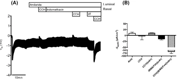
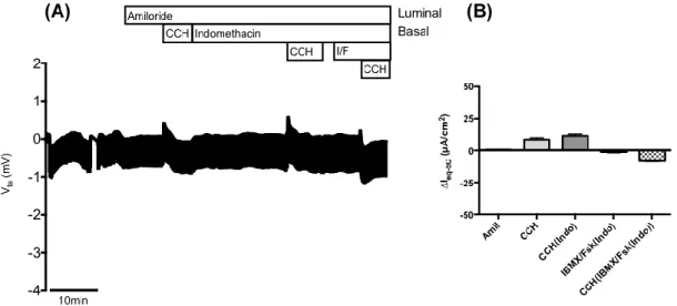
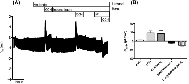
classic CF patients (Figure 1.6 C, third peak) and a biphased lumen-positive-then-negative response in tissues of residual function/non-classic CF patients (Figure 1.6 B, third peak).
Figure 1.6 – Representation of Ussing chamber measurements in rectal biopsies using the Cystic Fibrosis diagnosis protocol. Characteristic profiles of (A) non-CF controls, (B) non-classic and (C) classic CF patients. Vte values were referred to the basolateral side of the epithelium. Adapted from Sousa et al. (2012)50.
1.3.3. Newborn Screening
Currently implemented newborn screening (NBS) programs are especially important because early detection allows for the immediate administration of treatment before symptoms appear and improved disease outcome. For the diagnosis of CF in the NBS, the immunoreactive trypsinogen (IRT) test is performed, based on the observation that elevated blood levels of IRT are characteristic of all newborn CF patients41,51. However, these tests are only screening and thus they ultimately refer potential CF
patients to specialized CF centers for diagnosis confirmation52.
1.3.4. CFTR genotyping
Genotyping of mutations in the CFTR gene is usually performed to confirm a CF diagnosis. Although this works well for the set of mutations which have been established as CF-causing40,
genotyping does not always provide a clear diagnosis of CF vs. non-CF, since not all of the identified sequence variations in the CFTR gene have been confirmed as CF-causing variants. Also, most methods used in CFTR genetic testing are targeted at known mutations and so, many unidentified mutations and those that lie within the introns or regulatory regions of the gene may remain undetected unless extensive scanning methods are used53.
In addition, different patients, even those sharing the same CFTR genotype, show variable CF clinical phenotype due to the contribution of genetic modifiers, environmental factors and other undefined mechanisms (Figure 1.7), making a genotype/phenotype/therapy correlation hard to establish. This is even more challenging for patients that carry orphan variants. To overcome this issue, several cellular tools have been developed to help measure individual CFTR activity, as well as to establish drug responsiveness, and ultimately to help predict the best therapy for each individual, contributing to a personalized medicine approach for all CF patients, independent of their CFTR mutations.
Figure 1.7 – Key features of Cystic Fibrosis and relative contribution of genetic modifiers and other factors to phenotype variability in CF. CF is a multysistemic disease and the degree of each organ system disfunction varies considerably amongst
affected individuals. This is influenced by genetic factors such as the CFTR genotype and other modifier genes (such as TGFB1 and SLC26A9), and also by non-genetic determinants like environmental and stochastic factors (e.g. smoking). The CFTR genotype is the primary explanation for pancreatic exocrine insufficiency in CF. BMI = body mass index. Retrieved from
1.4.
Towards personalized therapies
1.4.1. 3D-intestinal organoids
A recently developed primary intestinal culture method54 has allowed for the generation of human
intestinal organoids, by isolating the stem cell-containing crypts of rectal biopsies and culturing them in the appropriate conditions. These stem cells expand in vitro into closed 3D-organoids that contain crypt-like structures and these can be used in the so-called Fsk-induced swelling (FIS) assay55, that uses Fsk
as a CFTR agonist. Fsk increases intracellular levels of cAMP, leading to the activation of CFTR in the apical membrane of the organoid cells, which in turn induces secretion of electrolytes and fluids to the inside of the organoids, resulting in their 3D-swelling. This CFTR-dependent fluid secretion has been shown to provide an indirect measurement of CFTR activity, by analysis of CFTR-deficient organoids – derived from CFTR-knockout mice and CF-patients – and the usage of CFTR-specific inhibitors55
(Figure 1.8).FIS in the organoids has been shown to correlate quantitatively to Fsk-induced currents measured in the Ussing chamber in freshly excised ex vivo rectal biopsies. Also, the fact that these cells can expand indefinitely in vitro allows for functional studies and drug response testing for individual patients in their own-derived material55,56.
Figure 1.8 – Representative images of the CFTR-dependent FIS assay. Representative confocal fluorescence microscopy
images of calcein green-labelled intestinal organoids from a CF patient carrying the F508del/F508del CFTR genotype in control conditions (Fsk only), after incubation with VX-809 + VX-770 (Orkambi®) and after incubation with Orkambi® and CFTR-specific inhibitors, and from a healthy control (HC) in control conditions, for t = 0 and t = 60 min of incubation with Fsk. Scale bar, 100 µm. Adapted from Dekkers et al. (2013)55.
1.4.2. Primary human nasal/bronchial epithelial cells (HNECs/HBECs)
Primary cultures of human bronchial epithelial cells (HBECs) derived from lung explants are another currently used and clinically relevant model for CFTR function analysis and drug testing, for e.g., through Ussing chamber measurements57. More recently, it was shown that it is also possible to
grow cells derived from patients’ nasal brushings (HNECs) and that stem cell hyperplasia can be induced in vitro58. Also, these cultures have been shown to maintain the ability to differentiate into functional
tissues and to recapitulate many features of the native epithelium when grown in permeable supports in appropriate air-liquid interface (ALI) conditions58. However, patient material is restricted and the
long-term expansion of some nasal/bronchial cells is still limited, making reproducible results difficult to achieve.
1.4.3. Cystic Fibrosis Bronchial Epithelial (CFBE) 41o- cell line
Besides intestinal organoids and primary airway cells, many other model systems have been developed to study the different aspects of CF pathology. Because most mutations exist in compound heterozygosity, it is hard to establish the contribution of each allele for the observed molecular and functional defects. To overcome this issue, cellular models were designed in order to characterize the molecular effects elicited by individual mutations. The Cystic Fibrosis Bronchial Epithelial (CFBE) 41o- cell line was generated through immortalization of HBECs from a F508del homozygous CF patient59, and with increasing passages they have lost endogenous CFTR expression. CFBEs retain some
aspects of in vivo HBECs, such as the ability to polarize forming electrically tight cell monolayers with functional cell-cell contacts when grown under immersed culture conditions, and expressing several proteins relevant for pulmonary drug absorption60. Because of this, they have been engineered to express
different CFTR mutants and used to characterize these mutations and their response to approved therapeutics, as well as to search for novel rescuing strategies through high-throughput approaches61,62.
Despite this, more physiologically relevant models of the airway epithelium may be required. Particularly, CFBEs do not achieve a full epithelial phenotype when grown in ALI conditions, which are more representative of the respiratory epithelium (e.g., no cilia formation)60.
Overall, since CFTR activity may vary among patients with different genotypes, patients that share the same genotype and even between tissues of the same patient, it is extremely important to validate different biological models to be used as diagnosis, prognosis and drug response assessment tools, aiming to improve and personalize CF care.