UNIVERSIDADE DE LISBOA
FACULDADE DE MEDICINA VETERINÁRIA
Escherichia coli - host interactions in the pathogenesis of canine pyometra
Sofia Correia Rosa de Barros Henriques
Orientador(es): Doutora Luísa Freire Leal Mateus
Doutora Maria Elisabete Tomé Sousa Silva
Tese especialmente elaborada para obtenção do grau de Doutor em Ciências Veterinárias na Especialidade de Ciências Biológicas e Biomédicas
Júri:
Presidente: Doutor Rui Manuel de Vasconcelos e Horta Caldeira Vogais:
- Doutor Luís Filipe Lopes da Costa - Doutora Paula Maria das Neves Ferreira da Silva
- Doutora Luísa Maria Freire Leal Mateus - Doutora Maria Constança Matias Ferreira Pomba - Doutora Rita Maria Payan Martins Pinto Carreira
i
“Science is the great antidote to the poison of enthusiasm and superstition.” Adam Smith (in Wealth of Nations, 1776)
“It is not the strongest of the species that survives, not the most intelligent that survives. It is the one that is the most adaptable to change.”
Charles Darwin (in Origin of Species, 1859)
To my grandparents Mariette and Nuno, Lai and Luís to my parents Helena e Carlos and to my dear sister Ana for all the unconditional love and support
iii ACKNOWLEDGMENTS
This thesis is the result of 6 years of hard work, dedication, effort but also scientific and personal growth. After a year of internship at the Institute of Tropical Medicine, the first steps in research were taken under the scope of a project headed by Doctor Luisa Mateus, whom I would like to thank for the opportunity to be part of the research group. The fascination with the research work grew and I did not hesitate when the invitation to continue my work in the reproduction and obstetrics department as a PhD student came. In science, as in everyday life, everything achieved with hard work has a special taste. The completion of this challenge was worth all the failed experiments and all the hours of hard work and personal effort.
I would like to thank Doctor Luis Costa for giving me the chance to take the first steps in scientific work in the reproduction and obstetrics department in 2008. I would also like to thank Doctor Luisa Mateus for the scientific orientation and for helping me to grow as a scientist and as a person and Doctor Elisabete Silva for the co-orientation of this thesis and for all the scientific and technical expertise, but also for the personal relationship we have built over these six years of professional contact.
To my colleagues in the reproduction and obstetrics department: to Engineer Patrícia Diniz for all the technical support and for the hours spent in the lab, to Doctor Daniel Murta, Mariana Batista, Doctor Ana Catarina Torres and technician Cristina Valado.
To Doctor Abdelhak Lemsaddek from BioFIG department of Ciences Faculty of Lisbon University, for all the essential support in the clustering data analysis.
To Sara Brito, Silvia Cruz, Carolina Merino, Sandra Carvalho and Marta Silva for all the help and assistance in several experimental tasks.
I would also like to thank the team of the laboratory of Doctor Constança Pomba, not only for the assistance with some technical protocols, but also for the lending of some biological samples which were essential for the work presented in this thesis.
To the team of the laboratory of Doctor Conceição Peleteiro, namely MSc Sandra Carvalho and technician Maria do Rosário Luís for the histological processing and sections of tissue samples.
iv
To the team of the laboratory of Doctor Carlos Fontes, for allowing me to use their facilities and equipment, when necessary.
I would also like to thank Doctor Luis Tavares, president of the Faculty of Veterinary Medicine and the Centre for Interdisciplinary Research in Animal Health (CIISA) at the time of the beginning of my work, for hosting me in this institution and for financially supporting my attendances in courses and conferences, essential to my scientific growth and the divulgation of the work of my laboratory.
To Doctor Graça Ferreira Dias for all the scientific and personal support and friendship in the last years. She is a truly a great scientist and a great friend.
To my lab colleagues and friends, Rita, Carina, Joana, Catarina, Liliana, Marina, Ana Tavares, Alexandre, Samuel and Carla Carneiro thank you for all the moments spent together, and for all the scientific experiences we shared.
To the Fundação para a Ciência e a Tecnologia (FCT) for the funding of my PhD studies and research projects in the reproduction and obstetrics lab.
To my family, especially to my dear mother Helena, for all the unconditional support, for always having given me the strength and encouragement to do better, and for always believing in me, this would never have been possible without you. To my father Carlos, not only for all the support and encouragement, but mainly for setting my greatest example of dedication in scientific work. To my grandparents, who helped me build the person that I am today. To my dear sister Ana, who always supported and believed in me, always knowing what I was capable of. To my stepfather Jorge and stepmother Ana. To my nieces, uncles and cousins.
To my dear friends Marta Batista and Mariana Batista for always supporting me and for the unconditional friendship that binds us together. Thank you for listening and understanding me and for all the support in the preparation of this thesis. Also, to my dear Ana Amaral for the friendship of these past years and for all the support in the lab. To my dear friend Sandra Albano, a longtime friend, who always believed what I was capable of.
v FUNDING
The present work was funded by the PhD Fellowship SFRH/BD/65044/2009 and project grant PTDC/CVT/66587/2006 from the Portuguese Foundation for Science and Technology (FCT) and by the research project 74.CIISA/FMVgranted by CIISA.
vii
Thesis title – Escherichia coli - host interactions in the pathogenesis of canine pyometra Abstract
Canine pyometra develops as a result of a complex interaction of etiological and physiopathological factors, such as the virulence and type of the bacteria and the individual host defence mechanisms. Since Escherichia coli is the most common bacterium isolated from uterus of bitches with pyometra, one main objective of this work was to characterize E. coli virulence potential, and to evaluate the role of its virulence factors (VF) and traits in the pathogenesis of canine pyometra (Chapters IV, V and VI). A second main objective was to evaluate the innate immune mechanisms within the uterus and their role in E. coli recognition (Chapters III and VI).
Results indicate that: i) although no single VF genes or virulence traits were associated with E. coli pyometra isolates, these isolates were mainly from the highly virulent phylogenetic group B2, which are characterized by a high number of uropathogenic E. coli VF genes and pathogenicity-associated islands markers; ii) Toll-like receptors were involved in the activation of the inflammatory response associated with pyometra; iii) β-hemolytic E. coli infection was associated with the occurrence of metritis and with an higher uterine tissue damage; iv) α-hemolysin (HlyA) contributes to the virulence of β-hemolytic E. coli, by inducing endometrial epithelial and stromal damage and a compromised early uterine immune response.
Overall, these findings provide new relevant insights into the role of the pathogen-specific modulation of host immunity, which may influence the severity of disease and its clinical outcomes. Also, HlyA is a promising target for a vaccine, with the objective to induce an immunity that can block the binding and action of this toxin.
Keywords: Escherichia coli, virulence traits, α-hemolysin, innate immunity, canine pyometra.
viii
Título da dissertação – Interacão hospedeiro-Escherichia coli na patogenia da piómetra na cadela
Resumo
A piómetra é uma doença comum do trato genital de cadelas adultas, durante a fase de diestro. A piómetra desenvolve-se como resultado de uma complexa interação de fatores etiológicos e fisiopatológicos. Entre estes, incluem-se a influência hormonal no útero, alterações estruturais no endométrio - como a hiperplasia quística (HQE) -, o tipo de bactérias e o seu potencial de virulência, e os mecanismos de defesa do hospedeiro. O trabalho desenvolvido nesta tese baseou-se no estudo do potencial de virulência de Escherichia coli (E. coli) (Capítulos IV e V) e nos mecanismos de imunidade inata do útero (Capítulos III e VI).
Tendo em conta a elevada prevalência de E. coli nos casos de piómetra (82-100% dos casos) e nas infeções do trato urinário (54 – 68%) e o facto de aquelas estirpes serem provenientes da flora fecal do animal e não de um clone específico disseminado entre animais, procedeu-se à comparação do potencial de virulência de E. coli, isolada de piómetra, de cistites e de fezes de cadela (Capítulo IV). Os resultados indicam que as estirpes de E. coli, que colonizam o útero, têm um elevado potencial de virulência, possuindo um grande número de genes que codificam para fatores de virulência (FV) e ilhas de patogenicidade (PAIs). No entanto, existem estirpes de E. coli isoladas de cistite e de origem fecal com as mesmas características, o que sugere que poderão induzir piómetra, em cadelas suscetíveis. De particular importância, foi a observação de que cerca de 50% das estirpes de E. coli isoladas de piómetra eram β-hemolíticas. A prevalência dos isolados pertencentes ao grupo filogenético B2 foi maior nos casos de piómetra (94%) do que nos casos de cistite (48%) ou do que nos de origem fecal (39%). No entanto, independentemente da origem dos isolados, o número médio de PAIs e de genes que codificam para FV foi maior nos isolados pertencentes ao grupo filogenético B2, comparativamente aos outros grupos filogenéticos. Verificou-se também que o reto poderá funcionar como um reservatório de estirpes potencialmente patogénicas dos grupos filogenéticos B2 e D. Esta observação tem especial importância pois sabe-se que as estirpes de E. coli uropatogénicas isoladas de cães e humanos são similares em relação ao seu
ix
serotipo, tipo clonal, grupo filogenético e perfil de virulência. Isto sugere que os cães podem servir como reservatórios de bactérias potencialmente virulentas que podem ser transmitidas ao homem.
Na primeira semana pós-parto, E. coli é a bactéria mais frequentemente isolada do conteúdo uterino de vacas de leite que desenvolvem infeções uterinas puerperais. No entanto, a associação, entre o perfil de virulência de E. coli e o desenvolvimento de metrite puerperal ou clinica, é controverso e, em muitos dos casos, a infeção resolve-se espontaneamente. Na cadela, as piómetras por E. coli estão associadas, em 50% dos casos, à síndrome de resposta inflamatória sistémica, a qual é potencialmente letal na ausência de terapêutica adequada. Numa tentativa de relacionar o potencial de virulência de E. coli com as diferentes evoluções da metrite clinica na vaca e da piómetra na cadela, compararam-se características genómicas dos isolados de E. coli (Capítulo V). Os resultados mostram que as estirpes de E. coli isoladas de vacas com metrite clinica pertencem maioritariamente aos grupos filogenéticos B1 e A, são geneticamente distintas das estirpes de piómetra e apresentam um menor número de genes que codificam para fatores de virulência, sendo por isso consideradas estirpes de menor potencial de virulência.
A resposta uterina à infeção é composta por mecanismos da imunidade inata e adaptativa. A resposta inata é desencadeada pelo reconhecimento de padrões moleculares associados aos agentes patogénicos, por recetores do tipo Toll (TLRs), induzindo uma reacção inflamatória. Os resultados apresentados no Capítulo III permitem concluir que o útero da cadela tem capacidade de reconhecer uma grande variedade de ligandos - através da activação dos TLRs - e desenvolver uma resposta inflamatória contra vários tipos de microorganismos. Verificou-se, também, que a transcrição e expressão dos TLRs 2 e 4 encontram-se significativamente diminuídas no início de diestro, o que pode contribuir para a maior susceptibilidade do útero à infeção por E. coli, nesta fase. Os resultados apresentados no Capítulo VI demonstram que, nos casos de piómetra a resposta inflamatória, mediada pelos TLRs, foi caracterizada por uma reação inflamatória exuberante, demonstrada pelo influxo de células de reação inflamatória no útero e por um aumento na transcrição de genes que codificam para citocinas pró- inflamatórias (IL-1β, IL-6, IL-8) e anti-inflamatórias (IL-10 e TGFβ). Observação relevante foi que, nos casos de piómetra por E. coli β-hemolítica, há um aumento significativo da
x
ocorrência de metrite e uma maior destruição do endométrio, bem como um aumento significativo da transcrição dos genes que codificam para as citocinas IL-1β e IL-8, quando feita a comparação com as piómetras por E. coli não hemolítica.
Tendo em conta os resultados apresentados anteriormente, bem como a elevada percentagem de isolados de E. coli β-hemolítica nos casos de piómetra, foi avaliado o papel da α-hemolisina (HylA) na patogenia da piómetra. Para este fim, recorreu-se à estimulação in vitro de culturas primárias de células epiteliais e do estroma do endométrio canino com uma estirpe não hemolítica (Pyo14), uma estirpe β-hemolítica (Pyo18) e um mutante isogénico da HylA (Pyo18ΔhlyA, perda de função). Os resultados obtidos mostram que o efeito citopático da HylA é maior nas células do estroma do que nas células do epitélio do endométrio. Este facto poderá estar associado à maior destruição do endométrio e ao maior número de casos de metrite por nós observado. Foi também demonstrado que estas estirpes de E. coli induzem respostas imunitárias diferentes nos dois tipos celulares. Nas células do estroma, a HylA esteve associada a uma diminuição da transcrição dos genes que codificam para as citocinas IL-1β, TNFα e IL-10. Pelo contrário, a estimulação celular com o mutante isogénico levou a um aumento da transcrição destas citocinas. A ação da HylA na inibição da transcrição destes mediadores da inflamação poderá permitir a multiplicação e invasão do endométrio por E. coli β-hemolítica, antes que se estabeleça uma reação inflamatória adequada.
Em súmula, os trabalhos que compõem esta tese permitem concluir que: i) as estirpes de E. coli isoladas de piómetra são principalmente do grupo filogenético B2, sendo caracterizadas por uma maior número de FV e PAIs; ii) os recetores do tipo Toll estão envolvidos na ativação da resposta inflamatória nos casos de piómetra; iii) a infecção por E. coli β-hemolítica está associada a metrite e a uma maior destruição do tecido uterino; iv) a α-hemolisina contribui para a virulência de E. coli β-hemolítica ao induzir uma destruição das células epiteliais e do estroma do endométrio e comprometer precocemente a resposta imunitária uterina.
Estes resultados aportam conhecimento novo sobre o papel de E. coli na modulação da resposta imunitária uterina e na patogenia da piómetra. Pelo seu papel na expressão da virulência de E. coli, a HlyA é uma potencial candidata para o desenvolvimento de uma
xi
vacina. Assim, os TLRs poderão ser considerados promissores alvos terapêuticos, dado o seu envolvimento na resposta inflamatória associada à piómetra.
Palavras-chave: Escherichia coli, perfil de virulência, α-hemolysina, imunidade inata, piómetra de cadela
xii TABLE OF CONTENTS
CHAPTER I – GENERAL INTRODUCTION AND OBJECTIVES ... 1
CHAPTER II - LITERATURE REVIEW... 7
1. Canine Pyometra ... 9 1.1.Hormonal component ... 10 1.2.Bacteriologic component ... 12 2.Escherichia coli ... 12 2.1.Virulence factors ... 14 2.1.1.Adhesins ... 14 2.1.2. Toxins... 18 2.1.2.1. Lipopolysaccharide ... 20 2.1.3. Siderophores ... 22
2.1.4. Other virulence factors ... 24
2.1.5. Pathogenicity islands ... 25
2.1.6. Phylogenetic groups ... 26
2.2.Antimicrobial resistance ... 26
3. Immune system ... 28
3.1. Innate immunity: Toll-like receptors ... 29
3.2. TLRs signaling pathways ... 30
3.3. Innate immunity in the uterus ... 34
3.4. Cytokines and Chemokines ... 35
CHAPTER III - OESTROUS CYCLE-RELATED CHANGES IN PRODUCTION OF TOLL-LIKE RECEPTORS AND PROSTAGLANDINS IN THE CANINE ENDOMETRIUM ... 39
1. Abstract ... 41
2. Introduction ... 42
3. Materials and methods ... 43
3.1. Animals and sample collection ... 43
xiii
3.3. Reverse transcription polymerase chain reaction and quantitative real time RT-PCR..45
3.3.1. Endometrial RNA extraction and cDNA synthesis ... 45
3.3.2. mRNA transcription of toll-like receptors genes and prostaglandin synthesis genes ... 46
3.4. Prostaglandin measurement ... 48
3.4. Immunohistochemical staining of TLR2, TLR4 and PTGS2 ... 48
3.6. Statistical analysis ... 49
4. Results ... 49
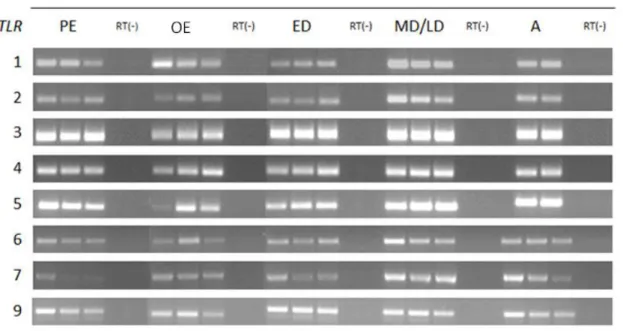
4.1. Presence of TLR1–7 and 9 gene transcripts in the canine endometrium throughout the oestrous cycle... 49
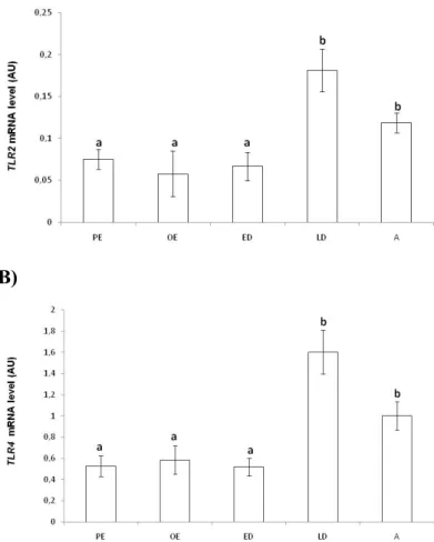
4.2. Transcription levels of TLR2 and TLR4 genes in the canine endometrium throughout the oestrous cycle... 50
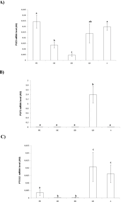
4.3. Transcription levels of PGES, PGFS and PTGS2 genes throughout the oestrous cycle ... 51
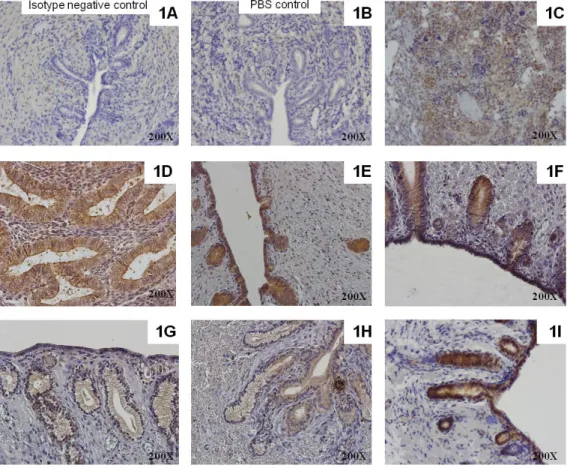
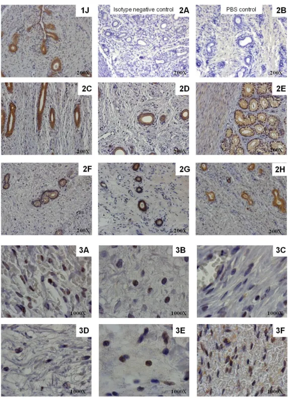
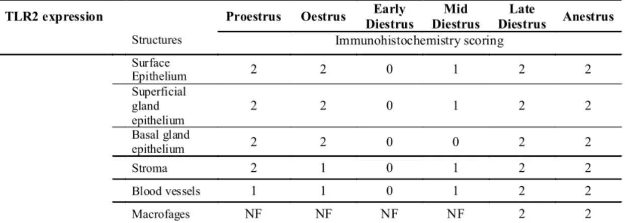
4.4. Protein expression of TLR2 and TLR4 and PTGS2 in the canine endometrium throughout the oestrous cycle and in pyometra cases ... 53
4.5. Prostaglandin production by endometrial explants after LPS or LTA stimulation ... 59
5. Discussion ... 60
CHAPTER IV - VIRULENCE GENOTYPES OF ESCHERICHIA COLI CANINE ISOLATES FROM PYOMETRA, CYSTITIS AND FECAL ORIGIN ... 65
1. Abstract ... 67
2. Introduction ... 68
3. Materials and methods ... 68
3.1. E. coli strains ... 68
3.2. Detection of PAI markers, virulence factors genes and phylogenetic groups ... 69
3.3. Cluster analysis of virulence genotype ... 69
3.4. Statistical analysis ... 69
xiv
CHAPTER V - GENOTYPIC AND PHENOTYPIC COMPARISON OF ESCHERICHIA COLI FROM UTERINE INFECTIONS WITH DIFFERENT OUTCOMES: CLINICAL METRITIS IN THE COW AND PYOMETRA IN THE
BITCH ... 68
1. Abstract ... 77
2. Introduction ... 78
3.Materials and methods ... 79
3.1. E. coli isolates ... 79
3.2. Detection of virulence factors genes and phylogenetic groups ... 79
3.3. Pulsed-field gel electrophoresis ... 82
3.4. Antimicrobial susceptibility tests ... 83
4. Results ... 83
4.1. Colony morphology of E. coli isolates ... 83
4.2. Phylogenetic grouping ... 84
4.3. Characterization of virulence factor gene profiles ... 84
4.4. PFGE analysis ... 85
4.5. Antimicrobial susceptibility test ... 88
5. Discussion ... 89
CHAPTER VI - IMMUNOMODULATION IN THE CANINE ENDOMETRIUM BY UTEROPATHOGENIC ESCHERICHIA COLI ... 93
1. Abstract ... 95
2. Introduction ... 96
3. Materials and methods ... 97
3.1. Experiment 1 ... 97
3.1.1. Immunohistochemistry ... 98
3.2. Experiment 2 ... 98
3.2.1. Cell culture ... 98
3.2.2. E. coli strains ... 100
3.2.3. E. coli cell stimulation ... 101
xv
3.2.5 Immunofluorescence ... 104
3.2.6 Statistical analysis ... 104
4. Results ... 105
4.1. Hemolytic E. coli induces a more extensive uterine damage and inflammatory cell infiltration than non-hemolytic E. coli (Experiment 1) ... 105
4.2. Hemolytic and non-hemolytic E. coli pyometra endometria have different transcription levels of genes coding pro-inflammatory cytokines (Experiment 1) ... 108
4.3 Cytotoxicity due to α-hemolysin is mainly targeted to stromal endometrial cells (Experiment 2) ... 109
4.4 Hemolytic and non-hemolytic E. coli strains induce differential immune response in endometrial epithelial and stromal cells ... 95
5. Discussion ... 117
CHAPTER VII - GENERAL DISCUSSION, CONCLUSIONS AND FUTURE PERSPECTIVES ... 123
General Discussion ... 125
Conclusions ... 137
Future Perspectives ... 137
xvi LIST OF FIGURES
Figure 1 - Structure of Escherichia coli cell envelope…………...……….……… 21 Figure 2 - LPS sensing via LBP to CD14/MD-2/TLR4 receptor complex………..22
Figure 3 – Mammalian TLR signaling pathways………....….……... 33
Figure 4 - Presence of TLR 1-7 and 9 gene transcripts in the canine endometrium throughout the oestrous cycle………. 50 Figure 5 - mRNA level (Arbitary Units, AU) evaluated by real-time PCR for the genes TLR2 (A) and TLR4 (B) in the endometrium of the bitch during the oestrous cycle (n = 25)……….….……..… 51 Figure 6 - Relative mRNA level (Arbitary Units, AU), evaluated by real-time PCR for the genes PGES (A) PGFS (B) and PTGS2 (C), in the endometrium of the bitch during the oestrous cycle (n=19)………..…….……… 52 Figure 7 - Immunostaining of TLR4 in healthy bitch endometrium during the oestrous cycle……….……… 53 Figure 8 - Immunostaining of TLR2 in healthy bitch endometrium during the oestrous cycle……….…………...…. 56 Figure 9 - Immunostaining of TLR4 (I-III) and TLR2 (IV-VI) and PTGS2 in canine pyometra endometrium……….……... 58 Figure 10 - Prostaglandin production (pg/mg of tissue) by endometrial explants (n=26) after 24 hours of incubation without (C) or with LPS (1 µg/mL) and LTA (1 µg/mL)…..………. 59 Figure 11 - Clustering of the eighty E. coli isolates based on the presence or absence of the virulence factor genes and PAI markers………...………... 74 Figure 12 - Cluster analysis of virulence factor genes profile of E. coli uterine isolates of canine pyometra (n=29) and bovine puerperal metritis (n=20) origin………..…... 86 Figure 13 - PFGE clonal type analysis of E. coli uterine isolates of canine pyometra (n=29) and bovine puerperal metritis (n=16) origin……….………... 87 Figure 14 - Histological section of uterine pyometra samples (A) and immunostaining of calprotectin positive cells (myeloid cells), CD79 αcy positive cells (B lymphocytes) and CD3 positive cells (T lymphocytes) in pyometra uterine sections (B)………….…….………… 107
xvii
Figure 15 - Representative PCR detection of transcripts of TLRs signaling components and of cytokines in diestrous and pyometra endometria (A). Relative mRNA expression level of IL-1β, IL-6, IL-8, IL-10, and TGFβ evaluated by real-time PCR (B)……….…....…… 108 Figure 16 - Morphology of endometrial epithelial (a-f) and stromal (g-l) cell cultures stained with Giemsa after 1 and 4 h of incubation……….……… 111 Figure 17 - Morphology of endometrial stromal cells after 1 and 4 h of incubation, evaluated after Giemsa staining (a-c) or under phase-contrast (d-l)………..…….…. 112 Figure 18 - Nuclear detection of NFκB (A-H) and IRF3 (I-P) in endometrial epithelial and stromal cells by immunofluorescence……… 114 Figure 19 - Escherichia coli or LPS-induced cytokines gene transcription in cultured canine endometrial epithelial and stromal cells.IL-1β (A), IL-1α (B), IL-6 (C), TNFα (D), IL-8 (E), CXCL10 (F), IL-10 (G), INFβ (H) in cultured canine endometrial epithelial and stromal cells in response to hemolytic E. coli (Pyo18), non-hemolytic E. coli (Pyo14) and the isogenic mutant of Pyo18 (Pyo18ΔhlyA)...……….……… 115 Figure 20 - Figure 20 - Schematic illustration of the proposed initial endometrial inflammatory events following infection with non-hemolytic (A) and hemolytic Escherichia coli (B) strains………...………..135
xviii LIST OF TABLES
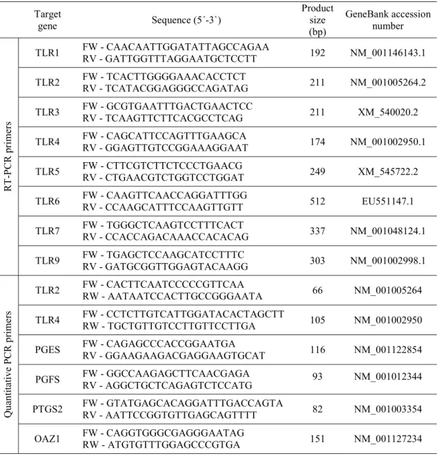
Table 1 - Parameters used for oestrous cycle phase determination………..…...…..……..… 44 Table 2 - Primer sequences for mRNA of target genes……….………….. 47 Table 3 - The mean expression score for TLR2 and TLR4 in different structures of the canine endometrium throughout the estrous cycle……….………. 55 Table 4 - PGF2α and PGE2 production by tissue explants after LPS (1 μg/mL) or LTA (1 μg/mL) challenge……….……….... 60 Table 5 - Comparison of prevalence of eight pathogenicity islands markers and 15 virulence factor genes between pyometra (n=31), cystitis (n=23) and fecal isolates (n=26)………..………. 72 Table 6 - Distribution of pathogenicity islands markers (PAI) in pyometra (n=31), cystitis (n=23) and fecal isolates (n=26), classified according to phylogenetic group……….... 73 Table 7 - Comparative prevalence of virulence factor genes in E. coli isolates from canine pyometra and bovine metritis origins ……….…………. 80 Table 8 - Distribution of the minimum inhibitory concentrations (MICs) of antimicrobials for canine (n=29) and bovine (n=20) Escherichia coli isolates……….……… 88 Table 9 - Primer sequences used for RT-PCR…………..…………..……….. 102 Table 10 - Primers sequences used for quantitative real time PCR (qRT-PCR)….…….… 103 Table 11 - Haematological and biochemical parameters in bitches with hemolytic E. coli pyometra (n=8) and with non-hemolytic E. coli pyometra (n=10)………….………. 106 Table 12 - Effect of bacterial and LPS incubation on endometrial epithelial and stromal cell numbers……….…. 110
xix
LIST OF ABBREVIATIONS AND SYMBOLS
% - percentage µg - microgram µL - microliter a - anestrus
ABC - ATP-binding cassete afa - afimbrial adhesins AP-1 - activator protein 1
APEC - avian pathogenic E. coli APC - antigen presenting cells
astA - heat-stable cytotoxin associated with enteroaggregative E. coli toxin ATP - Adenosine triphosphate
Bp - base pairs
BSA - bovine serum albumin cDNA - complementary DNA cdt - cytolethal distending toxin CEH - cystic endometrial hyperplasia CFU - colony-forming unit
CHEF - clamped homogenous electric field chuA - E. coli heme transport protein
CLSI - Clinical and Laboratory Standards Institute cnf1 - cytotoxic necrotizing factor 1
CO2 - carbon dioxide csg - curli fibres cvaC - microcin CoIV
DAB - substrate diaminobenzidine DAEC - diffusely adherent E. coli DAF - decay-accelerating factor DCs - dendritic cells
xx DNA - deoxyribonucleic acid
dra - dr fimbriae
dsRNA - double stranded RNA eaeA - intimin
EAEC - enteroaggregative E. coli E. coli - Escherichia coli
ED - early diestrus
EHEC - enterohamerrhagic E. coli EIEC - enteroinvasive E. coli
EnPEC - endometrial pathogenic E. coli ent - enterobactin
EPEC - enteropathogenic E. coli ETEC - enterotoxigenic E. coli ExPEC - extra-intestinal E. coli fecA - ferric citrate
fepA - iron-enterobactin outer membrane transporter fhuA - yersiniabactin system
fhuE - coprogen fhuF - ferrioxamine B fim - type 1 fimbriae foc - F1C fimbriae
fyuA - yersiniabactin system g - grams
h – hour H - hemolytic
HBSS - Hanks' Balanced Salt Solution hlyA - alpha-hemolysin
hlyE - hemolysin E
Hma - heme acquisition protein HPI - high-pathogenicity island
xxi HYP - hypervariable domain
ibeA - invasion of brain endothelium IFN - interferon
IgG - immunoglobulin G IHC – immunohistochemistry IKK - inhibitor of κ B kinase IL - interleukin
IM - inner membrane
iNOS - inducible nitric oxide synthase IPEC - enteric/diarrheagenic E. coli IRAK - IL-1 receptor-associated kinase IRF - IFN regulatory factor
iroN – catecholate siderophore receptor ITS - Insulin-Transferrin-Selenium iucD – aerobactin system
iutA – aerobactin
kpsMT - capsular polysaccharide synthesis LBP - LPS binding protein
LH - luteinizing hormone LPS – Lipopolysaccharide LRR - leucine-rich repeats HSP - heat shock proteins LH - luteinizing hormone LTA - lipoteichoic acid
MAP - mitogen-activated protein M - mucoide
MAPK – mitogen-activated protein kinase mCD14 - membrane-bound CD14
MD/LD - mid/late diestrus
xxii mg - milligrams
MHC - major histocompatibility complex MIC - minimum inhibitory concentration min – minutes
ml - milliliter
MPEC - mammary pathogenic E. coli mM – milimol
MRHA - mannose-resistant hemagglutination mRNA – messenger RNA
MSHA - mannose-sensitive hemagglutination
MyD88 - myeloid differentiation primary response 88 N - number
NAPs - natural antimicrobial peptides NF-κB - nuclear factor kappa B NH - non-hemolytic
NMEC - neonatal meningitic E. coli ◦C – grad Celsius
OAZ1 - ornithine decarboxylase antizyme 1 OE - oestrus
OHE – ovariohysterectomy OM - outer membrane
OTUs - operational taxonomic units OVX – ovariohysterectomy
P4 - progesterone
PAIs - pathogenicity-associated islands
PAMPs - pathogen associated molecular patterns pap - P fimbriae adhesin
PBP - periplasmic binding protein PBS - Phosphate-buffered saline PCR - polymerase chain reaction
xxiii PE - proestrus
PFGE - pulsed-field gel electrophoresis Pg - picogram
PG – prostaglandins PGE2 - prostaglandin E2
PGES - prostaglandins E2 synthases PGF2α - prostaglandin F2α metabolite PGFM - prostaglandin F2α metabolite PGFS - prostaglandins F2α synthases pH - potential of Hydrogen PMN - polymorphonuclear leukocyte pmol – picomol
PRRs - specialized pattern recognition receptors PTGS2 - prostaglandin-endoperoxide synthase 2
qRT-PCR - quantitative reverse transcription polymerase chain reaction R - rough
RLHs - rig-like helicases receptor RNA - ribonucleic acid
RT-PCR - reverse transcription polymerase chain reaction RTX - repeats-in-toxin
S - smooth
SEPEC - septicaemia associated E. coli
SIRS - systemic inflammatory response syndrome sitA - periplasm iron binding protein
sfa - S fimbriae spp - species
ssRNA - single stranded RNA stx - shiga toxin
TcpC - Toll/interleukin-1 receptor (TIR) domain-containing protein TGF - Transforming growth factor
xxiv TIR - translocated intimin receptor
TIRAP/MAL - TIR-containing adaptor protein TLRs - Toll-like receptors
TLR2 - Toll-like receptor 2 TLR4 - Toll-like receptor 4 TNF - tumor necrosis factor
TNFR - tumor necrosis factor receptor TRAF - TNFR-associated factor
TRIF - TIR-domain-containing adapter-inducing interferon-β TRAF6 - TNF receptor-associated factor 6
TRAM - TIR-domain-containing adaptor molecule 2 tRNA - Transfer ribonucleic acid
traT - serum survival factor U - units
u/ml - units/milliliter
UPEC - uropathogenic E. coli
UPGMA - unweighted average linkage usp - uropathogenic specific protein UTI - urinary tract infection
VF - virulence factor
1
3
Pyometra is a common and serious uterine disorder that develops in 25% of all intact female dogs, causing a variety of clinical and pathological signs. Escherichia coli (E. coli) is the most common bacterium isolated from the uterus of bitches with pyometra (82-100%). Its presence is normally associated with severe systemic signs. If left untreated is lethal and patients may develop endotoxemia, sepsis or septic shock. Apart its relevance for the canine species, studies on pyometra disease mechanisms may provide important insights into the mechanisms operating in human bacterial infection and sepsis. Despite several studies on the etiology of this disease, the pathogenesis of pyometra is still not completely understood. In particular, little is known regarding the host-pathogen interactions occurring in the pathogenesis of pyometra. The current knowledge on the pathogenicity of E.coli isolates from canine pyometra cases is based on the genomic identification of classical virulence factor genes (VFGs) (Chen et al., 2003; Siqueira et al., 2009). Although pyometra E. coli share virulence factor (VF) of uropathogenic E. coli (UPEC) strains, key VF essential for E. coli pathogenicity in pyometra cases are still unknown. Similarly, the role of E coli in the pathogenesis of the puerperal uterine infection of the cow is also unclear. It has been implicated in early ovarian disturbances and appears to increase the susceptibility of the uterus to subsequent infections with Trueperella pyogenes and Gram-negative anaerobes, the former being relevant to the establishment and persistence of uterine infection (Mateus et al., 2002; Sheldon et al., 2009). However the outcomes of these two conditions are very different and may be linked to different E. coli virulence traits, among other reasons.
The endometrium is the first line in defending against invading microbial pathogens. It recognizes pathogen-associated molecular patterns (PAMPs) shared by pathogens via innate immune receptors. Toll-like receptors (TLRs) are a large class of ancient innate immunity receptors which can recognize conserved components of pathogens or PAMPs and initiate innate immune response (Horne et al., 2008). An enhanced understanding of innate immune mechanisms within the female reproductive tract and their role in bacterial recognition may provide insights into the pathogenesis of uterine diseases and associated deleterious sequelae. The understanding of the underpinning mechanisms of pyometra and the potential role by which E. coli subvert host innate immunity, disrupting uterine function, is crucial to develop surrogate measures to alleviate the negative impacts of uterine infection.
4
Therefore, the research work presented in this thesis aimed to improve our understanding of the host-pathogen interactions in canine pyometra, and more specifically:
1. To evaluate if changes in toll-like receptor 2 (TLR2) and toll-like receptor 4 (TLR4) transcription and expression during the oestrous cycle may be associated with predisposition to the development of pyometra in the bitch.
2. To characterize the virulence potential of pyometra, UTI and feces E. coli in order to explain their ability to colonize and infect either the uterus or the urinary tract.
3. To compare the molecular and phenotypic characteristics of E. coli isolates recovered from the uterus of cows with clinical metritis and of bitches with pyometra, in an attempt to correlate their virulence potential with the different clinical outcomes of the diseases. 4. To evaluate the inflammatory response of canine endometrial cells to uteropathogenic E. coli, focusing on the role of α- hemolysin in epithelial and stromal cells.
The above studies were converted into four articles, submitted for publication in international refereed and indexed journals, and constitute the four chapters of the experimental work included in this thesis as follow:
1. Oestrous cycle-related changes in production of toll-like receptors and prostaglandins in the canine endometrium.
Silva, E.1, Henriques, S.1, Brito, S., Ferreira-Dias, G., Lopes-da-Costa, L., Mateus, L. (2012). Journal of Reproductive Immunology, 96 (1-2): 45-57. doi:10.1016/j.jri.2012.07.003
5
2. Virulence genotypes of Escherichia coli canine isolates from pyometra, cystitis and fecal origin.
Mateus, L., Henriques, S., Merino, C., Pomba, C., Lopes-da-Costa, L., Silva, E. (2013). Veterinary Microbiology, 166 (3-4): 590-594. doi:10.1016/j.vetmic.2013.07.018
3. Genotypic and phenotypic comparison of Escherichia coli from uterine infections with different outcomes: Clinical metritis in the cow and pyometra in the bitch. Henriques, S., Silva, E., Lemsaddek, A. Lopes-da-Costa, L., Mateus, L. (2014). Veterinary Microbiology, 170 (1-2): 109-116. doi:10.1016/j.vetmic.2014.01.021
4. Immunomodulation in the canine endometrium by uteropathogenic Escherichia coli Henriques, S.1, Silva, E.1, Silva, M.F., Carvalho, S., Diniz, P., Lopes-da-Costa, L.,
Mateus, L. (2016). Submitted to Veterinary Research
7
9 1. Canine Pyometra
Canine pyometra is a chronic disease characterized by the accumulation of pus in the uterine lumen of sexually mature intact bitches. The disease is associated with a variety of clinical and systemic signs, and occurs during a phase of progesterone dominance, usually diagnosed from 4 weeks to 4 months after estrus (Fransson & Ragle, 2003; Pretzer, 2008; Smith, 2006). Around 25% of all intact female dogs develop pyometra by 10 years of age (Dow, 1958; Egenvall et al., 2001; Niskanen & Thrusfield, 1998). Reported age range at time of development of pyometra is 8 months to 15 yearswith amean age of 8.6-9.9 years (Bigliardi et al., 2004; Niskanen & Thrusfield, 1998).
Pyometra develops as a result of a complex interaction of etiological factors. These factors include changes within the endometrium, the hormonal influence on uterine environment, the virulence and type of the bacteria and the individual defense mechanisms (Mateus & Eilts, 2010).
Cystic endometrial hyperplasia (CEH) develops after repeated estrous cycles in the bitch and predisposes uterus to a secondary infection that leads to pyometra (Fransson, 2003). In this way, for several years the term CEH/pyometra complex was used when referring to these two entities. However pyometra can also occur without CEH, and clinical and histopathologic findings suggest that CEH and pyometra should be classified separately into two entities. Both CEH and pyometra have similarities with each other, except for the inflammatory reaction observed in pyometra (De Bosschere, Ducatelle, Vermeirsch, Van Den Broeck, & Coryn, 2001).
Clinical signs in bitches with pyometra vary depending on cervical patency (open or closed pyometra) and with the type of bacteria. Vulvar discharge is often evident if the cervix is open (reported in 58 to 98 % of bitches), and the discharge’s characteristics may vary depending on the bacteria: muco-purulent, purulent (associated more often with Streptococcus spp) or sanguinopurulent (similar to tomato soup; associated more often with mucoid or haemolytic E. coli), white to red-brown in color and foul smelling (Gilbert, Nothling & Oettlé, 1989; Hagman et al., 2006a). A bitch with a closed cervix pyometra more commonly has abdominal distension. The onset of clinical signs may be acute or gradual, and in general, are more severe with a closed cervix pyometra (Smith, 2006). Common clinical signs associated with the pyometra are lethargy and anorexia,
10
polydipsia and polyuria, abdominal pain, vomiting or diarrhea and dehydration(Bigliardi et al., 2004; England, Freeman, & Russo, 2007; Gilbert et al., 1989; Hagman et al., 2006a).
Although less frequent, pyometra also can appeared associated with the systemic inflammatory response syndrome (SIRS) (Fransson, Lagerstedt, Hellmen, & Jonsson, 1997; Purvis & Kirby, 1994). Uncontrolled production of inflammatory mediators, such as tumor necrosis factor (TNF), interleukin-1 (IL-1) and interleukin-6 (IL-6), and platelet activating factor may provoke irreversible damage to internal organs or septic shock, which in some cases may lead to death (Manfra, Matthiesen & Nichols, 1989; Purvis & Kirby, 1994).
Bitches having higher plasma concentrations of endotoxins are associated with poorer prognosis for survival (Okano, Tagawa, & Takase, 1998). Endotoxins strongly stimulate prostaglandin synthesis, with prostaglandin E2 (PGE2) further contributing to the suppressed activity of cellular immunity during diestrus (Silva et al., 2010). Measuring blood concentration of the prostaglandin F2α (PGF2α) metabolite (PGFM) also provides a good indicator of endotoxin release in bitches with pyometra, and helps in the differentiation between pyometra and CEH (Hagman et al., 2006a). The increased systemic PGFM observed with pyometra probably originates from the endometrial synthesis of prostaglandins, which results from the stimulation of the uterus by bacteria (Silva et al., 2010). The safest treatment of pyometra is ovariohysterectomy (OHE), normally performed a soon as the general condition of the dog is stable. Medical treatment is an option in some selected cases to preserve fertility (Verstegen et al., 2008) . All bitches with pyometra, whether treated medically or with OHE, should be treated with systemic antibiotics.
1.1. Hormonal Component
Over the years, the association between pyometra and diestrus has been well established however the exact mechanism that leads to pyometra development is still unknown (Mateus & Eilts, 2010).
Canine pyometra disease occurs during diestrus stage when uterus is under progesterone influence. Bacteria may gain access into the uterus during pro-estrous and estrous phases
11
(J. R. Watts, Wright, & Lee, 1998). It is suggested that low-grade uterine infection is partially responsible for some of the endometrial proliferation that occurs early in the pathogenesis of CEH-complex (Arora, Sandford, Browning, Sandy, & Wright, 2006; Noakes, Dhaliwal & England, 2001; Nomura, 1983; Schlafer & Gifford, 2008).
The progesterone-sensitized uterus is suitable, not only for pregnancy, but also for bacterial infection, since progesterone stimulates endometrial glandular secretions, decreases myometrial contractions¸ induces functional cervical closure (Nelson & Feldman, 1986; Fransson & Ragle, 2003; Mateus & Eilts, 2010) and depress the local immune response by creating an anti-inflammatory environment (Sugiura et al., 2004; Tibbetts, Conneely, & O’Malley, 1999). Also, progesterone causes an increased binding of E. coli to the endometrium (Ishiguro et al., 2007; Leitner, Aurich, Galabova, Aurich, & Walter, 2003). Recent research involving the induction of canine pyometra by inoculating E. coli into the uterus demonstrated that, on days 11-20 and 21-30 after the luteinizing hormone (LH) peak, the uterus was most susceptible to infection (Tsumagari et al., 2005). In contrast, estrogens induce proliferation of endometrial glands, reduce the susceptibility of the endometrial epithelium to E. coli adhesion (Nishikawa & Baba, 1985) and induce a pro-inflammatory response within the uterus (Tibbetts, Conneely, & O’Malley, 1999) by increasing the production of bactericidal lactofferrin, the major estrogen-inducible protein in the uterus, that kills bacteria and modulates inflammatory and immune response (Teng et al., 2002) . Although estrogen seems to play a less important role, it appeared to enhance the endometrial response to progesterone (Teunissen, 1952; Nelson &
Feldman, 1986).
At moment, there is no conclusive evidence that pyometra is caused by a disturbance in either hormone production or in the uterine response to progesterone and estrogen (De Bosschere, Ducatelle, Vermeirsch, Simoens & Coryn, 2002; Dhaliwal, England & Noakes, 2002). Peripheral blood concentrations of estrogen and progesterone in bitches with CEH/pyometra complex are not abnormally elevated compared to normal bitches (Chen, Wright & Lee, 2001). This has led to speculation that hormone receptors may thus play a role in the pathogenesis of CEH/pyometra (De Cock, Vermeirsch, Ducatelle & De Schepper, 1997; Dhaliwal, England & Noakes, 1999; De Bosschere et al., 2002; Nomura, Kawasoe & Shimada, 1990). Estrogen and progesterone receptor expression has been shown to be increased in the uteri of bitches with CEH, but not in the uteri of bitches with
12
pyometra, when compared with healthy bitches (De Bosschere et al., 2002; De Cock et al., 1997; Ververidis et al., 2004). In contrast, an overall reduction in both receptors in CEH/pyometra cases has been described (Dhaliwal et al. 1999).
1.2. Bacteriologic component
The bacteria most commonly isolated from the uterus in cases of pyometra of bitches are E. coli (around 90% of the cases). Its presence is normally associated with highly severe systemic signs and a potentially life-threatening situation. However, other bacteria that may be isolated include Streptococcus spp, Klebsiella spp, Staphylococcus aureus, Pasteurella spp, Proteus spp and Pseudomonas spp. (Chen, Wright, Lee, & Browning, 2003; Dhaliwal, Wray, & Noakes, 1998; Fransson et al., 1997; Hagman & Kühn, 2002). These organisms are also those that are most commonly isolated from the vagina of normal bitches (Watts, Wright, & Whithear, 1996) and can ascend to the uterus during pro-estrus and estrus (Kustritz, 2006). E. coli associated with canine pyometra originates from the normal intestinal flora of bitches and is not derived from any specific clonal type that is epidemically spread between animals (Chen et al., 2003; Hagman & Kühn, 2002; Wadås, Kühn, Lagerstedt, & Jonsson, 1996). E. coli associated with pyometra are characterized by the presence of several virulence genes normally found in UPEC strains (Chen et al., 2003; Siqueira et al., 2009). In cases of E. coli pyometra with a concurrent subclinical urinary tract infection, the urinary tract and the uterus are likely to be infected with the same bacterial strain(Hagman & Kühn, 2002; Wadås et al., 1996).
2. Escherichia coli
E. coli strains can be commensal as part of normal microbial flora or can cause various infection diseases in immunosuppressed host like intestinal infections (enteric/diarrheagenic E. coli, IPEC) or extra-intestinal infections (extra-intestinal E. coli, ExPEC) (Bélanger et al., 2011; reviewed by Bien, Sokolova, & Bozko, 2012; Picard et al., 1999). Intestinal infections can be caused by different E. coli pathotypes such as enterotoxigenic (ETEC), enteropathogenic (EPEC), enterohamerrhagic (EHEC), enteroaggregative (EAEC), enteroinvasive (EIEC) and diffusely adherent (DAEC) E. coli
13
(Bélanger et al., 2011; Constantinou, Young, Clements, & Frankel, 2012; reviewed by Köhler & Dobrindt, 2011). The ExPEC includes UPEC E. coli, neonatal meningitic (NMEC), septicaemia associated (SEPEC) and avian pathogenic E. coli (APEC) (reviwed by Antão, Wieler, & Ewers, 2009; reviewed by Köhler & Dobrindt, 2011; Siqueira et al., 2009). Two new animal pathogenic subgroups have recently been proposed: mammary pathogenic (MPEC) (Shpigel, Elazar, & Rosenshine, 2008) and endometrial pathogenic E. coli (EnPEC) (Sheldon et al., 2010, reviewed by Köhler & Dobrindt, 2011). UPEC strains are associated with UTIs like cystitis and pyelonephritis in humans (Bélanger et al., 2011; reviewed by Bien et al., 2012; Johnson, Owens, Gajewski, & Kuskowski, 2005a), dogs and cats (Chen et al., 2003; Hagman & Kühn, 2002; Johnson, Kaster, Kuskowski, & Ling, 2003a; Siqueira et al., 2009) as well with pyometra in dogs (Chen et al., 2003). UPEC isolated from dogs, cats and humans are in some cases phylogenetically close related (Bélanger et al., 2011; Guardabassi, Schwarz, & Lloyd, 2004; Johnson et al., 2003a) and have a similar virulence gene profile (Johnson, Stell, & Delavari, 2001a). E. coli strains involved in cases of canine pyometra displays great similarity with those involved in canine UTIs, probably because in both cases bacteria are originated from the host’s vaginal or intestinal flora (Chen et al., 2003; Hagman & Kühn, 2002).
In cattle, ExPEC are responsible for metritis, endometritis and mastitis. (Bélanger et al., 2011). E. coli is the most prevalent bacterium isolated during the first week postpartum from the uterus of cows that developed puerperal uterine infection (Mateus, Lopes Da Costa, Bernardo, & Robalo Silva, 2002; Sheldon, Noakes, Rycroft, Pfeiffer, & Dobson, 2002). Infection of the endometrium with Gram-negative E. coli is the first step in the disease process for developing uterine disease in cattle, preceding infection by the other bacteria such as Trueperella pyogenes and Gram-negative anaerobes such as Fusobacterium necrophorum and Prevotella melaninogenicus (Sheldon et al., 2002). However, the association between E. coli virulence traits and the development of puerperal or clinical metritis in the cow is controversial (Bicalho, Machado, Oikonomou, Gilbert, & Bicalho, 2012; Sheldon et al., 2010; Silva et al., 2009a).
14 2.1.Virulence Factors
Commensal and pathogenic bacteria typically differ with respect to phylogenetic background and virulence profiles (Johnson & Stell, 2000b). The number of ExPEC VFGs on a strain is proportional to its pathogenic potential (Picard et al., 1999). The ExPEC E. coli carry virulence determinants, like adhesins, toxins, fimbriae, iron acquisition factors, extracellular lipopolysaccharides, capsule and serum resistance, associated or not in pathogenicity-associated islands (PAIs). Some of these VFGs contribute to fitness during asymptomatic intestinal colonization in the host in which, eventually, given the right conditions, can lead to an extra-intestinal infection (reviewed by Antão et al., 2009; Diard et al., 2010; Oelschlaeger, Dobrindt, & Hacker, 2002). Some strains of E. coli can diverge from their commensal cohorts, taking on a more pathogenic nature through the acquisition of specific VFs via DNA horizontal transfer of transposons, plasmids, bacteriophages and PAIs, which increased bacteria ability to adapt to new niches and cause a broad spectrum of diseases (reviewed by Bien et al., 2012; Escobar-Páramo et al., 2004). Infection implies adhesion to host cells, colonization or internalization, multiplication and release of bacterial products that leads to infection and dissemination to other tissues or to persistence (Azawi, 2008;reviewed by Bien et al., 2012). The balance between the immune system of the host and the presence and expression of virulence factors of the bacteria determines the barrier between commensalism and virulence (Picard et al., 1999). 2.1.1. Adhesins
Bacterial adhesion to host cells is a crucial step for the establishment of infection. Bacterial adhesins contribute to virulence by directly triggering host and bacterial cell signaling pathways, facilitating the delivery of bacterial products to host tissues, and promoting bacterial invasion (reviewed by Bien et al., 2012)
The primary bacterial adherence factors are filamentous adhesive organelles known as fimbriae (pili) typically presented on bacterial surface or as afimbrial anchored within the bacterial outer membrane (Bower, Eto, & Mulvey, 2005). Fimbriae include type 1 fimbriae, present in more than 90% of all E. coli strains, P fimbriae presents in 40% - 60% of all E. coli strains and S fimbriae presents in 30% - 60% of all E. coli strains. The
15
afimbrial adhesins were only present in 0% - 12,5% of all E. coli strains (Blanco et al., 1997; Miyazaki et al., 2002). The best characterized fimbriae are able to agglutinate erythrocytes and this agglutination can be classified in mannose-resistant hemagglutination (MRHA) (S, P fimbriae and afa adhesins) or in mannose-sensitive hemagglutination (MSHA) (type I fimbriae), depending whether D-Mannose can inhibit the hemagglutination (Blanco et al., 1997; Van Den Bosch, Verboom-Sohmer, & Postma, 1980).
Type 1 fimbriae are highly conserved and commonly expressed by both commensal and pathogenic E. coli strains. This fimbriae is encoded by fim gene cluster, mediate adhesion to mannosides, are classified as MSHA and enable the bacteria to colonize a variety of host's epithelial surfaces (reviewed by Antão et al., 2009; reviewed by Johnson, 1991; Klemm, 1986). The expression of type 1 fimbriae is phase variable, which is associated with the inversion of a 314-bp DNA fragment located upstream to the fimA gene containing the fimA promoter (Klemm, 1986). Bacteria shift between a fimbriate (ON) and non-fimbriate state (OFF) that is controlled by the products of two regulatory genes, fimB and fimE (Klemm, 1986; Bergsten et al., 2005; Wullt, 2003). Moreover, differences in fim switching between UTI and commensal isolates or between cystitis and pyelonephritis isolates support the idea that regulation of fim expression may influence both overall pathogenicity and anatomical site tropism (Johnson & Russo, 2005b).
Type 1 fimbriae act as virulence factors in the human and murine urinary tract and improve bacterial attachment to the mucosa bladder through FimH adhesin (reviewed by Bien et al., 2012; Pizarro-Cerdá & Cossart, 2006; Ragnarsdóttir, Lutay, Grönberg-Hernandez, Köves, & Svanborg, 2011). Type 1 fimbriae mediates not only the adhesion but also facilitate the invasion and internalization of uroepithelial cells and trigger TLR4 signaling pathway (Pizarro-Cerdá & Cossart, 2006; Ragnarsdóttir et al., 2011).
Although found in several ExPEC strains (Bélanger et al., 2011), FimH plays an important role in UTIs caused by UPEC and so it has been tested as a vaccine candidate (reviewed by Antão et al., 2009). In canine pyometra, it was demonstrated that E. coli binding to endometrial epithelium is facilitated by FimH ( Krekeler et al., 2012).
Similarly to type 1 fimbriae, P fimbriae genes are commonly found in E. coli isolates from canine pyometra (Chen et al., 2003; Siqueira et al., 2009).These fimbriae are encoded by pyelonephritis-associated pili genes cluster (pap gene cluster), that contain 11 genes
16
which 6 encodes to fimbriae structural proteins, typically found on the chromosome of strains isolated from human urinary tract infections (Bergsten et al., 2005; Wiles, Kulesus, & Mulvey, 2008). P fimbriae mediates attachment through PapG adhesion located at the tip of the fimbria, by binding to a glicolipid receptor that contains a digalactoside (Galα1-4Gal) core linked by a β-glucose (Glc) residue to a ceramide group that anchors the receptor in the membrane (reviewed by Antão et al., 2009; Bergsten et al., 2005; Ragnarsdóttir et al., 2011). P fimbrial expression also undergoes phase variation (Bergsten et al., 2005). PapG adhesin molecule can be found in three molecular variants, PapG I, II and III, encoded by three different alleles of the papG gene. papG allele II is associated with human pyelonephritis and bacteremia isolates (Johnson et al., 2000a; Lane & Mobley, 2007) and papG allele III is associated with women and children cystitis isolates (Johnson et al., 2000a; Wiles et al., 2008), and with canine pyometra isolates (Chen et al., 2003). More than 80%, 60% and 20% of all pyelonephritis, cystitis and faecal E. coli strains, respectively, express P fimbriae (Wullt, 2003). P fimbriae and type 1 fimbriae activate different intracellular signaling pathways: P fimbriated bacteria preferentially activate TLR4/Toll/interleukin-1 receptor (TIR)-domain-containing adapter-inducing interferon-β (TRIF) signaling while type 1 fimbriae trigger TLR4 responses mainly involving myeloid differentiation primary response 88 (MyD88) signaling (Wullt, 2003; Yadav et al., 2010). The role of the P fimbriae in binding of E. coli to the canine endometrium is still unknown. However, more recently it was demonstrated that E. coli is able to fully compensate for the loss of two of its three known adhesin genes (fimH, papGIII and sfa), without a significant reduction in bacterial binding to canine endometrium. To obtain a significant decrease in binding to the endometrium it was necessary to inactivate all three known adhesin genes. This suggests that adhesins have functionally redundant properties (Krekeler et al., 2013).
Curli fibres are thin surface structures that are involved in biofilm formation and binds to fibronectin, laminin, plasminogen and to major histocompatibility complex (MHC) class I molecules (Gophna et al., 2001; Robinson, Ashman, Hultgren, & Chapman, 2006). Curli fibres (csg) are encoded on the csg gene cluster consisting of two different transcribed operons, one which encodes de csgB, csgA and csgC genes while the other encodes csgD, csgE and csgG genes and are expressed by pathogenic and non-pathogenic E. coli isolates, (reviewed by Antão et al., 2009; Gophna et al., 2001; Robinson et al., 2006).
17
S fimbriae adhesins recognize receptors containing sialic acid sugar moieties and have the capacity to agglutinate human and bovine erythrocytes (reviewed by Johnson, 1991; Khan et al., 2000; Marre, Kreft, & Hacker, 1990). Morphologically, S fimbriae are similar to type 1 or P fimbriae and were reported to be the most frequently found in E. coli strains associated with meningitis and neonatal sepsis although it was also detected in some pyelonephrogenic E. coli strains (reviewed by Antão et al., 2009). The sfa gene cluster consists of several subunits (sfaA, B, C, G, H and S) and sfaS, the minor subunit of S fimbriae was identified as the sialic acid-binding adhesion (reviewed by Antão et al., 2009; Prasadarao, Wass, Hacker, Jann, & Kim, 1993) and its expression is dependent on several environmental conditions like temperature, osmolarity and the presence of glucose (Prasadarao et al., 1993). Sfa is also an adhesin frequently detected in E. coli isolates associated to canine pyometra (Chen et al., 2003).
S fimbriae are genetically and immunologically related with F1C fimbriae (fimbriae encoded by the foc operon) (reviewed by Johnson, 1991; Marre et al., 1990). However, although contributing to adhesive properties of UPEC strains, F1C fimbriae (foc) confer no haemagglutination to erythrocytes from humans, oxen, horses, guinea-pigs or chickens (reviewed by Johnson, 1991; Khan et al., 2000; Riegman et al., 1990).
The first determinant to be identified that encodes for afimbrial adhesins was afa-1 operon (Labigne-Roussel et al., 1984). Since then other genes of afa operon has been described. There are at least four different afa operons, afa-1, afa-2, afa-3 and afa-4 which encode for AFA-I, AFA-II, AFA-III and AFA-IV adhesins, respectively (Servin, 2005). Afimbrial adhesins also named MRHA adhesins agglutinate human erythrocytes in the presence of D-mannose (reviewed by Antão et al., 2009). Other related operons have also been reported including the dra operons detected in uropathogenic isolates (Nowicki et al., 1987; Pham et al., 1997). afa and dra operons have very similar genetic organization and are closely related at DNA level. Some gene subtypes encode adhesins named afa/dr adhesins that can encode for both afimbrial (such as AfaE-I and AfaEIII) and fimbrial (such as F1845 and Dr) adhesive structures on the bacterial surface (reviewed by Le Bouguénec & Servin, 2006). Dr fimbriae have been shown to bind to type IV collagen and decay-accelerating factor (DAF) of the basement membranes of human and canine kidneys, Bowman’s capsule and bladder epithelium (reviewed by Bien et al., 2012; Goluszko et al., 1997; Nowicki et al., 1990; Van Loy, Sokurenko, & Moseley, 2002). Dr
18
adhesin encoding operon dra (draA, B, C, D and E) is required for full expression of the mannose resistant haemagglutinin phenotype (Goluszko et al., 1997; Servin, 2005). Dr fimbriae have been found to be prevalent among APEC, UPEC and NMEC isolates but in a lower percentage as compared to type 1 fimbriae, P fimbriae and S fimbriae (reviewed by Antão et al., 2009).
2.1.2. Toxins
Toxins are important virulence factors that may induce an inflammatory response and were often used to categorize ExPEC isolates (Marrs, Zhang, & Foxman, 2005) The toxins most frequently associated with UPEC E. coli strains are -haemolysin (HlyA) and cytotoxic necrotizing factor-1 (Cnf1). Both toxins promote permeabilization and destruction of host cells, disrupting the mucosal barrier and causing different extra-intestinal infections (Ragnarsdóttir et al., 2011; Siqueira et al., 2009).
hly operon is genetically linked to the locus encoding Cnf1 toxin (Dhakal & Mulvey, 2012) and both toxins seem to be delivered to target host cells primarily via outer membrane vesicles (Wiles et al., 2008). HlyA is a pore-forming exotoxin calcium dependent, belonging to the RTX (repeats-in-toxin) toxin family (Dhakal & Mulvey, 2012; Schmidt & Hensel, 2004), widespread among Gram-negative bacteria and associated with upper UTIs such as pyelonephritis (reviewed by Bien et al., 2012). HlyA production is controlled by the expression of four genes that constitute the hly operon (hlyCABD). The hlyA gene encodes to HlyA, which is activated by HlyC, prior its secretion and release from the outer membrane by a mechanism involving the HlyB and HlyD proteins, respectively (reviewed by Johnson, 1991). The hly operon can be located on either a plasmid or on the chromosome (reviewed by Johnson, 1991; Schmidt & Hensel, 2004). Plasmid and chromosomal hly regions differ with respect to flanking and regulatory sequences and to the precise sequence of hlyA (reviewed by Johnson, 1991). At high concentrations, HlyA has a pore-forming activity in the membrane of erythrocytes, nucleated host cells and immune cells such as granulocytes and monocytes (Schmidt & Hensel, 2004) leading to cell destruction and osmotic lysis. This process may allow extra-intestinal pathogens like UPEC to better cross mucosal barriers, facilitating the release of nutrients and other factors like iron that are essential for bacteria growth and survival
19
(reviewed by Bien et al., 2012; Martínez-Martínez, Fernández, & Perea, 1999; Wiles et al., 2008). At low concentrations, HlyA can induce apoptosis of target host cells, including neutrophils, T lymphocytes, and renal cells, and promote the exfoliation of bladder epithelial cells (reviewed by Bien et al., 2012). HlyA has also been shown to induce Ca2+oscillations in renal epithelial cells, resulting in increased production of IL-6 and interleukin-8 (IL-8) (reviewed by Bien et al., 2012). HlyA is encoded by 50% of UPEC isolates and its expression is associated with increased clinical severity in UTI patients (Wiles et al., 2008). In some strains, hemolysin production is suppressed in high-iron conditions and enhanced in low-high-iron conditions. Hemolytic activity is maximal in the supernatants of log-phase cultures of hemolytic strains and declines as cultures enter stationary phase (reviewed by Johnson, 1991).
Cnf1 is a chromosomally encoded UPEC toxin (Rippere-Lampe & O’Brien, 2001), that interferes with polymorphonuclear phagocytosis and promotes apoptosis of bladder epithelial cells, stimulating their exfoliation and increase bacterial access to host tissue (reviewed by Bien et al., 2012; Wiles et al., 2008). The toxicity of this protein is assigned to its ability to activate the Rho family GTPases that promote host cells membrane ruffling, actin stress fibers and DNA replication in the absence of cell division, a phenomenon that results in enlarged, multinucleated cells (Kouokam et al., 2006; Rippere-Lampe & O’Brien, 2001; Schmidt & Hensel, 2004; Wiles et al., 2008). Cnf1 also promotes changes in host cell gene signaling pathways involving certain nuclear transcription factors (Y. C. Smith, Rasmussen, Grande, Conran, & O’Brien, 2008). Cnf1 facilitate the dissemination and persistence of UPEC within the urinary tract and are involved in kidney invasion (reviewed by Bien et al., 2012; Wiles et al., 2008).
Some studies with E. coli mutants for hlyA and cnf1 in mouse model of ascending UTI conclude that these two proteins may be responsible for the signs and symptoms of cystitis in humans (Marrs et al., 2005; Y. C. Smith et al., 2008). hlyA and cnf1 are also virulence factor genes frequently detected in E. coli isolates from canine pyometra (Chen et al., 2003; Ghanbarpour & Akhtardanesh, 2012; Siqueira et al., 2009).
20 2.1.2.1. Lipopolysaccharide
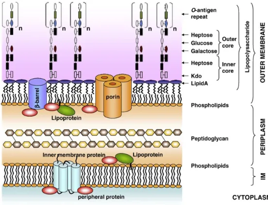
Lipopolysaccharide (LPS) is a major component of the outer membrane of Gram-negative bacteria and acts as a potent endotoxin inducer of pro-inflammatory cytokines, such as TNF-α, IL-6, IL-1 and interleukin-12 (IL-12) and inflammatory substances such as prostanoids, leukotrienes and nitric oxide in monocytes, macrophages, leucocytes, dendritic cells (DCs), epithelial and stromal cells (Ulevitch & Tobias, 1995; Van Amersfoort, Van Berkel, & Kuiper, 2003; Werling & Jungi, 2003; Yamamoto & Akira, 2005).
LPS consists of three parts: lipid A, a core oligosaccharide and O side chain (Lu, Yeh, & Ohashi, 2008; Wang & Quinn, 2010), works as a glycolipid complex essential for bacterial viability (Sperandeo, Dehò, & Polissi, 2009; Van Amersfoort et al., 2003) (Figure 1). Lipid A is the most essential and highly conserved hydrophobic portion of LPS and it is synthesized at the cytoplasmic side of the inner membrane (IM) and translocated to the outer leaflet of the outer membrane (OM) (Sperandeo et al., 2009; Wang & Quinn, 2010). It is responsible for the majority of the complications associated with Gram-negative bacterial infection such as endotoxin shock (reviwed by Akira, Uematsu, & Takeuchi, 2006; Sperandeo et al., 2009; Van Amersfoort et al., 2003). The second part of LPS molecule is the core oligosaccharide which connects to the lipid A through the inner core and to the O-antigen repeats through the outer core (Wang & Quinn, 2010). The third part of LPS molecule, consists of common sugars that provide the attachment site for the O-polysaccharide chain (Sperandeo et al., 2009; Van Amersfoort et al., 2003).
Several unique structural features of LPS contribute to the effective permeability barrier function of the OM. As a result, Gram-negative bacteria are protected from many toxic compounds such as bile salts, detergents, antibiotic and antimicrobial peptides (Sperandeo et al., 2009). Colonies with LPS molecules containing O-antigen are denoted S-LPS and have a smooth (S) appearance on agar plate whereas bacteria express an O-antigen-lacking LPS have a rough (R) appearance (Van Amersfoort et al., 2003). There are over 160 distinct O antigens in E. coli (Drummelsmith & Whitfield, 1999).
21
Figure 1 - Structure of Escherichia coli cell envelope. In (Sperandeo et al., 2009)
LPS is a complex glycolipid that can be structurally divided in three parts: lipid A, the hydrophobic moiety that anchors LPS to the outer membrane (OM), the oligosaccharide region named core, and the O-antigen polysaccharide chain. The core oligosaccharide of LPS can be divided into inner core, composed of Kdo (3-deoxy-D-manno-oct-2- ulosonic acid) and heptose, and outer core, which exhibits a greater structural diversity and provides the attachment site for the O-polysaccharide chain
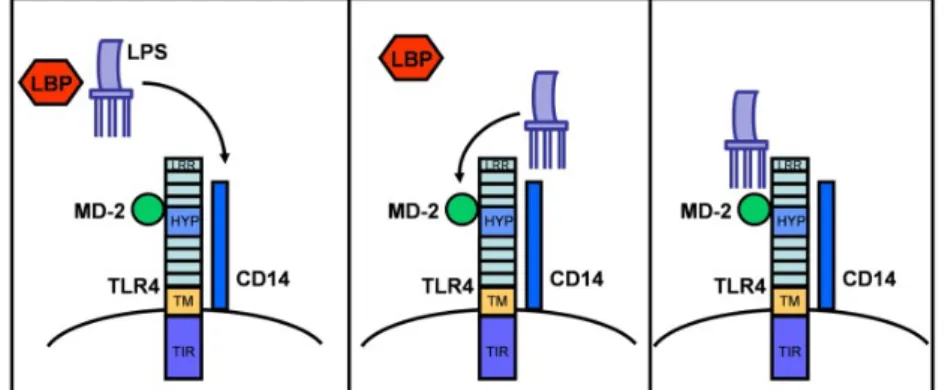
LPS is recognized by LPS-receptor complex CD14, TLR4, LPS binding protein (LBP) and myeloid differentiation factor-2 (MD-2) (Werling, Jann, Offord, Glass, & Coffey, 2009; Werling & Jungi, 2003).
LPS released from Gram-negative bacteria associates with LBP via lipid A (Ulevitch & Tobias, 1995). LBP is an acute-phase protein present in the bloodstream, that mediates the transfer of LPS to CD14, a protein expressed on the surface of inflammatory (monocytes, macrophages, polymorphonuclear leukocyte (PMN) and B cells) and non-inflammatory cells, like liver parenchymal cells, gingival fibroblasts and bovine stromal and epithelial endometrial cells (reviewed by Akira et al., 2006; Herath et al., 2006; Van Amersfoort et al., 2003). LPS is then transferred to MD-2, which associates with the extracellular