UNIVERSIDADE DE LISBOA
Faculdade de Medicina
Effect of GluN2B-containing NMDA receptors in the dorsal and
ventral hippocampus on the behavioral consequences of Chronic
Unpredictable Stress
Ricardo Filipe da Silva Ferreira
Orientadores:
Professor Doutor João José Fernandes Cardoso Araújo Cerqueira, ICVS, Escola de Ciências da Saúde, Universidade do Minho
Professora Doutora Raquel Alice da Silva Baptista Dias, IMM, Faculdade de Medicina, Universidade de Lisboa
Dissertação especialmente elaborada para obtenção do grau de Mestre em
Neurociências
A impressão desta dissertação foi aprovada pelo Conselho Científico da Faculdade de Medicina de Lisboa em reunião de 21 de Março de 2017.
Resumo
O stress pode ser definido como um conjunto de desafios, quer reais quer imaginários, à homeostasia do indivíduo. O quotidiano está repleto de tais desafios e, como tal, existem vários mecanismos compensatórios de resposta que, envolvendo alterações neuronais, autonómicas, endócrinas e comportamentais, tentam recuperar o equilíbrio. Assim, estes mecanismos fornecem formas de resposta a uma grande variedade de estímulos e desafios agudos. Contudo, apesar de serem fundamentais para a sobrevivência do indivíduo, está claramente descrito que uma exposição crónica pode levar a uma sobrecarga e uma má adaptação destes mecanismos, com consequências negativas para a saúde. As consequências mais comuns da exposição a stress crónico incluem alterações neuropsiquiátricas, como ansiedade, défices na memória de trabalho e na flexibilidade comportamental, formação de hábitos, e comportamento depressivo. Estas doenças neuropsiquiátricas estão associadas a alterações sequenciais de vias neuronais, provocadas principalmente por uma sobre-ativação do eixo hipotálamo-hipófise-adrenais (HPA) e consequente libertação excessiva de corticosteroides.
Os corticosteroides afetam as vias neuronais atuando em dois tipos distintos de recetores: os recetores de mineralocorticoides (MR), com alta afinidade; e os recetores de glucocorticoides (GR), que possuem baixa afinidade. Esta diferença de afinidades leva a um padrão de ativação específico destes recetores, que varia consoante a concentração hormonal libertada. Além disso, estes recetores apresentam também uma distribuição heterogénea no encéfalo, sendo que as suas diferentes distribuições, densidades e rácio provocam assim efeitos distintos em vias neuronais diferentes.
O hipocampo é uma região límbica que, em quase todas as espécies, apresenta uma elevada co-expressão de recetores GR e MR. Deste modo é interveniente numa das principais e mais estudadas vias inibitórias do eixo HPA, atuando quer em condições fisiológicas e em situações de stress. Contudo, está descrito que exposições prolongadas a concentrações elevadas de corticosteroides provocam uma reorganização morfológica nas vias neuronais do hipocampo, com alterações nos recetores excitatórios e inibitórios, na densidade das espinhas sinápticas, défices na neurogénese, e alterações na excitabilidade e plasticidade sináptica. Deste modo é provável que estas alterações das vias neuronais do hipocampo estejam relacionadas com as alterações cognitivas e emocionais observadas após exposição crónica a stress.
Além disso, vários estudos sustentam uma segmentação anatómica e funcional do hipocampo em dois compartimentos: um dorsal (posterior nos primatas), que desempenha principalmente funções cognitivas; e um ventral (anterior nos primatas), relacionado com a emoção, afeto e ansiedade.
O glutamato é o principal neurotransmissor excitatório do cérebro, e intervém na plasticidade sináptica e comportamental. O seu recetor N-metil D-aspartato (NMDA) é um recetor ionotrópico, permeável ao cálcio, e envolvido no desenvolvimento e função cerebral, mas também na neurotoxicidade. Estes recetores são proteínas tetraméricas, sendo que a sua maioria é constituída por duas subunidades GluN1 e duas subunidades GluN2. Para a função do canal a existência das subunidades GluN1 é obrigatória e, como tal, a distinção destes recetores deve-se principalmente à expressão específica de diferentes tipos de GluN2 (A-D). No córtex cerebral e no hipocampo os subtipos expressos são principalmente o GluN2A e GluN2B. Destes, foi demonstrado que os recetores que contêm subunidades GluN2B estão relacionados com comportamentos ansiosos, depressivos e influenciados pelo stress. Além disso, a exposição ao
stress foi associada a uma diminuição de expressão dos recetores GluN2B, com uma perda seletiva destes na superfície sináptica. Adicionalmente, a inativação farmacológica ou genética dos recetores com subunidades GluN2B foi capaz de inibir o fenótipo comportamental observado em consequência da exposição crónica ao stress.
De facto, experiências anteriores realizadas pelo nosso grupo, em que foi utilizado um modelo animal com um knockout (KO) condicional e pós-natal dos recetores GluN2B, foi capaz de alterar a resposta comportamental ao stress crónico, diminuindo principalmente o aumento induzido pelo stress de comportamento ansioso. Foram usados murganhos GluN2B-KO obtidos através do cruzamento de murganhos C57BL/6 com o gene que codifica a proteína GluN2B (GRIN2B) entre locais LoxP e murganhos que expressam a Cre-recombinase. Assim, após administração de tamoxifeno (um modulador específico dos recetores de estrogénio) foi possível obter uma perda de função do gene e uma deleção da expressão de recetores com o subtipo GluN2B. Estes murganhos foram depois submetidos a um protocolo de stress crónico imprevisível (Chronic Unpreditable Stress - CUS) e o seu comportamento foi testado e comparado a controlos (apenas manuseados gentilmente durante o período experimental), assim como a grupos wildtype controlo e submetidos ao mesmo protocolo de stress. Deste modo foi possível constatar que a deleção de expressão de GluN2B impediu as alterações biométricas e o aumento de comportamento ansioso, induzidos pelo stress. Contudo, o knockout genético utilizado é inespecífico, impedindo a expressão dos recetores GluN2B em várias estruturas encefálicas, cuja responsabilidade nas alterações comportamentais secundárias ao stress crónico ainda está por esclarecer.
Assim, no presente trabalho, utilizamos o mesmo modelo de murganhos C57BL/6 com o gene GRIN2B entre locais LoxP, mas administrámos a
Cre-recombinase através de um vetor viral, por cirurgia estereotáxica, no hipocampo dorsal e no hipocampo ventral, de modo a investigar o papel específico dos recetores GluN2B nestas estruturas. Foram então selecionados grupos de murganhos com 8 semanas de idade, sendo que um grupo foi inoculado com vírus-adeno-associado com Cre-recombinase no hipocampo dorsal e outro grupo inoculado no hipocampo ventral, e submetidos posteriormente a um protocolo de stress crónico imprevisível. Este protocolo, com duração de 4 semanas, foi iniciado 10 dias após a cirurgia, e consistiu na exposição diária, aleatória, a um dos seguintes fatores de stress: imobilização; agitação da jaula; derrota social; fluxo de ar quente; iluminação durante a noite; inversão do ciclo de luz; ou a inclinação da jaula. Em paralelo, dois outros grupos inoculados de forma similar foram apenas manuseados gentilmente durante o mesmo período e utilizados como controlos. De seguida testou-se o comportamento destes animais, com 13 semanas de idade, no Elevated Plus Maze, Open Field e Morris Water Maze.
Deste modo, foi possível observar que os animais com inativação dos recetores com subunidades GluN2B no hipocampo ventral demonstraram um fenótipo diferente à exposição crónica ao stress.
Foram observadas diferenças comportamentais entre os animais controlo e os submetidos a stress com inativação dos GluN2B no hipocampo dorsal. Nestes, alterações nos resultados dos testes comportamentais demonstraram um aumento marcado no comportamento ansioso dos animais submetidos a CUS. Contudo, tais diferenças não foram observadas nos murganhos com inativação no hipocampo ventral, o que nos permite inferir que os recetores que contêm o subtipo GluN2B nesta estrutura deverão modular a via neuronal responsável pelo aumento da ansiedade induzido pelo stress.
Adicionalmente, apesar da inativação de um subtipo de recetores NMDA do hipocampo, não foram observados défices nas capacidades de aprendizagem e memória espacial dos animais. Assim, é possível formular a hipótese que estas capacidades são independentes do processo de potenciação a longo prazo (LTP) descrito classicamente, respeitante aos recetores de glutamato GluN2B do hipocampo, incluindo os existentes no seu compartimento dorsal.
Assim, o presente trabalho permite confirmar o papel dos recetores NMDA que contêm a subunidade GluN2B do hipocampo nas alterações comportamentais consequentes à exposição crónica ao stress, evidenciando particularmente o papel destes recetores do hipocampo ventral no aumento de comportamento ansioso induzido pelo stress. Palavras-chave: NMDA; GluN2B; Ansiedade; Stress crónico; Hipocampo dorsal; Hipocampo ventral.
Abstract
Glutamate and NMDA receptor dysfunction is strongly implicated in the pathophysiology of behavioral changes that occur in response to chronic stress exposure. These behavioral impairments have been attributed to actions at the NMDAR GluN2B subunit, which influences the excitatory postsynaptic currents and synapse maturation. Furthermore, pharmacologic or genetic inactivation of GluN2B subunits is able to inhibit the behavioral phenotype observed in consequence to chronic stress, particularly the stress-induced increase in anxious-like behavior.
Nevertheless, the contribution of the several forebrain structures where these receptors are present, on the stress-induced changes, is yet to be fully understood. Still, the hippocampus has been shown to be implicated in stress regulation and in the pathophysiology of some changes observed after chronic stress exposure. As such, in the present work, we investigated the role of the GluN2B receptors of the dorsal and ventral hippocampus. To this end, we used GRIN2B C57BL/6 floxed mutant mice and specifically delivered Cre-recombinase in the dorsal and ventral hippocampus via viral vector–mediated delivery, which led to a restricted and specific deletion of GluN2B expression. A group of animals was inoculated in the dorsal hippocampus and another group was inoculated in the ventral hippocampus, and then submitted to a Chronic Unpredictable Stress protocol; while two other similarly treated groups were only gently handled to serve as controls.
We observed a distinct response to stress in our ventral hippocampus groups. Opposite to animals with GluN2B inactivation in the dorsal hippocampus, those whose receptor was inactivated in the ventral hippocampus did not show the
characteristic stress-induced increase in anxious-like behavior. Moreover, deletion of hippocampus’ GluN2B receptors did not affect spatial learning and memory capabilities.
Our data confirms that GluN2B-contaning NMDARs are involved in mediating the behavioral consequences of chronic stress. Particularly, receptors present in the ventral hippocampus seem to modulate the characteristic stress-induced increase in anxious-like behavior.
Keywords: NMDA; GluN2B; Anxiety; Chronic stress; Dorsal hippocampus; Ventral hippocampus.
Table of Contents
Resumo ... v Abstract ... xi Table of Contents ... xiii Figures Index ... xv Tables Index ... xvi Abbreviations List ... xvii 1 - Introduction ... 1 1.1 - Stress and HPA-axis ... 1 1.2 - GluN2B-contaning N-methyl D-aspartate Glutamate receptor ... 4 1.3 - Hippocampus and Stress ... 7 1.4 - Previous work ... 10 2 - Objectives ... 14 3 - Materials and Methods ... 15 3.1 - Subjects ... 15 3.2 - Inactivation of Hippocampal receptors ... 16 3.3 - Chronic Unpredictable Stress ... 21 3.4 - Behavioral tests ... 22 3.4.1 - Elevated Plus Maze ... 22 3.4.2 - Open Field ... 23 3.4.3 - Acoustic Startle ... 23 3.4.4 - Morris Water Maze ... 24 3.5 - Brain dissection ... 25 3.6 - Statistical Analysis ... 25 4 - Results ... 264.1 - Biometrics ... 26 4.2 - Behavioral tests ... 29 4.2.1 - Elevated Plus Maze ... 29 4.2.2 - Open Field ... 30 4.2.3 - Acoustic Startle ... 31 4.2.4 - Morris Water Maze ... 32 5 - Discussion ... 35 6 - Bibliography: ... 43
Figures Index
Figure 1 – Representation of the Hypothalamus-Pituitary-Adrenal Axis…...………2
Figure 2 – Immunofluorescent images for the MR and GR in the hippocampus……….……….4
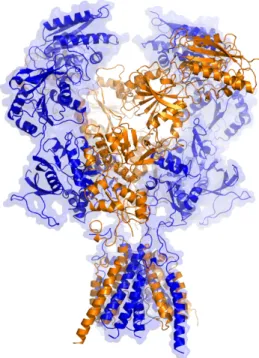
Figure 3 – Representation of a NMDA receptor...5
Figure 4 – The hippocampal formation, as drawn by Santiago Ramon y Cajal………...8
Figure 5 – Thymus and Adrenals weight (g) of animals of our previous experience……….11 Figure 6 – Percentage of time spent in the open arms and number of open arms’ entries in the EPM, of the animal of our previous work………12 Figure 7 – Ratio between the distance covered at the OF center / periphery, of the animals of our previous work………13 Figure 8 – PCR results for a set of animals……….16 Figure 9 – Stereotaxic Setup used in AAV-Cre inoculation surgeries………18 Figure 10 – Immunofluorescence microscopy image of a ventral hippocampus innoculated with AAV-Cre-GFP (x200)………...19
Figure 11 – Relative GluN2B protein levels, normalized to Actin, obtained by Western Blot analysis……….20 Figure 12 – Animal weight gain during the experimental period………27 Figure 13 – Average of thymus weight of sacrificed animals (g) ± SEM……….28 Figure 14 – Average of adrenals weight of sacrificed animals (g) ± SEM………28 Figure 15 – Average of the percentage of time spent in the open arms of the Elevated Plus Maze apparatus (time in open arms / total time) ± SEM……….29 Figure 16 – Open Field results………30
Figure 17 – Response amplitude (arbitrary units) in response to acoustic startle stimulus………31 Figure 18 – Average latency to reach platform on Morris Water Maze test………33 Figure 19 – Probe trial results………34
Tables Index
Table 1 – Distribution of the different cohorts of tested animals………....21 Table 2 – Response amplitude in response to acoustic startle stimulus...32Abbreviations List
AAV - Adeno-associated-virus ACTH - Adenocorticotropic hormone bp - Base pair CA - Cornu Amonis (Ammon’s horn) CaMKII - Calcium/calmodulin-dependent protein kinase II cm - Centimeters CRH - Corticotropin-releasing hormone CUS - Chronic Unpreditable Stress DAPI - 4',6-diamidino-2-phenylindole dHip - Dorsal hippocampus DNA - Deoxyribonucleic acid dB - Decibel DG - Dentate Gyrus EPM - Elevated Plus Maze G - Gram GFP - Green Fluorescent Protein GluN1 - Ionotropic glutamate receptor subunit 1 GluN2 - Ionotropic glutamate receptor subunit 2 GluN2B - Ionotropic glutamate receptor subunit 2B GluN3 - Ionotropic glutamate receptor subunit 3 GR - Glucocorticoid receptor GRIN2B - Ionotropic glutamate receptor subunit 2B encoding gene HPA - Hypothalamus-pituitary- adrenal KO - KnockoutLTD - Long Term Depression LTP - Long Term Potentiation mg/Kg - Milligram per Kilogram mL - Milliliter mm - Millimeter MR - Mineralocorticoid receptor ms - Millisecond mV - milivolt MWM - Morris Water Maze NaCl - Sodium chlorite nL - Nanoliter NMDA - N-methyl-D-aspartate NMDAR - N-methyl-D-aspartate receptor ºC - Celsius degrees OF - Open Field PCR - Polymerase Chain Reaction PSD95 - Postsynaptic Density Protein 95 rpm - Rotations per minute SEM - Standard Error of the Mean µL - Microliter vHip - Ventral hippocampus Vmax - Startle amplitude WT - Wildtype
1 - Introduction
1.1 - Stress and HPA-axis Everyday life is filled with a variety of real or perceived threats and challenges to one's homeostasis, which require compensatory responses to return to equilibrium. Any type of these threats can be defined as stress.Responses to stress exposure implicate neuroendocrine, autonomic and behavioral changes. And although these responses are imperative for the survival of the individual, by helping meet the demands of a variety of acute challenges, it is widely described that repeated exposure to stress has nocent effects on health (Dias-Ferreira et al 2009, Radley et al 2015).
The most common consequences of repeated stress exposure are neuropsychiatric disorders, namely concerning anxious-like behavior (Maes et al 1998, Pêgo et al 2006, Shin & Liberzon 2010); deficits in working memory and behavioral flexibility (Cerqueira et al 2007); promotion of habit formation (Dias-Ferreira et al 2009); and depressive-like behavior (Strekalova et al 2004) (Bessa et al 2009) (Peng et al 2016).
These behavioral changes in response to chronic stress are attributed to sequential structural modulation of the brain networks, mainly due to stress-induced release of corticosteroids (De Kloet et al 2005, McEwen 2007), but also owing to release of excitatory amino acids and many others cellular mediators (reviewed by (McEwen et al 2015). In a stressful situation the paraventricular
nucleus of the hypothalamus is activated and releases Corticotrophin-Releasing-Hormone (CRH), which then stimulates pituitary neurons to secrete Adrenocorticotrophin (ACTH) to the bloodstream (Herman & Cullinan 1997). ACTH induces the secretion of glucocorticoids by the adrenal cortex (Tsigos & Chrousos 2002). This chain of interactions constitutes the Hypothalamus-Pituitary-Adrenal (HPA) axis (Figure 1).
Glucocorticoids are then the end-products of the HPA axis, and play a fundamental role in the maintenance of both resting and stress-related homeostasis (Nicolaides et al 2015). This maintenance includes various
Figure 1 – Representation of the Hypothalamus – Pituitary – Adrenal Axis. From the Integrative Therapeutics webpage: http://www.integrativepro.com
CRF: Corticotropin-releasing hormone ACTH: Adenocorticotropic hormone
peripheral effects, such as regulation of the immune response or metabolism, but also has important consequences on the brain (McEwen et al 2015).
The stress-induced release of glucocorticoids mediates its effects by two different types of receptors. These two receptors bind the same hormone (cortisol in humans and corticosterone in rodents), but with differential affinity: the Type I, high-affinity, mineralocorticoid receptor (MR) and the Type II, low-affinity, glucorticoid receptor (GR) (Reul & Kloet 1985). Such differential affinity leads to a particular pattern of activation of GR and MR: While MR are tonically activated by circulating glucocorticoids, implicated in the onset of the stress response; GR are only activated by large concentrations of glucocorticoid, as in stress exposure or during the circadian peak (De Kloet et al 2005, Joëls 2006, Tsigos & Chrousos 2002). These distinct receptors have also a heterogeneous distribution in the brain, with substantial co-expression in the hippocampus of almost all species (Han et al 2005, Patel et al 2000) (Figure 2). Thus, the particular distribution, density and ratio of MR and GR will mediate distinct effects of glucocorticoids in different neuronal circuits (Joëls 2006).
As referred, acute responses to a stressful situation are indispensable to restoration of homeostasis, but persistent stress exposure can lead to a dysregulation and maladaptation to stressors (McEwen et al 2015). As such, there are multiple inhibitory routes to control the HPA-axis activity, including neuronal pathways. One of the most studied arises from the hippocampus, a sensible area to glucocorticoids, that provides an inhibitory output to the HPA-axis in physiological conditions (Kim et al 2006). However, it has been reported that the effects of persistent high levels of glucocorticoids include dendritic and synaptic reorganization in brain structures that have a role in the regulation of HPA-axis activity during stress, such as the hippocampus (Sousa et al 2000) and the medial prefrontal cortex (Cerqueira et al 2007) (Radley et al 2004).
1.2 - GluN2B-contaning N-methyl D-aspartate Glutamate receptor Both synaptic and behavioral plasticity are mediated by glutamate, which is the brain's major excitatory neurotransmitter. N-methyl-D-aspartate glutamate receptors (NMDAR) belong to the family of ionotropic glutamate receptors, which are glutamate-gated ion channels. NMDA receptors are critically involved in brain development and function, and also in neurotoxicity (Tajima et al 2016). The NMDA receptor is a tetrameric membrane-inserted protein complex comprising two GluN1 and GluN2, or GluN3 subunits (von Engelhardt et al 2008) (Figure 3). The majority of NMDA receptors has two GluN1 and two GluN2 subunits, which bind glycine and L-glutamate, respectively (Tajima et al 2016) although triheteromeric receptors have also been described (Tovar et al 2013). The GluN3 subunit binds both glycine and D-serine and is expressed throughout the nervous system. However, the role of GluN3 is not yet as well-defined as that of GluN1 and GluN2 subunits (Lee et al 2014). The binding of both glycine
Figure 2 – Immunofluorescent images for the mineralocorticoid receptor (MR) and glucorticoid receptor (GR) in the hippocampus. The merged image showed the co-localization of MR (red; A) and GR (green; B), indicated by yellow color (C). From (Han et al 2005)
and glutamate, with concurrent membrane depolarization to relieve magnesium block (Nowak et al 1984), activates the NMDA receptor. An active receptor opens a cation-selective, calcium permeable channel, thus causing further depolarization of the cell membrane and influx of calcium (Mayer & Westbrook 1987). The consequent calcium influx causes a cascade of signal transduction necessary for synaptic plasticity (Granger & Nicoll 2014). GluN1 is an obligate receptor subunit for channel function. As such, differentiation and development is targeted primarily toward specific GluN2 and GluN3 expression (Traynelis et al 2010). Each of the existing GluN2 subunits (GluN2A-D) displays a characteristic regional and developmental expression profile (Balsara et al 2014, Monyer et al 1994). GluN2A and GluN2B are the subunits mainly expressed in adult neocortex and hippocampus (Monyer et al 1994, von Engelhardt et al 2008). In early postnatal life, GluN2B-containing NMDARs are critical for circuit formation and
Figure 3 – Representation of a NMDA receptor. X-ray crystal structure of the receptor, with receptor subunits in different colors. From (Lee et al 2014).
development, promoting maturation of glutamatergic synapses (Kelsch et al 2014) (Traynelis et al 2010). After the first weeks of postnatal life a switch occurs, triggered by agonist binding (Barria & Malinow 2002), long-lasting potentiation stimulus (Bellone & Nicoll 2007) or sensory experiences (Barth & Malenka 2001), towards GluN2A-contaning receptors, resulting in NMDA receptor-mediated excitatory postsynaptic currents with a faster decay (Traynelis et al 2010). This switch seems to imply the importance of subunit composition to neuroplasticity, and the possibility of a differential role of the subunits in neuronal changes such as the ones observed in response to chronic stress exposure. In fact, GluN2B-contaning NMDARs have been shown to mediate anxiety (von Engelhardt et al 2008) and stress-related behaviors (Kiselycznyk et al 2011). The exposure to chronic stress has also been shown to be associated with a decrease in the total expression of GluN2B receptor and PSD95 (an anchoring protein of GluN2B), with selective loss of GluN2B receptors from the cell surface membrane and, therefore, a loss of surface receptors on synaptic sites (Jiang et al 2013). Similar finding were also observed in patients with Major Depressive Disorder (Feyissa et al 2009). Additionally, previous works described that extrasynaptic NMDA receptors are mainly composed of the GluN2B type and promote cell death (Hardingham et al 2002, Li & Ju 2012).
Furthermore, the use of pharmacological antagonists with differential affinity for GluN2B-contaning NMDARs is able to inhibit the behavioral changes observed in response to chronic stress exposure (Li et al 2011, von Engelhardt et al 2008), and have been shown to exert anti-depressant (Ibrahim et al 2012, Li et al 2011, Lima-Ojeda et al 2013) and anxiolytic-like effects (Li et al 2011). In fact, there are clinical trials registered that aim to assess the efficacy of selective GluN2B antagonists on the symptomatic improvement in patients with Major
Depressive Disorder (clinicaltrials.gov 2016), reflecting the potential therapeutic value of modulating the activity of these receptors.
Mice lacking GluN2B expression by genetic knock-out, or those expressing this subunit in a C terminally truncated form, die at birth, reflecting the importance of GluN2B-contaning NMDARs for perinatal brain functions (Mohrmann et al 2002). However, genetic manipulation that allows for a post-natal, restrictive deletion of GluN2B receptors is also able to inhibit the behavioral phenotype observed in consequence to chronic stress (von Engelhardt et al 2008) (Please see chapter 1.4 – Previous Work, of present thesis).
This genetic knock-out is, however, highly unspecific, deleting GluN2B expression in several forebrain structures, such as the prefrontal cortex, orbitofrontal cortex, dorsal and ventral hippocampus, cortex and striatum (Balsara et al 2014). The specific role of the receptors in each of these structures in the behavioral changes that are observed in chronic stress situations is not yet established. 1.3 - Hippocampus and Stress Over the past several decades, the hippocampus has been the quintessential limbic region implicated in stress regulation. As mentioned, the hippocampal formation is one of the most studied neuronal pathway-mediated inhibitor of HPA-axis activity. It has the capacity to inhibit the hypothalamo-pituitary-adrenal axis under both basal and stress conditions, is a prominent site for glucocorticoid receptor-mediated feedback, and is essential for restoring glucocorticoids to baseline levels following the cessation of stress (Fanselow & Dong 2010, Radley et al 2015).
The hippocampus is a limbic structure approximately 5 cm long in humans, within the para-hippocampal gyrus inside the inferior (temporal) horn of the lateral ventricle. The hippocampal formation includes the dentate gyrus, the hippocampal proper (divided into three distinct fields: CA1, CA2 and CA3), subicular complex (subiculum, presubiculum, parasubiculum) and entorhinal cortex (Standring 2015) (Figure 4).
Hippocampal neuronal circuits are well studied, with a well recognized unidirectional circuit. The input arises from the entorhinal cortex to the Dentate Gyrus, axons of the DG (mossy fibers) innervate CA3 neurons, which project through Schaffer collaterals axons to CA1 and this back to the entorhinal cortex closing the unidirectional circuit of input integration (Da Silva et al 1990).
Figure 4 - The hippocampal formation, as drawn by Santiago Ramon y Cajal (Histologie du Système nerveux de l'Homme et des Vertébrés). DG: dentate gyrus. Sub: subiculum. EC: entorhinal cortex. CA1-CA3: hippocampus proper. Image from wikiwand web page: www.wikiwand.com
Various behavioral, anatomical, and gene expression studies support a functional segmentation into two hippocampal compartments: the dorsal hippocampus, which corresponds to the posterior hippocampus in primates, performs primarily cognitive functions; the ventral (anterior in primates) relates to stress, emotion and affect (Bannerman et al 2003, Padilla-Coreano et al 2016) (reviewed by (Fanselow & Dong 2010)).
Persistent stress and, consequently, persistent high levels of systemic glucocorticoids have multiple described effects in the hippocampus (Conrad 2008, Joels et al 1994, Karst et al 1994). Several studies have described morphological changes, such as dendritic atrophy (Magarin & McEwen 1995, Sousa et al 2000); changes in the levels of excitatory and inhibitory receptors (Joëls et al 2004, Reagan et al 2004) and spine density (Tata et al 2006); downregulation of GR (Wright et al 2006); impairments in neurogenesis (Pham et al 2003, Rosenbrock et al 2005); and alterations on excitability and synaptic plasticity, such as impairments of long term potentiation (LTP) and long term depression (LTD) (Joëls et al 2004, Pavlides et al 2002, Rosenbrock et al 2005). Thus, it is highly plausible that all of the described hippocampal changes occurring in response to high levels of glucocorticoids are related to the cognitive and emotional deficits observed after chronic stress exposure.
1.4 - Previous work
The evidence above supports the hypothesis that the GluN2B-Containing NMDA receptors are of major importance to mediate the behavioral changes observed after exposure to chronic stress. The work behind the present thesis was elaborated following previous experiments, yet unpublished, from our group, that tried to clarify the role of GluN2B-contaning NMDARs in the behavioral changes observed in C57BL/6 mice exposed to a Chronic Unpredictable Stress (CUS) protocol.
As mentioned, animal models lacking expression of GluN2B by genetic knock-out die at birth (Mohrmann et al 2002). As such, we utilized an inducible, conditional, deletion method of the gene that encodes the GluN2B protein. Thus, GluN2B-knockout mice were generated by crossing GRIN2B (the gene that encodes the GluN2B protein) C57BL/6 floxed mutants with transgenic mice expressing Cre recombinase driven by the Calcium/calmodulin-dependent protein kinase II (CaMKII) promoter. This intercross led to a restricted, postnatal deletion of GluN2B, after administration of tamoxifen (a selective estrogen-receptor modulator). The binding of tamoxifen to the estrogen receptor results in the translocation of the Cre recombinase into the nucleus, where it can recombine its loxP-flanked DNA substrate (Feil et al 2009). The administration of tamoxifen during week 7 resulted in a complete ablation of GluN2B expression that lasted more than 8 weeks.
Four groups of mice were selected. One group was treated with tamoxifen as explained above, in order to achieve deletion of the GRIN2B gene, and a similarly treated group of wildtype animals were submitted to 4 weeks of a Chronic Unpredictable Stress (CUS) protocol (please see chapter 3.3 of present thesis),
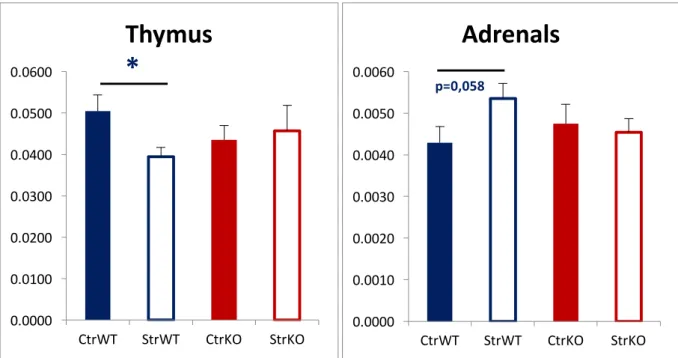
while a group of transgenic and wildtype animals was only gently handled to serve as controls. At 12 weeks of age mice were behaviorally tested in the Open Field (chapter 3.4.2 of present thesis), Elevated Plus Maze (chapter 3.4.3 of present thesis) and Morris Water Maze (chapter 3.4.4). There was a clear distinction in the behavioral response to stress in our GluN2B-Knock-out (KO) group. Contrary to the wildtype group, these animals did not show biometric changes in response to stress, neither an increase in anxiety. We did not observed some of the characteristic biometric changes, described as consequences of persistent elevated high levels of glucocorticoids (as in chronic stress situations), such as a decrease in thymic volume (Domínguez-Gerpe & Rey-Méndez 2001) or an increase in adrenal gland volume (Nemeroff et al 1992) between our transgenic groups (Figure 5), suggesting a different response to stress. 0.0000 0.0100 0.0200 0.0300 0.0400 0.0500 0.0600 CtrWT StrWT CtrKO StrKO
Thymus
*
0.0000 0.0010 0.0020 0.0030 0.0040 0.0050 0.0060 CtrWT StrWT CtrKO StrKOAdrenals
p=0,058 Figure 5 – Thymus and Adrenals weight (g) of animals of our previous experiment. Results are shown as mean ± SEM. CtrWT: Control wildtype group. StrWT: Stressed wildtype group. CtrKO: Control knock-out group. StrKO: Stressed knock-out group. * p < .05The difference between wildtype groups (control/stressed) (Thymus: t25 = 2.617; p = .015; d = 1.025; Adrenals: t25 = -1.987; p = .058; d = .778) was not observed in Knock-out animals.
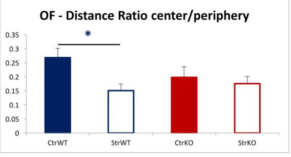
Moreover, KO groups did not show the characteristic increase in anxious-like behavioral after exposure to the chronic unpredictable stress protocol, contrary to wildtype animals (Maes et al 1998, Pêgo et al 2006, Shin & Liberzon 2010). This was demonstrated in the percentage of time spent in the Open Arms and in the number of Open Arms entries in the Elevated Plus Maze (EPM) (Figure 6), and also in the ratio between the distance covered in the center and in the periphery of the Open Field (Figure 7). 0.0 5.0 10.0 15.0 20.0 25.0 30.0 35.0 40.0 45.0 50.0 CtrWT StrWT CtrKO StrKO
EPM - Open Arms %
*
0 2 4 6 8 10 12 14 CtrWT StrWT CtrKO StrKOOpen Arms Entries
***
Figure 6 – Percentage of time spent in the open arms and number of open arms’ entries in the EPM, of the animals of our previous work. Results are shown as mean ± SEM. CtrWT: Control wildtype group. StrWT: Stressed wildtype group. CtrKO: Control knock-out group. StrKO: Stressed knock-out group. * : t25 = 2.853; p = .009; d = 1.005 *** : t25 = 4.575; p < .001; d = 1.792
Our data confirmed that GluN2B-contaning NMDARs are implicated in the pathophysiology of the behavioral changes observed in response to chronic stress, especially those related with anxious-like behavior. However, this GluN2B-KO conditional model deletes GluN2B expression in several forebrain structures, such as the prefrontal cortex, orbitofrontal cortex, dorsal and ventral hippocampus, cortex and striatum (Balsara et al 2014). More research was needed to further clarify the specific role of each of these structures on the behavioral changes seen in consequence of chronic stress. 0 0.05 0.1 0.15 0.2 0.25 0.3 0.35 CtrWT StrWT CtrKO StrKO
OF - Distance Ratio center/periphery
*
Figure 7 – Ratio between the distance covered at the OF center / periphery, of the animals of our previous work. Results are shown as mean ± SEM.CtrWT: Control wildtype group. StrWT: Stressed wildtype group. CtrKO: Control knock-out group. StrKO: Stressed knock-out group.
* : t24 = 2.871; p = .008; d = 1.157
2 - Objectives
The aforementioned evidences support the hypothesis that GluN2B-contaning NMDARs are implicated in the pathophysiology of the behavioral changes observed in response to chronic stress, especially those related with stress-induced increase in anxious-like behavior.
In fact, the conditional, post-natal, GluN2B-knock out animal model used in our previous work showed a distinct reaction to stress, especially in the behavioral tests that are influenced by anxious-behavior. However, this technique showed to be unspecific, deleting the expression of the GluN2B protein in several forebrain structures, whose accountability for the stress-induced changes is yet to be fully understood. Nevertheless, the hippocampus has been shown to be implicated in stress regulation and in the pathophysiology of some changes observed after chronic stress exposure. Also, several studies support a functional segmentation of the hippocampus in two compartments: the dorsal and the ventral hippocampus.
Based on these data, the specific aim of the work presented in this thesis was proposed: Study the behavioral changes observed in consequence to chronic stress exposure, of which the GluN2B-contaning NMDARs of the dorsal and ventral hippocampus may be held accountable. To this end, we used the same animal model of GRIN2B C57BL/6 floxed mutant mice, but specifically delivered the Cre-recombinase in the dorsal and ventral hippocampus via viral vector–mediated gene delivery (Ahmed et al 2004, Betley & Sternson 2011, Kaspar et al 2002, Royo et al 2008). These animals were then behaviorally tested in order to check for differences between control and stressed cohorts.
3 - Materials and Methods
3.1 - Subjects
Experiments were conducted in accordance with local regulations (European Union Directive 86/609/EEC) and Portuguese national authority for animal experimentation, Direção Geral de Veterinária (ID:DGV9457) guidelines on animal care and experimentation.
We used GRIN2B (the gene that encodes GluN2B protein) floxed C57BL/6 mice. In order to confirm the genotype of the tested mice, we collected a small tissue sample from newborn mice (postnatal day 5-7), either by toe clipping or tail extremity clipping. The genomic extraction from the tissue sample was performed as previously described (Castelhano-Carlos et al 2010). Quantification of the DNA was performed by spectrophotometry using NanoDrop ND-1000 (NanoDrop Technologies, LLC, DE, USA).
To assess the genotype, we performed Polymerase Chain Reactions (PCR) for the flox GluN2B product (326bp) and selected only the homozygous (flox GluN2B +/+) animals (Figure 8).
The administration of adeno-associated-virus with Cre-recombinase in a specific, localized region, led to a recombination between LoxP sites of the flanked gene, inducing a restricted, localized, postnatal deletion of the GRIN2B gene. The mice were housed in same-sex groupings in a temperature and humidity controlled vivarium under a 12-hour light/dark cycle and ad libitum access to food and water. In order to assess behavioral performance we used male mice only, thus avoiding any potential interference by the female estrous cycle. 3.2 - Inactivation of Hippocampal receptors
Inactivation of the GluN2B-contaning NMDARs of the dorsal and ventral hippocampus was achieved by viral vector–mediated gene delivery. Figure 8 – PCR results for a set of animals. L – DNA ladder. Band marked as “A” corresponds to a Homozygous wildtype animal (wildtype GluN2B product ≈ 412bp); bands marked as “B” correspond to Homozygous transgenic animals (Floxed GluN2B product ≈326bp). All remaining bands correspond to Heterozygous animals. L A B B B B B B B B B
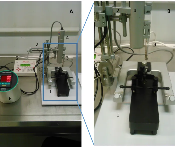
We performed the direct injection of adeno-associated-virus (AAV) -Cre into the dorsal and ventral hippocampi using stereotaxic guidance. To perform the surgery, the mice were pre-medicated with buprenorphine 0,05mg/Kg, subcutaneously, 10 to 20 minutes prior the administration of the anesthetics (medetomidine 1mg/Kg and ketamine 75mg/Kg), intraperitoneally. After placement in the stereotaxic setup (Figure 9), the coordinates of the ventral and dorsal hippocampus were calculated by adding or subtracting the appropriate lateral and anterior/posterior values from bregma, using a reference atlas (Paxinos & Franklin 2004). For the administration of the viral vector in the dorsal hippocampus the following coordinates were used: -2mm antero-posterior; +/- 1,5mm lateral; -2mm ventral. For the administration in the ventral hippocampus the following coordinates were selected: -3,6mm antero-posterior; +/- 2,5mm lateral; -2,3mm ventral. General surgical procedures and protocol was done as described by (Cetin et al 2006). Administration of the AAV-Cre solution was performed in a slow rate (75nL per minute) in order to prevent tissue damage. Post-operative analgesia in the three days following surgery was assured by administration of buprenorphine (0,05mg/Kg) and carprofen (5mg/Kg), subcutaneously.
Being a new procedure in our lab, validation and calibration of the dosage and dynamics of the AAV-Cre inoculation was needed. To this end, several 8-week-old GRIN2B floxed C57BL/6 mice were inoculated 1uL AAV2-Cre- Green
Figure 9 – Stereotaxic Setup used in AAV-Cre inoculation surgeries.
A – Overview of the different components used for stereotaxic surgery. 1 – Stereotaxic apparatus (animal restraint). 2 – Stereotaxic arm, with microsyringe holder. 3 – Microsyringe (10µL). 4 –Microsyringe pump. 5 – Digital microsyringe pump controller. 6 – Digital coordinates display. B – Close-up picture of the stereotaxic apparatus and microsyringe.
A
B
6 1 2 3 5 1 3 4 4 2Fluorescent Protein (GFP) (University of North Carolina Vector Core), with a concentration of 3,7 x 10¹² viral molecules / mL; and allowed to recover during different periods of time (1week/10days). Afterwards, the expression was evaluated using Immunofluorescence and Western Blot techniques (Figures 10 and 11). Figure 10 – Immunofluorescence microscopy image of a ventral hippocampus innoculated with AAV-Cre-GFP (x200). Co-immunofluorescent staining with DAPI. A portion of the ventral dentate gyrus is visible, with cells marked with the transfected virus (presenting a green glow).
This allowed us to determine the pertinent timeframe and dosage of the inoculation. We were able to determine that a inoculation of 1µL of adeno-associated-virus – Cre, was able to trigger a specific and substantial reduction of GluN2B receptor levels, 10 days after the procedure.
Following this validation process, we then selected four different cohorts of mice. One was inoculated with adeno-associated virus with Cre in the dorsal hippocampus (n=5) while another cohort was inoculated in the ventral hippocampus (n= 7), and submitted to a Chronic Unpreditable Stress protocol, as explained below. Two other control cohorts of mice were inoculated with AAV-Cre in the dorsal (n= 4) and ventral hippocampus (n= 5), respectively, but were only subject to gentle handling during the experimental period (Table 1). 0 0.2 0.4 0.6 0.8 1 1.2 1.4 1.6 1.8
Control (AAV-Cre -) AAV-Cre + 7d AAV-Cre + 10d
Gl uN 2B p ro te in le ve l / A ct in p ro te in le ve l Figure 11 – Relative GluN2B protein levels, normalized to Actin, obtained by Western Blot analysis of hippocampal tissue without AAV-Cre inoculation (Control AAV-Cre -), 7 and 10 days after AAV-Cre inoculation.
AAV - Cre inoculation site Stress Control
Dorsal Hippocampus N = 5 N = 4 Ventral Hippocampus N = 7 N = 5 3.3 - Chronic Unpredictable Stress
Two different groups of mice (dorsal Hippocampus AAV-Cre and ventral Hippocampus AAV-Cre) were then submitted to a chronic unpredictable stress protocol, for a 4-week period. This protocol was performed to mice 10 days after the surgical procedure, thus with 9 weeks of age. The chronic unpredictable stress protocol consisted in exposure, once daily in a random order and in an unpredictable fashion, to one of the following aversive stressors: Restraint – mice were placed in a 50 mL plastic tube with openings in both sides for breathing, for 1 hour; Shaking – mice were placed in a plastic box container and placed in an orbital shaker for 1 hour at 150 rpm; Social defeat – mice were introduced in a cage of an aggressive mouse and, after being defeated, they were placed in a transparent and perforated plastic container, to avoid further physical contact, inside the resident homecage for 30 minutes;
Hot air stream – mice were exposed to a hot air stream from a hairdryer for 10 minutes;
Overnight illumination – mice were exposed to regular room light during the night period;
Inverted light cycle – regular room light was off during daytime and on during nighttime;
Tilted cage – homecages were tilted in a 45° angle for 1 hour.
This type of chronic stress paradigm, mixing different stressors (including physical and psychological components) presented in an unpredictable schedule, was previously shown to result in chronically elevated serum corticosterone titres, and to better mimic the variability of stressors encountered in daily life (Monteiro et al 2015, Sousa et al 1998). 3.4 - Behavioral tests 3.4.1 - Elevated Plus Maze
After exposure to chronic unpredictable stress, 13-week-old mice were evaluated for behavioral performance. Similarly, mice that were only gently handled during the experimental period were also evaluated for behavioral performance at the same age.
Anxious-like behavior was assessed using the Elevated Plus Maze test.
The test setting consists of a plus-shaped apparatus with two open and two enclosed arms, each with an open roof, elevated from the floor. Mice’s aversion of open spaces leads to avoidance of open areas by confining movements to enclosed spaces or to the edges of a bounded space.
Anxiety reduction in the plus-maze is indicated by an increase in the proportion of time spent in the open arms (time in open arms/total time in open or closed
arms), or an increase in the proportion of entries into the open arms (entries into open arms/total entries into open or closed arms) (Komada et al 2008). Animals were tested during 5 minutes in a black polypropylene “plus” shaped maze (ENV-560, Med Associates Inc.), elevated 72.4cm above the floor. The maze consisted of two opposite open arms (50.8cm x 10.2cm) and two enclosed arms (50.8cm x 10.2cm x 40.6cm). We recorded and evaluated the number of closed and open arms entries, and the ratio of time spent in open arms over closed arms. 3.4.2 - Open Field Locomotor and exploratory activities were assessed using the OF. Each mouse was left in the center of a squared arena (43.2cm x 43.2cm), which the mouse was free to explore for 5 minutes. Activity was detected and registered using a monitor software (Med Associates Inc.). Distance traveled was used as a measure of locomotor activity, and the ratio of the distance and time spent between the center and the periphery of the arena was used as a measure of anxious-like behavior.
3.4.3 - Acoustic Startle
The acoustic startle consists of a rapid and involuntary extension and then flexion of a series of muscles, in response to an abrupt and unexpected loud sound. Although the startle response is a reflex, it can be modulated by many different stimuli, including fear, pain or anxiety (Davis, Walker e Lee 1997). We measured the startle response in our mice using an automated startle chamber, with detectors recording whole-body reaction. After a habituation
period to the apparatus (5 minutes daily, 1 day before testing), startle stimuli were presented with an unpredictable and random intensity, ranging from 70dB to 120dB. Startle magnitudes were sampled each millisecond during a period of 200ms, beginning at the onset of the startle stimulus. A startle response was defined as the peak response during this 200-ms period. 3.4.4 - Morris Water Maze To assess spatial reference memory, mice were tested in a white circular pool filled with water (24–25°C) placed in a dimly lit room, with spatial cues placed in the walls around the pool (square, stripes, triangle, and a cross). The pool is divided into four imaginary quadrants and a hidden transparent platform was placed in one of the quadrants. Data were collected via a fixed camera placed in the ceiling and connected to a video-tracking system (Viewpoint, Champagne-au-Mont-d’Or, France). Mice had to learn the position of a hidden platform over a period of 4 days. Each day, mice were placed facing the wall of the pool at different quadrants as a starting point for each trial, for a total of 4 trials. Each trial was considered completed whenever the mouse reached the platform or when 60 seconds elapsed. Latency to reach the platform was recorded for each trial during the 4 days. On the fifth day, the platform was removed and a single trial of 60 seconds was performed (probe trial). The percentage of time that each mouse swam in each quadrant was also recorded.
3.5 - Brain dissection
Mice from the different groups were sacrificed at 15 weeks of age. Under deep anesthesia (medetomidine and ketamine), they were transcardially perfused with sodium chlorite (NaCl 0,9%). The thymus and adrenals were dissected and weighted. After decapitation, the brain was rapidly removed from the brain cavity and the hippocampi were dissected and separated in their dorsal and ventral portions. These hippocampal areas were separately frozen at -80ºC for potential further processing.
3.6 - Statistical Analysis
All values were calculated as means ± standard error of mean (SEM). Body weight, Acoustic Startle and Morris Water Maze results were compared between groups using ANOVA repeated-measures on the average results for each week, decibel range or day, respectively. For all the other data, differences amongst groups were analyzed using Student’s t-test for independent samples. Differences were considered statistically significant if p<0.05. All statistical analysis was performed using IBM SPSS Statistics 23.
4 - Results
4.1 - Biometrics Analysis of our weight gain data showed that both time (different weeks) (F (2.470-41.991) = 2.992; p = .051; ηp2 = .150) and exposure to CUS (F(1, 17) = 4.591; p = .047; ηp2 = .213) had an impact on weight gain. Furthermore, there was a statistical significant interaction between the factors time and local of AAV-Cre inoculation (dorsal hippocampus (dHip) / ventral hippocampus (vHip)) (F(2.470-41.991) = 6.620; p = .002; ηp2 = .280); although not between the factors time and exposure to CUS, nor between local of AAV inoculation and exposure to CUS, nor even between time, local of inoculation and exposure to CUS.This means that in our experimental groups, stressed and control animals displayed a statistical different body weight gain, overall. Moreover, the local of AAV-Cre inoculation was able to cause a statistical significant difference in the evolution of weight gain during the experimental period (Figure 12).
Our results showed no statistical significant difference between the adrenals (Figure 14) and thymus weight (Figure 13) of the different groups of animals, measured at the moment of sacrifice. -1 -0.5 0 0.5 1 1.5
Week 1 Week 2 Week 3 Week 4
Di ff er en ce in Bo dy W ei gh t b et w ee n w ee ks (w ee k x -we ek x -1) Weight Gain
Control dHip Stress dHip Control vHip Stress vHip
Figure 12 – Animal weight gain during the experimental period. Results are presented as mean ± SEM. Control dHip: group of control animals inoculated in the dorsal hippocampus. Stress dHip: group of stress animals inoculated in the dorsal hippocampus. Control vHip: group of control animals inoculated in the ventral hippocampus. Stress vHip: group of stress animals inoculated in the ventral hippocampus.
0 0.005 0.01 0.015 0.02 0.025 0.03 0.035 0.04 0.045
Control dHip Stress dHip Control vHip Stress vHip
We ig ht (g ) Thymus 0 0.0005 0.001 0.0015 0.002 0.0025 0.003 0.0035 0.004 0.0045
Control dHip Stress dHip Control vHip Stress vHip
We ig ht (g ) Adrenals Figure 13 – Average of thymus weight of sacrificed animals (g) ± SEM. Figure 14 – Average of adrenals weight of sacrificed animals (g) ± SEM.
4.2 - Behavioral tests
4.2.1 - Elevated Plus Maze
Although not reaching statistical significance, there was a clear tendency towards an increase in anxious-like behavior after exposure to chronic stress in animals that were inoculated in the dorsal hippocampus (dHip), demonstrated by a decrease in the percentage of time spent in the open arms of the EPM (t7 = 1.949; p = .092; d = 1.307). Furthermore, stressed animals presented different behavioral in the EPM depending on the local of GluN2B inactivation, with animals inoculated in the ventral hippocampus (vHip) spending more time in the open arms, when compared with those inoculated in the dorsal hippocampus (t7.323 = 2.300; p = .053; d = 1.154) (Figure 15). 20.00 22.00 24.00 26.00 28.00 30.00 32.00 34.00 36.00 38.00
Control dHip Stress dHip Control vHip Stress vHip
Ti m e in O pe n Ar m s / To ta l Ti m e % time Open Arms p = .053 p = .092 Figure 15 – Average of the percentage of time spent in the open arms of the Elevated Plus Maze apparatus (time in open arms / total time) ± SEM.
4.2.2 - Open Field Analysis of the Open Field results showed a decrease in the distance performed at the center area of the arena of the stressed animals, when compared with the controls, in the groups that were inoculated in the dorsal hippocampus. This difference was almost statistical significant (t7 = 2.294; p = .056; d = 1.538), and was not observed between the groups of animals that were inoculated in the ventral hippocampus (Figure 16). 0 0.2 0.4 0.6 0.8 1 1.2 1.4
Control dHip Stress dHip Control vHip Stress vHip
Open Field
Distance ratio center/periphery
p = .056
Figure 16 – Open Field. Results are shown as mean of the ratio (distance performed in the center / peripheral areas of the OF arena) ± SEM.
4.2.3 - Acoustic Startle
No statistically significant differences were observed in the results of the Acoustic Startle test. The amplitude of response to the acoustic stimulus with variant intensity was, on average, similar between our different groups of animals (Figure 17) (Table 2). 0 100 200 300 400 500 600 70 dB 80 dB 90 dB 100 dB 110 dB 120 dB Vm ax Acoustic Startle - Amplitude of Response
control dHip Stress dHip control vHip Stress vHip
Figure 17 – Startle amplitude (Vmax) measured in units of voltage change (mV). Results are presented as mean ± SEM.
Startle Amplitude (Vmax)
dHip vHip
Noise (dB) Control Stress Control Stress 70 55.53 ± 21.81 22.00 ± 4.23 38.80 ± 6.81 33.28 ± 5.08 80 59.56 ± 10.86 49.92 ± 18.52 51.65 ± 2.81 54.28 ± 6.52 90 56.16 ± 8.56 65.00 ± 35.63 65.78 ± 13.15 63.63 ± 12.99 100 94.09 ± 23.26 95.92 ± 12.90 112.67 ± 32.24 230.84 ± 34.99 110 347.72 ± 71.16 388.33 ± 58.01 365.15 ± 82.97 459.38 ± 88.28 120 409.31 ± 104.20 459.89 ± 45.95 404.62 ± 85.96 502.97 ± 118.96 4.2.4 - Morris Water Maze Morris Water Maze results showed that both time (different days) (F(2.183-37.110) = 23.617; p = .035; ηp2 = .581) and Stress (F(1-17) = 5.266; p < .001; ηp2 = .237) had a statistically significant impact on the latency to reach the platform. However, none of the interactions between conditions showed any statistical significant difference. In fact, all our groups were able to reduce the time needed to reach the platform with the passing days. Moreover, animals that were subject to stress spent, on average, more time to reach the platform but reacted statistically similarly during the experimental period (Figure 18).
Table 2 – Response amplitude (Vmax) in response to acoustic startle stimulus. Results are presented as mean ± SEM.
In the probe trial (Figure 19), all of the animals spent more than 25% of time and distance on the area that previously had the platform. Moreover, animals whose GluN2B receptors were inactivated in the dorsal hippocampus and not submitted to stress, performed statistically significant better than those who were inoculated in the ventral hippocampus, both control and stressed animals - (time: t7 = 2.590; p = .036; d = 1.737 ; distance: t7 = 3.340; p = .012; d = 2.240) and (time: t9 = 3.898; p = .004; d = 2.443 ; distance: t9 = 4.747; p = .001; d = 2.976), respectively. Although not reaching the statistical significance threshold, it was also observed a tendency for these animals who were inoculated in the dorsal hippocampus and only gently handled to serve as controls, to spend more time in the area that previously contained the platform, when compared with the stressed dorsal hippocampus group (time: t7 = 1.741; p = .125; d = 1.168; distance: t7 = 1.730; p = .127; d = 1.160). 0 10 20 30 40 50 60
Day1 Day2 Day3 Day4
se co nd s Morris Water Maze Latency to platform
control dHip Stress dHip control vHip Stress vHip
Figure 18 – Average latency to reach platform on Morris Water Maze test, in the different days of the experimental period, ± SEM.
25.00 30.00 35.00 40.00 45.00 50.00 55.00 60.00 65.00 70.00 Total % time Total % distance Pe rc en ta ge o n pl at fo rm a re a Probe
Control dHip Stress dHip Control vHip Stress vHip
Figure 19 – Probe trial. Results are shown as mean of percentage of time and distance spent in the platform that previously contained the platform ± SEM. *: p<0,05
5 - Discussion
Our data confirms that GluN2B-contaning NMDARs are clearly involved in mediating the behavioral consequences of chronic stress. Particularly, receptors present in the ventral hippocampus seem to modulate, at least some aspects of the characteristic stress-induced increase in anxious-like behavior.
It is widely described that exposure to persistent levels of glucocorticoids can lead to systemic changes including an increase in adrenal gland weight (Nemeroff et al 1992); a thymic involution (Domínguez-Gerpe & Rey-Méndez 2001); and a decrease in overall body weight gain (Martí et al 1994). However, such changes are notoriously hard to evaluate in mice. Studies have reported that some stressors, such as social stress (Henry et al 1993) or restraint (Jones et al 1998), were not able to produce changes in adrenal and thymic mass of C57BL/6 mice; with factors such as age, sex and genetic differences also influencing such stress-induced changes (Bielohuby et al 2007, Jones et al 1998). Moreover, the relative small volume and weight of these mice’s organs make their measurement difficult and prone to errors. Changes in food intake are also modulated by various other peripheral stimuli and brain regions (Ahima & Antwi 2008) and, as such, are an unpredictable marker of the impact of chronic stress (Smagin et al 1999). These aforementioned facts may explain why we didn’t observe either of the stress-induced changes in thymic and adrenal weight in our animal groups. Also, control and stressed animals reacted statistically similarly in body weight evolution during the experimental period. Interestingly, we observed a statistically significant difference in body weight gain in the interaction between
time (different weeks) and local of GluN2B inactivation (dorsal/ventral hippocampus), with animals with dorsal hippocampal GluN2B inactivation displaying a less prominent decrease in body weight gain. As such, it is possible that these dorsal hippocampal NMDA receptors are an integrative part of the neuronal pathway that modulates food intake inhibition, described as a characteristic of some chronic stressors (Marin et al 2007, Martí et al 1994).
In the Elevated Plus Maze, animals whose GluN2B receptor expression was inactivated in the dorsal hippocampus showed a tendency for a reduction in time spent in the open arms after exposure to CUS, when compared with control animals. However, such difference was not observed between the groups inoculated in the ventral hippocampus, implicating that they showed a different response to stress.
Exposure to chronic stress is related to an increase in anxious-like behavior (Maes et al 1998, Pêgo et al 2006, Shin & Liberzon 2010). In the Elevated Plus Maze the mice’s tendency to be thigmotaxic (aversion of open spaces) leads to avoidance of open areas and confining of movements to enclosed spaces or to the edges of a bounded space (Komada et al 2008, Korte & De Boer 2003). As such, increase in anxiety in the plus-maze is indicated by a reduction in the proportion of time spent in the open arms (time in open arms/total time in open or closed arms). Moreover, anxiety-altering drugs have been shown to alter EPM performance. Specifically, anxiolytic drugs increase, and anxiogenic drugs decrease, the time spent in the open arms (Korte & De Boer 2003, Rodgers & Dalvi 1997).
As such, animals inoculated in the ventral hippocampus did not display the characteristic increase in anxious behavior. It is, therefore, plausible that the GluN2B receptors of the ventral hippocampus are an integrative component of
the neuronal pathway responsible for the stress-induced increase in anxiety. This is corroborated by the described functional segmentation of the hippocampus in its dorsal and ventral portion, with the latter being associated to emotion and anxiety (Bannerman et al 2003, Padilla-Coreano et al 2016). The same mice’s tendency to thigmotaxis may explain the results of the Open Field test. An increase in anxious-like behavioral tends to decrease the amount of distance and time that mice spend in the center area of the OF arena (Bailey & Crawley 2009, Seibenhener & Wooten 2015). Again, stressed animals whose GluN2B-contaning NMDA receptors were inactivated in the ventral hippocampus did not show an increase in anxious behavioral in the OF, contrary to the dorsal hippocampus groups.
Since our previous results had already showed a significant reduction in stress-induced anxious-like behavioral in a conditional full GluN2B-Knockout mice model, in the present work we added the Acoustic Startle as another behavioral test to evaluate anxious behavior, non dependent of locomotor activity. However, no significant differences were observed between neither of our groups, reflecting a non-existing effect of our CUS protocol or the local of GluN2B inactivation on startle response amplitude. Different studies have produced many different results concerning the Acoustic Startle test (Beck & Catuzzi 2013), however, it is generally accepted that an increase in anxiety leads to a greater response in startle amplitude (Poli & Angrilli 2015). Nevertheless, several confounding factors have been described, such as different sensibilities of different mice strains (Conti & Printz 2003), level of habituation (Conti & Printz 2003), and the difficulty in distinguishing close emotional states as are fear, anxiety or pain (Lee & Davis 1997). Therefore, it is possible that the
Acoustic Startle results of the present work were influenced by some of these confounding factors, or that the method used was not able to distinguish between different components of the startle response, such as the fear-potentiated or the anxiety-potentiated startles. Furthermore, it isn’t possible to exclude the possibility that, anxiety being a complex and heterogeneous disorder, the impact of GluN2B receptors of the ventral hippocampus is mainly in the locomotor components of anxious behavior.
Most studies report that chronic stress decreases spatial learning, impairing performance in the Morris Water Maze (Abidin et al 2004, Radecki et al 2005, Song et al 2006). However, all of the animal groups were able to successfully learn the task in the MWM, therefore exhibiting an intact spatial learning ability. Despite the statistically significant overall difference between stressed and control animals, all responded statistically similarly during the learning period. It is supported by several studies that the effects of chronic stress on spatial learning are more subtle, and not always verified in the MWM (Cerqueira et al 2007, Monteiro et al 2015), whose task is also dependent of motivational factors (reviewed by (Conrad 2010)). Furthermore, recent studies using hippocampal specific NMDARs-knockout mice models showed no deficits in spatial learning and memory, providing evidence that disputes the popular belief that NMDA-dependent long-term potentiation process underlie spatial learning and memory capabilities (Bannerman et al 2012, Bannerman et al 2014, Niewoehner et al 2007, Taylor et al 2014, von Engelhardt et al 2008). Instead, it is proposed that hippocampal NMDARs may mediate a comparator system, detecting mismatched or ambiguous spatial information, in order to resolve conflict or uncertainty (Bannerman et al 2014).
Interestingly, in the probe trial, control animals with deletion of GluN2B expression in the dorsal hippocampus performed statistically better than the remaining groups. There is a wide agreement that spatial learning and spatial memory abilities are dependent on normal hippocampal function (Broadbent et al 2006, Clark et al 2007), particularly its dorsal component (Fanselow & Dong 2010). The probe trial tests the animal’s spatial memory by evaluating the amount of time and distance that they spend in the pool area that previously contained the escape platform. In fact, hippocampal GluN1-knockout mice have also been described as better performers in the probe trial, when compared with controls (Bannerman et al 2012). As mentioned, hippocampal NMDARs are assumed to be implicated in a conflict detection and resolving system. As such, they may be crucial mediators of the neuronal pathways that use spatial information in decision-making between alternative responses. Therefore, it is possible that the inactivation of the GluN2B receptors in the dorsal hippocampus decrease the behavioral inhibition that arises from the ambiguity of spatial clues in the water maze, providing an increased performance in the trial, with an intact spatial memory. However, this increased performance was not resistant to chronic stress exposure, probably explained by several stress-induced changes in other brain regions that, as mentioned, also greatly interfere with spatial learning and memory.
These results seem to indicate that the shift from adaptive to maladaptive alterations in response to chronic stress exposure may include mediation by GluN2B-contaning NMDARs present in the hippocampus.
In this work we are able to observe the influence of GluN2B-contaning NMDARs in the behavioral consequences of chronic stress. Particularly, the inactivation of GluN2B expression in the ventral hippocampus led to a different