This Accepted Author Manuscript is copyrighted and published by Elsevier. It is posted here by agreement between Elsevier and University of Brasilia. Changes resulting from the publishing process - such as editing, corrections, structural formatting, and other quality control
mechanisms - may not be reflected in this version of the text. The definitive version of the text was subsequently published in [FEMS Microbiology Letters, Volume 203, Issue 1, 11 September 2001, Pages 29–33, doi:10.1016/S0378-1097(01)00332-9].You may download, copy and
otherwise use the AAM for non-commercial purposes provided that your license is limited by the following restrictions:
(1) You may use this AAM for non-commercial purposes only under the terms of the CC-BY-NC-ND license.
(2) The integrity of the work and identification of the author, copyright owner, and publisher must be preserved in any copy.
(3) You must attribute this AAM in the following format: [agreed attribution language, including link to CC BY-NC-ND license + Digital Object Identifier link to the published journal article on Elsevier’s ScienceDirect® platform].
________________________________________________________________________
Este Manuscrito do Autor Aceito para Publicação (AAM) é protegido por direitos autorais e publicado pela Elsevier. Ele esta disponível neste Repositório, por acordo entre a Elsevier e a Universidade de Brasília. As alterações decorrentes do processo de publicação - como a edição, correção, formatação estrutural, e outros mecanismos de controle de qualidade - não estão refletidas nesta versão do texto. A versão definitiva do texto foi posteriormente publicado em [FEMS Microbiology LettersVolume 203, Número 1, 11 de Setembro de 2001, Páginas 29–33, doi:10.1016/S0378-1097(01)00332-9]. Você pode baixar, copiar e utilizar de outra forma o AAM para fins não comerciais , desde que sua licença seja limitada pelas seguintes restrições:
(1) Você pode usar este AAM para fins não comerciais apenas sob os termos da licença CC- BY- NC-ND.
(2) A integridade do trabalho e identificação do autor, detentor dos direitos autorais e editor deve ser preservado em qualquer cópia.
Binding of lactoferrin and free secretory component to enterotoxigenic
Escherichia coli
Inaiara Rosa de Oliveira Andréa Nascimento de Araújo Sonia Nair Báo
Loreny Gimenes Giuglianoa
Abstract
The ability of two glycoproteins of human milk, lactoferrin and free secretory component, to bind to Escherichia coli colonization factors (CFAs) was investigated using immunocytochemistry assays of enriched fimbrial extracts. The results revealed that lactoferrin binds to fimbrial CFA I adhesin but not to CFA II adhesin (CS1 and CS3), while free secretory component interacts with both CFA I and CFA II adhesins. Our data indicate that lactoferrin and free secretory component, which are very abundant proteins of human milk, could play an important role against infant enteric disease by blocking bacterial adhesion.
Keywords: Diarrhea; Human milk; Lactoferrin; Free secretory component; Colonization factor antigen; Enterotoxigenic Escherichia coli
1. Introduction
Enterotoxigenic Escherichia coli (ETEC) has been considered one of the major cause of
diarrhea in children living in developing countries [1] and [2]. The ability of many strains of
ETEC to adhere and colonize the intestinal mucous membrane of humans has been correlated
with the presence of specific fimbrial antigens, called colonization factor antigens (CFA) [3].
CFA I was the first colonization factor described from human ETEC [3] and it is
composed of only one type of subunit [4]. The second colonization factor from ETEC, CFA II [5],
is composed of three distinct E. coli surface antigens (CS), which may be expressed in different
combinations: CS1 and CS3, CS2 and CS3, CS3 only or some rare cases CS2 only [6] and [7].
Most of the ETEC strains isolated in Brazil have been shown to possess CFA I or CFA II [8] and
[9].
Epidemiological studies of diarrhea have show that breast feeding protects infants
from intestinal infections [10] and [11]. The protective effect of human milk has been
attributed to its immunoglobulin content, mainly to secretory immunoglobulin A (sIgA) [12]
and [13], and to non-specific defence factors such as lactoferrin, free secretory component
(fSC), lysozyme, bifidus factor and oligosaccharides [14], [15] and [16].
Lactoferrin is an 80-kDa glycoprotein, found in high concentrations in human milk [15].
Several workers have suggested that lactoferrin has the ability to interact with various
[18]. In addition, a study investigating the lactoferrin binding capacity of different groups of E.
coli reveals that ETEC strains had a significantly higher lactoferrin binding than others groups
[19].
The SC can be found in several secretions complexed with sIgA or as free glycoprotein
[20]. It has been shown that fSC is an 80-kDa glycoprotein, which consists of a single
polypeptide chain and large amounts of carbohydrate (20%) [21]. There is little information
about the role of fSC in secretions but some workers suggest that it may have a protective role
against diarrhea [16] and [22].
Previously, we demonstrated that both lactoferrin and fSC inhibit adhesion to
erythrocytes by ETEC CFA I+ strains [16]. To further analyze the role of these compounds, we
investigated the ability of lactoferrin and fSC to bind to CFA I and CFA II.
2. Materials and methods
2.1. Bacterial strains and culture conditions
The ETEC CFA I+ and CFA II+ strains used were TR50/3 (CFA I+ ST I+/LT I+ O63:H−) and
PB176 (CFA II+ CS1+ CS3+ ST I+/LT I+ O6:H16), kindly provided by Dr B.C. Guth, Escola Paulista
de Medicina, São Paulo, Brazil. These strains have been isolated from children suffering from
diarrhea and stored frozen. The bacterial cells were grown in CFA agar [3] at 37°C overnight.
2.2. Fimbrial purification
The fimbrial purification was performed as described by Mynott et al. [23], with some
modifications. CFA-positive bacteria were harvested and suspended in phosphate-buffered
saline (PBS). Fimbriae were detached from bacteria by heat treatment in a shaking water bath
(60 rev min−1) at 65°C for 20 min. Bacterial cells were removed and fluid supernatant was
centrifuged at 39 000×g for 2 h at 4°C to remove outer-membrane contaminants. The resultant fluid supernatant was stored for 48 h at 4°C to allow fimbrial aggregation and centrifuged at
167 000×g for 2 h in a Beckman L5-50B (fixed rotor 50Ti) ultracentrifuge. The pelleted material
containing the partially purified fimbriae, was suspended in 2 ml of PBS supplemented with
2.3. SDS
–
PAGE and immunoblot analyses
The protein content was measured by method of Bradford [24] and analyzed by SDS–
PAGE on acrylamide 12% as described by Laemmli [25]. Gels were stained with Coomassie
brilliant blue G (Bio-Rad Laboratories, Hercules, CA, USA).
After electrophoresis, proteins were transferred to nitrocellulose membranes
(Trans-Blot SD Semi-Dry CELL, Bio-Rad Laboratories, Hercules, CA, USA) by method of Towbin et al.
[26]. The membranes were incubated with specific anti-CFA IgG, anti-rabbit IgG
peroxidase-conjugated (Sigma, St. Louis, MO, USA) and reaction was analyzed using ECL detection system
(Amersham Life Science, Buckinghamshire, England, UK).
Quantitative analyses of electrophoretic protein profiles were obtained by scanning
the gels at 340 nm in a CS-9301 PC SHIMADZU computing densitometer.
2.4. Antibodies
Polyclonal monospecific rabbit antiserum against CFA I and CFA II (CS1CS3) were also
kindly provided by Dr. B.C. Guth, Escola Paulista de Medicina, São Paulo, Brazil. Rabbit
anti-lactoferrin serum, goat anti-fSC serum and the secondary antibodies peroxidase-conjugated
and gold-labeled were purchased (Sigma, St. Louis, MO, USA).
2.5. Purification of human milk fSC
Fractionation and purification of fSC was performed as described previously [16].
Briefly, lipids and casein were removed, and proteins were concentrated by adding ammonium
sulfate to 70% final saturation. The sample obtained was dialyzed and then applied to a
Sephacryl S-200 HR column (2.6×80 cm; Pharmacia Biotech AB, Uppsala, Sweden) equilibrated
with Tris–HCl buffer, pH 7.6. Thereafter, the fractions of the second peak were applied to a
DEAE cellulose column (2.6×30.0 cm; Sigma, St. Louis, MO, USA) equilibrated with the same
buffer. For further purification of fSC, the material eluted in the first peak after DEAE cellulose
chromatography was concentrated, dialyzed and applied to a Heparin–Sepharose affinity
column (1.0×10.0 cm; Pharmacia Biotech AB, Uppsala, Sweden) equilibrated with Tris–HCl
2.6. Immunolabeling assay and electron microscopy
For immunogold labeling, the CFA I and CFA II fimbrial preparations were placed on
Formvar-carbon-coated grids (200 mesh) (Electron Microscopy Sciences, Fort Washington,
USA). Then, grids were treated with antibody anti-CFA I or anti-CFA II and sequentially
incubated with anti-rabbit IgG gold-labeled (10 nm gold; Sigma, St. Louis, MO, USA).
Thereafter, fimbrial preparations were examined by negative staining.
Binding of glycoproteins to CFAs were determined by an adapted immunological
labeling assay using lactoferrin commercially acquired or fSC from human milk at 0.2 mg ml−1
and 0.08 mg ml−1, respectively. Enriched CFA I (0.7 mg ml−1) and CFA II (1.6 mg ml−1) fimbrial
preparations were previously exposed to lactoferrin (Sigma, St. Louis, MO, USA) or fSC for 1 h
at room temperature. Subsequently, fimbrial suspension were extensively washed with 10 mM
Tris–HCl buffer, pH 8.0, to remove lactoferrin or fSC that did not bind. Prior to immunolabeling
assay, coated grids were incubated for 1 h at room temperature with a blocking solution
containing bovine serum albumin 1%, NaCl 0.85% and Tween 20 1% in 10 mM Tris–HCl buffer.
Thereafter, the grids were placed on a drop of each fimbrial preparation, incubated with rabbit
lactoferrin serum or goat fSC serum (Sigma, St. Louis, MO, USA), followed by
anti-rabbit IgG gold-labeled or anti-goat IgG gold-labeled (10 nm gold; Sigma, St. Louis, MO, USA)
and negatively stained with phosphotungstic acid 2%, pH 7.2. Subsequently, the grids were
examined with a Jeol Jem 100 C electron microscope (Jeol, Japan).
3. Results and discussion
Electrophoretic analyses of ultracentrifuge extract obtained from CFA I strain exhibited
a major protein band (Fig. 1A, lane 1), with a molecular mass similar to that of CFA I with a
purity of about 93%. The identity of the CFA I band was verified by immunoblotting assay.
Results demonstrated that the protein band with a molecular mass of 16 kDa was recognized
by CFA I antiserum (Fig. 1A, lane 2). SDS–PAGE and immunoblot analyses of enriched fimbrial
extract CFA II strain (Fig. 1B, lanes 1 and 2, respectively) revealed that CFA II antiserum reacted
with proteins of 18.8 kDa and 16.4 kDa, showing that these proteins correspond, respectively,
to the CS1 and CS3 components of CFA II. The purity was about 53% to the CS1 component and
Fig. 1. Electrophoretic and immunoblot analyses of fimbrial preparations. A: Fimbrial extracts from TR50/3 CFA I+ strain: M, molecular mass markers; lane 1, SDS–PAGE; lane 2, immunoblot using anti-CFA I serum. B: Fimbrial extracts from PB176: M, molecular mass markers; lane 1, SDS–PAGE; lane 2, immunoblot using anti-CFA II serum. CFA I and CFA II (CS1 and CS3) are indicated (arrows).
In this study, to determine if lactoferrin binds to CFA I and CFA II, both fimbrial
preparations were exposed to lactoferrin and afterwards treated with rabbit anti-lactoferrin
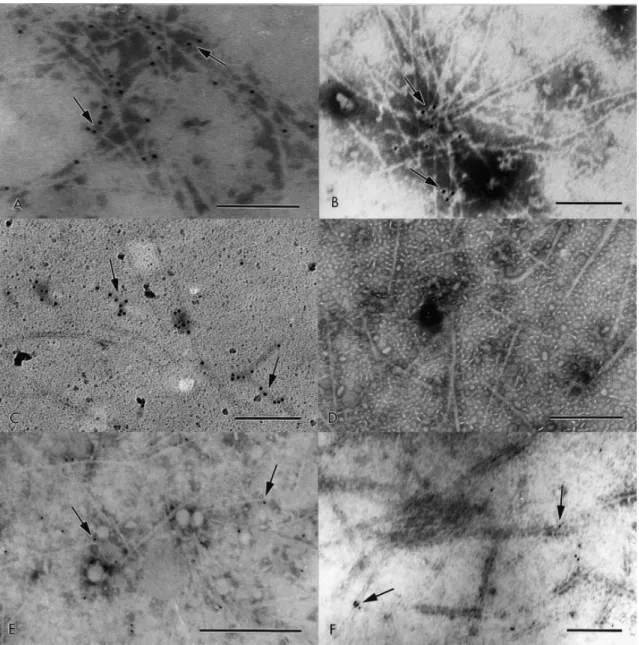
serum. The CFA I fimbrial preparation showed a scattered labeling on the fimbriae (Fig. 2C).
This binding pattern was similar to those obtained after the treatment of fimbriae with CFA I
antiserum (Fig. 2A). When whole bacteria were exposed to lactoferrin, similar results were
observed. These results revealed that lactoferrin have ability to bind to CFA I fimbriae,
indicating that this compound may act as a receptor analogue in the inhibition of adhesion of
CFA I to the host cell. On the other hand, the binding of lactoferrin to CFA II fimbriae (CS1 and
CS3) was not demonstrated (Fig. 2D).
Immunogold assay performed with CFA I and CFA II fimbrial preparations exposed to
fSC, showed that both CFA I and CFA II fimbriae were labeled (Fig. 2E,F). The observed pattern
of gold beads was similar to that found with specific fimbrial antibody (Fig. 2B). A similar result
was obtained when the whole bacteria was exposed to fSC. Previously, it was shown that fSC
inhibits the adhesion of E. coli CFA I to host cells [16]. These findings indicate that this
glycoprotein could prevent CFA I and CFA II binding to host cells. Furthermore, the ability of
fSC to act as a bacterial cell receptor analogue has been reported by Falk et al. [27] and Dallas
Fig. 2. Immunogold labeling assay of the enriched fimbrial preparations. CFA I (A) and CFA II fimbriae (B) recognized by anti-CFA I and anti-CFA II serum, respectively. Binding of lactoferrin on CFA I (C). Lactoferrin did not bind to CFA II fimbriae (D). The fSC binds on CFA I (E) and CFA II (F) fimbrial preparations. The arrows indicate the gold particles on fimbriae. Bar, 0.2 μm (A,B,C); bar, 0.5 μm (D,E,F).
In conclusion, our results indicate that lactoferrin and fSC, which are very abundant
proteins of human milk, could contribute to protection against infant enteric disease by
blocking bacterial adhesion, therefore supporting the eventual use of these compounds for
prophylactic or therapeutic purposes.
Acknowledgments
This work received nancial support from Funda o de Apoio esquisa do Distrito Federal (FA DF) and estl Brasil Ltda. e thank the Conselho acional de Desenvolvimento Cien co
[1] Paniagua, M., Espinoza, F., Ringman, M., Reizenstein, E., Svennerholm, A.-M. and Hallander, H. (1997) Analysis of incidence of infection with enterotoxigenic Escherichia coli in a
prospective cohort study of infant diarrhea in Nicaragua. J. Clin. Microbiol. 35, 1404-1410.
[2] Faruque, A.S., Salam, M.A., Faruque, S.M. and Fuchs, G.J. (1998) Aetiological, clinical and epidemiological characteristics of a seasonal peak of diarrhoea in Dhaka, Bangladesh. Scand. J. Infect. Dis. 30, 393-396.
[3] Evans, D.G., Silver, R.P., Evans Jr., D.J., Chase, D.G. and Gorbach, S.L. (1975) Plasmide controlled colonization factor associated with virulence in enterotoxigenic Escherichia coli for humans. Infec. Immun. 12, 656-667.
[4] Bühler, T., Hoschu«tzky, H. and Jann, K. (1991) Analysis of colonization factor antigen I, an adhesin of enterotoxigenic Escherichia coli O78:H11: Fimbrial morphology and location of the receptor-binding site. Infect. Immun. 59, 3876-3882.
[5] Evans, D.G. and Evans Jr., D.J. (1978) New surface-associated heatlabile colonization factor antigen (CFA II) produced by enterotoxigenic Escherichia coli of serogroups 06 and 08. Infect. Immun. 21, 638-647.
[6] Cravioto, A., Scotland, S.M. and Rowe, B. (1982) Hemagglutination activity and colonization factor antigens I and II enterotoxigenic and non-enterotoxigenic strains of Escherichia coli isolated from humans. Infect. Immun. 36, 189-197.
[7] Smyth, C.J. (1982) Two mannose-resistent haemagglutinins of enterotoxigenic Escherichia coli of serotype O6:H16 or H-isolated from travellers' and infantile diarrhoea. J. Gen. Microbiol. 128, 2081-2096.
[8] Reis, M.H.L., Guth, B.E.C., Gomes, T.A., Murahovschi, J. and Trabulsi, L.R. (1982) Frequency of Escherichia coli strains producing heat-labile toxin or heat-stable or both in children with and without diarrhea in São Paulo. J. Clin. Microbiol. 15, 1062-1064.
[9] Guth, B.E.C., Aguiar, E.G., Gri¤n, P.M., Ramos, S.R.T.S. and Gomes, T.A.T. (1994) Prevalence of colonization factor antigens (CFAs) and adherence to HeLa cells in enterotoxigenic
Escherichia coli isolated from feces of children in São Paulo. Microbiol. Immunol. 38, 695-701.
[10] Young, H.B., Buckley, A.E., Hamza, B. and Mandarano, C. (1982) Milk and lactation: Some social and developmental correlates among 1,000 infants. Pediatrics 69, 169-175.
[11] Kovar, M.G., Serdula, M.K., Marks, J.S. and Fraser, D.W. (1984) Review of the
epidemiologic evidence for an association between infant feeding and infant health. Pediatrics 74, 615-638.
[12] Glass, R.I., Svennerholm, A.-M., Stoll, B.J., Khan, M.R., Hossain, K.M.B., Huq, M.I. and Holmgren, J. (1983) Protection against cholera in breast-fed children by antibodies in breast milk. New Engl. J. Med. 308, 1389-1392.
[14] Brock, J.H. (1980) Lactoferrin in human milk: its role in iron absorption and protection against enteric infection in the newborn infant. Arch. Dis. Child. 55, 417-421.
[15] Ashkenazi, S. and Mirelman, D. (1987) Nonimmunoglobulin fraction of human milk inhibits the adherence of certain enterotoxigenic Escherichia coli strains to guinea pig intestinal tract. Pediatr. Res. 22, 130-134.
[16] Giugliano, L.G., Ribeiro, S.T.G., Vainstein, M.H. and Ulhoa, C.J. (1995) Free secretory component and lactoferrin of human milk inhibit the adhesion of enterotoxigenic Escherichia coli. J. Med. Microbiol. 42, 3-9.
[17] Appelmelk, B.J., An, Y.-Q., Geerts, M., Thijs, B.G., De Boer, H.A., MacLaren, D.M., De Graa¡, J. and Nuijens, J.H. (1994) Lactoferrin is lipid A-binding protein. Infect. Immun. 62, 2628-2632.
[18] Erdei, J., Forsgren, A. and Naidu, A.S. (1994) Lactoferrin binds to porins Omp/F and Omp/C in Escherichia coli. Infect. Immun. 62, 1236-1240.
[19] Naidu, S.S., Erdei, J., Cziro¨k, Eè., Kalfas, S., Gado¨, I., Thore¨n, A., Forsgren, A. and Naidu, A.S. (1991) Speci¢c binding of lactoferrin to Escherichia coli isolated from human intestinal infections. APMIS 99, 1142-1150.
[20] Mestecky, J. and Mcghee, J.R. (1987) Immunoglobulin A (IgA): Molecular and cellular interactions involved in IgA biosynthesis and immune response. Adv. Immunol. 40, 153-245.
[21] Kobayashi, K. (1971) Studies on human secretory IgA comparative studies of the IgA-bound secretory piece and the free secretory piece protein. Immunochemistry 8, 785-800.
[22] Buts, J.-P., Bernasconi, P., Vaerman, J.-P. and Dive, C. (1990) Stimulation of secretory IgA and secretory component of immunoglobulins in small intestine of rats treated with
Saccharomyces boulardii. Dig. Dis. Sci. 35, 251-256.
[23] Mynott, T.L., Luke, R.K.J. and Chandler, D.S. (1995) Detection of attachment of enterotoxigenic Escherichia coli (ETEC) to human small intestinal cells by enzyme immunoassay. FEMS Immunol. Med. Microbiol. 10, 207-218.
[24] Bradford, M.M. (1976) A rapid and sensitive method for the quantitation of microgram quantities of protein utilizing the principal of protein-dye binging. Anal. Biochem. 72, 248-254.
[25] Laemmli, U.K. (1970) Cleavage of structural proteins during the assembly of the head of bacteriophage T4. Nature 227, 680- 685.
[26] Towbin, H., Staehelin, T. and Gordon, J. (1979) Electrophoretic transfer of proteins from polyacrylamide gels to nitrocellulose sheets: Procedure and some applications. Proc. Natl. Acad. Sci. USA 76, 4350-4354.
[27] Falk, P., Roth, K.A., Bore¨n, T., Westblom, T.U., Gordon, J.I. and Normark, S. (1993) An in vitro adherence assay reveals that Helicobacter pylori exhibits cell lineage-speci¢c tropism in the human gastric epithelium. Proc. Natl. Acad. Sci. USA 90, 2035-2039.