A multiply substituted G–H loop from foot-and-mouth disease
virus in complex with a neutralizing antibody : a role for
water molecules
Wendy F. Ochoa,
1Susana G. Kalko,
1Mauricio G. Mateu,
2Paula Gomes,
3David Andreu,
3Esteban Domingo,
2Ignasi Fita
1and Nuria Verdaguer
11Instituto Biologı!a Molecular de Barcelona, Consejo Superior de Investigaciones Cientı!ficas, Jordi-Girona 18-26, 08034 Barcelona, Spain
2Centro de Biologı!a Molecular ‘Severo Ochoah, Consejo Superior de Investigaciones Cientı!ficas, Universidad Auto!noma de Madrid,
28049 Cantoblanco, Madrid, Spain
3Departament de Quı!mica Orga!nica, Universitat de Barcelona, 08028 Barcelona, Spain
The crystal structure of a 15 amino acid synthetic peptide, corresponding to the sequence of the
major antigenic site A (G–H loop of VP1) from a multiple variant of foot-and-mouth disease virus
(FMDV), has been determined at 2
n3 A/ resolution. The variant peptide includes four amino acid
substitutions in the loop relative to the previously studied peptide representing FMDV C-S8c1 and
corresponds to the loop of a natural FMDV isolate of subtype C
1. The peptide was complexed with
the Fab fragment of the neutralizing monoclonal antibody 4C4. The peptide adopts a compact fold
with a nearly cyclic conformation and a disposition of the receptor-recognition motif Arg–Gly–Asp
that is closely related to the previously determined structure for the viral loop, as part of the virion,
and for unsubstituted synthetic peptide antigen bound to neutralizing antibodies. New structural
findings include the observation that well-defined solvent molecules appear to play a major role in
stabilizing the conformation of the peptide and its interactions with the antibody. Structural results
are supported by molecular-dynamic simulations. The multiply substituted peptide developed
compensatory mechanisms to bind the antibody with a conformation very similar to that of its
unsubstituted counterpart. One water molecule, which for steric reasons could not occupy the same
position in the unsubstituted antigen, establishes hydrogen bonds with three peptide amino acids.
The constancy of the structure of an antigenic domain despite multiple amino acid substitutions has
implications for vaccine design.
Introduction
Foot-and-mouth disease virus (FMDV) is the causative
agent of the most economically important disease of
cloven-hoofed animals worldwide (Pereira, 1981 ; Brown, 1994). The
virus belongs to the genus Aphthovirus of the family
Picorna-viridae (Rueckert, 1996). Control of the disease has been based
on large-scale vaccinations with whole-virus inactivated
vaccines, limitation of animal movements and destruction of
herds exposed to the virus (the ‘ stamping-out ’ procedure)
(reviews in Bachrach, 1968 ; Brown, 1994 ; Domingo et al.,
1990 ; Timoney et al., 1992). The available vaccines show
Author for correspondence : Nuria Verdaguer.
Faxj34 93 2045904. e-mail nvmcri!cid.csic.es
generally good protection against infection with homologous
virus and with antigenically related isolates. Difficulties facing
the eradication of FMD include the antigenic diversity of
FMDV in nature, which has been reflected in the identification
of seven serotypes (A, O, C, SAT1, SAT2, SAT3 and Asia1),
65 subtypes, until subtyping was interrupted (Pereira, 1977),
and multitudes of antigenic variants that often co-circulate in a
given geographical area (Mateu et al., 1988). Antigenic
variation imposes a periodic updating of vaccine strains,
antigenic properties of which must match those of the
circulating viruses. Furthermore, some FMD outbreaks have
been traced to vaccine strains of the virus (Beck & Strohmaier,
1987). For these reasons, it would be highly desirable to
develop effective, synthetic FMD vaccines.
Neutralizing antibodies are important determinants of
Table 1. Amino acid sequences of site A peptide variants
Peptides A15-C"-Brescia, A15-C-S8c1 and A15-C-S30 represent sequences of FMDV isolates belonging to subtype C". MAb 4C4, which was raised against C"-Brescia, reacts with all three peptides as shown by immunochemical and structural analyses (Mateu et al., 1990, 1992 ; Verdaguer et al., 1995, 1997). Synthetic peptide
A15-C-S8c1\LV presents a decrease in binding affinity for most site A-specific
MAbs, including 4C4 (Mateu et al., 1992).k, Less than 20% of the
binding observed with the other peptides (j) in a quantitative
enzyme immunodot assay described by Mateu et al. (1992). No virus has been reported with a site A sequence corresponding to peptide
A15-C-S8c1\LV. The one-letter amino acid code is used.
Peptide Sequence Binding to MAb 4C4 A15-C"-Brescia YTASTRGDLAHLTAT j A15-C-S8c1 YTASARGDLAHLTTT j A15-C-S8c1\LV YTASARGDLAHVTTT k A15-C-S30 YTTSTRGDLAHVTAT j
protection against FMD and other picornavirus diseases
(Mateu, 1995 ; McCullough et al., 1992 ; Misbah et al., 1992).
An understanding of the types of interactions between
antibodies and viruses that lead to virus neutralization is
essential for vaccine design. One of the major antigenic sites of
FMDV is located in the G–H loop of capsid protein VP1 (Bittle
et al., 1982 ; Pfaff et al., 1982 ; Strohmaier et al., 1982). This loop
is disordered on the surface of FMDV particles (Acharya et al.,
1989 ; Curry et al., 1996 ; Lea et al., 1994, 1995 ; Logan et al.,
1993). For FMDV of serotype C, this antigenic site has been
termed site A, and it behaves as an independent unit, with very
limited influence of other capsid residues regarding the
interaction of site A with antibodies (Hewat et al., 1997 ; Lea et
al., 1994 ; Mateu, 1995 ; Verdaguer et al., 1999). The behaviour
of this antigenic loop in FMDV particles in its interaction with
antibodies can be mimicked faithfully with synthetic peptides
that represent the relevant amino acid sequences found in
authentic virus (Clarke et al., 1983 ; Mateu et al., 1989,
1990 ; Mateu, 1995 ; Rowlands et al., 1983). Interestingly,
antigenic site A includes a highly conserved Arg–Gly–Asp
(RGD) triplet that serves as the recognition site of an integrin
receptor (Berinstein et al., 1995 ; Fox et al., 1989 ; Herna
! ndez et
al., 1996 ; Jackson et al., 1997 ; Mason et al., 1994 ; Neff et al.,
1998).
The structure of a synthetic peptide representing antigenic
site A of FMDV S8c1 [a biological clone of natural isolate
C-Sta Pau Sp70 (Sobrino et al., 1983), a virus representative of the
European subtype C" FMDVs] in a complex with the Fab of
neutralizing monoclonal antibodies (MAbs) raised against the
virus has been studied by X-ray crystallography (Verdaguer et
al., 1995, 1996, 1998). The synthetic peptide spanned positions
136–150 of capsid protein VP1 (peptide A15, Table 1). Two
complexes were studied, involving MAbs SD6 (Verdaguer et
al., 1995, 1996) and 4C4 (Verdaguer et al., 1998). The two
neutralizing MAbs were raised against two different FMDV
type C isolates (Mateu et al., 1990) : SD6 against C-S8c1 and
4C4 against C"-Brescia, a virus that differs from C-S8c1 at two
positions (140 and 149) within site A (Table 1). In the two
complexes, the peptide antigen acquired a very similar
quasi-circular conformation and the RGD motif participated directly
in the interaction with residues of the
complementarity-determining regions (CDRs) of the antibodies (Verdaguer et al.,
1998). The RGD triplet appeared in an open turn conformation,
very similar to that of reduced FMDV particles of serotype O
(Logan et al., 1993), and also similar to the conformation in
RGD-containing integrin ligands (Pfaff, 1997). Remarkably,
the Gly-142 and Asp-143 residues of the RGD triplet appear to
be critical for interaction both with an integrin receptor and
with neutralizing antibodies directed to site A (Mateu et al.,
1996 ; Verdaguer et al., 1995). Both structural and biochemical
evidence suggest that MAbs SD6 and 4C4 neutralize by
monovalent binding to antigenic site A (Verdaguer et al., 1997,
1998 ; review in Domingo et al., 1999).
The quasi-circular shape of the peptide antigen in the
complexes was stabilized by peptide–antibody interactions
and also through intrapeptide hydrogen bonds and van der
Waalsh interactions (Verdaguer et al., 1995, 1996, 1998). If a
structure similar to the one found in these complexes was also
maintained with variant antigens (including one or several
amino acid substitutions), the structure would gain ground as
the basis for the design of new anti-FMD immunogens. Several
natural isolates of FMDV of serotype C have been
charac-terized previously with regard to the amino acid sequence at
antigenic site A and reactivity with MAbs (Martı!nez et al.,
1991 ; Mateu et al., 1988, 1989, 1990 ; Verdaguer et al., 1995,
1996, 1998 ; Villaverde et al., 1991). One of the isolates
analysed, termed C-S30 (or C"-Barcelona Sp\81), included four
replacements within antigenic site A. Two of the changes were
coincident with those found in the C"-Brescia virus (Ala-140
Thr, Thr-149
Ala) and two were new (Ala-138
Thr,
Leu-147
Val) (Table 1). In spite of the presence of four
amino acid substitutions in the G–H loop of VP1, the reactivity
of FMDV C-S30 with MAb 4C4 was indistinguishable from
that of FMDV C-S8c1 (Mateu et al., 1990). Single substitutions
at positions 138 and, especially, 140 and 149 did not have a
significant effect on the interaction of synthetic peptide
antigens with most MAbs, including 4C4, in ELISA (Verdaguer
et al., 1998). However, the single replacement of residue
Leu-147, highly conserved among FMDVs of serotype C, by valine
diminished greatly the reactivity of synthetic peptides with
most MAbs, including 4C4, in enzyme immunodot assays and
competitive ELISA (Mateu et al., 1992 ; Verdaguer et al., 1998).
That is, the four amino acid replacements found at the antigenic
site A of C-S30 relative to C-S8c1 (Mateu et al., 1989, 1990)
appear to exert a compensatory effect that restores the affinity
of C-S30 (Mateu et al., 1990) and of a synthetic antigen
representing its antigenic site A (Mateu et al., 1992) for MAb
4C4. Therefore, the substituted antigen offered an opportunity
to investigate the structure that a variant form of antigenic site
A could acquire as a complex with antibody. The results
described in this report reaffirm the quasi-circular conformation
for this antigenic loop and reveal new features about the forces
involved in the stability of site A and about antigen–antibody
interactions, in particular, key interactions mediated by water
molecules.
Methods
MAb and synthetic peptides. The origin and characterization of the anti-FMDV MAb 4C4 (Mateu et al., 1987, 1989, 1990) and the preparation and purification of the Fab fragment of MAb 4C4 (Verdaguer
et al., 1998) have been reported previously. Peptides were synthesized by
solid-phase procedures, purified and analysed by using procedures that
have been described previously (Carren4 o et al., 1992; Mateu et al., 1996).
Crystallization and data collection. Crystals of the complex between the Fab fragment of the MAb 4C4 and the variant peptide A15-CS30 (Table 1) were obtained by the hanging drop-vapour diffusion
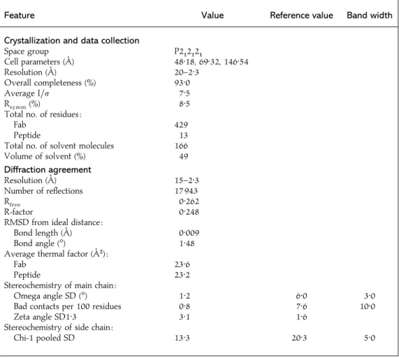
Table 2. Features of crystals of the antigen–4C4 Fab complex analysed in the present
study
Feature Value Reference value Band width
Crystallization and data collection
Space group P2"2"2"
Cell parameters (A/) 48n18, 69n32, 146n54
Resolution (A/) 20–2n3
Overall completeness (%) 93n0
Average I\σ 7n5
Rsymm(%) 8n5
Total no. of residues :
Fab 429
Peptide 13
Total no. of solvent molecules 166
Volume of solvent (%) 49 Diffraction agreement Resolution (A/) 15–2n3 Number of reflections 17 943 Rfree 0n262 R-factor 0n248
RMSD from ideal distance :
Bond length (A/) 0n009
Bond angle (m) 1n48
Average thermal factor (A/#):
Fab 23n6
Peptide 23n2
Stereochemistry of main chain :
Omega angle SD (m) 1n2 6n0 3n0
Bad contacts per 100 residues 0n8 7n6 10n0
Zeta angle SD1n3 3n1 1n6
Stereochemistry of side chain :
Chi-1 pooled SD 13n3 20n3 5n0
technique and successive micro- and macroseedings (Stura & Wilson, 1992). Typically small, twined needles, obtained with 18 % PEG 4K, were used for microseeding, which produced larger needles at 16 % PEG 4K.
Finally, these larger needles were used for macroseedings in 2µl droplets
containing 7 mg\ml Fab, 1n8 mg\ml peptide, 6n5% PEG 4K, 0n2 M LiCl
with 50 mM Tris–HCl (pH 9), equilibrated against a reservoir containing 13 % PEG 4K equally buffered at room temperature. Crystals were orthorhombic, space group P2"2"2" with unit cell parameters al48n2 A/,
bl 69n3 A/ and c l 146n5 A/, containing one molecule of the complex
per asymmetric unit, which corresponds to a solvent content volume of 49 %. A data set was collected, at 100 K with 20 % glycerol as a cryoprotectant, by using a MarReseach imaging plate on a Rigaku rotating anode. Intensities were evaluated and scaled internally with programs Denzo and Scalepack, respectively (Otwinowski & Minor,
1996). Data were 93 % complete at 2n3 A/ resolution, with an internal
agreement factor (Rsymm) of 8n5% (Table 2).
Structure solution and refinement. Crystals of the complex
between peptide A15-C-S8c1\LV and the Fab of MAb 4C4 seemed
related to crystals formed with the same Fab and peptide A15-C-S8c1 (the names and amino acid sequence of peptide antigens are listed in Table 1), whose structure had been solved previously (Verdaguer et al., 1998). However, the unit cell parameter c differed by about 10 A/ and the structure was newly determined by molecular replacement by using the
Fig. 1. (A) Stereo view of the (2Fo-Fc) electron density map corresponding to the antigenic peptide A15-C-S30. The peptide model and nearby solvent molecules (isolated dots) are also shown. (B) Stereo view of the Fab–peptide interactions. Only the Fab residues in direct contact with the peptide are depicted (open rods). Water molecules (W) in direct contact with the peptide or mediating the antibody recognition are also shown. Hydrogen bonds are indicated by broken lines. (C) Super-position of the A15 peptide conformations found in the 4C4 Fab complexes with the S30 (filled) and the A15-C-S8c1 (open) peptides.
AMoRe package with the 4C4 Fab coordinates as searching model (Navaza, 1994). The initial solution was then optimized by allowing to move as four separated rigid bodies the variable heavy, variable light, constant heavy and constant light domains. The resulting R-factor was
0n36% in the resolution range of 15 to 4 A/. Examination of the electron
density maps, calculated at this stage, clearly showed extra density corresponding to the oligopeptide occupying the antigen-binding site. The final model for the structure of the complex was obtained by iterative cycles of model rebuilding by using the program O (Jones et al., 1991)
and positional refinement with XPLOR (Bru$ nger, 1992), including bulk
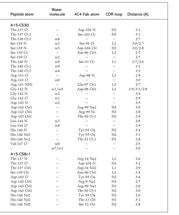
Table 3. Hydrogen bonds between the A15-CS30 and A15-CS8 peptides and the 4C4
Fab
The participants in the various hydrogen bonds are shown. Crystallographic data are described in Table 2 and the three-dimensional structures of the two complexes are shown in Fig. 2. The CDR loops are given to which the 4C4 Fab atoms listed belong.
Peptide atom
Water
molecule 4C4 Fab atom CDR loop Distance (A/)
A15-CS30 Thr-137 O – Asp-104 N H3 3n2 Thr-137 Oγ1 – Ser-103 Oγ H3 3n1 Thr-138 Oγ1 w8 – – 2n7 Ser-139 N w3 Ser-95 O L3 3n0\2n7 Ser-139 N w3 Asp-104 Oδ H3 3n0\2n8 Ser-139 Oγ – Asn-96 Oδ1 L3 2n7 Ser-139 O w2 – – 2n7 Thr-140 N w9 Ser-31 Oγ L1 2n7\2n6 Thr-140 Oγ1 w9 – – 3n2 Thr-140 Oγ1 w4 – – 3n3 Arg-141 O – Asp-98 N L3 2n9 Arg-141 O w6 – – 3n0 Arg-141 NH1 – Glu-97 Oε1 L3 2n7 Gly-142 N w1\w5 Asp-98 Oδ1 L3 2n9\3n1\2n8 Gly-142 N w2 – – 3n4 Gly-142 O w1 – – 2n9 Asp-143 N w2 – – 3n3 Asp-143 Oδ2 – Arg-99 Nη2 H3 3n0 Asp-143 Oδ1 – Arg-99 Nε H3 2n8 Asp-143 Oδ1 – Thr-50 Oγ1 H2 2n9 Leu-144 N w2 – – 2n8 Leu-144 O w8 – – 2n9 His-146 N – Tyr-59 Oη H2 3n4 His-146 Nδ1 – Tyr-59 Oη H2 3n1 His-146 Nε2 – Thr-33 Oγ1 H1 2n8 Val-147 O w8 – – 2n5 – w7\w1 – – 3n0 A15-CS8c1 Thr-137 N – Arg-54 Nη2 L2 2n6 Thr-137 O – Asp-104 N H3 3n2 Thr-137 Oδ1 – Asp-34 Nδ2 L1 2n9 Ser-139 Oδ – Asn-96 Oδ1 L3 3n4 Asp-143 O – Tyr-59 Oη H2 3n4 Asp-143 Oδ1 – Arg-9 Nη2 H3 2n7 Asp-143 Oδ2 – Arg-99 Nη1 H3 2n8 Asp-143 Oδ2 – Thr-50 Oγ1 H2 3n0 His-146 Nδ1 – Tyr-59 Oη H2 2n6 His-146 Nδ2 – Thr-33 Oδ H1 3n2 His-146 Nδ2 – Ser-52 Oδ H2 2n8
solvent correction. The refined model converged to crystallographic
agreement factors R and Rfreeof 24 % and 26 %, respectively, for 17 943
reflections in the resolution shell 15n0–2n3 A/ (Table 2).
Molecular dynamics (MD) simulation. Three sets of MD simulations, corresponding to the variable module (Fv) of the 4C4 Fab
interacting with peptides A15-C-S8c1, A15-C-S8c1\LV and A15-C-S30
(Table 1), were performed by using the GROMOS96 package with its
standard protein and water force fields (van Gunsteren et al., 1996). Crystallographic coordinates of peptides A15-C-S8c1 and A15-C-S30 (residues 137–148) were used as starting models in the corresponding simulations. Thr-148, not visible in the electron density maps of the A15-C-S8c1 complex, was added to avoid end effects in the critical Leu-147 residue, by using the information available in the A15-C-S30 structure.
Starting coordinates for the A15-C-S8c1\LV peptide were then derived,
with the graphic program TURBO (Rousel & Cambillau, 1989), by
replacing Leu-147 with valine. Crystallographic water molecules de-termined in the present work for the complex of 4C4 Fab with the A15-C-S30 peptide were not included in the simulations.
In every simulation, the structure of the corresponding complex was placed initially at the centre of a truncated octahedron, the dimensions of which were chosen such that the minimum distance of any protein atom from the closest wall was 7 A/. The edge lengths of the corresponding cubic boxes were about 73 A/. Systems were treated as immersed into an equilibrium configuration of bulk simple point charge (SPC) water (Berendsen et al., 1986). Water molecules outside the box or with a distance to a solute atom of less than 2n3 A/ were removed. The numbers of water molecules considered in the three simulations were 5271, 5272 and 5264, respectively. To relax strong water–water and water–protein non-bonded interactions, steepest-descent energy minimization was performed until stabilization was reached. After that, counter-ions were added to neutralize charged protein sites, with a subsequent energy minimization. Simulations were performed at constant volume and temperature (300 K) with periodic boundary conditions and an in-tegration step of 2 fs. Temperature was kept constant by weak coupling to an external bath (Berendsen et al., 1984, 1986). Bond lengths were constrained to equilibrium values by using the SHAKE algorithm (Rickaert et al., 1977). Equilibrations were achieved within 50 ps and, afterwards, the three systems were simulated for a total time of 300 ps with the total potential energy remaining essentially constant. Only amino acids from the peptides and all water molecules were allowed to move during calculations. Analysis were performed, mainly with programs contained in the GROMOS96 package, using the structures
generated every 0n1 ps during the interval spanning from equilibration
until the end (from about 50 to 350 ps).
Coordinates. Coordinates have been deposited in the Brookhaven protein database under accession number 1EJO.
Results
Structure of the 4C4 Fab in complex with the
A15-CS30 peptide
The final electron density maps of the complex between the
4C4 Fab and the A15-C-S30 peptide, at 2n3 A/ resolution,
allowed the positioning of the main and side chains of 431 of
the total of 436 4C4 Fab residues (216 from the light chain and
220 from the heavy chain) and of 12 of the 15 peptide residues
(Fig. 1 A). Heavy-chain Fab residues 136–143, in the constant
domain, and terminal peptide residues Ala-149, Thr-150 and
the side chain of Tyr-136 were disordered, and their
co-ordinates have not been introduced in the present model. The
quality of the final maps is also reflected in the 166 well-defined
solvent molecules found, nine of which participate in the
interactions between the peptide and the Fab CDRs (Table 3),
and three water molecules establish bridges among peptide
residues (Fig. 1 B).
The A15-C-S30 peptide shows an overall conformation
closely related to the one found in other site A complexes
determined previously (Verdaguer et al., 1995, 1998) (Fig. 1 C).
The RGD motif, residues 141–143, is located in an open turn
conformation preceded by an extended region, residues
136–140, and followed by a short helix, positions 144–148
(Fig. 1). All of the peptide residues incorporated into the model
100 75 50 25 0 B Residue number
Contact area (% of residue surf
ace) 100 75 50 25 0 A 136 Residue number Contact area (Å 2) 137 *138 139 *140 141 142 143 144 145 146 *147 148 136 137 *138 139 *140 141 142 143 144 145 146 *147 148
Fig. 2. Contact areas for the A15 peptides in the 4C4 Fab complexes with the A15-C-S30 (filled bars) and the A15-C-S8c1 (open bars) peptides expressed as absolute values (A) or as percentages of the residue surface (B). Asterisks indicate A15-C-S30 residues that differ in A15-C-S8c1. Peptide sequences are shown in Table 1.
interact directly with the hypervariable regions of the Fab
(Table 3 and Fig. 2). In particular, the entire molecular surfaces
of Asp-143, from the RGD motif, and Leu-144 are in contact
with the antibody, similar to what had been observed in
complexes involving Fab A15-C-S8c1 (Fig. 2) (Verdaguer et al.,
1995, 1998). However, the substituted residues Thr-138 and,
particularly, Thr-140 and Val-147 do not present extensive
interactions with the antibody (Fig. 2). The fourth substitution,
Ala-149, not visible in the electron density and not included in
the present model, must also remain exposed to the solvent
and far from the antibody.
Comparisons of the site A variant peptides A15-CS30
and A15-CS8c1
The root-mean-square deviation (RMSD) between the
main-chain peptide atoms in the A15-C-S30 and A15-C-S8c1
Fab 4C4 complexes is only 0n4 A/ for the common residues
137–147. The Arg-141 side chain has different dispositions in
the two peptides (Fig. 1 C), although the electron density
corresponding to this residue was weak in both complexes
(Fig. 1 A ; see also Verdaguer et al., 1995, 1998). The replaced
residues, Thr-138 and Val-147, show the largest main-chain
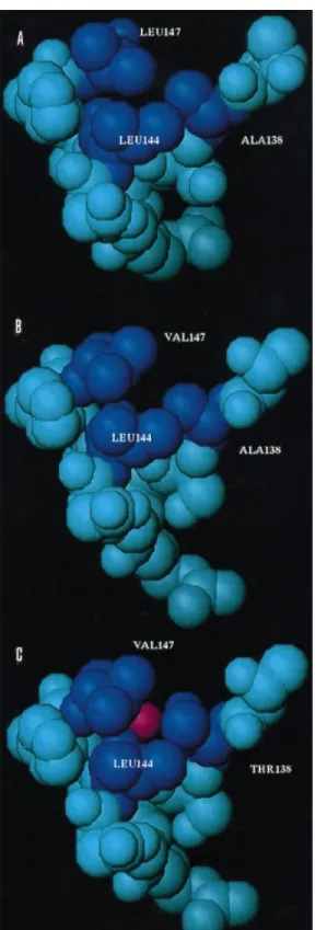
Fig. 3. Space-filling representation of the three peptide models A15-C-S8c1 (A), A15-C-S8/LV (B) and A15-C-S30 (C). Peptide residues that contribute to intrapeptide hydrophobic interactions are highlighted in dark-blue. The water molecule found in contact with the substituted Thr-138 in the A15-C-S30 peptide is displayed in red (C).
deviations of 0n7 and 1n0 A/, respectively. Despite the high
structural similarity between the two peptides A15-C-S30 and
A15-C-S8c1, differences in the main-chain conformational
angles are important, particularly around the RGD motif (Fig.
2). Thus, in the A15-C-S30 peptide, residues Thr-140 and
Arg-141 are situated in the Ramachandran region corresponding to
α
-helices while, in the A15-C-S8c1 peptide, Ala-140 and
Arg-141 are in the regions of
β-strands and left-handed helices,
respectively. A third situation is found in the structure reported
for the G–H loop of the reduced FMDV serotype O" (Lea et al.,
1993, 1995), where the corresponding residues Leu-144 and
Arg-145 are both found in the Ramachandran region
cor-responding to
β-strands. The flexibility of Gly-142 allows
compensatory main-chain torsional angles that result in the
overall structural similarity of the three peptides. It is important
to notice the presence, in the A15-CS30 structure, of one water
molecule (named w8 in Fig. 1) that is hydrogen-bonded with
the side chain of Thr-138 and the main-chain oxygen atoms of
residues Leu-144 and Leu-147. This water molecule cannot be
present, for steric reasons, in the A15-C-S8c1 structure, where
the bulkier Leu-147 side chain fills the available space (Fig. 3).
MD analysis
Three MD simulations corresponding to complexes of the
Fv 4C4 with peptides C-S8c1, C-S8c1\LV and
A15-C-S30 were carried out as described in Methods. The main
difference between the averaged conformations of the three
peptides is the opening of the C-terminal region in the
simulation of the single-substituted A15-C-S8c1\LV peptide
(Fig. 4 A). Other significant differences can also be seen at the
amino termini and for the Arg-141 side chain. The stability of
the averaged conformations was analysed by evaluating the
RMSD values of the peptide fluctuations in every simulation.
The lowest values correspond to the A15-C-S30 peptide, while
the more constant main-chain region includes the RGD motif
and Leu-144 in the three peptides, with Asp-143 having the
smallest variation. The side chains of Asp-143 and Leu-144
also appear extremely invariant, probably reflecting the extent
and the stability of the interactions of these residues with the
antibody. The stability of the tetra-substituted peptide
A15-C-S30 observed during the simulation is contributed by a
hydrogen-bonded water molecule located in a site very close
to the position of the bridging crystallographic water (w8)
(Figs 1 B and 4 B). This water site presents a high occupation
and a low RMSD of about 0n1 A/. Hydrogen-bond interactions
take place between the water molecule that occupies this site
and polar atoms from the substituted residues Thr-138,
Leu-144 and Val-147 (Table 3). The increased hydrophobic surface
exposed to the solvent appears to explain the departure from
the ‘ correcth site A-like conformation observed by MD with
the A15-CS8\LV peptide (Fig. 3).
Fig. 4. (A) Stereo view of the superposition of the A15-C-S8c1 (filled), A15-C-S8/LV (shaded) and A15-C-S30 (open) peptides corresponding to the averaged conformations calculated in 300 ps of MD simulations of the corresponding
Fv–peptide complexes. (B) Stereo view of the superposition of the A15-C-S30 coordinates from the crystal (filled) and the MD simulation (open). The water molecule found in the crystal structure (shaded) is situated close to the water site determined in the MD simulation (open).
Discussion
The effects of single or multiple amino acid substitutions at
antigenic site A of FMDV of serotype C on the reactivity of
site A-specific neutralizing MAbs have been studied
ex-tensively by immunochemical assays employing variant
viruses and substituted synthetic peptides (Mateu et al., 1989,
1990, 1992 ; Martı!nez et al., 1997; Novella et al., 1993;
Verdaguer et al., 1998). Replacements at position 147 altered
the binding of most antibodies substantially, including 4C4,
even though residue 147 had only marginal interactions with
that antibody. In particular, substitution of Leu-147 by valine
diminished the binding of most site A-specific MAbs analysed
(Mateu et al., 1992). However, the effect of this single
substitution Leu-147
Val was apparently compensated for,
at least in part, by the replacement Ala-138
Thr (Mateu et
al., 1992). These observations and the fact that natural FMDV
isolate C-S30 (Martı
!nez et al., 1991; Villaverde et al., 1991)
contains these critical replacements in its VP1 G–H loop
relative to clone C-S8c1 (Table 1) encouraged the present
study.
In the structure of the 4C4 Fab complexed with the variant
peptide A15-C-S30 reported here, the two amino acids
Thr-138 and Val-147 that were replaced in the peptide show only
minor interactions with the Fab (Table 2 and Fig. 2). Differences
in binding affinities for peptides with substitutions in those
two residues should therefore be due mainly to free-energy
differences when adopting a ‘ correcth site A-like loop structure,
the one recognized by antibodies or by the receptor (Fig. 5).
The reduced stability of that conformation in the peptide with
the single substitution Leu-147
Val would then explain the
reduced affinity observed for the antibody. Instead, the
double-substituted peptide could recover binding affinity by
re-stabilizing the ‘ correcth conformation with the additional water
molecule (w8) that bridges the chain hydroxyl group of the
replaced Thr-138 with the main-chain oxygen atoms of
residues Leu-144 and Leu-147 (Table 3 and Fig. 3 C). In the MD
simulation of the A15-C-S30 variant peptide, a water molecule
with a low RMSD is placed in almost exactly the same location
as the one found in the crystal structure (Fig. 4 B). These
structural results provide a rationale for how a general change
in specificity to a large diversity of antibodies can be achieved
by a destabilization of the original antigen structure. In those
cases, further substitutions can restore the affinity for the
antibodies by increasing the stability of the original antigen
conformation, even when none of the residues substituted
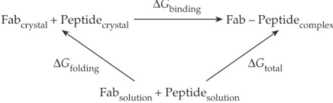
Fabcrystal + Peptidecrystal Fab – Peptidecomplex
Fabsolution + Peptidesolution ∆Gbinding
∆Gtotal
∆Gfolding
Fig. 5. A simplified thermodynamic cycle of a Fab interacting with an antigenic peptide can be described as a two-step process. In the first step, the peptide adopts individually the structure seen in the complex that is formed in the second step, so that∆Gtotall ∆Gfoldingj∆Gbinding, where ∆Gfoldingrefers to the free-energy difference between the peptide structure in solution and that found in the complex (the Fab reorganization, if required, can also be included in this term) and∆Gbindingrefers to the free-energy difference due to the formation of the interactions between the peptide and the Fab. A ‘ lock-and-key ’ recognition mechanism, in which structures remain unchanged on complex formation, corresponds to ∆Gfoldingl 0. Instead, ∆Gfolding 0 describes an ‘ induced-fit ’ mechanism, where the complex would only be stabilized by a favourable∆Gbinding contribution. Analysis of Fab 4C4 complexes with a number of A15 peptides, both from the available crystal structures and with MD
simulations, suggest that∆Gbinding(A15-C-S30)$ ∆Gbinding(A15-C-S8/LV), while the experimental data on the relative stabilities of these complexes (Mateu et al., 1992) imply that∆Gtotal(A15-C-S30) ∆Gtotal (A15-C-S8/LV), which would allow us to conclude that∆Gfolding(A15-C-S30) ∆Gfolding(A15-C-S8/LV). The reduced stability of the ‘ correct ’ site A-like conformation for the A15-C-S8/LV peptide would explain the lack of affinity of most site A MAbs for this variant peptide (see text).
participates in direct antigen–antibody interactions (Freire,
1999).
Recognition of continuous epitopes, like the VP1 G–H
loop of FMDV studied in this work, depends not only on the
amino acid sequence but also on the spatial conformation of
the epitope, where solvent molecules often play a critical role.
Interpretation of mechanisms of virus escape from antibody
recognition, essential in RNA virus evolution and vaccine
design, must also take into consideration the effects of amino
acid replacements in connection with the solvent
rearrange-ments imposed. In spite of difficulties in predicting the effects
of amino acid replacements in antigen–antibody recognition,
the present results for FMDV encourage the design of
quasi-cyclic structures reproducing antigenic site A. Data for the
G–H loop from a number of different antibodies, including the
multiply substituted peptide reported here, suggest a robust
and strongly immunogenic structure despite the mobility of
this antigenic site on the virion surface (Parry et al., 1990 ;
Logan et al., 1993). Cocktails of quasi-circular peptides
representing the most frequent escape mutants found at site A
should be candidates for incorporation into vaccine
for-mulations. Such formulations should include additional B-cell
and T-cell epitopes to ensure a broad immune response to
minimize the selection of escape mutants and vaccine failures
(Domingo & Holland, 1992 ; Taboga et al., 1997).
We wish to thank E. Brocchi for the generous supply of MAb 4C4 and her continuous help with our research. Work in Barcelona was funded by DGICYT grants PB95-0218 and PB97-0873. Work in Madrid was
supported by grants from DGES PM97-006-C02-01 and Fundacio! n
Ramo! n Areces. W.F.O. is a recipient of a fellowship from the Ministerio
de Educacio! n y Cultura (Spain). P.G. thanks the Fundaçao C. Gulbenkian
for a PhD fellowship and the University of Porto for temporary leave.
References
Acharya, R., Fry, E., Stuart, D., Fox, G., Rowlands, D. & Brown, F. (1989).The three-dimensional structure of foot-and-mouth disease virus at 2n9 A/ resolution. Nature 337, 709–716.
Bachrach, H. L. (1968).Foot and mouth disease virus. Annual Review of
Microbiology 22, 201–244.
Beck, E. & Strohmaier, K. (1987).Subtyping of European foot-and-mouth disease virus strains by nucleotide sequence determination. Journal
of Virology 61, 1621–1629.
Berendsen, H. J. C., Postma, J. P. M., van Gunsteren, W. F., DiNola, A. & Haak, J. R. (1984).Molecular dynamics with coupling to an external bath. Journal of Chemical Physics 81, 3684–3690.
Berendsen, H. J., Van Gunsteren, W. F., Zwinderman, H. R. & Geurtsen, R. G. (1986).Simulations of proteins in water. Annals of the
New York Academy of Sciences 482, 269–286.
Berinstein, A., Roivainen, M., Hovi, T., Mason, P. W. & Baxt, B. (1995). Antibodies to the vitronectin receptor (integrin alpha V beta 3) inhibit binding and infection of foot-and-mouth disease virus to cultured cells.
Journal of Virology 69, 2664–2666.
Bittle, J. L., Houghten, R. A., Alexander, H., Shinnick, T. M., Sutcliffe, J. G., Lerner, R. A., Rowlands, D. J. & Brown, F. (1982). Protection against foot-and-mouth disease by immunization with a chemically synthesized peptide predicted from the viral nucleotide sequence. Nature 298, 30–33.
Brown, F. (1994). Foot-and-mouth disease. In Synthetic Vaccines, pp. 416–432. Edited by B. H. Nicholson. Oxford : Blackwell Scientific. Bru$nger, A. T. (1992).XPLOR manual, version 3.0. Yale University, New Haven, CT, USA.
Carren4o, C., Roig, X., Cairo, J., Camarero, J., Mateu, M. G., Domingo, E., Giralt, E. & Andreu, D. (1992).Studies on antigenic variability of C strains of foot-and-mouth disease virus by means of synthetic peptides and monoclonal antibodies. International Journal of Peptide and Protein
Research 39, 41–47.
Clarke, B. E., Carroll, A. R., Rowlands, D. J., Nicholson, B. H., Houghten, R. A., Lerner, R. A. & Brown, F. (1983).Synthetic peptides mimic subtype specificity of foot-and-mouth disease virus. FEBS Letters 157, 261–264.
Curry, S., Fry, E., Blakemore, W., Abu-Ghazaleh, R., Jackson, T., King, A., Lea, S., Newman, J., Rowlands, D. & Stuart, D. (1996).Perturbations in the surface structure of A22 Iraq foot-and-mouth disease virus accompanying coupled changes in host cell specificity and antigenicity.
Structure 4, 135–145.
Domingo, E. & Holland, J. J. (1992). Complications of RNA het-erogeneity for the engineering of virus vaccines and antiviral agents. In
Genetic Engineering, Principles and Methods, vol. 14, pp. 13–32. Edited by
J. K. Setlow. New York : Plenum.
Domingo, E., Mateu, M. G., Martı!nez, M. A., Dopazo, J., Moya, A. & Sobrino, F. (1990).Genetic variability and antigenic diversity of foot-and-mouth disease virus. In Applied Virology Research, pp. 233–266. Edited by E. Kurkstak, R. G. Marusyk, S. A. Murphy & M. H. V. Van Regenmortel. New York : Plenum.
Domingo, E., Verdaguer, N., Ochoa, W. F., Ruiz-Jarabo, C. M., Sevilla, N., Baranowski, E., Mateu, M. G. & Fita, I. (1999).Biochemical and structural studies with neutralizing antibodies raised against foot-and-mouth disease virus. Virus Research 62, 169–175.
Fox, G., Parry, N. R., Barnett, P. V., McGinn, B., Rowlands, D. J. & Brown, F. (1989).The cell attachment site on foot-and-mouth disease virus includes the amino acid sequence RGD (arginine-glycine-aspartic acid). Journal of General Virology 70, 625–637.
Freire, E. (1999).The propagation of binding interactions to remote sites in proteins : analysis of the binding of the monoclonal antibody D1.3 to lysozyme. Proceedings of the National Academy of Sciences, USA 96, 10118–10122.
Herna!ndez, J., Valero, M. L., Andreu, D., Domingo, E. & Mateu, M. G. (1996).Antibody and host cell recognition of foot-and-mouth disease virus (serotype C) cleaved at the Arg-Gly-Asp (RGD) motif : a structural interpretation. Journal of General Virology 77, 257–264.
Hewat, E. A., Verdaguer, N., Fita, I., Blakemore, W., Brookes, S., King, A., Newman, J., Domingo, E., Mateu, M. G. & Stuart, D. I. (1997). Structure of the complex of an Fab fragment of a neutralizing antibody with foot-and-mouth disease virus : positioning of a highly mobile antigenic loop. EMBO Journal 16, 1492–1500.
Jackson, T., Sharma, A., Abu-Ghazaleh, R., Blakemore, W. E., Ellard, F. M., Simmons, D. L., Newman, J. W., Stuart, D. I. & King, A. M. (1997). Arginine-glycine-aspartic acid-specific binding by
foot-and-mouth disease viruses to the purified integrinαvβ3 in vitro. Journal of
Virology 71, 8357–8361.
Jones, T. A., Zou, J.-Y., Cowan, S. W. & Kjeldgaard, M. (1991). Improved methods for building protein models in electron density maps and the location of errors in these models. Acta Crystallographica A47, 110–119.
Lea, S., Herna!ndez, J., Blakemore, W., Brocchi, E., Curry, S., Domingo, E., Fry, E., Abu-Ghazaleh, R., King, A., Newman, J., Stuart, D. & Mateu, M. G. (1993).Structure of a major immunogenic site on foot-and-mouth disease virus. Nature 362, 566–568.
Lea, S., Herna!ndez, J., Blakemore, W., Brocchi, E., Curry, S., Domingo, E., Fry, E., Abu-Ghazaleh, R., King, A., Newman, J., Stuart, D. & Mateu, M. G. (1994).The structure and antigenicity of a type C foot-and-mouth disease virus. Structure 2, 123–139.
Lea, S., Abu-Ghazaleh, R., Blakemore, W., Curry, S., Fry, E., Jackson, T., King, A., Logan, D., Newman, J. & Stuart, D. (1995). Structural comparison of two strains of foot-and-mouth disease virus subtype O" and a laboratory antigenic variant, G67. Structure 3, 571–580. Logan, D., Abu-Ghazaleh, R., Blakemore, W., Curry, S., Jackson, T., King, A., Lea, S., Lewis, R., Newman, J., Parry, N., Rowlands, D., Stuart, D. & Fry, E. (1993).Structure of a major immunogenic site on foot-and-mouth disease virus. Nature 362, 566–568.
McCullough, K. C., De Simone, F., Brocchi, E., Capucci, L., Crowther, J. R. & Kihm, U. (1992).Protective immune response against foot-and-mouth disease. Journal of Virology 66, 1835–1840.
Martı!nez, M. A., Herna!ndez, J., Piccone, M. E., Palma, E. L., Domingo, E., Knowles, N. & Mateu, M. G. (1991).Two mechanisms of antigenic diversification of foot-and-mouth disease virus. Virology 184, 695–706. Martı!nez, M. A., Verdaguer, N., Mateu, M. G. & Domingo, E. (1997). Evolution subverting essentiality : dispensability of the cell attachment Arg-Gly-Asp motif in multiply passaged foot-and-mouth disease virus .
Proceedings of the National Academy of Sciences, USA 94, 6798–6802.
Mason, P. W., Rieder, E. & Baxt, B. (1994).RGD sequence of foot-and-mouth disease virus is essential for infecting cells via the natural receptor but can be bypassed by an antibody-dependent enhancement pathway.
Proceedings of the National Academy of Sciences, USA 91, 1932–1936.
Mateu, M. G. (1995). Antibody recognition of picornaviruses and escape from neutralization : a structural view. Virus Research 38, 1–24. Mateu, M. G., Rocha, E., Vicente, O., Vayreda, F., Navalpotro, C.,
Andreu, D., Pedroso, E., Giralt, E., Enjuanes, L. & Domingo, E. (1987). Reactivity with monoclonal antibodies of viruses from an episode of foot-and-mouth disease. Virus Research 8, 261–274.
Mateu, M. G., Da Silva, J. L., Rocha, E., De Brum, D. L., Alonso, A., Enjuanes, L., Domingo, E. & Barahona, H. (1988).Extensive antigenic heterogeneity of foot-and-mouth disease virus of serotype C. Virology 167, 113–124.
Mateu, M. G., Martı!nez, M. A., Rocha, E., Andreu, D., Parejo, J., Giralt, E., Sobrino, F. & Domingo, E. (1989).Implications of a quasispecies genome structure : effect of frequent, naturally occurring amino acid substitutions on the antigenicity of foot-and-mouth disease virus.
Proceedings of the National Academy of Sciences, USA 86, 5883–5887.
Mateu, M. G., Martı!nez, M. A., Capucci, L., Andreu, D., Giralt, E., Sobrino, F., Brocchi, E. & Domingo, E. (1990).A single amino acid substitution affects multiple overlapping epitopes in the major antigenic site of foot-and-mouth disease virus of serotype C. Journal of General
Virology 71, 629–637.
Mateu, M. G., Andreu, D., Carren4o, C., Roig, X., Cairo!, J. J., Camarero, J. A., Giralt, E. & Domingo, E. (1992).Non-additive effects of multiple amino acid substitutions on antigen–antibody recognition. European
Journal of Immunology 22, 1385–1389.
Mateu, M. G., Valero, M. L., Andreu, D. & Domingo, E. (1996). Systematic replacement of amino acid residues within an Arg-Gly-Asp-containing loop of foot-and-mouth disease virus and effect on cell recognition. Journal of Biological Chemistry 271, 12814–12819. Misbah, S. A., Spickett, G. P., Ryba, P. C., Hockaday, J. M., Kroll, J. S., Sherwood, C., Kurtz, J. B., Moxon, E. R. & Chapel, H. M. (1992). Chronic enteroviral meningoencephalitis in agammaglobulinemia : case report and literature review. Journal of Clinical Immunology 12, 266–270. Navaza, J. (1994). AMoRe : an automated package for molecular replacement. Acta Crystallographica A50, 157–163.
Neff, S., Sa!-Carvalho, D., Rieder, E., Mason, P. W., Blystone, S. D., Brown, E. J. & Baxt, B. (1998).Foot-and-mouth disease virus virulent
for cattle utilizes the integrinαvβ3 as its receptor. Journal of Virology 72,
3587–3594.
Novella, I. S., Borrego, B., Mateu, M. G., Domingo, E., Giralt, E. & Andreu, D. (1993).Use of substituted and tandem-repeated peptides to probe the relevance of the highly conserved RGD tripeptide in the immune response against foot-and-mouth disease virus. FEBS Letters 330, 253–259.
Otwinowski, Z. & Minor, W. (1996).Processing of X-ray diffraction data collected in an oscillation mode. Methods in Enzymology 35, 307–326. Parry, N., Fox, G., Rowlands, D., Brown, F., Fry, E., Acharya, R., Logan, D. & Stuart, D. (1990).Structural and serological evidence for a novel mechanism of antigenic variation in foot-and-mouth disease virus. Nature 347, 569–572.
Pereira, H. G. (1977). Subtyping foot-and-mouth disease virus.
Developments in Biological Standardization 35, 167–174.
Pereira, H. G. (1981).Foot-and-mouth disease virus. In Virus Diseases
of Food Animals, pp. 333–363. Edited by R. P. G. Gibbs. New York :
Academic Press.
Pfaff, E. (1997). Recognition sites of RGD-dependent integrins. In
Integrin Ligand Interactions, pp. 101–121. Edited by J. A. Eble & K. Ku$ hn.
Austin, TX : R. G. Landes.
Pfaff, E., Mussgay, M., Bohm, H. O., Schulz, G. E. & Schaller, H. (1982). Antibodies against a preselected peptide recognize and neutralize foot and mouth disease virus. EMBO Journal 1, 869–874. Rickaert, J. P., Ciccotti, G. & Berendsen, H. J. C. (1977). Numerical integration of cartesian equations of motions of a system with constraints :
molecular dynamics of n-alkanes. Journal of Computational Physics 23, 327–341.
Rousel, A. & Cambillau, C. (1989).TURBO-Frodo. In Silicon Graphics
Geometry Partners Directory, pp. 77–79. Mountain View, CA : Silicon
Graphics.
Rowlands, D. J., Clarke, B. E., Carroll, A. R., Brown, F., Nicholson, B. H., Bittle, J. L., Houghten, R. A. & Lerner, R. A. (1983).Chemical basis of antigenic variation in foot-and-mouth disease virus. Nature 306, 694–697.
Rueckert, R. R. (1996).Picornaviridae : the viruses and their replication.
In Fields Virology, 3rd edn, pp. 609–654. Edited by B. N. Fields, D. M. Knipe & P. M. Howley. Philadelphia : Lippincott–Raven.
Sobrino, F., Da!vila, M., Ortı!n, J. & Domingo, E. (1983). Multiple genetic variants arise in the course of replication of foot-and-mouth disease virus in cell culture. Virology 128, 310–318.
Strohmaier, K., Franze, R. & Adam, K.-H. (1982). Location and characterization of the antigenic portion of the FMDV immunizing protein. Journal of General Virology 59, 295–306.
Stura, E. A. & Wilson, I. A. (1992).Seeding techniques. In Crystallization
of Nucleic Acids and Proteins. A Practical Approach, pp. 99–126. Edited by
A. Ducruix & R. Giege. Oxford : IRL Press.
Taboga, O., Tami, C., Carrillo, E., Nu!n4ez, J. I., Rodrı!guez, A., Saı!z, J. C., Blanco, E., Valero, M. L., Roig, X., Camarero, J. A., Andreu, D., Mateu, M. G., Giralt, E., Domingo, E., Sobrino, F. & Palma, E. L. (1997).A large-scale evaluation of peptide vaccines against foot-and-mouth disease : lack of solid protection in cattle and isolation of escape mutants.
Journal of Virology 71, 2606–2614.
Timoney, J. F., Gillespie, J. H., Scott, F. N. & Barlough, J. E. (1992). Foot-and-mouth disease. In Hagan and Bruner’s Microbiology and Infectious
Diseases of Domestic Animals, pp. 647–667. Ithaca, NY : Cornell University
Press.
van Gunsteren, W. F., Billeter, S. R., Eising, A. A., Hu$nenberg, P. H.,
Kru$ger, P., Mark, A. E., Scott, W. R. P. & Tironi, I. G. (1996).
Biomolecular simulation. In GROMOS96 Manual and User Guide. Zu$ rich:
Biomolecular Software.
Verdaguer, N., Mateu, M. G., Andreu, D., Giralt, E., Domingo, E. & Fita, I. (1995). Structure of the major antigenic loop of foot-and-mouth disease virus complexed with a neutralizing antibody : direct involvement of the Arg-Gly-Asp motif in the interaction. EMBO Journal 14, 1690–1696.
Verdaguer, N., Mateu, M. G., Bravo, J., Domingo, E. & Fita, I. (1996). Induced pocket to accommodate the cell attachment Arg-Gly-Asp motif in a neutralizing antibody against foot-and-mouth disease virus. Journal of
Molecular Biology 256, 364–376.
Verdaguer, N., Fita, I., Domingo, E. & Mateu, M. G. (1997).Efficient neutralization of foot-and-mouth disease virus by monovalent antibody binding. Journal of Virology 71, 9813–9816.
Verdaguer, N., Sevilla, N., Valero, M. L., Stuart, D., Brocchi, E., Andreu, D., Giralt, E., Domingo, E., Mateu, M. G. & Fita, I. (1998).A similar pattern of interaction for different antibodies with a major antigenic site of foot-and-mouth disease virus : implications for intratypic antigenic variation. Journal of Virology 72, 739–748.
Verdaguer, N., Schoehn, G., Ochoa, W. F., Fita, I., Brookes, S., King, A., Domingo, E., Mateu, M. G., Stuart, D. & Hewat, E. A. (1999). Flexibility of the major antigenic loop of foot-and-mouth disease virus bound to a Fab fragment of a neutralizing antibody : structure and neutralization. Virology 255, 260–268.
Villaverde, A., Martı!nez, M. A., Sobrino, F., Dopazo, J., Moya, A. & Domingo, E. (1991).Fixation of mutations at the VP1 gene of foot-and-mouth disease virus. Can quasispecies define a transient molecular clock ?
Gene 103, 147–153.
Received 2 December 1999 ; Accepted 22 February 2000