PACAP system evolution and its role in melanophore function in teleost fish skin
1
2
João CR Cardoso*, Rute C Félix, Rute ST Martins, Marlene Trindade, Vera G Fonseca+, Juan
3
Fuentes and Deborah M Power
4
5
6
Comparative Endocrinology and Integrative Biology, Centre of Marine Sciences, Universidade
7
do Algarve, Campus de Gambelas, 8005-139 Faro, Portugal
8
+Present address: Zoological Research Museum Alexander Koenig (ZFMK), Centre for
9
Molecular Biodiversity Research, Adenauerallee 160, 53113 Bonn, Germany
10
11
Running Title: PACAP system and fish skin pigmentation
12
13
Corresponding author:
14
João Carlos dos Reis Cardoso
15
Comparative Endocrinology and Integrative Biology
16
Centre of Marine Sciences
17
Telephone: +351 289 800 05018
Fax: +351 289 800 05119
email: [email protected]20
21
Highlights
22
• PACAP receptor genes duplicated and acquired distinct functions during the teleost
23
radiation
24
• PACAP receptor expression in tilapia skin is modified by a light/dark challenge
25
• PACAP stimulates pigment aggregation in tilapia skin melanophores
26
• PACAP activation of Pac1a increases cAMP but RAMP1 attenuates the increase
27
28
29
Abstract
30
Pituitary adenylate cyclase-activating polypeptide (PACAP) administered to tilapia
31
melanophores ex-vivo causes significant pigment aggregation and this is a newly identified
32
function for this peptide in fish. The G-protein coupled receptors (GPCRs), adcyap1r1a
33
(encoding Pac1a) and vipr2a (encoding Vpac2a) are the only receptors in melanophores with
34
appreciable levels of expression and are significantly (p < 0.05) down-regulated in the absence
35
of light. Vpac2a is activated exclusively by peptide histidine isoleucine (PHI), which suggests
36
that Pac1a mediates the melanin aggregating effect of PACAP on melanophores. Paradoxically
37
activation of Pac1a with PACAP caused a rise in cAMP, which in fish melanophores is
38
associated with melanin dispersion. We hypothesise that the duplicate adcyap1ra and vipr2a
39
genes in teleosts have acquired a specific role in skin and that the melanin aggregating effect of
40
PACAP results from the interaction of Pac1a with Ramp that attenuates cAMP-dependent PKA
41
activity and favours the Ca2+/Calmodulin dependent pathway.
42
43
44
Keywords: gene duplication; family B GPCRs; melanin aggregation; functional divergence;
45
PACAP; RAMPs; skin melanophores; teleost
46
47
48
1. Introduction
49
Pituitary adenylate cyclase-activating polypeptide (PACAP) is an important
50
neuropeptide with well-conserved functions in vertebrates. PACAP and its receptors have a
51
widespread tissue distribution and act on the central nervous system (CNS), cardiovascular,
52
gastrointestinal, respiratory, reproductive and immune systems and skin (Halvorson,
53
2013,Reglodi et al., 2012,Sherwood et al., 2007,Sherwood et al., 2000,Tamas et al.,
54
2012,Vaudry et al., 2009,Vaudry et al., 2000). PACAP is a member of the secretin-like peptide
55
family and shares sequence, structure and functional similarities with vasoactive intestinal
56
peptide (VIP) (Sherwood et al., 2000,Cardoso et al., 2007,Cardoso et al., 2010). In mammals,
57
two mature PACAP peptide isoforms have been described: the predominant form is PACAP-38,
58
and a second form PACAP-27, results from post-translational processing of the longer peptide
59
(Vaudry et al., 2009,Arimura et al., 1991,Cox, 1992). The biological action of PACAP is
60
triggered when it binds to class 2 subfamily B1 (a.k.a secretin-like receptors or class II)
G-61
protein coupled receptors (GPCRs), PAC1, VPAC1 and VPAC2. PAC1 is the specific receptor for
62
PACAP but the peptide also activates VPAC1 and VPAC2 and this explains why its functions
63
overlap with VIP. PACAP receptors are typical GPCRs with seven transmembrane domains but,
64
in contrast to other GPCR families in which extracellular loops (ECs) are involved in ligand
65
recognition, its large N-terminal domain (N-ted) is the most important region for peptide
66
binding (Couvineau and Laburthe, 2012).
67
Splice variants of both PAC1 and VPAC exist in mammals, although the functions of
68
VPAC variants are still unknown (Dickson and Finlayson, 2009,Poyner and Hay, 2012). PAC1
69
splice variants have modified mechanisms of signal transduction and their divergent patterns of
70
expression in the nervous system are indicative of different functions (Dickson and Finlayson,
71
2009,Blechman and Levkowitz, 2013,Journot et al., 1995,Spengler et al., 1993). PAC1 and
72
VPAC signalling cascades are triggered by intracellular trimeric G-protein complexes that,
73
when coupled to the receptor C-terminal domain, activate either the adenylate-cyclase (AC)
74
pathway increasing cAMP production or the intracellular calcium (iCa2+) mobilization pathway
75
involving phospholipase C and inositol 1,4,5-triphosphate (IP3) activity (Harmar et al.,
76
2012,Laburthe and Couvineau, 2002,Langer, 2012). Activation of subfamily B1 GPCRs may
77
also involve the assembly of homo- or hetero-receptor dimers and interaction with accessory
78
proteins that regulate receptor function and cellular response (Couvineau and Laburthe,
79
2012,Ng et al., 2012,Yu et al., 2012). Receptor activity-modifying proteins (RAMPs) are a class
80
of accessory membrane proteins that interact with and modulate the activity of class 2 B1
81
GPCRs by changing receptor pharmacology (Archbold et al., 2011). RAMPs are single
82
transmembrane proteins with a large extracellular amino terminal domain and a short
83
cytoplasmic domain. In mammals, three related proteins RAMP1, RAMP2 and RAMP3 have
84
been characterized (Sexton et al., 2001) and their association with VPACs has been
85
demonstrated in mammals. Interaction of RAMP2 with VPAC1 enhances phosphoinositol (PI)
86
but not cAMP and co-transfection of RAMPs with VPAC2 suggests that they do not interact
87
(Archbold et al., 2011,Christopoulos et al., 2003,Couvineau and Laburthe, 2012,Wootten et al.,
88
2013).
89
Teleost fish possess duplicate PACAP genes, adcyap1a (protein; Pacapa) and adcyap1b
90
(protein; Pacapb) and six PACAP receptor genes adcyap1r1a (protein; Pac1a) and b (protein;
91
Pac1b), vipr1a (protein; Vpac1a) and b (protein; Vpac1b) and vipr2a (protein; Vpac2a) and b
92
(protein; Vpac2b) that are homologues of mammalian ADCYAP (protein; PACAP) and PACAP
93
receptor genes ADCYAP1R (protein; PAC1), VIPR1 (protein; VPAC1) and VIPR2 (protein;
94
VPAC2). Recent studies of fish Pac1a and Pac1b paralogues indicate that their function
95
resembles human PAC1 but that the duplicate gene copies acquired specialized functions early in
96
the teleost radiation (Cardoso et al., 2007,Roch et al., 2009). Activation of teleost Pac1, Vpac1
97
and Vpac2 triggers the same signalling pathways as in mammals, birds and amphibians. It
98
remains to be established if the conservation of the PACAP system in teleosts and mammals
99
extends to their interaction with RAMPs. Although Ramps have been identified in teleosts (Nag
100
et al., 2007,Nag et al., 2012) their association with Pac1, Vpac1 or Vpac2 signalling has not been
101
explored.
102
PACAP regulates skin colour in frogs (Tang et al., 2014). PACAP in the CNS modifies
103
amphibian, Xenopus laevis, skin melanophore cell activity and stimulates pituitary
104
proopiomelanocortin (POMC) biosynthesis and a-MSH (melanocyte-stimulating hormone)
105
release that activates the melanocortin receptors (MCs), which stimulate a cAMP dependent
106
mechanism (Kidane et al., 2007,Koch and Lutz-Bucher, 1992,Rene et al., 1996). PACAP also
107
has a direct action on amphibian skin and provokes melanin pigment granule dispersal via
108
activation of a specific endogenous VPAC2 receptor (Marotti et al., 1999,Pereira et al., 2002).
109
Human skin, expresses RAMPs, although their association with the ligand preference of class 2
110
B1 GPCRs is restricted to studies of the calcitonin peptide receptor subfamily, which are
111
involved in keratinocyte cell growth, cutaneous immunity and skin vasodilation (Hasbak et al.,
112
2006,Mikami et al., 2011,Roggenkamp et al., 2013).
113
In teleosts, body colour change is regulated by a rapid response of the sympathetic
114
system and also by the endocrine system. The a-Msh released from the pituitary and
115
melanocyte-concentrating hormone (Mch) from the hypothalamus have opposing effects on
116
pigment migration in melanophores (reviewed by (Mizusawa et al., 2013). Fish skin also
117
responds to PACAP and juvenile catfish (Clarias gariepinus) exposed to water borne PACAP
118
over several weeks became redder (Lugo et al., 2008). In sea bream skin only the gene for
119
adcyap1r1a is expressed (Cardoso et al., 2007) although in rainbow trout both adcyap1r1 and
vipr2 are expressed (Lugo et al., 2011) but their role in fish skin physiology remains elusive.
121
The presence of Ramps in fish skin is also largely unstudied.
122
The function of PACAP in pigmentation in fish was evaluated by studying its role in
123
melanophore regulation. Furthermore, the hypothesis that the divergent expression of the two
124
adcyap1r1 transcripts in fish skin is an example of their functional specialization after
125
duplication was tested. In analogy with the situation in mammalian skin, the presence of Ramps
126
in fish skin and their involvement in Pac1 receptor signalling and physiology was assessed in the
127
melanophores.
128
129
2. Material and methods
130
2.1 Peptides
131
All peptides were purchased from Sigma-Aldrich (Spain) and were of human (h) origin
132
with the exception of peptide histidine isoleucine (PHI) that was from rat (r). Human secretin
133
(SCT) peptide was purchased from Bachem (Germany). The mammalian PACAP, VIP and PHI
134
peptides are identical, or share high sequence identity with the predicted fish homologues
135
(Supplementary Figure 1 A, B and C) and interact with, and stimulate the fish PACAP receptors
136
(Cardoso et al., 2007,Kwok et al., 2006,Wong et al., 1998)).
137
138
2.2 Animal maintenance
139
Animal maintenance and manipulation was in accordance with Portuguese legislation
140
and covered by a “Group-1” license obtained from the Direção-Geral de Veterinária, Ministério
141
da Agricultura, do Desenvolvimento Rural e das Pescas, Portugal. Juvenile immature tilapia
142
(Oreochromis mossambicus) of 9 ± 1 g body weight/ 7 ± 2 cm body length (a stock bred and
143
reared in the PRODEP Marine Experimental Station, University of Algarve) were kept in a
144
fresh water closed circuit system (approximately 60 litres) at 26 ºC+ 1ºC under a constant
145
photoperiod (12 light (L): 12 dark (D)) and fed ad libitum once a day with commercial cichlid
146
food (Nutrafin basix®; Rolf C. Hagen, Inc, Canada). Food was withheld for 24 hours before
147
sampling.
148
149
2.3 Skin ex-vivo pigment motility assays
150
For ex-vivo skin melanophore assays, fish (n = 6) under normal photoperiod were
151
netted, wrapped in a damp cloth and scales (harbouring melanocytes) were plucked from below
152
the dorsal fin using fine tweezers and washed with tilapia saline solution (TSS) (140 mM NaCl,
153
4 mM KCl, 1 mM CaCl2, 1 mM MgCl2, 10 mM NaHCO3, 2 mM NaH2PO4, 5.5 mM glucose, pH
154
= 7.8) (McCormick et al., 1992) and maintained on ice prior to peptide incubations. Ex-vivo
155
peptide assays were performed in eppendorf tubes and tilapia scales with skin were incubated
156
with PACAP-27 (1 μM), hPACAP-38 (1 μM), hVIP (1 μM), rPHI (1 μM) and hSCT (1 μM)
157
diluted in 1 x TSS, for 60 min at RT. After peptide stimulation, scales were mounted on clean
158
glass slides (75 x 25 mm; Industrial Quality, Germany) using a press-seal hybridization
159
chamber (19 x 32 x 0.15 mm; Sigma-Aldrich, Spain) filled with the peptide solution and
160
changes in melanin pigment translocation were observed by phase-contrast microscopy
161
(Olympus BH-2, UK). Control experiments were carried out in parallel with 1 x TSS without
162
peptides.
163
Skin melanophore assays were also performed in the presence of drugs known to
164
interfere with signalling pathways that regulate pigment motility in melanophores (Thaler and
165
Haimo, 1992). These included IBMX (2.5 mM, 3-Isobutyl-1-methylxanthine, Sigma-Aldrich,
166
Spain) a potent non-specific inhibitor of cAMP phosphodiesterases that convert cAMP to
5’-167
AMP, (Beavo et al., 1970) or W7 (100 μM
N-(6-aminohexyl)-5-chloro-1-168
naphthalenesulfonamidean, Sigma-Aldrich, Spain) an inhibitor of Ca2+/Calmodulin
(CaM)-169
activated phosphodiesterase (Hidaka et al., 1981). Tilapia skin was incubated for 10 min with
170
the drugs diluted in 1 x TSS and subsequently challenged with 1 μM PACAP-27 in the presence
171
of the drugs for 60 min at RT. As a control of the effect of the drug treatment on pigment
172
transport, epinephrine, a potent melanin aggregator (1μM for 5 minutes, Sigma-Aldrich, Spain),
173
was used (Clark et al., 1987,Rozdzial and Haimo, 1986) in the presence or absence of IBMX
174
(2.5 mM) or W7 (Thaler and Haimo, 1992,Thaler and Haimo, 1990). To confirm the
175
responsiveness of skin melanophores (pigment dispersion) to an increase in intracellular cAMP
176
levels, tilapia skin was incubated for 10 min at RT with forskolin (10 μM), an activator of
177
adenylyl cyclase (Seamon et al., 1981).
178
Skin melanophore assays were performed with scale obtained from 6 different fish in 4
179
independent assays each using 2-4 scales/treatment. Bias in the analysis of melanin aggregation
180
state was removed by carrying out blinded trials. The total number of scales assayed and the
181
total melanophores analysed for each experiment is described in Table 1. Digital images of
182
scales were captured using a Leica DFC 480 camera (Germany) and Leica IM50 Image Master
183
Software and assembled using Adobe Photoshop. Image J software was used to quantify
184
individual melanophore response to the peptide treatments in the presence and absence of the
185
drugs. Melanin translocation (aggregation or dispersion) response was scored using the
186
melanophore index (MI) (Hogben and Slome, 1931). All the assays were performed using fish
187
skin with > 95% MI5 melanophores and in the dark to eliminate the effect of the light that
188
favours melanin pigment aggregation.
189
190
2.4 Fish skin assays
191
To induce morphological changes in skin pigmentation (designated hereafter as
192
darkening skin), tilapia maintained in the conditions indicated above (section 2.2) were exposed
193
for 8 days to total darkness (n = 5, 0:24 LD) or maintained under a standard photoperiod (n = 5,
194
12:12 LD) and the skin from experimental and control fish was collected for RNA isolation and
195
cDNA synthesis. Sampling of dark challenged fish skin was performed under red light.
196
Approximately 1 cm2 of skin was removed from below the dorsal fin and snap frozen in liquid
197
nitrogen.
198
199
2.5 Tilapia melanophore enriched skin cell cultures
200
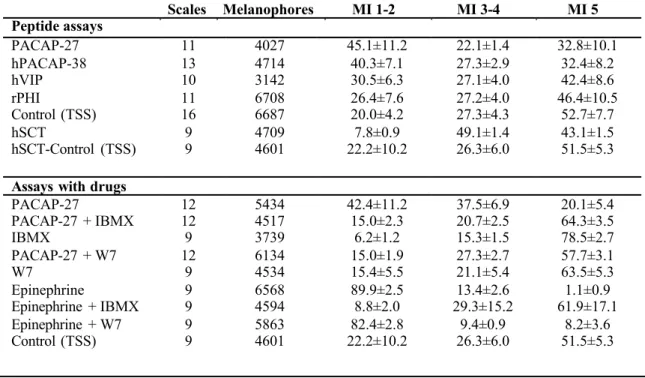
Scales from the dorsal fin region were collected and melanophore enriched cultures
201
were derived from the skin attached to the scale (12-15 scales) from different fishes. The skin
202
was isolated and finely sliced and the scale-skin fragments were digested with 400 μl of a 0.25
203
% trypsin/EDTA solution for 90 min at 37 ºC with agitation. The released cells were added to
204
1600 ml of L-15 medium (Sigma Aldrich, Spain) supplemented with 10 % sterile foetal bovine
205
serum and 0.1 % penicillin: streptomycin antibiotic mixture (10.000 U:10 mg/ml, Sigma
206
Aldrich, Spain) and 250 μg/ml (1:100) sterile filtered amphotericin B solution (Sigma-Aldrich,
207
Spain) in a 6 well plate. Melanophores were identified by their dark colour in phase-contrast
208
microscopy and were transferred with the aid of a micropipette to a new well of a 6 well-plate to
209
generate melanophore-enriched cultures. Other cell types that were transferred with
210
melanophores were subsequently removed by medium aspiration. Melanophore-enriched
211
cultures containing approximately 40 to 50 melanophores were snap frozen and stored at -80 ºC
212
for total RNA extraction and cDNA synthesis.
213
214
2.6 RNA isolation and cDNA synthesis
215
Tilapia cDNA was used to amplify receptors, peptide precursors and ramps. The cDNA
216
was synthesized from total RNA (tRNA) extracts of several tissues to confirm in silico gene
217
predictions and characterize the tissue distribution. For tissue collection, fish were anaesthetized
218
with 2-phenoxyethanol (1 ml.l-1 water) and killed by decapitation. Skin and other tissues were
219
collected and immediately frozen in liquid nitrogen and stored at -80 oC. Total RNA from tilapia
220
tissues was isolated using Tri-Reagent (Sigma Aldrich, Spain) and treated with 1 U DNase
221
(DNA-free Kit, Ambion, UK) for 30 min at 37 °C in accordance with the manufacturer’s
222
instructions. DNase treated total RNA (500 ng) was denatured at 65 °C for 5 min, quenched on
223
ice for 5 min and used for cDNA synthesis in a 20 μl reaction volume containing 10 ng of
224
pd(N)6 random hexamers (GE Healthcare, UK), 2 mM dNTPs, 100 U of MMLVRT and 20 U
225
RNasin® Plus RNase inhibitor (Promega, Spain). cDNA was synthesized for 10 min at 20 °C
226
followed by 60 min at 42 °C and 72 °C for 5 min. The quality and quantity of the cDNA was
227
assessed by PCR amplification of ribosomal subunit 18s rRNA (18s) using the following cycle:
228
94 ºC for 3 min; 25 cycles of 94 ºC for 35 sec, 57 ºC for 30 sec, 72 ºC for 45 sec, followed by 72
229
ºC for 5 min.
230
2.7 Database mining
232
Adcyap1, vip and adcyap1r, vipr1 and vipr2 genes were identified and retrieved from
233
the cichlid, Nile tilapia (Oreochromis niloticus) genome ENSEMBL annotation
234
(http://www.ensembl.org/Oreochromis_niloticus/Info/Index) (July 2014 release) using the
235
tblastn algorithm. Sequence queries were performed with the deduced mature peptides of
236
Takifugu rubripes Pacapa (ABG73207), Pacapb (ABG73208), Vip (DQ659330) and the
237
zebrafish peptides (Vipa, ABV83048 and Vipb, NP_001108027). Tilapia receptors were
238
retrieved using the Takifugu PACAP receptor sequence homologues: Pac1a (Q5WML1); Pac1b
239
(Q5WML0); Vpac1a (CAC82588); Vpac1b (CAC82587); Vpac2a (CAC83861); Vpac2b
240
(CAC82587). Searches for PACAP and PACAP receptors genes and transcripts were extended
241
to other fish genomes (12 genome assemblies, November 2014) available in ENSEMBL
242
(http://www.ensembl.org), the recently published European sea bass (Dicentrarchus labrax)
243
genome (Tine et al., 2014), the cartilaginous fish, the elephant shark (Callorhinchus milii)
244
genome (http://esharkgenome.imcb.a-star.edu.sg/blast/), the Japanese lamprey (Lethenteron
245
japonicum) genome (http://jlampreygenome.imcb.a-star.edu.sg) and an “in house” sea bream
246
(Sparus auratus) gene transcript nucleotide database. The available genome of the Mexican
247
tetra (Astyanax mexicanus) in ENSEMBL corresponds to its caveform and is designated
248
hereafter as cavefish. The tilapia genome and other fish databases were also used to search for
249
homologues of human RAMP1 (NP_005846.1), RAMP2 (NP_005845.2) and RAMP3
250
(NP_005847.1). The sequences of Takifugu obscurus ramp1a (BAE45310.1), ramp1b
251
(BAE45306.1), ramp2tv1 (BAE45308.1), ramp2tv2 (BAE45311.1), ramp3 (BAE45305.1) and
252
ramp5 (BAE45307.1) gene transcripts were used to interrogate the databases. The Nile tilapia
253
(O. niloticus) sequences were aligned with the other fish and tetrapod homologues and used to
254
design primer pairs for isolation of adcyap1r, vipr1, vipr2, adcyap1, vip and ramp transcripts
255
from Mozambique tilapia (O. mossambicus).
256
257
2.8 In silico comparisons, phylogeny and gene synteny analysis
258
Multiple sequence alignments were generated using the Clustal X (version 2) software
259
programme (Larkin et al., 2007,Thompson et al., 1997) and the GeneDoc software
260
(http://iubio.bio.indiana.edu/) was used to annotate the alignment and to calculate percentage of
261
sequence identity/similarity of the deduced amino acid sequences. The PAC/VPAC receptor
262
sequence alignment was submitted to ProtTest (2.4) to select the best model that characterizes
263
receptor protein evolution according to the Akaike Information Criterion (AIC) statistical model
264
(Abascal et al., 2005). The Jones-Taylor-Thornton (JTT) (Jones et al., 1992) matrix based
265
model was chosen and phylogenetic analysis was performed with 88 vertebrate receptor
266
sequences that included the tilapia receptors, the orthologues from other fish, Xenopus and
267
human. Phylogenetic trees were built using the Maximum Likelihood (ML) method in PhyML
268
3.0 implemented in ATGC (http://www.atgc-montpellier.fr/phyml/) (Guindon et al., 2010) and
269
using the Neighbor-Joining (NJ) (Saitou and Nei, 1987) method in Mega 5.2 (Tamura et al.,
270
2011) with 100 and 1000 bootstrap replicates, respectively (Felsenstein, 1985). ML trees were
271
searched using the Nearest-Neighbor-Interchange (NNI) heuristic method and constructed with
272
4-substitution rate categories gamma shape (0.941). The vertebrate SCT receptor sequences
273
(Cardoso et al., 2014) were also included in the analysis and both trees were rooted with the
274
STCR clade. The NJ and ML methods generated trees with similar topologies.
275
A similar strategy to that outlined above was used with the teleost Ramps. Phylogenetic
276
tree was performed with the Jones-Taylor-Thornton (JTT) matrix selected after ProtTest (2.4).
277
Analysis included 69 sequences and the ML tree was constructed with 4-substitution rate
278
categories gamma shape (1.116) and proportion of invariable sites (0.019) with 100 bootstrap
279
replicates. The ML and NJ trees generated similar tree topologies and a hypothetical root was
280
added between the RAMP1/3 and RAMP2/5 clades based upon the model proposed for RAMP
281
family evolution (Benítez-Páez and Cárdenas-Brito, 2008).
282
The gene environment of the teleost adcyap1r1, vipr2, adcyap1 and vip was
283
characterized and the neighbouring gene homologues were identified based upon the
284
ENSEMBL database gene annotation and complemented with sequence homology searches.
285
286
2.9 Quantitative PCR (q-PCR)
287
Receptor and peptide transcript abundance in tilapia skin (with no scales) and
288
melanophore enriched cultures was quantified using quantitative real-time PCR (q-PCR).
289
Primer sequences and annealing temperatures are described in Supplementary Table 1. Four
290
reference genes b-actin, glyceraldehyde-3-phosphate dehydrogenase (gapdh1), 18s, and
291
melanocortin 1 receptor (mc1r, a.k.a. melanocyte-stimulating hormone receptor) were used to
292
calculate relative expression units of the tilapia transcripts. Transcript absolute values were
293
normalized for differences in reverse transcription efficiencies against the geometric mean of
294
the reference genes. The geometric mean of β-actin and 18s was used to normalize the results
295
for tissue distribution of PACAP receptor transcripts. For normalization of PACAP receptor
296
transcripts in skin collected from fish under a light/dark challenge the geometric mean of tilapia
297
β-actin and gapdh1 was used. Gene expression analysis on the melanophore cell cultures was
298
normalized using the mc1r (a marker of melanophores) (Higdon et al., 2013,Kobayashi et al.,
299
2012,Selz et al., 2007). Reference genes were selected taking into consideration their stable
300
expression level in the tissues analysed. Q-PCR analysis was performed in duplicate (< 5%
301
variation between replicates) using an Icycler iQTMReal-Time PCR Detection System (Bio-Rad,
302
Portugal) and SsoFast EvaGreen supermix (Bio-Rad, Portugal) and 300 nM of the forward and
303
reverse primer in 96-well micro plates (Bio-Rad, Portugal). Optimized cycling conditions
304
consisted of 95 ºC for 30 s, followed by 45 cycles of 95 ºC for 5 s and 58 - 62 ºC for 10 s.
305
Melting curves were performed to detect nonspecific products and primer dimers. Standard
306
curves were prepared from M13 amplified PCR products of each transcript cloned in
pGEMT-307
easy (Promega, Spain). Control reactions were included in all runs to confirm the absence of
308
genomic DNA.
309
310
2.10 Cloning and recombinant vector construction
311
A sea bream (Sparus auratus) Pac1a construct was available in house (Cardoso et al.,
312
2007) and was used to infer the activity of the tilapia receptor homologue as they shared over
313
90% aa sequence identity (Supplementary Figure 2). The complete coding sequence of tilapia
314
vipr2a was amplified from brain cDNA using specific primers and proofreading DNA
315
polymerase (iProof, BioRad, Portugal, Supplementary Table 1). The PCR thermocycle
316
consisted of: 98 ºC for 30 sec, followed by 45 cycles (98 ºC 10 sec, 64 ºC for 30 sec, 72 ºC for 2
317
min) with a final extension of 72 ºC for 10 min. The PCR product was cloned into the
318
pcDNA3.1 vector (Directional TOPO Expression Kit, Invitrogen, USA) and used to transform
319
TOP10 competent bacteria. Positive bacterial colonies were screened by PCR using receptor
320
specific primers. Plasmid DNA was isolated from those giving a PCR product of the expected
321
size using the standard alkaline lysis method and was sequenced to confirm identity. Human
322
RAMP1 (conceded by Dr Vanessa Schein, UFRGS, Brazil) was amplified with proofreading
323
DNA polymerase (iProof, BioRad, Portugal) using specific primers (Supplementary Table 1)
324
and cloned into pcDNA3.1.
325
326
2.11 Mammalian cell cultures and transfections
327
HEK293 cells (ECACC collection, UK) were maintained in complete Dulbecco’s
328
modified Eagle’s medium (DMEM, Sigma-Aldrich, Spain) with 4.5 g/L glucose, 110 mg/L
329
sodium pyruvate and L-glutamine supplemented with 10 % sterile foetal bovine serum and 0.1
330
% penicillin: streptomycin antibiotic mix (10.000 U:10 mg/ml, Sigma) and 250 μg/ml sterile
331
filtered 1:100 amphotericin B solution (Sigma, Spain) in a humid 5 % CO2 incubator (Heraeus,
332
Portugal) at 37 °C. One day prior to transfection, 2–3 x 10⁵ cells were seeded into 6 well plates
333
(Sarstedt, Portugal) and cells were transiently transfected using Fugene 6 transfection reagent (1
334
: 6 DNA : Fugene, Roche, Germany) following the manufacturer’s protocol. Simultaneous
335
transient transfections of a vector expressing green fluorescent protein were performed to
336
estimate the efficiency and success of cell transfections. The capacity of the PACAP peptides
337
PACAP-27 and hPACAP-38 to activate the teleost receptor cAMP-signalling pathway was
338
assayed 72 hours after cell transfections. HEK293 stable cell lines expressing human RAMP1
339
were generated to assess the effect of transmembrane accessory proteins on the pharmacology
340
of PACAP receptor function.
341
342
2.12 cAMP and Ca2+ intracellular signalling
343
The capacity of PACAP-27 and hPACAP-38 peptides to stimulate intracellular cAMP
344
production by binding Pac1a and Vpac2a and RAMP1-Pac1a complex in the transfected cells was
345
measured using a cAMP dynamic 2 kit (Cisbio, France) following the manufacturer’s protocol.
346
Approximately 15,000 cells were assayed per well and incubations were performed in white 384
347
well small Volume™ HiBase Polystyrene microplates (Greiner, Germany). Prior to the assay,
348
cells were ressuspended in 1 x PBS with 1 mM of 3-isobutyl-1-methylxantine (IBMX, Sigma)
349
and incubated for 5 min at 37 ºC. Peptides (1 μM - 0.01 nM) diluted in 1 x PBS / 1 mM IBMX
350
were added to the cells for 30 min at 37 ºC. cAMP production was monitored using a Biotek
351
Synergy 4 plate reader (Biotek, USA). Data was normalized following the manufacturer’s
352
recommendations.
353
Intracellular Ca2+ (iCa2+) release (RFU) was also measured for Pac
1a in the presence of
354
PACAP peptides using the Ca2+ sensitive fluorescent dye Fluo-4 NW (Molecular Probes,
355
Invitrogen). Prior to the assay, plates (96 well black/plates, μClear bottom, Greiner, Germany)
356
were coated with sterile poly-L-lysine (0.1 mg/ml, Sigma, Spain). Assays were performed with
357
approximately 50,000 cells according to manufacture’s instructions. Background RFU of
358
transfected cells was measured prior to stimulation and dose-response curves were determined
359
for PACAP-27 and hPACAP-38 (1 μM to 0.01 nM). Calcium mobilization stimulated by the
360
peptide was measured every 10 sec over a total period of 2 min using a Biotek Synergy 4 plate
361
reader (Biotek, USA).
362
363
2.13 Statistical analysis
364
Significant changes in melanin response to the peptides in ex-vivo assays were assessed
365
using a two-tailed Student t-test. Q-PCR expression data are presented as mean ± SEM.
366
Significant differences in the abundance of receptor distribution in tissue was assessed with a
367
One-way Anova and Tukey’s multiple comparison test. Significant difference in receptor
368
expression in the skin of light/dark challenged tilapia was determined using a two-tailed Student
369
t-test. Receptor activation assays are presented as the mean ± SEM of three independent
370
experiments carried out in triplicate and significant differences in cAMP or iCa2+ was assessed
371
using a two-tailed Student t-test. All the analyses were performed using Prism GraphPad
372
version 5. Statistical significance was considered at p < 0.05, p < 0.01 and p < 0.001.
373
374
3. Results
375
3.1 Effect of PACAP and related peptides on melanin mobilization in the skin
376
Incubations of tilapia skin with PACAP and VIP and also with peptide histidine
377
isoleucine (PHI) and secretin (SCT), which are all members of the secretin-like peptide family,
378
indicated that PACAP was the most potent inducer of melanin aggregation in melanophores
379
(Figure 1A, Table 1). The aa sequence of hPACAP-27 is 100% identical with most of the fish
380
homologues (PACAP-27a, Supplementary Figure 1 A) and hereafter the peptide is designated
381
PACAP-27.
382
PACAP-27 or hPACAP-38 peptides added to fish skin had a similar effect on pigment
383
aggregation and 45.1 ± 11.2 % (p < 0.05) and 40.3 ± 7.1 % (p < 0.05), respectively, of the total
384
melanophores were fully aggregated (MI 1-2) compared to 20.0 ± 4.2 % MI 1-2 in the control.
385
The melanin aggregating effect of hVIP (1 µM) and rPHI (1 µM) were not significantly
386
different from the control (30.5 ± 6.3 % and 26.4 ± 7.6 %, respectively) (Figure 1A). Secretin
387
(SCT), which is absent from teleost genomes (Cardoso et al., 2014), did not modify the state of
388
melanin aggregation (Table 1). The results suggest that PACAP regulated pigment translocation
389
in fish skin melanophores and stimulated melanin aggregation. Forskolin (10 μM, a stimulator
390
of cAMP production) promoted melanin pigment dispersion in tilapia skin melanophores (data
391
not shown, Logan et al., 2006,Sheets et al., 2007). The results, suggest that the action of
392
PACAP-27 and hPACAP-38 on tilapia skin pigment aggregation was unlikely to be triggered by
393
a rise in the intracellular levels of cAMP.
394
The signalling pathways involved in the melanin aggregating effect of PACAP were
395
assessed using IBMX (2.5 mM) and W7 (100 μM) (Table 1, Figure 1B). IBMX (a non-specific
396
inhibitor of cAMP phosphodiesterases) caused dispersion of melanin in 78.5 ± 2.7% of skin
397
melanophores (MI5) compared to 51.5 ± 5.3 % in the control (Figure 1B, Table 1). Incubation
398
of skin with W7 (an inhibitor of Ca2+/CaM-activated phosphodiesterase) did not change the
399
pigment translocation pattern in relation to the control (Table 1, Figure 1B). Incubation of skin
400
melanophores with PACAP-27 (1 μM) and IBMX significantly (p < 0.05) decreased the
401
melanin aggregating effect of the peptide (42.4 ± 11.2% to 15.0 ± 2.3). Incubation of skin with
402
PACAP-27 and W7 also caused a significant (p < 0.05) reduction in the number of aggregated
403
melanophores (42.4 ± 11.2% to 15.0 ± 1.9). Epinephrine (1μM) had a significant (p < 0.01)
404
melanin aggregating effect (89.9 ± 2.5% of melanophores) compared to the control. IBMX (2.5
405
mM) blocked the effect of epinephrine (1μM) and melanin aggregation occurred in only 8.8 ±
406
2.0 % of the melanophores. W7 did not affect the melanin aggregating effect of epinephrine
407
(1μM) (Table 1).
408
409
3.2 PACAP system and RAMPs
410
Two adcyap1 (a and b), two vip (a and b), six Pacap receptors (adcyap1r1a and b,
411
vipr1a and b and vipr2a and b) genes and five ramp genes were identified in the Nile tilapia (O.
412
niloticus) genome and amplified in the Mozambique tilapia (O. mossambicus). The deduced
413
mature peptide sequence of the genes and transcripts in both species of tilapia were 100%
414
identical. A similar gene number was also retrieved from other teleost genomes (Supplementary
415
Table 2, 3 and 4).
416
417
3.2.1 PACAP and VIP in fish
418
In tilapia and other teleosts, duplicates of the human ADCYAP1 and VIP genes were
419
identified (Supplementary Table 2, Supplementary Figure 3, (Cardoso et al., 2007, Ng et al.,
420
2012). The fish adcyap1 genes (adcyap1a and b) produced paralogue Pacap-27 and Pacap-38
421
peptides (a and b) and in most fishes (including tilapia and other teleosts, elephant shark and
422
coelacanth) Pacap-27a was 100% identical to human and Xenopus PACAP-27 and was the most
423
conserved peptide of this family in vertebrates (Supplementary Figure 1A). The exceptions were
424
cod, cavefish and zebrafish in which the predicted paralogue Pacap-27b was most similar to
425
human PACAP-27a. Teleost Pacap-27b only differed from the tetrapod homologues (96% aa
426
identical) at a single aa residue but in the cavefish it was 100% identical (Supplementary Figure
427
1A). The teleost duplicate Pacap-38 was also highly conserved and Pacap-38a and Pacap-38b
428
differed at 3 and 4 aa residues and shared 92% and 89% identity, respectively with the human
429
peptides. The cod, cavefish and zebrafish Pacap-38 was less conserved.
430
In tilapia, cavefish and zebrafish duplicate vip genes (vipa and b) were identified but in
431
other fishes only a single gene copy existed (Supplementary Table 2). In teleosts the predicted
432
mature peptides of Vip shared less conservation than Pacap with the human homologue
433
(Supplementary Figure 1B). The predicted tilapia Vipa and Vipb peptides shared 81% aa
434
identity with human VIP and the other teleosts shared between 77% to 85% identity with the
435
exception of the Amazon molly that shared only 59% identity. In the early ray-finned fish the
436
spotted gar, the cartilaginous elephant shark and the coelacanth (a lobe-finned fish) a single
437
adcyap1 and vip1 gene was retrieved. The genome regions of the spotted gar and duplicate
438
adcyap1 and vip1 genes in teleost shared high synteny with the homologue genome region in
439
human (Supplementary Figure 3) indicating that duplication of the peptide genes was teleost
440
specific (Supplementary Figure 3). Adcyap1 and vip genes were retrieved from the Japanese
441
lamprey genome and the deduced mature peptides shared 85% and 66% aa identity with the
442
human homologue (Supplementary Figure 1A and B).
443
444
3.2.2 PACAP receptor genes in fish
445
The deduced tilapia Pac1a and b mature protein sequence shared 92 % and 89 % aa
446
sequence similarity with the Takifugu homologues (Table 2). The alternatively spliced-exon in
447
the fish Pac1a hop isoform (adcyap1a-hop) was also amplified from tilapia and caused an 82 bp
448
insertion in the C-terminal intracellular domain (Cardoso et al., 2007,Kwok et al.,
449
2006,Fradinger et al., 2005).
450
The teleost analysed also contained duplicate adcyap1r1, vipr1 and vipr2 genes and
451
phylogenetic analysis suggested that receptor gene duplication was a consequence of the teleost
452
genome tetraploidization event (Figure 2). The exception was the cavefish that lacked the gene
453
homologue for adcyap1r1a and the zebrafish that lacked the vipr2b gene (Supplementary Table
454
3, Figure 2). The absence of adcyap1r1b and vipr2b genes in the cod was presumably a
455
consequence of the incomplete genome assembly. The genome region flanking adcyap1r1a was
456
conserved in several fishes (Supplementary Figure 4) and mapped to short contigs in the
457
cavefish genome. The genes, c2cd4c, chico, ccdc94 and shda in linkage with zebrafish
458
adcyap1r1a mapped to KB882255.1 in cavefish but adcyap1r1a was not identified and further
459
studies will be required to confirm if specific gene loss occurred in the cavefish lineage.
460
The conserved gene environment that flanked the teleost paralogue vipr2b gene (esyt2a,
461
pflkpb and ptpnr2) was distributed on chr 2 and chr 7 in the zebrafish genome suggesting that
462
this gene may have been lost (Supplementary Figure 4). Single copies of adcyap1r1, vipr1 and
463
vipr2 genes were retrieved from the spotted gar and with the exception of vipr2 were also
464
identified in the coelacanth. In the Japanese lamprey genome two putative receptor genes for
465
PACAP and VIP have been described (Cardoso et al., 2014,Ng et al., 2012) and in the
466
cartilaginous elephant shark genome, putative vipr1 and vipr2 genes were identified but the
467
adcyap1r1 gene was missing (Supplementary Table 3).
468
469
3.2.3 RAMP genes in fish
470
The tilapia and other teleost genomes contained five predicted gene homologues of
471
human and T. obscurus RAMPs (Supplementary Figure 5, Supplementary Table 4). The
472
deduced mature protein sequence of tilapia Ramp1 and Ramp4 were 52% identical and shared
473
42% aa identity with human RAMP1. Phylogenetic analysis of piscine ramp1 and ramp4
474
indicated that they were duplicate genes in the teleost lineage and we have designated them
475
ramp1a and ramp1b, respectively. Ramp genes were also identified in other fish genomes such
476
as the cartilaginous elephant shark, the spotted gar and the coelacanth but were not found in the
477
Japanese lamprey genome assembly (Supplementary Table 4). In the elephant shark orthologues
478
of vertebrate RAMP1, RAMP3 and RAMP2/RAMP5 were identified and in the spotted gar a
479
single gene of RAMP2 and RAMP5 was identified. Homologues of the fish ramp5 genes were
480
not found in human or Xenopus genomes and the fish members tended to group with the
481
RAMP2 clade. Overall the results support the notion that RAMP2/RAMP5 shared a common
482
origin and that both genes emerged during an early gene duplication event in the vertebrate
483
lineage (Benítez-Páez and Cárdenas-Brito, 2008) and ramp5 gene was subsequently deleted in
484
tetrapods. In the coelacanth, five putative ramp genes with sequences divergent from vertebrate
485
homologues were identified; it was unclear if this was a species-specific evolutionary event or a
486
problem with the genome assembly.
487
488
3.3 Expression of the PACAP system and RAMPs in tilapia
489
Q-PCR analysis revealed that the six predicted PACAP receptors and adcyap1r1a-hop
490
had a widespread tissue distribution in tilapia (Figure 3). PACAP receptor transcripts were low
491
abundance with the exception of adcyap1r1a, which was highly abundant in brain and vipr1a
492
that had a moderate expression in all the tissues analysed (Figure 3). In tilapia adcyap1r1a and
493
adcyap1r1a-hop (p < 0.001) and the paralogue adcyap1r1b (p < 0.05) transcripts were
494
significantly more abundant in brain relative to other tissues. The duplicate vipr1 and vipr2
495
transcripts were present in brain, kidney, duodenum, gills, liver and white muscle. Vipr1b
496
transcripts were significantly (p < 0.001) higher in the kidney compared to the gills and brain.
497
Vipr2b transcripts were significantly (p < 0.05) higher in brain relative to all other tissues
498
(Figure 3).
499
In the skin PACAP receptor transcript abundance was low and changed in normal and
500
darkening tilapia skins (Figure 4). In normal tilapia skin, transcripts for the paralogues vipr1a,
501
vipr1b and vipr2a were more abundant relative to other receptors and vipr2b was low
502
abundance/undetectable and was excluded from further analysis. Adyap1r1a and vipr2a were
503
significantly down-regulated (p < 0.05) in darkening skin compared to normal skin. In contrast,
504
vipr1b transcripts were significantly up-regulated (p < 0.05) in darkening skin suggesting that
505
its function in skin diverged from that of adcyap1r1a and vipr2a (Figure 4). Adcyap1r1a-hop,
506
adyap1r1b and vipr1a transcript abundance did not change in response to a light/dark challenge
507
and were also excluded from further analysis. Of the duplicate PACAP and VIP peptide
508
encoding transcripts, adcyap1a and vip (a and b) were low abundance/undetectable in skin and
509
only adcyap1b was detected at quantifiable levels (Figure 4). Most of the ramps, with the
510
exception of ramp1b, showed low abundance in skin and ramp5 was undetectable. No
511
significant differences in ramp expression between normal and darkening skins were observed
512
(Figure 5). In melanophore enriched tilapia cell cultures (Figure 6), adcyap1r1a was relatively
513
more abundant than vipr1b and vipr2a (Figure 6A). Ramp1b was the most abundant transcript in
514
melanophores (Figure 6B).515
516
3.4 Intracellular signalling517
Activation by PACAP of sbPac1a and tiVipr2a expressed in HEK293 cells was
518
characterised (Figure 7). PACAP peptides were tested because; i) they have the most potent
519
effects on pigment aggregation in tilapia melanophores in vitro and ii) adcyap1b transcripts
520
were detected in fish skin. Both PACAP-27 and hPACAP-38 promoted a dose-dependent
521
increase in intracellular levels of cAMP when they were added to human HEK293 cells
522
transiently transfected with fish sbPac1a (Figure 7A). PACAP-27 and hPACAP-38 were
523
equipotent when they activate sbPac1a and had EC50 values of 1.422 e-8 M and 1.457 e-8 M,
524
respectively. PACAP-27 and hPACAP-38 binding to sbPac1a also stimulated an increase in
525
intracellular Ca2+ and had an EC
50 respectively of 1.243 e-8 M and 4.175 e-8 M (Figure 7B).
526
TiVipr2a transiently transfected in HEK293 cells was not stimulated by PACAP (Figure 7A)
527
and was only activated by PHI (data not shown and (Wu et al., 2008).
528
The association between a rise in cAMP and melanin dispersion in tilapia melanophores
529
and the melanin aggregating effect of PACAP suggested that fish Pac1a, may use an alternative
530
signalling mechanism that leads to a decrease in intracellular cAMP levels. To test this
531
hypothesis the effect of PACAP on HEK293 cells co-transfected with sbPac1a and RAMP1
532
from human was evaluated by measuring the change in cAMP. PACAP-27 and hPACAP-38
533
stimulated cAMP production in a HEK293 cell line co-expressing sbPac1a and hRAMP1 but at
534
10-fold lower levels than in a HEK293 cell line expressing sbPac1a alone (Figure 7A). The
535
results indicated that PACAP mediated activation of sbPac1a in the presence of hRAMP1
536
modified cAMP pathway signalling.
537
538
4. Discussion
539
Hormones and neurotransmitters regulate the function of pigment containing cells in
540
skin and eye and GPCRs are major signal transducers in this process. PACAP is an important
541
vertebrate neuroendocrine factor that activates GPCRs and regulates pigment dispersion in
542
amphibians via an increase in cAMP. In teleost fish, gene duplicates are suggested to have
543
contributed to their exuberant skin colour and complex pigment patterns (Braasch et al.,
544
2008,Braasch et al., 2009). The present study investigated if genes duplicates of the PACAP
545
system in fish are involved in skin colour change and revealed PACAP as a new player in the
546
endocrine regulation of fish pigment mobilization. PACAP receptors are differentially
547
expressed in skin, receptor abundance is responsive to light exposure and PACAP-27
548
significantly stimulates melanin pigment aggregation ex-vivo. However, the activation of
549
PACAP receptors by PACAP-27 causes a rise in intracellular cAMP, which is normally
550
associated with skin melanin pigment dispersal. We hypothesize that the physiological pigment
551
translocation response caused by PACAP-27 most likely involves fish Pac1a with RAMP in
552
skin, which attenuates or inhibits the rise in intracellular cAMP associated with activation of
553
Pac1a alone. Furthermore, the reduction of cAMP-dependent PKA activity favours a Ca2+/CaM
554
dependent phosphatase that promotes melanin aggregation.
555
556
4.1 Evolution of PACAP and its receptors in fish
557
Teleosts are the most diverse and successful group of vertebrates and more than 23,000
558
species have been identified (Venkatesh, 2003). Their genomes duplicated early in evolution
559
and the presence of gene duplicates in fish has been associated with the physiological plasticity
560
of this group of vertebrates (Braasch et al., 2009,Glasauer and Neuhauss, 2014). In teleost
561
genomes, duplicates of class 2 B1 GPCRs and ligands have been extensively studied (Sherwood
562
et al., 2000,Cardoso et al., 2007,Cardoso et al., 2010,Roch et al., 2009,Nag et al., 2012,Cardoso
563
et al., 2014,Wu et al., 2008,Cardoso et al., 2005,Cardoso et al., 2004,Martins et al.,
564
2014,Cardoso et al., 2014,Hwang et al., 2013) but their functional role in fish physiology
565
remains unclear. PACAP and PACAP receptors emerged early in the evolution of vertebrates
566
and members of the gene family are present in representatives of early vertebrate genomes, such
567
as the lamprey, and both peptide and receptor genes duplicated in the ancestral teleost and
568
persisted during the fish radiation (Sherwood et al., 2000,Cardoso et al., 2007,Cardoso et al.,
569
2014,Ng et al., 2012,Wu et al., 2008,Cardoso et al., 2004). Adcyap1r1 genes were only
570
identified in bony vertebrates and two PACAP peptides and two receptors genes were present in
571
most of the teleost genomes analysed. In fish with genomes that did not undergo
572
tetraploidization, such as the spotted gar, a representative of the ray-finned fish that preceded
573
the teleost radiation, and the coelacanth, basal lobe-finned fish, a single receptor gene copy was
574
found. Within the teleosts, the exceptions were cod and the cavefish that, in common with other
575
teleosts, underwent genome tetraploidization (Glasauer and Neuhauss, 2014) and retained
576
duplicate adcyap1 but only a single adcyap1r1 was identified and in the cavefish no adcyap1r1a
577
was found (Figure 8). The cavefish lacks melanin pigmented cells and eyes (McCauley et al.,
578
2004,Protas et al., 2006,McGaugh et al., 2014) and this intriguing observation seems to support
579
the link between PACAP receptors and pigmentation. However, the incomplete nature of the
580
cavefish genome assembly makes the loss of adcyap1r1a during evolution uncertain and further
581
studies are required to confirm that they lack duplicate PACAP receptors.
582
583
4.2 PACAP regulates melanophore pigment motility in fish skin
584
In tetrapods, PACAP is a central endocrine factor that regulates the secretion of
585
pituitary hormones involved in skin melanophore cell activity. Studies in amphibians have
586
demonstrated that PACAP is a key factor regulating vertebrate skin colour and that this action
587
has been conserved in tetrapods (Marotti et al., 1999,Kidane et al., 2008). In tilapia, the two
588
PACAP peptides and six receptor genes identified in this study are the duplicates of the human
589
and amphibian homologues and the present study indicates that in common with tetrapods,
590
elements of the piscine PACAP system are expressed in fish skin and involved in melanin
591
pigment translocation. Duplicates that are homologues of human ADCYAP1R1 and VIPR are
592
expressed in fish skin but only adcyap1r1a, vipr1b and vipr2a have modified expression in
593
darkening skin and adcyap1r1a is the most abundant receptor transcript in piscine melanophore
594
enriched skin cultures. The results suggest that adcyap1r1a, vipr1b and vipr2a acquired a
595
specific function in fish skin and that the former receptors may regulate the status of melanin
596
pigment in tilapia. It remains to be established if their functions are conserved across teleosts as
597
homologue receptors were identified in all jawless, cartilaginous, ray-finned and lobe-finned
598
fish genomes explored and further experiments directed at confirming this hypothesis are
599
required.
600
PACAP has a direct action on frog skin and provokes pigment granule dispersal in X.
601
laevis melanophores via activation of an endogenous VPAC2 (Marotti et al., 1999,Pereira et al.,
602
2002,Yamaguchi et al., 2007). The effect of PACAP on melanophore granule status in frog
603
(melanin dispersion) is opposite to that observed in tilapia (melanin aggregation) and highlights
604
that gene sequence homology and apparent similarity of physiological systems across evolution
605
does not guarantee conserved function. The divergent functions of vertebrate PAC1 may result
606
from differences in the mechanisms that govern pigment translocation in fish and frog
607
melanophores. Fish skin possesses larger and faster-responding melanophores than frog skin
608
and the cellular microtubule motors that translocate pigment work differently (Zaliapin et al.,
609
2005). For example, in fish pigment movements are assisted predominantly by kinesin 1, while
610
in frog kinesin 2 has this role. Further work will be required to establish at the cellular level how
611
Pac1a activation in fish melanophores is coupled to pigment translocation.
612
The functional divergence observed between PAC1 in frog and fish may be related to
613
the remarkable percentage of duplicate genes that have been retained in the genome of fish most
614
likely as a consequence of acquisition of new functions (neofunctionalization) or partitioning of
615
the ancestral molecule function between the duplicated gene copies (subfunctionalization) (Sato
616
and Nishida, 2010,Taylor et al., 2003,Postlethwait et al., 2004). The PACAP endocrine system
617
in fish is duplicated and receptor transcript expression in sea bream was taken to suggest that
618
the persistence of duplicate adcyap1r1a (widespread) and adcyap1r1b (brain specific) genes is a
619
consequence of their distinct function in fish (Cardoso et al., 2007). In tilapia, adcyap1r1a and
620
adcyap1r1b are both brain-specific but the former receptor is approximately 40 times more
621
abundant than adcyap1r1b, supporting the notion first observed in sea bream that the paralogues
622
may have acquired distinct functions. The different expression pattern of the duplicate PACAP
623
receptors in tilapia skin after a light/dark challenge, the abundance of adcyap1r1a in enriched
624
melanophore cultures, and the role of PACAP, the ligand, on melanin aggregation ex-vivo
625
suggest that Pac1a has acquired a specific role in fish skin pigmentation.
626
627
4.3 PACAP signalling in fish skin
628
Rapid changes in pigment motility can occur in amphibian and teleost cells. In
629
amphibians and fish an increase in intracellular cAMP induces pigment dispersion and a
630
decrease promotes pigment aggregation (Logan et al., 2006,Busca and Ballotti, 2000,Nery and
631
Castrucci, 1997,Tuma and Gelfand, 1999). In contrast, the role of the iCa2+-dependent pathway
in melanophores is controversial, this pathway is not directly involved in melanin translocation
633
and a species-dependent skin pigmentation response occurs in fish (Tuma and Gelfand,
634
1999,Sammak et al., 1992,Svensson et al., 1997). Perturbations in cAMP levels interfere with
635
microtubule (MT) (dynein and kinesin) and actin filament (AF) (myosin V) proteins that govern
636
the movement of melanin pigment. A decline in cAMP increases the frequency of minus-end
637
motility episodes that are associated with aggregation (Tuma and Gelfand, 1999,Rodionov et
638
al., 2003). In tilapia melanophores melanin pigment aggregation by epinephrine,
nor-639
epinephrine and Mch depends on the activity of PKA and of a protein phosphatase (eg. PP2B or
640
calcineurin) (Thaler and Haimo, 1992,Thaler and Haimo, 1990,Oshima et al., 2001,Oshima and
641
Wannitikul, 1996).
642
Functional characterisation of fish Pac1a revealed that both PACAP-27 and hPACAP-38
643
peptides are equipotent in stimulating cAMP production (Cardoso et al., 2007,Wong et al.,
644
1998) and that the presence of RAMP1 attenuates the production of intracellular cAMP. Since
645
high cAMP is associated with pigment dispersion this suggests that the melanophore response
646
observed in tilapia skin in the present study (melanin aggregation) is a result of the change in
647
receptor pharmacology (Figure 9 A). We hypothesise that the combination of attenuated levels
648
of cAMP and the probable increase in iCa2+ leads to stimulation of CaM and protein
649
phosphatases (PP) that promote melanin aggregation (Figure 9 B). The similar functional
650
characteristics of the PACAP receptor from goldfish, zebrafish and sea bream despite the
651
differences in their amino acid sequence suggests this may be a common mechanism in teleosts
652
(Cardoso et al., 2007,Kwok et al., 2006,Wong et al., 1998,Fradinger et al., 2005,Wu et al.,
653
2008). It also supports the notion that the characteristics of the sea bream Pac1a tested in vitro
654
will be similar for the tilapia orthologue and so can be related to the in vivo response in tilapia.
655
In vertebrates, MSH and MCH peptides are classical endocrine factors that regulate skin
656
pigmentation. In tilapia (and other fish), background adaptation does not modify plasma or
657
pituitary
α-Msh
levels or Mc1r expression and Msh/Mc1r seem to play a minor role in the658
regulation of skin pigmentation (van der Salm et al., 2005). In contrast, an increase in plasma
659
Mch levels and up-regulation of the peptide in the hypothalamus is observed when fish are
660
adapted to a light (white) background (Mizusawa et al., 2013,Kishida et al., 1989,Suzuki et al.,
661
1995). In fish skin, rapid changes in colour also involve the neurotransmitter norepinephrine
662
(NE) released by the sympathetic system and this stimulates pigment aggregation (Mizusawa et
663
al., 2013). Our study suggests that PACAP is a regulatory factor of fish skin and the presence of
664
PACAP transcripts in skin and the action of PACAP-27 on melanin status in melanophores
665
suggests that the PACAP system may also participate in the local and rapid response of skin
666
colour change (Figure 9 A). If the PACAP system interacts with Msh/Mch to modulate skin
667
pigmentation in fish still remains to be established. In tetrapods, PACAP modulates central
668
MSH secretion but in fish this remains to be demonstrated, as does the potential effect of
669
PACAP on Mch release (Tanaka et al., 2009).
670
PACAP, MSH and MCH peptides and also NE activate GPCRs and regulate
671
intracellular levels of cAMP coupled to distinct G-protein complexes. In fish skin, Msh
672
produced in the adenohypophysis or locally, binds to Mch-r1 that is associated with the
673
intracellular Gs-protein complex and increases melanophore cAMP production (Oshima et al.,
674
2001). Fish Mch is mainly secreted by the neurohypophysis and activates Mch-r2 that is
675
coupled to the Gi-protein complex that inactivates adenylate-cyclase and signals via the
676
inhibition of cellular cAMP levels (Hamamoto et al., 2011,Kawauchi, 2006). However, Mch
677
actions in fish are not conserved and tilapia and catfish skin contain another receptor type,
Mch-678
r1, that signals via cAMP and promotes and stimulates melanin dispersion suggesting that
679
regulation of skin colour pattern in fish may be species dependent (Oshima et al., 2001). In the
680
studies of duplicate PACAP receptors in fish they mainly signal via Gs (cAMP release) and Gq
681
(calcium release) (Cardoso et al., 2007,Kwok et al., 2006,Wong et al., 1998,Fradinger et al.,
682
2005,Wu et al., 2008), although it is possible that other intracellular signalling pathways are
683
activated in parallel particularly if accessory proteins are involved. For example, the association
684
of human VPAC1 with S-SCAM (synaptic scaffolding molecule) inhibits cAMP production via
685
Gi-coupling and favours receptor internalization (Gee et al., 2009).
686
The interaction of RAMPs with class 2 B1 GPCRs was first demonstrated for the
687
calcitonin receptors and was associated with promiscuity of ligand-binding and increased
688
signalling diversity and physiological responses (Couvineau and Laburthe, 2012). RAMPs have
689
also been described to interact with the mammalian glucagon, parathyroid and VPAC receptors,
690
although no changes in the cAMP intracellular signalling response have been reported
691
(Archbold et al., 2011). To date the effect of RAMP on fish Pac1a pharmacology has not been
692
explored and to our knowledge the present study represents the first evidence that they may
693
modify the receptors intracellular signalling response as previously reported for the calcitonin
694
receptors. It was not possible to establish if in the present study the attenuated cAMP response
695
associated with PACAP stimulation of cells co-transfected with Pac1a and RAMP in vitro also
696
occurs in vivo particularly since an heterologous expression system was used (fish receptor,
697
mammalian RAMP and mammalian cell line). Despite differences between fish and human
698
RAMPs (only 43% identical) the results from the cell transfection studies suggest that RAMP
699
interacts with the fish Pac1a and reduces the potency of PACAP to increase cAMP. We
700
hypothesise that functional conservation between the vertebrate RAMP systems exists and it is
701
possible that the homologue system may be more effective at modifying receptor signalling
702
potentially via the recruitment of other intracellular signalling molecules.Future studies with a
703
homologous system should contribute to resolve how the interaction of Pac1a and RAMP
704
modifies the response to PACAP and will contribute to more precisely establish how this affects