1
Microspheres loaded with polysaccharide nanoparticles for pulmonary delivery:
1Preparation, structure and surface analysis
2Sonia Al-Qadi1, Ana Grenha2, Carmen Remuñán-López1* 3
1
Department of Pharmaceutical Technology, University of Santiago de Compostela, Faculty of
4
Pharmacy, Campus Vida, 15782-Santiago de Compostela, Spain; 2CBME-Centre for Molecular and
5
Structural Biomedicine, IBB-Institute for Biotechnology and Bioengineering, University of Algarve,
6
Campus de Gambelas, Faro, Portugal.
7 8 * Corresponding author: 9 Tel.: +34 981563100 – Extn. 15045 10 Fax.: +34 981547148 11 E-mail: [email protected] 12 13 14
Abbreviations:
15A459: adenocarcinoma human alveolar basal epithelial cells 16
Calu-3: human airway epithelial cell line 17
CLSM: confocal laser scanning microscopy 18
CS: chitosan sodium hydrochloride 19
GlcA: glucoronic acid 20
GlcNAc: N-acetyl glucose amine 21
Glc: glucose amine 22
1D-CPMAS 13C: cross polarization magic angle spinning NMR 23
HA: hyaluronic acid 24
16HBE14o: human bronchial epithelial cell line 25
M: mannitol microspheres 26
M-NPs: microencapsulated nanoparticles 27
Mix: physical mixture of chitosan with hyaluronic acid 28
NMR: nuclear magnetic resonance 29
Revised manuscript
2 NPs: nanoparticles
30
SEM: scanning electron microscope 31
TEM: transmission electron microscopy 32
TOF-SIMS: time-of-flight secondary ion mass spectroscopy 33
TPP: pentasodium tripolyphosphate 34
XPS: X-ray photoelectron spectroscopy 35 36 37 38
Abstract
39In this work, we report the preparation of a nanoparticle-based dry powder for pulmonary 40
administration. Hybrid chitosan/hyaluronic acid nanoparticles were produced by ionotropic gelation 41
and characterized for their physicochemical properties, being further studied by solid nuclear 42
magnetic resonance (NMR). Using mannitol as carrier, nanoparticles were microencapsulated by 43
spray drying, resulting in a dry powder with appropriate aerodynamic properties for lung delivery. 44
In order to investigate the nanoparticles distribution within the carrier matrix, several techniques 45
were applied that permitted an in-depth analysis of the system structure and surface, such as 46
confocal laser scanning microscopy (CLSM) and X-ray photoelectron spectroscopy (XPS) in 47
combination with time-of-flight secondary ion mass spectroscopy (TOF-SIMS). Overall, the studies 48
conducted revealed that nanoparticles are homogeneously distributed through mannitol 49
microspheres, suggesting the success of the microencapsulation process. In the light of these 50
findings, it was concluded that the developed delivery system holds great potential for lung delivery 51
of macromolecules. 52
53
Key-words:
Chitosan; Hyaluronic acid; Microspheres; Nanoparticles; Pulmonary administration; 54Spray drying; TOF-SIMS; XPS. 55
3
1. Introduction
57
Presently, there is particular research interest in pulmonary delivery of drugs, specially peptides, 58
proteins and genes, not only for local, but also for systemic effect. This is primarily due to the 59
important advantages offered by the pulmonary route, such as the large alveolar surface available 60
for absorption, very thin diffusion path to the blood stream, extensive vascularisation, relatively low 61
metabolic activity compared to other routes and avoidance of gastrointestinal degradation and 62
hepatic metabolism (Agu, Ugwoke, Armand, Kinget, & Verbeke, 2001; Courrier, Butz, & 63
Vandamme, 2002). 64
In spite of this, in order to succeed in the pulmonary delivery of any therapeutic molecules, 65
many obstacles and lung defense mechanisms, that could hinder the path of foreign substances, 66
must be overcome, such as the effect of the airways’ structure, mucociliary clearance and 67
phagocytosis by alveolar macrophages (Courrier, Butz, & Vandamme, 2002; Hastings, Folkesson, 68
& Matthay, 2004). Thus, to evade the impact of such barriers and to assure optimal drug delivery to 69
the desired site, it is critical to develop the appropriate drug carriers. In this respect, specific 70
characteristics are required which provide the drug delivery system with the ability to reach the 71
alveolar region, if a systemic effect is desired, or another specific site, or if a local action is intended 72
(Pandey, & Khuller, 2005). Considering the specific anatomy of the airways, it is traditionally 73
believed that droplets and/or particles with an aerodynamic diameter within the range of 1-3 μm 74
will present appreciable deposition in the alveolar region, while those with a higher aerodynamic 75
diameter will mainly deposit in the upper regions (Chrystyn, 1997). Therefore, size and density of 76
the delivery system are the most critical parameters in obtaining adequate therapeutic effects. 77
However, notwithstanding the referred aerodynamic requirements, nanoparticles have also been 78
recently proposed for the same end (Dailey et al., 2003; Sung et al., 2007; Yang et al., 2008, Bailey 79
et al., 2009) due to their ability to delay or avoid mucociliary clearance and macrophage capture 80
(Schürch, Gehr, Im Hof, Geiser, & Green, 1990). 81
4
The selection of suitable biocompatible materials (polymers, lipids, sugars) used for the 82
preparation of lung carriers has been shown to be an essential consideration and, in this context, the 83
polysaccharides chitosan and hyaluronan are particularly attractive polymers. Chitosan, a natural 84
polysaccharide derived from chitin and is one of the most promising materials for transmucosal 85
drug delivery, given its reported low toxicity, biodegradability and biocompatibility(Hirano, Seino, 86
Akiyama, & Nonaka, 1988; Issa, Koping-Hoggard, & Artursson, 2005; Varshosaz, 2007), as well as 87
mucoadhesivity (Lehr, Bouwstra, Schacht, & Junginger, 1992; Agnihotri, Mallikarjuna, & 88
Aminabhavi, 2004) and enhancement of macromolecules permeation (Bernkop-Schnürch, Kast, & 89
Guggi, 2003), thus being extensively employed in the development of micro- and nanocarriers 90
(Grenha et al., 2008). In fact, chitosan is known to be degraded by mammalian enzymes such as -91
amylase (Muzzarelli, 1997) and lysozyme and has been demonstrated to induce low or absent 92
toxicity in cell lines representative of the pulmonary route (16HBE14o-, Calu-3 and A549) (Lim, 93
Forbes, Martin, & Brown, 2001; Florea, Thanou, Junginger, & Borchard, 2005; Grenha, Grainger, 94
Dailey, Seijo, Martin, Remuñán-López, & Forbes, 2007). Hyaluronic acid is a natural, linear and 95
non-sulfated glycosaminoglycan (Stern et al., 2007; Bastow et al., 2008; Theocharis et al., 2008; 96
Volpi et al., 2009),which is present in human tissues and fluids, mostly in soft connective tissue. 97
Interestingly, it can be found on the surface of alveolar epithelial cells, providing protection against 98
tissue damage and injury in a number of respiratory diseases (Jiang, Liang, & Noble, 2007) and 99
preventing pleural thickening in tuberculosis patients (Zhuo, Guo, & Tang, 2003; Cantor, & Turin, 100
2004). 101
Furthermore, it has been widely implicated in the development of drug and gene delivery 102
systems directed to different routes of administration ( Lim et al., 2002; Coradini et al., 2004; Peer 103
et al., 2004; Brown et al., 2005; Woo et al., 2007; Hwang et al., 2008; Gómez-Gaete et al., 2008; 104
Sahiner et al., 2008; Xin et al., 2010).This interesting profile of hyaluronic acid arises from its 105
unique characteristics, such as endogenicity, biodegradability, mucoadhesivity (Avitabile et al., 106
2001; Morimoto et al., 2001; Mayol et al., 2008; Sivadasa., 2008), the capacity to increase drug 107
5
circulation time in vivo (Peer et al., 2004; Jiang et al., 2008), and the ability to modify drug 108
dissolution and absorption (Chono, Li, Conwell, & Huang, 2008). Interestingly, hyaluronic acid 109
selectively binds to CD44 receptors expressed on lung epithelial cells and over-expressed in cancer 110
cells, a capacity that has prompted its use for targeting purposes (Akima et al., 1996; Taetz et al, 111
2009). More specifically, it has been employed in drug inhalation and gene therapy, which have 112
revealed encouraging in vitro and in vivo outcomes related to improved bioavailability and 113
transfection (Akima et al., 1996; Taetz et al, 2009; Surendrakumarm et al., 2003; Rouse; 2007; 114
Hwang et al., 2008). We recently proposed the preparation of chitosan/hyaluronic acid 115
nanoparticles using a mild gelation technique (De La Fuente, Seijo, & Alonso, 2008a), which 116
demonstrated great potential for ocular gene delivery (De La Fuente, et al., 2008a; De La Fuente, et 117
al., 2008b; De La Fuente, et al., 2010). Furthermore, these nanoparticles have shown to have a 118
potential application in the treatment of asthma, for heparin administration (Oyarzun-Ampuero, 119
Brea, Loza, Torres, & Alonso, 2009). 120
It is well known that delivering nanoparticles to the lungs is impractical due to their reduced 121
dimensions and, hence, low inertia (Sung, Pulliam, & Edwards, 2007; Yang, Peters, & Williams III, 122
2008). To address these limitations, we have recently proposed the microencapsulation of chitosan 123
nanoparticles within a micronsized mannitol inert carrier (Grenha, Seijo, & Remuñán-López, 2005) 124
as an attempt to improve the nanoparticles stability (nanoparticles are administered in solid state 125
and, therefore, more stable than the liquid counterpart) and aerosolization pattern, by conferring 126
adequate aerodynamic properties for proper particle deposition and drug delivery in the lungs 127
(Azarmi, Tao, Chen, Wang, Finlay, Löbenberg, & Roa, 2006; Sham, Zhang, Finlay, Roa, & 128
Löbenberg, 2004; Freitas, Müller, & 1998). 129
For a better understanding of the relation between surface properties and biological performance, 130
it is necessary to characterize in detail the surface of the developed carrier, which entails 131
determining the composition, structure and distribution of all components present on the surface. It 132
has been reported, specifically for dry powders, that having information on their surface 133
6
composition affords the possibility of controlling interparticulate interactions and, thus, enhancing 134
powder dispersion during inhalation (Bosquillon, Rouxhet, Ahimou, Simon, Culot, Préat, & 135
Vanbever, 2004; Bunkera, Daviesa, Chena, James, & Roberts, 2006; Chougule, Padhi, Jinturkar, & 136
Misra, 2007). Thus, the investigation of surface chemistry of dry powders may be beneficial in the 137
selection of optimal formulation and process parameters to maintain macromolecule integrity and 138
aerosolization efficiency which definitively result in high in vivo outcomes. 139
Herein, we report the preparation of hybrid chitosan/hyaluronic acid nanoparticles and their 140
characterization by NMR technique. These nanoparticles were microencapsulated in mannitol 141
microspheres using the spray drying technique, rendering them adequate for pulmonary delivery. 142
The mannitol microspheres´ structure was observed by confocal laser scanning microscopy (CLSM) 143
in order to investigate the nanoparticles´ spatial distribution within mannitol microspheres 144
following the microencapsulation process. Their surface was further analyzed by the application of 145
two surface-sensitive analytical techniques, X-ray photoelectron spectroscopy (XPS) and static 146
time-of-flight secondary ion mass spectrometry (TOF-SIMS), accurately characterizing their 147
surface chemical composition and determining the presence of nanoparticles. 148
149
2. Materials and methods
1502.1. Chemicals
151
Ultrapure chitosan in the form of hydrochloride salt (CS) (Protasan UP Cl 113, deacetylation 152
degree 75-90%, viscosity < 20 mPa.s, molecular weight < 150 KDa) was purchased from FMC 153
Biopolymers Norway; hyaluronic acid (HA) (molecular weight 166 KDa) was provided by 154
Bioiberica Spain; fluorescein sodium salt, phosphate buffered saline tablets (PBS) pH 7.4, 155
pentasodium tripolyphosphate (TPP) and D-mannitol were supplied by Sigma-Aldrich Spain and 156
Bodipy® 630/650-X was provided by Molecular Probes Netherlands. Ultrapure water MilliQ 157
plus, Millipore Ibérica, Spain was used throughout. All other chemicals were reagent grade. 158
7 159
2.2. Preparation of chitosan/hyaluronic acid nanoparticles
160
Chitosan/hyaluronic acid nanoparticles were produced by a slight modification of the ionotropic 161
gelation technique previously developed by our group (De La Fuente, Seijo, & Alonso, 2008a; 162
Calvo, Remuñán-López, Vila-Jato, & Alonso, 1997a; Calvo, Remuñán-López, Vila-Jato, & Alonso, 163
1997b). Electrostatic interactions were involved in the nanoparticles formation, where the positively 164
charged amino groups of CS interact with both negatively charged HA and TPP. Briefly, solutions 165
of TPP and HA in ultrapure water were prepared at concentrations of 0.4-2 mg/mL (w/v) and 2-4 166
mg/mL (w/v), respectively, and then, equal volumes of both solutions were mixed. Thereafter, 1 mL 167
of this mixture was added to 3 mL of CS solution whose concentration ranged from 1-1.25 mg/mL 168
(w/v) and the reaction was maintained for 10 minutes under mild magnetic stirring, resulting in 169
different formulations of nanoparticles as indicated in Table 1. Nanoparticles formed immediately 170
and were subsequently isolated for further analysis by centrifugation on a 10 µL glycerol layer 171
18,000×g, 30 min, 15C, Beckmann Avanti 30, Beckmann, USA, afterwards being re-suspended 172
in 100 µL of purified water after discarding the supernatants. 173
For confocal laser scanning microscopy (CLSM) study, CS was labeled with fluorescein 174
following the method described by De Campos et al (De Campos, Diebold, Carbalho, Sánchez, & 175
Alonso, 2004). Nanoparticles were also prepared on a large scale, where the final volume of 176
nanoparticles suspension was scaled to 40 mL. In this case, centrifugation was performed for 40 177
min at 18,000 × g and 15 C. 178
179
2.3. Determination of nanoparticles production yield
180
The nanoparticles production yield was calculated by gravimetry. Fixed volumes of 181
nanoparticles suspensions were centrifuged (18,000×g, 45 min, 15 C), supernatants were discarded 182
and sediments of nanoparticles were freeze-dried over 48 h (24 hours set at -34 C and gradual 183
ascent until 20 C), using a Labconco Freeze Dryer Labconco, USA (n=6). 184
8 The process yield was calculated as follows: 185
Nanoparticles weight 186
Process yield (%) = --- x 100 187
Total solids (CS + HA + TPP) weight 188
189
2.4. Physicochemical characterization of nanoparticles
190
The morphological appearance of nanoparticles was examined by transmission electron 191
microscopy (TEM) CM 12 Philips, Eindhoven, Netherlands. The samples were previously stained 192
with 2% phosphotungstic acid and placed on copper grids with Formvar films for viewing. 193
Measurements of nanoparticles size and zeta potential were performed on freshly prepared 194
samples, by photon correlation spectroscopy and laser Doppler anemometry, respectively, using a 195
Zetasizer Nano-ZS Malvern instruments, Malvern, UK. For particle size analysis, each sample of 196
isolated nanoparticles was diluted to the appropriate concentration with ultrapure water. Each 197
analysis was performed at 25C at a detection angle of 173C. For the determination of zeta 198
potential of the electrophoretic mobility, isolated nanoparticles samples were diluted with 0.1 mM 199
KCl and placed in an electrophoretic cell, where a potential of ± 150 mV was established. Size and 200
zeta potential of each formulation were analyzed in triplicate (n=3). 201
202
2.5. Solid NMR spectroscopy of nanoparticles
203
Solid-state 13C CP-MAS NMR spectroscopy experiments were performed at 298 K in an 11.7 T 204
Varian Inova-750 spectrometer (operating at 750 MHz proton frequency) equipped with a T3 205
Varian solid probe Varian, Inc, USA. Solid NMR samples were prepared in 3.2 mm rotors with an 206
effective sample capacity of 22 µL which corresponds to approximately 30 mg of the powder 207
sample. The spectra were processed and analyzed with MestreNova software (Mestrelab Research 208
Inc.). Carbon chemical shifts were assigned to the carbon methylene signal of solid adamantane at 209
28.92 ppm. 210
9
Four samples were analyzed: pure chitosan (CS), pure hyaluronic acid (HA), a physical mixture 211
of equal weights of CS and HA (Mix.) and chitosan/hyaluronic acid freeze-dried nanoparticles 212
(NPs) (CS/HA/TPP = 3.75/1/1). For each sample, a 1D-CPMAS 13C (cross polarization magic angle 213
spinning) spectrum was acquired under semi-quantitative experimental conditions. The inter-scan 214
delay was set to 3 s, the number of scans was 8000 and the MAS rate was 15 kHz. Heteronuclear 215
decoupling during acquisition of the FID was performed with Spinal-64 with the proton field 216
strength of 70 kHz. The cross polarization time was set to 3 ms. During cross polarization, the field 217
strength of the proton pulse was set constant to 75 kHz and that of the 13C pulse was linearly 218
ramped with a 20 kHz ramp near the matching sideband Prior to the acquisition of the 1D-CPMAS 219
spectra of the samples, the adamantane sample was used to calibrate the maximum 1H-13C cross-220
polarization under the experimental conditions. 221
222
2.6. Preparation of dry powders containing chitosan/hyaluronic acid nanoparticles
223
Sediments of chitosan/hyaluronic acid nanoparticles (CS/HA/TPP = 3.75/1/1), obtained 224
following centrifugation of the fresh nanoparticles suspensions, were resuspended in a mannitol 225
aqueous solution and the resultant suspension of nanoparticles in mannitol was spray dried. 226
Mannitol solutions were prepared with such concentrations that allowed final 227
mannitol/nanoparticles to be obtained at ratios of 90/10, 80/20, 70/30 (w/w) and suspensions with a 228
solid content of 3%. Dry powders were obtained in a one step process by spray drying either 229
aqueous solutions of mannitol or suspensions of nanoparticles in mannitol using a laboratory scale 230
drier Büchi Mini Spray Dryer, B-290, Switzerland. The spray drying operating conditions were: 231
two fluids external mixing 0.7 mm nozzle, feed rate of 2.5 mL/min and inlet temperature of 170 ± 2 232
C, resulting in outlet temperature of 111 ± 2 C. The air flow rate and the aspirator rate were 233
constant at 400 NI/h and 70%, respectively. The resultant spray dried powders were collected and 234
stored in a dessicator at room temperature until use. Preparation of microspheres for CLSM study 235
was performed with mannitol labeled with the fluorophore Bodipy 630/650-X, which was then 236
10
mixed with the fluorescently-labeled nanoparticles (described in section 2.2) and, then co-spray 237
dried. The labeling of mannitol with Bodipy was obtained by adding a solution of the fluorophore 238
in dimethyl sulfoxide to a mannitol solution (0.32 μg Bodipy/mg mannitol), which was then kept 239
under magnetic stirring for 1 hour. 240
241
2.7. Determination of spray drying process yield
242
Process yield of spray drying process was determined by gravimetry establishing a comparison 243
between the weight of resultant dry powder (microspheres) and that of the solids involved in the 244 formulation, as follows (n=3): 245 Microspheres weight 246 Process Yield (%) = --- x 100 247
Total solids (NPs + Mannitol) weight 248
249 250
2.8. Microspheres morphological and aerodynamic characterizations
251
Morphology of microspheres was viewed using a scanning electron microscope (SEM, Leo, 252
435VP, UK). Dry powders were placed onto metal plates and a 200 nm thick gold palladium film 253
was sputter-coated on to the samples High resolution Sputter Coater SC7640, Termo VG 254
Scientific, UK before viewing. 255
Aerodynamic diameter measurement was obtained using a TSI Aerosizer LD, equipped with an 256
AerodisperserAmherst process Instrument, Inc, Amherst, Ma, USA, whose measuring principle 257
is based on the measurement of particles time of flight in an air stream, according to the following 258 equation (n=3): 259 πd2 (Va-Vp) dVp 260 Cd ρa = 1/6 πd3ρp 261
11
4 2 dt 262
where Cd: drag coefficient, d: particle diameter, ρa: density of air, Va: velocity of air, Vp: velocity of 263
particle, and ρp: density of particle. 264
Real density was measured using a Helium Pycnometer Micropycnometer, Quanta Chrome, 265
Model MPY, 2, USA. Measurements were performed in triplicate (n = 3). 266
267
2.9. Structural characterization of nanoparticle-loaded microspheres using CLSM
268
Confocal laser scanning microscopy (CLSM) study was conducted to characterize the internal 269
structure of nanoparticles-loaded microspheres (CS/HA/TPP = 3.75/1/1, NPs/Mannitol = 30/70 270
(w/w)), using a TCS-SP2 vertical microscope Leica GmbH, Germany, which collects images 271
using different fluorescent detectors and using, in this case, two laser lines: argon at 488 nm and 272
helium-neon at 633 nm. Small samples of the dry powder composed of nanoparticles-loaded 273
microspheres (fluorescein-labeled nanoparticles and Bodipy®-labeled mannitol) were placed on a 274
glass slide and a drop of immersion oil was added to avoid particle displacement during viewing. 275
Laser excitation wavelengths of 488 and 633 nm were used to scan the powder, and fluorescent 276
emissions from fluorecein (emission λ = 492-550 nm) and Bodipy®
(emission λ = 650-725 nm) 277
were collected using separate channels. Images were acquired with a magnification of 63x, using an 278
oil immersion lens (HCX PL APO Ibd. BL 63x/1.40). The gray scale images obtained from each 279
scan were pseudo-colored green (fluorescein) and red (Bodipy®), and overlapped afterward (LCS 280
Lite, Leica Confocal Software, Leica GmbH, Germany) to obtain a multicoloured image. 281
282
2.10. Microspheres surface analysis using XPS and TOF-SIMS
283
The surface of microencapsulated nanoparticles (CS/HA/TPP = 3.75/1/1, NPs/Mannitol =30/70 284
(w/w)), mannitol microspheres and chitosan/hyaluronic acid nanoparticles (CS/HA/TPP = 3.75/1/1) 285
was analyzed by X-ray photoelectron spectroscopy (XPS) and time-of-flight secondary ion mass 286
spectrometry (TOF-SIMS). To do so, powder samples (microencapsulated nanoparticles and 287
12
mannitol microspheres) or a small drop of nanoparticles suspension were directly placed on a clean 288
polished monocrystalline silicon wafer, used as the sample holder. In the latter case, the droplet of 289
chitosan/hyaluronic acid nanoparticles was allowed to dry at room temperature. Mannitol 290
microspheres and chitosan/hyaluronic acid nanoparticles were used separately as controls. 291
XPS analysis of the samples was performed using a Thermo Scientific K-Alpha ESCA 292
instrument VG Escalab 250 iXL ESCA, VG Scientific, U. K, equipped with aluminum Ka1, 2 293
monochromatized radiation at 1486.6 eV X-ray source. Due to the non conductor nature of samples, 294
it was necessary to use an electron flood gun to minimize surface charging. Neutralization of the 295
surface charge was performed using both a low energy flood gun (electrons in the range 0 to 14 eV) 296
and a low energy Argon ions gun. The XPS measurements were carried out using monochromatic 297
Al-K radiation (λν=1486.6 eV). Photoelectrons were collected from a takeoff angle of 90о relative 298
to the sample surface. The measurement was carried out in a Constant Analyzer Energy mode 299
(CAE) with a 100 eV pass energy for survey spectra and 20eV pass energy for high resolution 300
spectra. Charge referencing was achieved by setting the lower binding energy C1s photopeak at 301
285.0 eV C1s hydrocarbon peak. Surface elemental composition was determined using the standard 302
scofield photoemission cross sections. 303
The static time-of-flight secondary ion mass spectrometry (TOF-SIMS) analysis was performed 304
where the mass spectra of the samples were recorded on a TOF-SIMS instrument (TOF-SIMS IV, 305
Ion-Tof GmbH Germany). Samples were bombarded with a pulsed Bismuth ion beam. The 306
secondary ions generated were extracted with a 10 KV voltage and their time of flight from the 307
sample to the detector was measured in a reflectron mass spectrometer. Typical analysis conditions 308
for this work were: 25 keV pulsed Bi3+ beam at 45 о
incidence, rastered over 500 x 500 um2. 309
Electron flood gun charge compensation was necessary during measurements. 310
311
3. Results and discussion
3123.1. Preparation and characterization of chitosan/hyaluronic acid nanoparticle
13
As described in the methodology section, chitosan/hyaluronic acid nanoparticles were prepared 314
by the ionotropic gelation of the positively charged CS, mediated by the interaction with oppositely 315
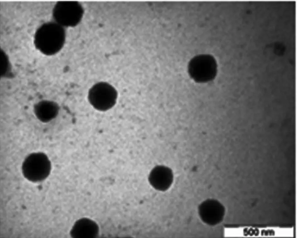
charged HA and TPP. As evidenced by TEM microphotographs displayed in Figure 1, nanoparticles 316
showed a spherical morphology. Table 1 depicts the physicochemical properties of the resultant 317
nanoparticles, which exhibited a positive zeta potential (+19 - +37 mV) and small sizes in the range 318
of 173-297 nm. The formulation CS/HA/TPP = 3.75/1/1 (w/w) was selected to conduct all 319
subsequent experiments as it displayed the highest production yield. 320
NMR is a well established technique for structural and dynamic characterization of molecules 321
and for the study of organic reactions and processes either in solution, semi-solid or solid states. 322
The NMR study of relatively high-molecular weight polymers, containing a number of carbon 323
atoms, usually benefits from the use of solid NMR techniques (Mi, Sung, & Shyu, 2000). In this 324
regard, the 1D-CPMAS 13Cspectra of these polymers may reveal detailed information relative to 325
their composition with semi-quantitative results (Metz, Ziliox, & Smith, 1996). ID solid-state NMR 326
experiments were employed here to verify the cross-linking reaction between CS and HA (Figure 2) 327
that contributes to nanoparticles formation. More specifically, the technique can be sensitive enough 328
to subtle changes in the electronic environments of the carbon atoms of CS and HA when they are 329
ionically cross-linked to generate the nanoparticles. To perform this study, four samples were 330
analyzed: nanoparticles (CS/HA/TPP = 3.75/1/1 (w/w)), pure CS and HA polymers and the physical 331
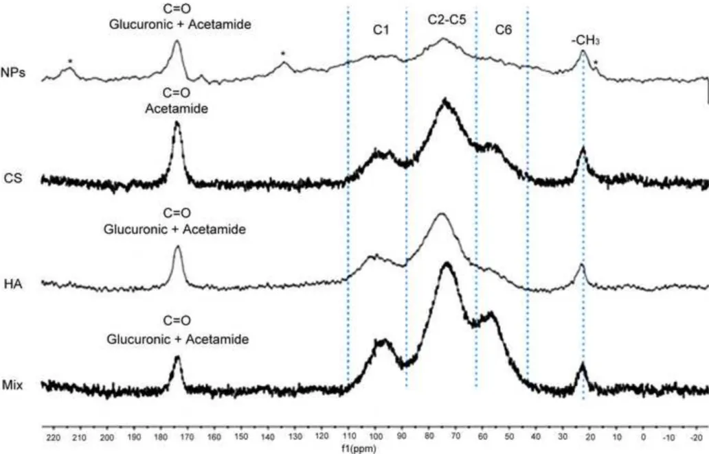
mixture of CS and HA (Mix). The corresponding 1D-CPMAS 13Cspectra are displayed in Figure 3, 332
each of which contains three broad signals that overlap in the band between 43-110 ppm, which is 333
the typical region of the sugar ring carbons from C1 to C6. The signals in this band can be assigned 334
as follows: i) region 95-110 ppm corresponds to the C1 anomeric carbons of the polymer, ii) region 335
70-95 ppm corresponds to the carbons C2 to C5 of the polymer and iii) region 43-70 ppm 336
corresponds to the C6 methylene carbons of the polymer. The four spectra also show the 337
characteristic peak at ca. 174 ppm corresponding to the carboxylate and/or carbonyl acetamide 338
carbons, as well as the carbon peak at ca. 24 ppm corresponding to the methyl group of the 339
14
acetamide group. According to these observations, all spectra elucidate the essential pattern of 340
signals related to polysaccharide structure, but are different in the relative intensities, as depicted in 341
Table 2. Interestingly, the integration of the signals, in Table 2, is consistent with the structures of 342
these polymers. 343
According to Figure 3, there is a remarkable difference between the spectrum of nanoparticles 344
and the other spectra, as signals in the first are considerably broader and extend over a larger region 345
than the corresponding signals in the other spectra. Moreover, some new signals appear in the 346
spectrum of nanoparticles, which are indicated with asterisk. The new carbon signals, resonating at 347
ca. 135 and 205 ppm, correspond to spinning sidebands from the CO carbonyl group at ca. 175 348
ppm. Their presence is indicative of enhanced chemical shift anisotropy of the CO group. There is 349
also an additional signal at ca. 19 ppm that is presumably due to a methyl group of acetamide. 350
These changes occurring to the acetamide group of CS could possibly result from re-organization 351
due to ionic interactions between the randomly cross-linked rings of CS and HA, with effects utterly 352
leading to a higher heterogeneity and broadening of signals in the NMR spectrum. Similar 353
observations were described by others for solid NMR spectra of gel based systems (Saiò, Tuzi, & 354
Naito, 1998). We can conclude, thereby, that the aforementioned changes detected in the 1D-355
CPMAS 13C spectra could confirm the hypothesis of the mechanism of nanoparticles formation, 356
which involves cross-linking via electrostatic and hydrophobic interactions between the CS and HA 357
in addition to that contributed by the TPP cross-linker through the gelation process. 358
359
3.2. Microspheres preparation and characterization
360
Nanoparticles were co-spray dried with mannitol in a one-step spray–drying process with yields 361
around 65-70 %. As stated in the introduction, the microencapsulation step envisages the 362
improvement of nanoparticles aerosolization pattern and lung deposition, which are mainly driven 363
by the aerodynamic parameters of inhaled particles (e.g. size, density) (Vanbever, Mintzes, Wang, 364
Nice, Chen, Batychy, Langer, & Edwards, 1999; Larhrib, Martin, Prime, & Marriott, 2003; Minne, 365
15
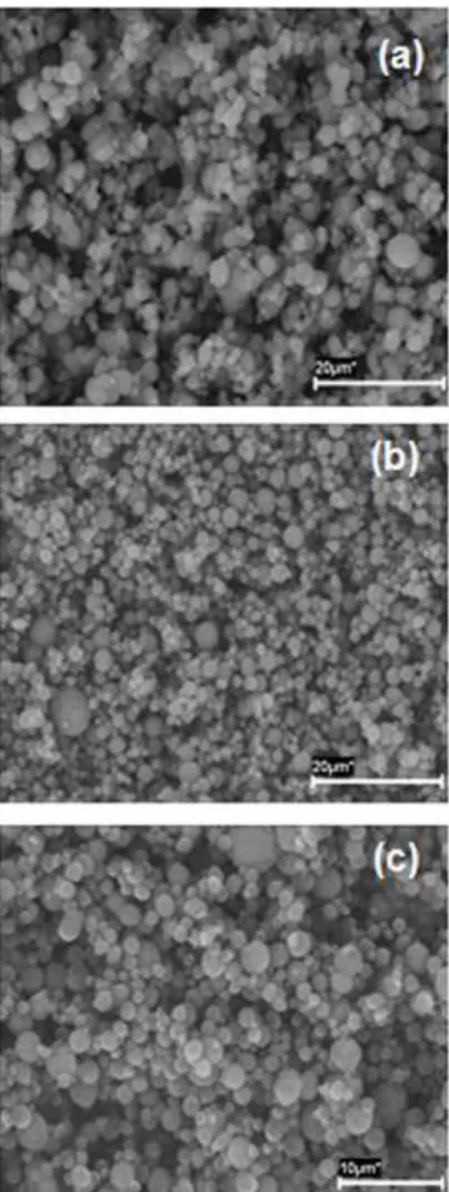
Boireau, Horta, & Vanbever, 2008). The resultant microencapsulated nanoparticles were viewed by 366
SEM (Figure 4), evidencing spherical morphology and demonstrating less tendency to aggregate as 367
the nanoparticles load increased with respect to mannitol. Microspheres exhibited a real density of 368
1.45 g/cm3 and an aerodynamic diameter of 2.6 µm (Table 3), which are suitable characteristics to 369
achieve deep lung deposition (Bosquillion, Lombry, Préat, & Vanbever, 2001; Mustante, Schroeter, 370
Rosati, Crowder, Hickey, & Martonen, 2002). 371
The application of sensitive techniques to characterize the structure of dry powders provides 372
important information that helps to elucidate the behavior of these drug delivery systems in 373
subsequent studies, both in vitro and in vivo. CLSM has been used to this end, since it allows us to 374
acquire high resolution optical sections of x-y scans along the z-axis, which are then reconstructed 375
into 3-D multicolored views, enabling a complete visualization of the dry powder external and 376
internal structure, as well as the spatial arrangement of the components (Lamprecht, Schäfer, & 377
Lehr, 2000). In our work, the acquisition of fluorescent images by CLSM enabled us to precisely 378
detect the nanoparticles location within the microspheres. This could not be attained by SEM, which 379
only provides information on the particles surface structure, rather than its internal structure. Figure 380
5(a-c) displays the images of microsphere encapsulating chitosan/hyaluronic acid nanoparticles. An 381
outer shell composed of mannitol (red channel) and an even distribution of chitosan/hyaluronic acid 382
nanoparticles throughout the microsphere matrix can be observed. The presence of a mannitol outer 383
shell is confirmed by Figure 5(d), which further evidences microspheres spherical shape as 384
previously observed by SEM. The homogeneous nanoparticles distribution within the mannitol 385
microspheres without detecting any punctuate green signals of aggregated particles in the 386
microspheres matrix may lead to the assumption that mannitol is almost entirely located at the 387
particles’ surface. This was also verified by the optical cross-sections of the confocal images (not 388
shown) which suggest that the microsphere matrix is almost occupied by the fluorescent 389
nanoparticles. It has been shown that sugar stabilizers, like mannitol, tend to preferentially adsorb at 390
the air/liquid interface during the drying process (Arakawa, & Timasheff, 19982; Wang, Chua, & 391
16
Wang, 2004). Therefore, we may hypothesize that non-specific interactions occurred between the 392
mannitol and the hydrophobic fluorophore (Bodiby), displacing the positively charged 393
nanoparticles inwards. It is noteworthy that these findings are similar to those reported in previous 394
studies for microparticles designed for inhalation therapy (Ely, & Finlay, 2007). 395
XPS and TOF-SIMS represent a complementary approach as non destructive and surface-396
sensitive analytic techniques. However, the particular interest of their application in the study of 397
drug delivery systems arises from the capability of these techniques to provide quantitative and 398
qualitative information of surface composition (De Vries, E, 1998), which provides valuable 399
information for the interpretation of kinetic and dynamic behavior, such as drug dissolution, 400
stability, distribution and release (Chesko, Kazzaz, Ugozzoli, Singh, O’hagan, Madden, Perkins, & 401
Patel, 2008; Dahlberg, Millqvist-Fureby, & Schuleit, 2008). Additionally, using these tools, the 402
encapsulation efficiency of microencapsulated drugs(Xie, Marijnissen, & Wang, 2006; Morales, 403
Ruiz, Oliva, Oliva, & Gallardo, 2007) or nanoparticles(Grenha, Seijo, Serra, & Remuñán- López, 404
2007).can also be assessed. This latter approach was our goal in the present study. Considering the 405
fact that microspheres have a lot of surface contact due to their powdery nature, the adsorption of 406
atmospheric natural contaminants such as nitrogen (N), is highly probable, therefore sample 407
surfaces were sputter cleaned using a soft argon ion beam (Ar+/1KV, /60 sec, 2X1 mm2). The 408
signals of the contaminating N in the powder samples almost disappeared (preliminary data not 409
shown), indicating that it was weakly bound (adsorbed) and, thus, easily removed. 410
As displayed in Table 4, however very weak N signals were detected in these samples (values 411
below 0.1 AT%), which could be explained on the basis that the ionic barrel is 45 degrees to the 412
surface, generating areas of shadow where the argon ions cannot reach. By contrast, the N signal in 413
the chitosan/hyaluronic acid nanoparticles spectrum persisted after the sputter cycle with a 414
relatively high value, suggesting that it is chemically bonded and which could be ascribed to CS. 415
Moreover and as expected, both Na and P, ascribed to TPP in the nanoparticles, were detected 416
solely on the surface of chitosan/hyaluronic acid nanoparticles; whereas were absent on the surface 417
17
of either mannitol microspheres or microencapsulated nanoparticles. Taking into account the 418
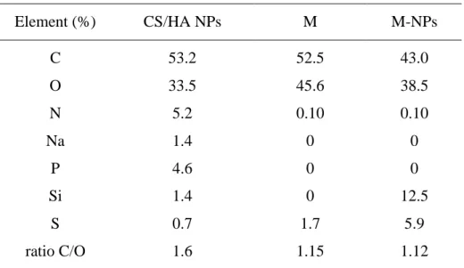
detection limit of XPS (all elements except H: ~ 0.01 monolayer, or ~ 0.1% bulk), this finding 419
indicates the absence of TPP on the powder surfaces. Our assumption of efficient nanoparticles 420
microencapsulation can be further reinforced by the C/O ratio, which is similar for mannitol 421
microspheres and the microencapsulated nanoparticles (1.15 and 1.12, respectively). Interestingly, 422
this ratio is different from that of chitosan/hyaluronic acid nanoparticles (1.6), suggesting that the 423
surfaces of both mannitol microspheres and the microencapsulated nanoparticles are similar in 424
terms of the atomic composition and concentrations of C and O. Additionally, the higher C/O ratio 425
for the chitosan/hyaluronic acid nanoparticles implies lower surface concentration of O, which is 426
due to the contribution of other elements detected on the surface. It is worth while to notice here 427
that a signal for silicon was identified in some spectra which could be originated from the silicon 428
wafer used as a sample support during the analysis (Grenha, Seijo, Serra, & Remuñán- López, 429
2007). 430
This result was also confirmed by deconvolution analysis of the spectra where the high 431
resolution spectra of carbon (C1s) signals, showing an envelope, were curve fitted using the 432
Gaussian distribution into a series of peaks corresponding to different functional groups. We have 433
assigned as reference the peak at the lowest binding energy (285.0 eV) to carbon atoms linked to 434
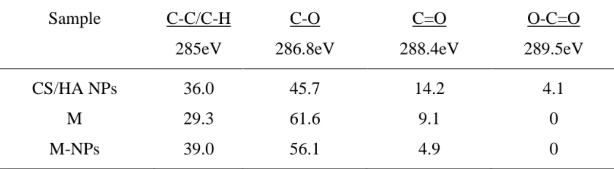
carbon and hydrogen atoms. Table 5 summarizes the relative peak area of each carbon environment. 435
As can be seen, the peak areas (%) and relative intensities of (C-C, 285 eV), (C-O, 286.8 eV) and 436
(C=O, 288.37 eV) are nearly similar in mannitol microspheres and microencapsulated 437
nanoparticles. More importantly, the peak of (O-C=O, 289.54 eV), unique for CS, was detected in 438
the spectrum of chitosan/hyaluronic acid nanoparticles but not in the other two samples. The 439
analysis of this result was also confirmed by deconvolution analysis of the spectra where the high 440
resolution spectra of carbon (C1s) signals, showing an envelope, were curve fitted using the 441
Gaussian distribution into a series of peaks corresponding to different functional groups. We have 442
18
assigned as reference the peak at the lowest binding energy (285.0 eV) to carbon atoms linked to 443
carbon and hydrogen atoms. 444
Table 5 summarizes the relative peak area of each carbon environment. As can be seen, the peak 445
areas (%) and relative intensities of (C-C, 285 eV), (C-O, 286.8 eV) and (C=O, 288.37 eV) are 446
nearly similar in mannitol microspheres and microencapsulated nanoparticles. More importantly, 447
the peak of (O-C=O, 289.54 eV), unique for CS, was detected in the spectrum of 448
chitosan/hyaluronic acid nanoparticles but not in the other two samples. The analysis of 449
deconvoluted C1s high resolution spectra re-affirms that chitosan/hyaluronic acid nanoparticles are 450
entirely encapsulated in mannitol microspheres. 451
TOF-SIMS analysis was conducted under non-destructive energetic conditions and under the 452
static limit (10 12 ions/cm3). In the mass spectra, positive ions were detected. According to the 453
spectra displayed in Figure 6, the nanoparticles spetrum differs from those of both microsphere 454
samples in the existence of some molecular fragments [m/z = 232(C16H24O), 205(C15H9O), 455
200(C10H16O4)]in addition to the main and most representative ions of mannitol observed at m/z = 456
183 (the molecular ion + H) and the two times molecular ion + H observed at m/z = 365. At the 457
same time, many identified molecular fragments of nanoparticles, not observed in the spectra of 458
microspheres and located at m/z = 189(C7H13O4Si), 202(C12H10O3), 215(C11H12O3Na), 459
239(C15H11O3) and 226(C18H10), could result basically from the fragmentation of both CS and HA. 460
Furthermore, other intensive signals for fragments containing N and O are clearly observed in the 461
nanoparticles but not in mannitol microspheres and microencapsulated nanoparticles, located at m/z 462
= 60(C2H6NO), 59(C2H5NO), 58(C2H4NO) and which could arise from the fragmentation of CS 463
(Figure 7-1). The N-containing fragment (C2H4NO, m/z = 58), identified as intensive in the 464
nanoparticles sample, was detected however in both microsphere samples but in one order of 465
magnitude lower, which is likely due to the atmospheric exposure as mentioned previously (Figure 466
7-2). Importantly, the PO3 fragment (m/z =79), attributed to TPP, is intensive and clearly observed 467
in the nanoparticles sample whereas it is not detected in that of mannitol microspheres and its 468
19
intensity in microencapsulated nanoparticles is at least one order of magnitude lower than that of 469
nanoparticles sample (Figure 8). 470
According to our observations from these spectra, the samples of mannitol microspheres and the 471
microencapsulated nanoparticles, if not identical, are very similar (intensity of the identified 472
fragments and also the distribution of the intensity between ions). These outcomes demonstrate that 473
chitosan/hyaluronic acid are efficiently encapsulated within the mannitol carrier, especially if we 474
refer to the fact that the TOF-SIMS technique is qualified with the highest surface sensitivity for 475
surface analysis (detection limit range of ppm-ppb, orders of magnitude better than XPS) and the 476
resolution depth of 1-3 monolayers. 477
478
4. Conclusion
479Chitosan/hyaluronic acid nanoparticles were prepared, characterized and microencapsulated in 480
mannitol microspheres, resulting in a dry powder that shows adequate aerodynamic properties for 481
deep pulmonary deposition. Following the encapsulation process, structural analysis of the dry 482
powder was provided by CLSM, which elucidated that the nanoparticles were homogeneously 483
distributed within the mannitol microsphere. The evidence that nanoparticles were completely 484
encapsulated within the carrier by means of the spray drying process, was achieved by application 485
of the sensitive surface analysis techniques, XPS and TOF-SIMS. These outcomes confirm the 486
success of nanoparticles microencapsulation by spray drying. We expect, thereby, that the 487
microencapsulated nanoparticles hold promise for pulmonary delivery of macromolecules such as 488
proteins and nucleic acids, as these nanoparticles have demonstrated great potential in gene 489
transfection in ocular cell lines (De La Fuente, Seijo, & Alonso, 2008b). Therefore, further work is 490
required to investigate the delivery potential of these developed carriers, in the form of dry powders 491
in pulmonary cell lines. 492
493
Acknowledgments
49420
The authors acknowledge funding from the Spanish Government (ISCIII, Acción Estratégica de 495
Salud, PS09/00816), XUNTA de Galicia (PGIDIT, 09CSA022203PR, NANOPULMOGENIC) and 496
IBB/CBME, LA, FEDER/POCI2010. MAEC-AECID fellowship of the Spanish Agency of 497
International Cooperation to S. Al-Qadi is gratefully recognized. Assistance from M. Pastor (NMR 498
service, University Santiago de Compostela) and C. Serra (Centre for Scientific and Technological 499
Support to Research, University of Vigo, E-36310,Vigo,Spain ) are highly appreciated. 500
501
References
502Agnihotri, S. A., Mallikarjuna, N. N., & Aminabhavi, T. M. (2004). Recent advances on chitosan-503
based micro- and nanoparticles in drug delivery. Journal of Controlled Release, 100, 5-28. 504
Agu, R. U., Ugwoke, M. I., Armand, M., Kinget, R., & Verbeke, N. (2001). The lung as a route for 505
systemic delivery of therapeutic proteins and peptides. Respiratory Research, 2, 198-209. 506
Akima, K., Ito, H., Iwata, Y., Matsuo, K., Watari, N., Yanagi, M., Hagi, H., Oshima, K., Yagita, A., 507
Atomi, Y., & Tatekawa, I. (1996). Evaluation of antitumor activities of hyaluronate binding 508
antitumor drugs: synthesis, characterization and antitumor activity. Journal of Drug 509
Targeting, 4, 1-8.
510
Arakawa, T., & Timasheff, S. N. (1982). Stabilization of protein structure by sugars. Biochemistry, 511
21, 6536-6544.
512
Avitabile, T., Marano, F., Castiglione, F., Bucolo, C., Cro, M., Ambrosio, L., Ferrauto, C., & 513
Reibaldi, A. (2001). Biocompatibility and biodegradation of intravitreal hyaluronan implants 514
in rabbits. Biomaterials, 22, 195-200. 515
Azarmi, S., Tao, X., Chen, H., Wang, Z., Finlay, W. H., Löbenberg, R., & Roa, W. (2006). 516
Formulation and cytotoxicity of doxorubicin nanoparticles carried by dry powder aerosol 517
particles. International Journal of Pharmaceutics, 319, 155–161. 518
Bailey, M. M., & Berkland, C. J. (2009). Nanoparticle formulations in pulmonary drug delivery. 519
Medicinal Research Reviews. 29, 196-212.
21
Bastow, E. R., Byers, S., Goluba, S. B., Clarkinc, C. E., Pitsillides, A. A., & Fosang, A. J. (2008). 521
Hyaluronan synthesis and degradation in cartilage and bone. Cellular and Molecular Life 522
Sciences, 65, 395–413.
523
Bernkop-Schnürch, A., Kast, C. E., & Guggi, D. (2003). Permeation enhancing polymers in oral 524
delivery of hydrophilic macromolecules: thiomer/GSH systems. Journal of Controlled 525
Release, 93, 95-103.
526
Bosquillion, C., Lombry, C., Préat, V., & Vanbever, R. (2001). Influence of formulation excipients 527
and physical characteristics of inhalation dry powders on their aerosolization performance. 528
Journal Controlled Release, 70, 329-339.
529
Bosquillon, C., Rouxhet, P. G., Ahimou, F., Simon, D., Culot, C., Préat, V., & Vanbever R. (2004). 530
Aerosolization properties, surface composition and physical state of spray-dried protein 531
powders. Journal of Controlled Release, 99, 357–367. 532
Brown, M. B., & Jones, S. A. (2005). Hyaluronic acid: a unique topical vehicle for the localized 533
delivery of drugs to the skin. Journal of the European Academy of Dermatology and 534
Venereology, 19, 308–318.
535
Bunkera, M. J., Daviesa, M. C., Chena, X., James, M. B., & Roberts, J.C. (2006). Single particle 536
friction on blister packaging materials used in dry powder inhalers. European Journal of 537
Pharmaceutical Sciences, 29, 405–413.
538
Calvo, P., Remuñán-López, C., Vila-Jato, J. L., & Alonso, M. J. (1997a) Chitosan and Chitosan/ 539
Ethylene Oxide-Propylene Oxide Block Copolymer Nanoparticles as Novel Carriers for 540
Proteins and Vaccines. Pharmaceutical. Research, 14, 1431-1436. 541
Calvo, P., Remuñán-López, C., Vila-Jato, J. L., & Alonso, M. J. (1997b) Novel hydrophilic 542
chitosan–polyethylene oxide nanoparticles as protein carriers. Journal of. Applied Polymer 543
Science, 63, 125–132.
544
Cantor, J. O., & Turin, G. M. (2004). Can exogenously administered hyaluronan improve 545
respiratory function in patients with pulmonary emphysema?. Chest, 125, 288–292. 546
22
Chesko, J., Kazzaz, J., Ugozzoli, M., Singh, M., O’hagan, D., Madden, C., Perkins, M., & Patel N. 547
(2008). Characterization of antigens adsorbed to anionic PLG microparticles by XPS and 548
TOF-SIMS. Journal of Pharmaceutical Sciences, 97, 1443-1453. 549
Chono, S., Li, S-D., Conwell, C. C., & Huang, L. (2008). An efficient and low immunostimulatory 550
nanoparticle formulation for systemic siRNA delivery to the tumor. Journal of Controlled 551
Release, 131, 64–69.
552
Chougule, M. B., Padhi, B. K., Jinturkar, K. A., & Misra, A. (2007). Development of dry powder 553
inhalers. Recent Patents on Drug Delivery and Formulation, 1, 11-21. 554
Chrystyn, H. (1997). Is total particle dose more important than particle distribution?. Respiratory 555
Medicine, suppl A, 17-9.
556
Coradini, D., Pellizzaro, C., Abolafio, G., Bosco, M., Scarlata, I., Cantoni, S., Stucchi, L., Zoezet, 557
S., Turrin, C., Sava, G., Perbellini, A., & Diadone, M. G. (2004). Hyaluronic-acid butyric 558
esters as promising antineoplastic agentsin human lung carcinoma: A preclinical study, 559
Investigational New Drugs, 22, 207–217.
560
Courrier H. M., Butz N., & Vandamme T. F. (2002). Pulmonary drug delivery systems: recent 561
developments and prospects. Critical Reviews in Therapeutic Drug Carrier Systems, 19, 425-562
498. 563
Dahlberg, C., Millqvist-Fureby, A., & Schuleit M. (2008). Surface composition and contact angle 564
relationships for differently prepared solid dispersions. European Journal of Pharmaceutics 565
and Biopharmaceutics, 70, 478–485.
566
Dailey, L. A., Kleemann, E., Wittmar, M., Gessler, T., Schmehl, T., Roberts, C., Seeger, W., & 567
Kissel. T. (2003). Surfactant-free, biodegradable nanoparticles for aerosol therapy based on 568
the branched polyesters, DEAPA-PVAL-g-PLGA. Pharmaceutical Research, 20, 2011–2020. 569
De Campos, A. M., Diebold, Y., Carbalho, E. L. S., Sánchez, A., & Alonso, M. J. (2004). Chitosan 570
nanoparticles as new ocular drug delivery systems: in vitro stability, in vivo fate and cellular 571
toxicity. Pharmaceutical Research, 21, 803-810. 572
23
De La Fuente, M., Seijo, B., & Alonso, M. J. (2008a). Novel hyaluronan-based nanocarriers for 573
transmucosal delivery of macromolecules. Macromolecular Bioscience, 8, 441–450. 574
De La Fuente, M., Seijo, B., & Alonso, M. J. (2008b). Novel hyaluronic acid-chitosan nanoparticles 575
for ocular gene therapy. Investigative Ophthalmology and Visual Science, 49, 2016-2024. 576
De La Fuente, M., Raviña, M., Paolicelli, P., Sanchez, A., Seijo, B., & Alonso, M. J. (2010). 577
Chitosan-based nanocarriers: A delivery platfom for ocular therapeutics. Advanced Drug 578
Delivery Reviews, 62, 100-117.
579
De Vries, E. (1998). Surface Characterization Methods-XPS, TOF-SIMS, and SAM A 580
Complimentary Ensemble of Tools. Journal of Materials Engineering and Performance 581
Contents, 7, 303-311.
582
Ely, L., Roa, W., & Finlay, W. H., R. (2007). Löbenberg, Effervescent dry powder for respiratory 583
drug delivery. European Journal of Pharmaceutics and Biopharmaceutics, 65, 346–353 584
Florea, B. I., Thanou, M., Junginger, H. E., & Borchard, G. (2005). Enhancement of bronchial 585
octreotide absorption by chitosan and N-trimethylchitosan shows linear in vitro/in vivo 586
correlation. Journal Control. Release, 110, 353–361. 587
Freitas, C., Müller, & R. H. (1998). Spray-drying of solid lipid nanoparticles (SLNTM). European 588
Journal of Pharmaceutics and Biopharmaceutics, 46, 145–151.
589
Gómez-Gaete, G., Tsapis, N., Silva, L., Bourgaux, C., Besnard, M., Bochot, A., & Fattal, E. (2008). 590
Supramolecular organization and release properties of phospholipid hyaluronan 591
microparticles encapsulating dexamethasone. European Journal of Pharmaceutics and 592
Biopharmaceutics, 70, 116–126.
593
Grenha, A., Seijo, B., & Remuñán-López, C. (2005). Microencapsulated chitosan nanoparticles for 594
lung protein delivery. European Journal of Pharmaceutical Sciences. 25, 427-437. 595
Grenha, A., Grainger, C. I., Dailey, L. A., Seijo, B., Martin, G. P., Remuñán-López, C., & Forbes, 596
B. (2007). Chitosan nanoparticles are compatible with respiratory epithelial cells in vitro. 597
European Journal of Pharmaceutical Sciences, 31, 73–84.
24
Grenha, A., Seijo, B., Serra, C., & Remuñán-López, C. (2007). Chitosan nanoparticle-loaded 599
mannitol microspheres: structure and surface characterization. Biomacromolecules, 8, 2072- 600
2079. 601
Grenha, A., Carrion-Recio, D., Teijeiro-Osorio, D., Seijo, B., Remuñán-López, C. (2008). Nano- 602
and microparticulate carriers for pulmonary drug delivery. In M. N. V. Kumar (Ed.); 603
Handbook of Particulate Drug Delivery (Applications) (pp.165-192). Valencia, CA: American 604
Scientific Publishers. 605
Hastings, R. H., Folkesson, H. G., & Matthay, M. A. (2004). Mechanism of alveolar protein 606
clearance in the intact lung, American Journal of Physiology- Lung Cellular and molecular 607
Physiology, 286,679-89.
608
Hirano, S., Seino, H., Akiyama, Y., & Nonaka, I. ( 1988). Biocompatibility of chitosan by oral and 609
intravenous administrations. Polymer Materials and Science Engineering. 59, 897-901. 610
Hwang, S. M, Kim, D., Chung, S. J., & Shim, C. K. (2008). Delivery of ofloxacin to the lung and 611
alveolar macrophages via hyaluronan microspheres for the treatment of tuberculosis. Journal 612
of Controlled Release, 129, 100–106.
613
Issa, M., Koping-Hoggard, M., & Artursson, P. (2005). Chitosan and the mucosal delivery of 614
biotechnology drugs. Drug Discovery Today: Technologies, 2, 1-6. 615
Jiang, D., Liang, J., & Noble, P. W. (2007). Hyaluronan in tissue injury and repair. Annual Review 616
of Cell and Developmental Biology, 23, 435–461.
617
Jiang, G., Park, K., Kim, J., Kim, K. S., Oh, E. J., Kang, H., Han, S. E., Oh, Y. K., Park, T. G., & 618
Hahn, S K. (2008). Hyaluronic acid–polyethyleneimine conjugate for target specific 619
hyaluronic intracellular delivery of siRNA. Biopolymers, 89, 635-642. 620
Lamprecht, A., Schäfer, U. F., & Lehr, C-M. (2000). Structural analysis of microparticles by 621
confocal laser scanning microscopy. AAPS Pharm Sci Tech, 1, 10-19. 622
Larhrib, H., Martin, G.P., Prime, D., & Marriott, C. (2003). Characterisation and deposition studies 623
of engineered lactose crystals with potential for use as a carrier for aerosolised salbutamol 624
25
sulfate from dry powder inhalers. European Journal of Pharmaceutical Sciences, 19,
211-625
221. 626
Lehr, C. M., Bouwstra, J. A., Schacht, E. H., & Junginger, H. E. (1992). In vitro evaluation of 627
mucoadhesive properties of chitosan and some other natural polymers. International Journal 628
of Pharmaceutics, 78, 43-48.
629
Lim, D. S. T., Forbes, B., Martin, G. P., & Brown, M. B. (2001). In vivo and in vitro 630
characterization of novel microparticulates based on hyaluronan and chitosan hydroglutamate. 631
AAPS PharmsciTech, 2, 1–14.
632
Lim, S. T., Forbes, B., Martin, G. P., & Brown, M. B. (2002). In vivo evaluation of novel 633
hyaluronan/chitosan microparticulate delivery systems for the nasal delivery of gentamicin in 634
rabbits. International Journal of Pharmaceutics, 231, 73-82. 635
Mayol, L., Quaglia, F., Borzacchiello, A., Ambrioso, L., La, & Rotonda, M. (2008). A novel 636
poloxamers/hyaluronic acid in situ forming hydrogel for drug delivery: Rheological, 637
mucoadhesive and in vitro release properties. European Journal of Pharmaceutics and 638
Biopharmaceutics, 70, 199–206.
639
Metz, G., Ziliox, M., & Smith, S.O. (1996). Towards quantitative CP-MAS NMR. Solid State 640
Nuclear Magnetic Resonance, 7,155- 160.
641
Mi, F-L., Sung, H-W., & Shyu, S-S. (2000). Synthesis and characterization of a novel chitosan-642
based network prepared using naturally occurring cross linker. Journal of Polymer Science: 643
Part A: Polymer Chemistry, 38, 2804–2814.
644
Minne, A., Boireau, H., Horta, M. J., & Vanbever, R. (2008). Optimization of the aerosolization 645
properties of an inhalation dry powder based on selection of excipients. European Journal of 646
Pharmaceutics and Biopharmaceutics, 70, 839-844
647
Morales, M. E., Ruiz, M. A., Oliva, I., Oliva, M., & Gallardo, V. (2007). Chemical characterization 648
with XPS of the surface of polymer microparticles loaded with morphine. International 649
Journal of Pharmaceutics, 333, 162–166.
26
Morimoto, M., Metsugi, K., Katsumata, H., Iwanaga, K., & Kakemi, M. (2001). Effects of low-651
viscosity sodium hyaluronate preparation on the pulmonary absorption of rh-insulin in rats. 652
Drug Development and Industrial Pharmacy, 27, 365-371.
653
Mustante, C. J., Schroeter, J. D., Rosati, J. A., Crowder, T. M., Hickey, A. J., & Martonen, T. B. 654
(2002). Factors Affecting the Deposition of Inhaled Porous Drug Particles. Journal of 655
Pharmaceutical Sciences. 91, 1590-1600.
656
Muzzarelli, R. A. (1997). Human enzymatic activities related to the therapeutic administration of 657
chitin derivatives. Cellular and Molecular Life Sciences, 53, 131-140. 658
Oyarzun-Ampuero, F. A., Brea, J., Loza, M. I., Torres, D., & Alonso M. J. (2009). Chitosan– 659
hyaluronic acid nanoparticles loaded with heparin for the treatment of asthma. International 660
Journal of Pharmaceutics, 381, 122–129.
661
Pandey R., & Khuller G. K. (2005). Antitubercular inhaled therapy: opportunities, progress and 662
challenges. Journal of Antimicrobial Chemotherapy, 55, 430-435. 663
Peer, D., & Margalit, R. (2004). Loading mitomycin c inside long circulating hyaluronan targeted 664
nano-liposomes increases antitumor its activity in three mice tumor models. International 665
Journal of Cancer, 108, 780–789.
666
Rouse, J. J; Whateley, T. L; Thomas, M; & Eccleston, G. M. (2007). Controlled drug delivery to the 667
lung: Influence of hyaluronic acid solution conformation on its adsorption to hydrophobic 668
drug particles. International Journal of Pharmaceutics, 330, 175–182. 669
Sahiner, N., & Jia, X. (2008). One-step synthesis of hyaluronic acid-based (sub) micron hydrogel 670
particles: process optimization and preliminary characterization. Turkish Journal of 671
Chemistry, 32, 397–409.
672
Saiò, H., Tuzi, S., Naito, A. (1998). Polysaccharides and biological systems. In I. Ando, & T. 673
Asakura (Eds.), Solid State NMR of Polymers: Studies in physical and theoretical chemistry 674
(pp 509-588). Elsevier Science 675
27
Schürch, S.; Gehr, P.; Im Hof, V.; Geiser, M.; & Green, F. (1990). Surfactant displaces particles 676
toward the epithelium in airways and alveoli. Respiratory Physiology, 80, 17-32. 677
Sham, J. O-H., Zhang, Y., Finlay, W. H., Roa, W. H., & Löbenberg, R. (2004). Formulation and 678
characterization of spray-dried powders containing nanoparticles for aerosol delivery to the 679
lung. International Journal of Pharmaceutics, 269, 457–467. 680
Sivadasa, N., O’rourke, D., Tobin, A., Buckley, V., Ramtoola, Z., Kellya, J. G., Hickey, A. J., & 681
Cryan, S-A. (2008). A comparative study of a range of polymeric microspheres as potential 682
carriers for the inhalation of proteins. International Journal of Pharmaceutics, 358, 159–167. 683
Stern, R., Kogan, G., Jedrzejas, M. J, & Šoltès L. ( 2007). The many ways to cleave hyaluronan. 684
Biotechnology Advances, 25, 537–557.
685
Sung, J., Pulliam, B., & Edwards, D. (2007). Nanoparticles for drug delivery to the lungs. Trend in 686
Biotehcnolog, 25, 563-570.
687
Surendrakumarm, K., G. P.Martyn, Hodgers, E.C. M., Jansen M., & Blair J. A. (2003). Sustained 688
release of insulin from sodium hyaluronate based dry powder formulations after pulmonary 689
delivery to beagle dogs. Journal of Controlled Release, 91, 385–394. 690
Taetz, S., Bochot, A., Surace, C., Arpicco, S., Renoir, J. m., Schaefer, U. f., Marsaud, V., Kerdine-691
Roemer, S., Lehr, C. M., Fattal E. (2009). Hyaluronic acid-modified DOTAP/DOPE 692
liposomes for the targeted delivery of anti-telomerase siRNA to CD44-expressing lung cancer 693
cells. Oligonucleotides. 19, 103-116. 694
Theocharis, D. A., Skandalis, S. S., Noulas, A. V., Papageorgakopoulou, N., Theocharis, A. D., & 695
Karamanos, N. K. (2008). Hyaluronan and chondroitin sulfate proteoglycans in the 696
supramolecular organization of the mammalian vitreous body. Connective Tissue Research, 697
49, 124-128.
698
Vanbever, R., Mintzes, J. D., Wang, J., Nice, J., Chen, D., Batychy, R., Langer, R., & Edwards, D. 699
A. (1999). Formulation and physical characterization of large porous particles for inhalation. 700
Pharmaceutical Research, 16, 1735-1742.
28
Varshosaz, J. (2007). The promise of chitosan microspheres in drug delivery systems. Drug 702
delivery, 4, 263-273.
703
Volpi, N., Schiller, J., Stern, R., & Soltès, L. (2009). Role, metabolism, chemical modifications and 704
applications of hyaluronan. Current Medicinal Chemistry, 16, 1718-1745. 705
Wang, J., Chua, K. M., & Wang, C. H. (2004). Stabilization and encapsulation of human 706
immunoglobulin G into biodegradable microspheres. Journal of Colloid and Interface 707
Science, 271, 92-101.
708
Woo, J. S., Piao, M. G., Li, D. X., Ryu, S., Choi, J. Y., Kim, J-A., Kim, J. H., Jin, S. G., Kim, D-709
D., Lyoo, W., & Yong, C. S. (2007). Development of cyclosporin A-loaded hyaluronic 710
microsphere with enhanced oral bioavailability. International Journal of Pharmaceutics, 345, 711
134–141. 712
Xie, J.; Marijnissen, J. C.; & Wang, C. H. (2006). Microparticles developed by electro 713
hydrodynamic atomization for the local delivery of anticancer drug to treat C6 glioma in vitro. 714
Biomaterials, 27, 3321-3332.
715
Xin, D., Wang, Y., & Xiang, J. (2010). The use of amino acid linkers in the conjugation of 716
paclitaxel with hyaluronic acid as drug delivery system: synthesis, self-assembled property, 717
drug release, and In Vitro Efficiency. Pharmaceutical Research, 27, 380-389. 718
Yang, W., Peters, J. I., & Williams III, R. O. (2008). Inhaled nanoparticles - A current review. 719
(2008). International Journal of Pharmaceutics, 356, 239–247. 720
Zhuo, A., Guo, L., & Tang, L. (2003). Effect of an intrathoracic injection of sodium hyaluronic acid 721
on the prevention of pleural thickening in excess fluid of tuberculous thoracic cavity. Clinical 722
and Experimental Pharmacology and Physiology, 30, 203–205.
1
Table 1. Process yields and physiochemical properties of chitosan/hyaluronic acid nanoparticles (CS/HA NPs), prepared with different concentrations of hyaluronic acid (HA), tripolyphosphate (TPP) and chitosan (CS) (mean ± S.D., n = 3).
CS/HA/TPP (w/w/w) Yield (%) Size (nm) Z potential (mV) 3.75/1/1 4.3/1.1/1 4.3/1.4/1 5.0/1.3/1 5.0/1.7/1 5.0/2.5/1 6.3/3.1/1 7.5/3.8/1 9.4/4.7/1 15.0/10/1 56 ± 8 44 ± 1 45 ± 2 41± 4 48 ± 2 56 ± 1 40 ± 4 41 ± 3 33 ± 1 23 ± 9 233 ± 3 239 ± 4 275 ± 20 297 ± 23 212 ± 4 254 ± 6 241 ± 5 219 ± 7 197 ± 4 173 ± 1 + 37 ± 2 + 35 ± 1 + 34 ± 1 + 34 ± 1 + 25 ± 1 + 23 ± 1 + 20 ± 1 + 26 ± 1 + 25 ± 1 + 19 ± 1 Revised tables
2
Table 2. Chemical shifts (ppm) and signal integrations obtained from the 13C 1D- CPMAS spectra of the samples studied: pure chitosan (CS), pure hyaluronic acid (HA), physical mixture of both polymers (Mix), and chitosan/hyaluronic acid nanoparticles (NPs) (CS/HA/TPP =3.75/1/1). The relative area of the 13C NMR signal is indicated between parentheses.
Chemical Shift (ppm) Simple C1 to C6 (sugar)a CO (acetamide)b CO (glucuronic)b CH3 (acetamide)c CO* d CS 43-110 (27.3) 174.2 (1.0) --- 22.8 (0.8) --- HA 43-110 (7.26) 173.6 (1.0) 22.6 (0.39) --- Mix 43-110 (7.75) 173.9 (1.0) 22.6 (0.67) --- NPs 43-110 (7.75) 174.2 (1.0) 22.6 (0.98) 214.0 (0.31) a: Integral from 43 to 110 ppm, b: Integral from 163 to 185 ppm, c: Integral from 12 to 34 ppm, d: Integral from 205 to 222
3
Table 3. Aerodynamic properties of dry powders prepared with different mannitol/ nanoparticles weight ratios and solids contents (CS/HA/TPP = 3.75/1/1, mean ± S.D., n = 3).
Mannitol/ nanoparticles ratio Solids contenta (%) Feret’s diameterb (µm) Real density (g/cm3) Aerodynamic diameter (µm) 70/30 3.0 2.3 ± 0.7 1.45 ±0.06 2.57 ± 0.08 80/20 2.8 2.7 ± 1.3 1.45 ±0.12 ND 90/10 3.0 2.2 ± 0.4 1.45 ±0.17 ND
a: Solids content represents the total solids concentration (%) of the spraying suspensions, b: Feret’s diameters are determined by optical microscopy
4
Table 4. Surface elemental composition (atomic %), determined by XPS, of chitosan/hyaluronic acid nanoparticles (CS/HA NPs), mannitol microspheres (M) and microencapsulated nanoparticles (M-NPs) (CS/HA/TPP = 3.75/1/1, mannitol/ nanoparticles = 70/30).
Element (%) CS/HA NPs M M-NPs C 53.2 52.5 43.0 O 33.5 45.6 38.5 N 5.2 0.10 0.10 Na 1.4 0 0 P 4.6 0 0 Si S 1.4 0.7 0 1.7 12.5 5.9 ratio C/O 1.6 1.15 1.12