Relatório Final de Estágio
Mestrado Integrado em Medicina Veterinária
Abdominal and Thoracic Focused Assessment using Sonography for
Trauma in Small Animal Practice
Ana Filipa Lopes Pereira Pinto
Orientadora
Professora Doutora Ana Colette Pereira de Castro Osório Maurício
Co-Orientadora
Anne MacGregor
A
BSTRACTThe sixteen weeks of the professional training period were performed in the United Kingdom in Ashlands Veterinary Centre & Animal Hospital in Skipton and Ilkley branches, a small animal medicine and surgery practice.
Before the professional training period, three weeks of training in abdominal ultrasound with Dr. José Graça and another three weeks in heart ultrasound with Dr. Claúdia Abreu were performed. During that time, several ultrasound examinations were observed and different cases seen. As a result, a special interest in sonography was developed.
Emergency cases are, without a doubt, an important part of the role of the veterinary surgeon, both in the United Kingdom and in Portugal. In the majority of emergency cases observed during the training period, after performing the physical examination, the veterinary surgeon would resort to an ultrasound scan to determine the best treatment approach.
Emergency ultrasound is a field that has been explored recently in veterinary medicine, and protocols have been developed, based on previous ones created in human medicine. Two of those protocols are TFAST and AFAST (thoracic and abdominal focused assessment using sonography for trauma). These two techniques were developed so that any veterinary surgeon with minimal skills in ultrasound could perform a reliable examination during emergency cases. During the present report, TFAST and AFAST protocols will be explained and their advantages and limitations discussed. Two clinical cases are included and described where the FAST techniques were explored.
A
CKNOWLEDGMENTIn first place, I want to thank to my parents for their quiet and unconditional love and support. I'm very proud to be your daughter.
Secondly, I want to thank to my second family, “Tia” Ana and “Tio” João. You are also responsible for the person I am today. Thank you for helping raise me.
A special thanks to Professor Colette, who said yes and agreed to be my mentor, even though I was making her life a little harder. Thank you so much for saying yes, I couldn't have chosen a better person.
I’m also grateful to Dr. Zé Graça with whom I learn a lot about... hum... Surf! And ultrasound, of course. In your greatness, you inspired me to be better and to want to learn more about ultrasound.
Dr. Claúdia Abreu, I honestly don't know how to thank you! Your echocardiography's skills are beyond impressive, but your heart it's even greater. I enjoyed every moment that I spent in your car. I've learn so much with you. I will never forget you and your precious gift. You will be an inspiration throughout my life.
I also want to express a special thanks to Ashland’s team, specially to the Skipton brunch. Every practical knowledge of being a vet, I learnt from you. And you made my learning experience in the UK amazing. Ruth, thank you for your patience, kindness and mother-like care. You gave me the confidence that I lacked in the theatre room. And Sam, you're a very impressive woman! If I could, I'll pick your brain every other day. But your heart is also impressive, you inspire with your devotion for each and every animal.
I’m grateful to Roger Wilkinson for letting me use his images on this paper and also for taking me with him to see some weird cases on days when most people were resting.
Still in the UK, I want to thank Tom for making my life in the UK more entertaining and helping me improve my English. A very special thank you to my British mum, Diane, for adopting me, for been always supportive when I most needed and for every practical advice she gave me.
To my friends and colleagues from the restaurant, I grown so much with every one of you. To the rest of my family, to my grandparents, to my aunties, to my uncles, to my cousins (Claúdia, Isabel, Raquel, Andreia, Filipe, Beatriz,…), and to the newest member of my family Margarida. I always took you for granted, but distance made the connection tighter. And you spoiled me with love.
Very special thanks, and a congratulation as well, to the best class ever: Ines Rei (since the very first semester), Cátia (since the very first Queima), Rita M., Rita L., Alice, Ines G., Joana, Cristiana, Mariana and Xana. We finally did it! And we did it together.
C
ONTENTS Abstract ... ii Acknowledgment ... iii List of Illustrations ... v Abbreviations ... vi Introduction ... 1How to perform an AFAST exam ... 3
Ultrasound settings and probe preferences ... 3
Patient position and preparation ... 3
Diaphragmatico-hepatic view ... 4
Spleno-renal view ... 6
Cysto-colic view ... 7
Hepato-renal view ... 8
How to use AFAST with Abdominal Fluid Scoring ... 9
How to diagnose anaphylaxis in dogs with AFAST ... 12
How to perform a TFAST exam ... 13
Ultrasound settings and probe preferences ... 13
Patient position and preparation ... 13
Chest tube site view ... 13
Pericardial site view ... 15
Diaphragmatico-hepatic view ... 16
How to diagnose pneumothorax with TFAST ... 17
How to diagnose lung contusions with TFAST... 18
How to diagnose pleural and pericardial effusion with TFAST ... 18
How to diagnose of cardiac tamponade with TFAST ... 19
How to diagnose diaphragmatic herniation with TFAST ... 19
Clinical Cases ... 20
Spike’s case ... 20
Ruby’s case... 24
Conclusion ... 29
L
IST OFI
LLUSTRATIONS Figure 1. ... 5 Figure 2 ... 5 Figure 3. ... 6 Figure 4 ... 6 Figure 5 ... 7 Figure 6 ... 7 Figure 7 ... 8 Figure 8 ... 8 Figure 9 ... 10 Figure 10 ... 11 Figure 11 ... 14 Figure 12. ... 14 Figure 13 ... 15 Figure 14. ... 15 Figure 15 ... 16 Figure 16. ... 16 Figure 17 ... 16 Figure 18 ... 18 Figure 19. ... 21 Figure 20 ... 21 Figure 21 ... 21 Figure 22 ... 21 Figure 23. ... 25 Figure 24 ... 25 Figure 25………25 Figure 26 ... 25 Figure 27 ... 26 Figure 28 ... 26 Figure 29 ... 26 Figure 30 ... 26A
BBREVIATIONSAFAST Abdominal-focused assessment using sonography for trauma
AFS Abdominal fluid scoring
ALT Alanine Transaminase
BID Twice daily
CC Cysto-colic
cm Centimetres
CRT Capillary refill time
CT Computerized tomography
CTS Chest tube sites
CVC Caudal vena cava
DH Diaphragmatico-hepatic
ECG Electrocardiogram
FAST Focused assessment with sonography for trauma
HR Hepato-renal
ICS Intercostal space
IV Intravenous
kg Kilograms
LA:Ao ratio
Left atrial aortic root ratio
LV Left ventricle
m/s Meters per second
mg Milligrams
MHz Megahertz
min minutes
mL Millilitres
mL/kg Millilitres per kilogram
mm Millimetre
mm/Hg Millimetre of mercury
PCS Pericardial site
PCV Packed cell volume
PO Orally
PP-line Pulmonary-pleural line
PTX Pneumothorax
RV Right ventricle
Se Test sensitivity
SID Once daily
Sp Test specificity
SR Spleno-renal
TFAST Thoracic-focused assessment using sonography for trauma
TID Three times a day
I
NTRODUCTIONThe FAST scan is a systematic procedure that searches in specific sites of the abdominal and thorax cavities for injury related abnormalities, such as free fluid. Since the 1990s, FAST has been used as a first line diagnostic test in human trauma centres in both Europe and North America (Lisciandro, 2011). In humans, the FAST sensitivity (Se) and specificity (Sp) is already well known (99% and 86% respectively) and it is superior to diagnostic peritoneal lavage (DPL) and comparable to computer tomography (CT) in detecting free fluid in the abdomen. Therefore, in human hospitals, FAST protocols have reduced the need for abdominal radiography and CT scans. Also, because the advantages are so clear in human medicine, they became part of the core curriculum for most physicians trained in American trauma centres (Boysen, et al., 2004). In animals, the first study applying FAST exam to blunt trauma reported a 45% rate of free fluid in the abdomen, which is higher than those reported previous to ultrasound examination (6% to 13%). This study also reported that the FAST scans performed by veterinary clinician with minimal prior ultrasonography training had a Se of 96% and a Sp of 100%, validated retrospectively by one board-certified radiologist. However, those aren’t the true Se and Sp because they didn’t use control group nor the gold standard (CT scan) to validate their results (Boysen, et al., 2004). Moreover, in a subsequent study, the rate was slightly lower (27%) and the main reason for that was the fact that 35% of the study group in the first article was referral cases. Therefore, the time between the trauma and the FAST scan was longer, and also the patients could had been through interventions, such as abdominocentesis or fluid resuscitation. This only enhances how important FAST scan can be and how lifesaving it is to know of the existence of an ongoing bleeding at the time of presentation. Because, traditional aggressive fluid resuscitation contributes to a further blood loss and increases morbidity and mortality in trauma (Lisciandro, et al., 2009).
Currently, there are no studies attesting the true Se and Sp of FAST exam in the detection of free abdominal fluid in trauma animals. Although, three decades ago, an experienced team of veterinary radiologist sonographers proved that at least 8.8 mL/kg of free fluid needed to be present in the peritoneal cavity to be detected on the x-rays and just 6.6 mL/kg on sonography examination (Henley & Ackerman, 1989).
On the other hand, abdominocentesis can be traumatic and has been previously reported as 49% accurate (Crowe, 1984) and a volume as 5.2 to 6.6 mL/kg for the detection of intra-abdominal fluid in blunt trauma (Mongil, et al., 1995). The CT scan, which is currently the gold standard, isn’t portable nor widely available, it is very expensive, and requires patient stabilization with sedation or anaesthesia, which may increase risk to an already critical situation (Lisciandro, et al., 2009). The two most notorious advantages of the FAST protocols are that it is a very quick procedure and it can be performed by a non-radiologist veterinary surgeon. In bibliography, the reported mean time for FAST scan was less than 6 min performed by clinicians with minimal skill in
imagiology techniques. Above all that, it is also cheaper, radiation free, point of care, non invasive, it requires a minimal patient restrain and it can be performed simultaneously with other procedures (e.g. IV catheter placement, oxygen supplementation, blood sample drawing, and treatment administration) (Lisciandro, 2014).
In its original form, the FAST procedure was a four-point ultrasound exam that aimed to detect the free fluid in abdominal, pleural and pericardium cavities in trauma patients. But, since its creation, FAST application and technique has expanded itself beyond trauma. Initially, as well as in human medicine, the first extension of FAST examination was the abdominal fluid score (AFS). This system predicts the need for blood transfusions or for exploratory laparotomy by anticipating the degree of anaemia in blunt trauma cases in dogs (Boysen & Lisciandro, 2013).
In humans, the thoracic cavity is surveyed during an AFAST scan through the liver acoustic window. So, the heart function can be easily assessed, because the pericardium sac in humans lies on the diaphragm. In cats and dogs, between this two organs, there’s lungs, which doesn’t allow a good image for examination, in most cases. Apart from that, there’s another life-threatening condition that affects a lot of trauma patients, which is pneumothorax (PTX). In order to face these demands, a new FAST protocol was created to survey the thorax and, as a result, new terminology was implemented: AFAST for abdominal focussed assessment with sonography for trauma, and TFAST for thoracic focussed assessment with sonography for trauma (Lisciandro, 2014).
Finally, and since FAST scans were a proven accurate tool to detected free fluid, in human medicine, they started to use it in non-traumatized unstable patients. This was rapidly implemented as well in animals to detect conditions such as pleura and pericardium effusion, cardiac tamponade and non-traumatic hemoabdomen (splenic mass rupture). And once again, new terminology was imposed, and the “T” from FAST3 stands now for trauma, triage and tracking (Lisciandro, 2014).
The AFAST protocol consists in four-point search for fluid, and each site was given the name of the target organs: 1) diaphragmatico-hepatic (DH) view; 2) spleno-renal (SR) view; 3) cysto-colic (CC) view; and hepato-renal (HR) view. Nowadays, the indications for the AFAST scan with AFS system are 1) blunt trauma cases; 2) all collapsed animals; 3) all anaemic cases; 4) all unstable animals unresponsive to treatment; 5) all post interventional at risk of bleeding patients or other effusions; 6) all peritonitis suspects. On the other hand, the TFAST scan is a five-point scan and those sites are: two bilateral chest tube sites (CTS), two bilateral pericardial sites (PCS) and the single diaphragmatico-hepatic view, that’s common to AFAST. Currently, the indications for the TFAST scan are 1) blunt and penetrating trauma; 2) all forms of respiratory distress; 3) monitoring PTX; 4) determine the degree of PTX; 5) monitoring pleural and pericardium effusions; 6) monitoring the diuretic treatment; and 7) survey at risk patients for PTX or pleural and pericardium effusion post-interventional procedures (Lisciandro, 2014).
H
OW TO PERFORM ANAFAST
EXAMUltrasound settings and probe preferences
A portable ultrasound machine is preferable, so the ultrasound exam can be performed where the animal is most comfortable. A curvilinear probe with a range between 5 and 10 MHz is acceptable for most animals, and the transducer can be set at 5 MHz for patients above 18.2 kg and 7.5 MHz for patients below 18.2 kg. Depending on animal size, the depths adjustments are set between 5 and 10 cm, for most of the views (McMurray, et al., 2016).
Since in a previous FAST study, 399 out of 400 views were a match in longitudinal and transversal planes, for beginners, it’s advised to perform the FAST scan with the probe directed to the patient’s head, in a longitudinal view, where the organs are easily identified (Boysen, et al., 2004).
Patient position and preparation
The fur is parted rather than shaved, and to enhance the skin-probe contact surgical alcohol/ disinfectant solution and/or gel is used. The use of alcohol is very controversial because: 1) if electrical defibrillation is anticipated, alcohol isn’t acceptable; 2) it can be physically cool and incite nausea in some patients; and 3) depending on the manufacturer of the ultrasound equipment, it can also cause probe head damage. The patient may be clipped if the practitioner can’t obtain a good image quality, but it will consume more time and the cosmetic appearance isn’t preserved (Lisciandro, 2011).
AFAST scan can be performed in lateral recumbecy or in modifed sternal recumbecy, for stressed patients, where the animal has the forelegs in sternal position and the hind legs together laterally (Lisciandro, 2014). For some authors, right lateral recumbecy is preferable because it’s a standard position for ECG and echocardiographic evaluation. Also, it’s better for abdominocentesis procedure, since the spleen in cats and dogs lies on the left side. In these species, even if it is ultrasound-guided, the risk is considerable. On the other hand, some studies proved that it might take longer to preformed the scans in right lateral recumbecy because additional time is spend trying to find the right kidney. Lisciandro and colleagues, on the other hand, referred that the right kidney should only be scanned if there’s suspicion of retroperitoneum space injury (McMurray, et al., 2016 and Lisciandro, 2014).
Currently, there aren’t any studies that prove which side is best, so the exam should be done on the side where the patient feels more comfortable. Dorsal recumbecy should never be used, in trauma, respiratory-compromised and hemodynamic fragile patients (Sigris & Adamik, 2011). For pedagogical proposes, the following protocol is described with the animal position in right lateral recumbecy.
Diaphragmatico-hepatic view
In the DH view, the probe is placed longitudinally (marker toward the animal’s head) just below the xiphoid process. If that’s not possible, because the animal can be only maintain in sternal recumbecy, the view may be accessed placing the probe paracostally either on the left or right side (or even both).
The gall bladder “kissing the diaphragm” view is obtained by scanning a little bit downward toward the table top. At this point, the gall bladder shape and wall should be recorded. Because if it’s not visualised, its rupture or displacement (in case of diaphragmatic herniation) should be considered, although the animal’s history should always be taking into consideration, when performing diagnostics.
At this point of the examination, the gain can also be adjusted for the rest of the procedure, changing it until the luminal contents of the gall bladder appear anechoic (Lisciandro, 2014). The next step is fanning the probe upward, away from the table top and towards the liver lobes, while keeping the diaphragm in view. Small volumes of fluid, if present, are typically seen between the liver and the diaphragm, between the liver and the falciform ligament (which is hyperechoic and has a coarse echotexture relative to the liver) or between liver lobes (Lisciandro, 2014). In the DH view, if the sonographer increases the depth (10 to 30 cm), it might be possible to observe into the thorax as far as the heart level. And, although the canine and feline heart don’t rest on the diaphragm (like in humans) and the air in the lungs may turn the image unreliable, a recent clinical study proved that 83% of pericardial and pleural effusions were diagnosed by this view. Nevertheless, if the thorax can’t be observed properly, a TFAST exam is recommended (Lisciandro, 2016).
Still in the DH view, and for a more experienced sonographer, the glide sign (the to-and-fro motion between pleura films) and a few ultrasound lung rockets (ULR – occurs at sites where water is immediately adjacent to air) might be seen along the diaphragm. If that happens, PTX can be ruled out. Although, since this view doesn’t represent the highest point on the thorax (where the air accumulates), the Se and Sp for detection of PTX in DH view is unknown (Lisciandro, 2014). Finally, this view allows to assess the patient volume status by imaging the caudal vena cava (CVC) as it passes through the diaphragm. This is possible by moving the probe from its original starting point, downward to the table top (on the patient’s right side, if in right lateral recumbecy) and searching deeper. The CVC, at this point, looks like a hyperechoic “equal sign” made by the near and the far field of the venous walls. Generally, if subjectively CVC diameter and hepatic veins are distended, the patient volume status and right-sided cardiac function should be investigated (Lisciandro, 2014).
Artefacts and false results
When looking at the thorax, a pericardial or a pleural effusion can be mistaken for normal or dilated cardiac chambers. Before making a diagnostic, the sonographer should keep in mind the saying “one view is no view” and perform a TFAST exam, so that different perspectives are taken into consideration. A wrong decision can lead to puncture the heart chamber unnecessarily. This view is also a classic site for artefacts like mirror image, acoustic enhancement, side-lobe and edge shadowing. The mirror image, that is caused by multiple echoes at a highly reflective interface, places the liver and its structures into the thorax (Barr & Gaschen, 2011). An inexperience sonographer can mistake the odd shape of the gallbladder for pleural effusion, but this artefact when present also rules out pleural effusion (Lisciandro, 2014).
The enhance artefacts happens when sound waves pass through a fluid-filled structure, like the gall bladder, and there’s less tissue reflectance and attenuation, and a bright area appears immediately deep to the structure (Barr & Gaschen, 2011).
The side lobe occurs when returning echoes from the side lobes position appear in the picture as if they were in the main beam. The edge shadowing happens when the sound waves are reflected or refracted on the edge of structures containing material of lower acoustic velocity. This produces an anechoic line deeper to the structure (Barr & Gaschen, 2011).
The most common mistake is confusing the gall bladder, the biliary system and the hepatic and portal veins for free fluid. On the other hand, these mistakes can be avoided 1) by fanning the probe and connecting the biliary tree to the gallbladder; 2) by recognising that free fluid acquires anechoic triangle shapes and not linear as the venous system; and 3) by using the colour flow Doppler.
Serial AFAST scans increase the Se and, therefore, the occurrence of false negatives is reduced (Lisciandro, 2014).
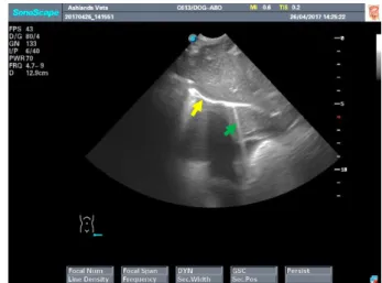
Figure 1 DH view site for an AFAST or TFAST exam. Figure 2 DH view during AFAST or TFAST scan. Presence of URLs (green arrow) which are typically present to a small extent (none to one or two ULRs) along the diaphragm (yellow arrow) in normal dogs and cats. (Lisciandro, 2014)
Spleno-renal view
In the SR view, the probe is positioned longitudinally more or less parallel with the spine just caudal to the costal arch and fanning cranially under the rib. Usually, the spleen and the kidney are brought into view immediately. If that doesn’t happen, the spleen can be used to locate the kidney by following it caudolaterally. If present, the free fluid is visualised as anechoic triangles between the spleen and the colon.
Finally, and back to the starting point, the probe should be moved away from the table top and dorsally, so the retroperitoneal space and its great vessels can be further observed. If retroperitoneal fluid is found in this area, cranially fluid sources would include the kidneys, vertebral bodies, great vessels and adrenal glands, and caudally the kidneys, ureters, vertebra bodies and pelvis (Lisciandro, 2014).
Artefacts and false results
The artefacts found in the SR view are most colon related, because if it’s filled with air, it is more difficult to see without air interference. However, in the right lateral recumbecy the animals have the colon fall away from this view.
In some cats and small dogs, a false mirror image appears, because both kidneys are seen. In this view, it is also common to mistake the great vessels and small intestines with free fluid. It’s important to remember that free fluid rarely appears like linear anechoic stripes, but as triangles. If the probe is turned transversally, the vessels may have a pulse and appear like a black circle and the small intestine like hamburgers. The colour flow Doppler can also be used. Regarding the differentiation between retroperitoneal and peritoneal fluid, in lateral recumbecy the retroperitoneal fluid remains static and not falling away from view. Increasing the depth, it might also be helpful (Lisciandro, 2014).
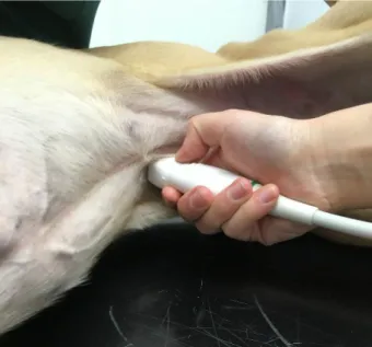
Figure 3 SR view site in an AFAST exam.
Figure 4 SR view during AFAST scan. It’s possible to visualize the left kidney (yellow arrow).
Cysto-colic view
In the CC view the probe is placed over the bladder region, ventral middle line cranial to the pubis, directing it downward to the tablet top. At this point, the bladder lays against the ventral abdominal wall. Small amounts of fluid, anechoic triangles, can appear between the bladder apex and the body wall. Also, if large amounts of fluid are present, usually it’s easier to identify them, because the small intestines and omentum are floating on it (Lisciandro, 2014).
Artefacts and false results
The CC view is also responsible for several imaging artefacts. The tissue distal to the urinary bladder will be brighter because of the acoustic enhancement. In addition, the air-filled colon can obscure the view in the far field making the bladder hard to see, but altering the position and the pressure of the probe will lead the colon out of the way. If the probe is directed dorsally, it’s possible to see the great vessels and the lymph nodes that can be mistaken by free fluid. But linear anechoic stripes usually don’t represent fluid, and, as said before, changing the probe transversally the vessels will acquire a round shape and may have a pulse. It’s also important to know that puppies and kittens sometimes have small amounts of fluid in this site, which is considered a physiologic imaging (Lisciandro, 2014).
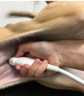
Figure 5 CC view site in an AFAST exam.
Figure 6 CC view during AFAST scan. It is possible to identify a small portion of the colon (green arrow), by its hyperechoic contents, dorsally to the bladder (yellow arrow)
Hepato-renal view
In the HR view, despite its name, it is most commonly seen are the small intestines and, only occasionally, the spleen. Unless, there’s suspicion of renal pathology, the kidney is not assessed in this view, because of the right kidney more cranially position under the rib cage. Therefore, the probe is just placed ventrally to the umbilicus and then moved downward to the table top. If it’s necessary to assess the right kidney and the retroperitoneal space, the animal can be moved dorsally enough to place the probe underneath it, just ventrally to the last rib. For heavier and larger animals, sometimes it’s necessary to change sides and move it to right lateral recumbecy. If small amounts of liquid are present, they are obviously identified as small anechoic triangles. If large amounts are present, then the small intestine and omentum appear floating in the free fluid. Since intestines can be mistaken for free fluid, in this view it is advised to change the probe angle transversally, so that the gastrointestinal tract acquires the shape of hamburgers.
The HR view is the most gravity dependent site in right lateral recumbecy, so this is the site of choice for abdominocentesis. Although, if there isn’t fluid in this site, it can be collected from the site where the animals present collection of fluid (Lisciandro, 2014).
Artefacts and false results
Necrotic fluid-filled masses present in the mid-abdomen can be confused with free fluid. So, it’s helpful to increase the depth and create a different perspective, where the margins of the mass can be observed. Likewise, it’s important to give special attention in non-neutered bitches. Since fluid-filled uterus can give the false impression of free fluid in the abdomen (Lisciandro, 2014).
Figure 7 HR view site in an AFAST exam.
Figure 8 HR view during AFAST scan. The right kidney isn’t observed, because of its cranial position under the ribs. There’s portions of small intestine in transversal, or hamburger like shape (green arrow), and longitudinal views (yellow arrow)
How to use AFAST with Abdominal Fluid Scoring
The most common causes of death in human trauma patients are undiagnosed abdominal injury and uncontrolled internal haemorrhage. Ongoing bleeding accounts for 80% of early deaths in hospitalized humans, specially, because they used to treat it with aggressive fluid therapy, which exacerbates the bleeding. The medical management in animals is very complicated, because routine measures, like initial packed cell volume (PCV) values and radiographic serosal detail, were found misleading in the detection of free abdominal fluid. In one study of 101 dogs that suffered blunt trauma, serosal radiography was performed in 97% of the cases and normal results were reported in 24% of animal’s positives for fluid in the abdominal cavity. Moreover, initial PCV in animals that had fluid in the abdomen (44%) wasn’t significant different from the ones without it (48%) (Lisciandro, et al., 2009).
Since ultrasound is a diagnostic tool readily available in most veterinary practices and FAST scan was proved a reliable technique in detecting free abdominal fluid, an AFS system has been developed. This system allows assessment of the initial degree and the progression of intra-abdominal injury, which helps choose a better treatment approach. In the same study mentioned above, almost all of the positive cases for free fluid went through a resuscitation fluid therapy of 45 mL/kg of crystalloids instead of the usual aggressive one of 90 mL/kg. This change of treatment was proved adequate by finding normal blood lactate levels and clinical improvement on those dogs (Lisciandro, et al., 2009).
In the standardized AFS system, the animals are scored as follows: 0, if negative for fluid at all of the 4 AFAST sites, to a maximum of 4, if positive in all 4 views. Each view receives a score of 1, if positive for fluid, and in the end all the views’ scores are added. In practice, the animals are qualified as small bleeders, if they scored 1 or 2 AFS and big bleeders, if they got 3 or 4 AFS. Big bleeders, according to research, are more predisposed to become anaemic, more precisely they are predicted to decrease 20-25% their admission PVC. Moreover, 20-25% of these animals, will become severe anaemic (PCV < 25%) and will need a blood transfusion or an emergency laparotomy. Big bleeders are also associated with higher alanine transaminase (ALT) and blood lactate values, which are injury serum markers (Lisciandro, et al., 2009).
On the other hand, small bleeders, if they don’t have any pre-existing anaemia, are unlikely to become anaemic unless they’re bleeding from somewhere else (retroperitoneal, pericardial, pleural, lungs or fracture sites) or experience haemodilution due to large fluid volumes. In blunt trauma, small amounts of free fluid were commonly found in non-gravity dependent sites (DH and CC) and larger amounts in the gravity dependent sites (HR if right lateral recumbecy and SR if left lateral recumbecy). Although in another study with non-traumatized patients, the results were slightly different. The fluid was more frequent present in SR and HR views, independently of the gravity-dependent site (McMurray, et al., 2016).
The application of AFS in serial procedure is very useful because animals can change their score between scans. For example, a 0 AFS can become a bleeder in the second exam or a small bleeder can become a big bleeder. It’s important to register the views that were positive to fluid, because it might help locate the source of injury. A serial AFAST scan also allows to check if the treatment is working (animal decreases his AFS) or needs to be changed. So, the indications for serial AFAST scans are: 1) If the patient is stable, a second AFAST scan is performed after 4h; 2) if it is unstable, less than 4h; 3) If the dog already has the highest score (AFS 4), the second scan is advised after 8/12h (because fluid takes at least 48h to clear) (Lisciandro, et al., 2009).
Figure 9 The template for registration of AFAST exam and AFS findings (Lisciandro, et al., 2009).
The use in blunt trauma
In blunt trauma, AFAST serial exams applied with AFS system is a powerful tool that helps the clinic assess the degree of the injury and to manage the patient in the first 4 hours of critical care. In these cases, AFAST scans are initially more reliable than other methods to detect hemoperitoneum, therefore it should be used as a standard care (Boysen & Lisciandro, 2013).
In cats, it hasn’t been found any correlation between AFS system and the degree of injury. But one author suggested that cats suffering from blunt trauma usually do not survive, based on the small sample that he had in one study (Lisciandro, 2014).
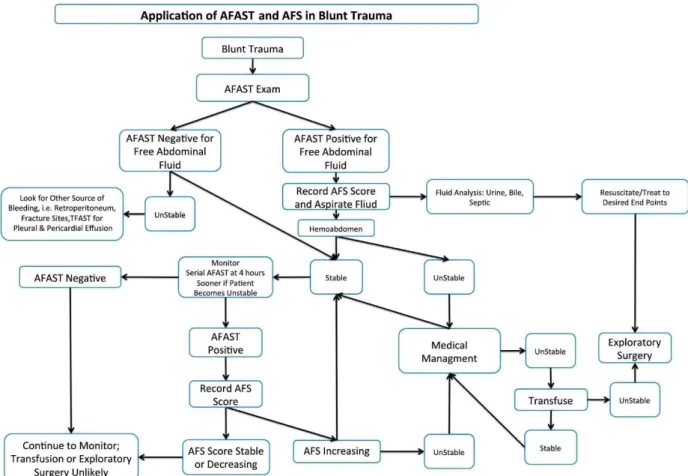
Figure 10 Algorithm for the use of AFAST and AFS in dogs after blunt trauma. (Boysen & Lisciandro, 2013)
The use in penetrating trauma
In penetrating trauma, physical examination and radiography are better tools than AFAST scan. Because in blunt trauma blood defibrinates, so it appears anechoically and as free fluid. In penetrating wounds, like bites, the coagulation cascade works differently and the blood clots, appearing echogenic and similar to soft tissue, are not readily detected by inexperienced sonographers (Lisciandro, et al., 2009).
The use in non-traumatic injury
The FAST scans have been demonstrated to enhance the detection of free fluid in non-traumatized humans, particularly those unstable with undifferentiated hypotension, shock, dyspnoea and sepsis. Therefore, this procedure narrows differential diagnoses, improves decision making accuracy and expedites patient management (McMurray, et al., 2016).
In animals, FAST scan can also be used as an extension of the physical exam and detect conditions that could be missed by traditional means, for example, hemoperitoneum, haemothorax, acute cardiac tamponade, rupture viscous, acute peritonitis, and anaphylaxis
(Lisciandro, 2011). Unlike traumatic patients, where the fluid found in the abdominal cavity is in most clinical cases, blood, in no traumatic animals, the type and the origin of the fluid presence cannot be easily predicted. Therefore, abdominocentesis has a greater diagnostic value than in trauma (McMurray, et al., 2016). Furthermore, AFAST scans can be applied in all patients at imminent risk, and post interventional procedures, like surgery (spay, liver lobectomy, splenectomy, adrenalectomy) and percutaneous or laparoscopic aspiration or biopsy. This clinical application in humans has already been well established as a valuable diagnostic procedure and it allows a quicker intervention before the patient decompensates, because in Humans the clinical signs like mucous membrane colour, heart rate, and pulse quality, are unremarkable until they lose up to 30% of their blood. On the other hand, dogs might be able to compensate even more by splenic contractions (Lisciandro, 2014).
How to diagnose anaphylaxis in dogs with AFAST
The organs, in dogs, most affected by the release of histamine during the anaphylactic shock are the liver and gastrointestinal tract because, histamine is released from the gastrointestinal tract into the portal vein, and from there to the hepatic vessels, creating arterial vasodilation and venous outflow obstruction. The venous drainage of the gallbladder is reduced and changes in the gallbladder wall are expected. The halo sign is one example of those changes and is characterise by wall thickening (more than 3 mm) with inner and outer hyperechoic walls with a dark middle layer (Quantz, et al., 2009). This gallbladder changes take less than 2 to 4 minutes and are faster than traditional markers such as ALT in serum. A halo sign isn’t restricted to anaphylaxis shock and can manifest in conditions that cause obstruction on the venous and lymphatic return (cardiac tamponade and congestive heart failure); in conditions that affect the regional anatomy (pancreatitis, cholangiohepatitis and primary gallbladder disease); and conditions that lead to third spacing (severe hypoalbuminemia and over-resuscitation). Thereby, is very important to complete the AFAST and TFAST scan before giving the final diagnosis, in order to rule out other causes for the halo sign and give the appropriate treatment. It is also common for anaphylactic dogs to have a marked hemoabdomen at presentation. So, in order to avoid taking anaphylactic dogs into surgery, it’s substantial to evaluate the clotting times, levels of ALT and haematocrit (Lisciandro, 2014).
H
OW TO PERFORM ATFAST
EXAMUltrasound settings and probe preferences
The same ultrasound machine and probe used in AFAST scan can be used in the TFAST. The transducer can be set at 7.5 MHz for all animals, and the depths are variable according to the view, which will be discussed ahead (McMurray, et al., 2016). An abdominal setting works well for a TFAST exam, but it’s necessary to flip the image when visualizing the heart chambers to obtain the standard images of that organ (Lisciandro, 2014).
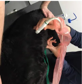
Patient position and preparation
If the patient is clinically stable, like for AFAST, the right lateral recumbecy is preferable for most of the exam. In this position, it’s easier to assess the gall bladder, CVC and the hepatic vessels. For respiratory-compromise animals is advised a modified sternal recumbecy, where the forelegs are in sternal position and the hind legs laterally to one side (right side preferable). For better heart visualisation, a rolled towel can be placed under the forelegs, which makes for easier probe movement. In opposition to AFAST scan, in TFAST the patient needs to be moved for the last view, so the two sides of the chest are covered. At this point, the animal can be moved to a sternal recumbecy. As for AFAST, no hair needs to be shaved and surgical alcohol or gel may be use to enhanced the skin-to-probe contact. (Lisciandro, 2014)
Chest tube site view
The chest tube site (CTS) view is done in the most dorsal region of the thorax, where accumulation of air might be present. The probe is placed dorsally to the xiphoid process, more precisely in the 8th or 9th intercostal space (ICS). Some deep-chested dogs and some small animals with pathologies in the abdomen that shrink the thoracic cavity (like pregnancy and large masses) might need the probe to be placed in the 7th and 8th ICS because it reduces the diaphragm, lung and liver interference during breathing, which make easier to see the glide sign (Lisciandro, 2014). The standard position for every lung ultrasound exam, is acquired with the transducer depth set at 2 to 4 cm and when the gator sign is visualised (McMurray, et al., 2016). The gator sign is composed with two ribs heads (the eyes of the alligator) and the ICS that sits between them (the nose of the alligator). In the ICS, a hyperechoic line is found and it represents the pulmonary-pleural line (PP-line). During inspiration and expiration, there’s a to-and-fro motion at the PP-line and that’s the glide sign. Sometimes when the ultrasound echoes strike the air interface directly, it becomes difficult to see the glide sign. So, one of the tricks is to move the probe a little bit dorsally or ventrally to change the angle where the echoes strike the lungs. Another trick is to change the orientation and get the rib at the centre, thereby the glide sign can
be seen on both sides. The final trick is to change the pre-set from abdominal to cardiac or the other way around (Lisciandro, 2014).
Artefacts and false results
The interpretation of artefacts on a lung scan is the most important part. So, when the echoes strike with aeration of normal lung (also called dry lung) the reverberation artefact goes beyond the PP-line and produces parallel lines called A-lines. Although they are a mirror of the PP-line, A-lines don’t produce a glide sign. Another artefact is the ultrasound lung rockets (ULR), also designated as B-lines. These lines occur when, in the lung periphery, fluid is immediately adjacent to air. They appear as a hyperechoic laser-like line that swings like a pendulum. Even though these lines can occupy all of the ultrasound image, they are produced in the first 1-3 mm of lung’s surface. The ULR are useful because: 1) they are easy to identify; 2) rules out PTX; and 3) the distribution patterns can be used to diagnose different lung conditions.
The last artefact in CTS view is the step sign. This artefact manifests as a discontinuity of the normal PP-line. Some of the conditions that create the step sign are: pleural effusion, intercostal tear and rib fracture, subcostal hematomas, diaphragmatic hernia, anterior mediastinal mass and severe left atrial enlargement. A false step sign is observed if the probe is far caudally, because of the depths change between the lungs, diaphragm and liver during respiration. If subcutaneous emphysema is present in the CTS, the air makes impossible to see the gator sign. However, usually this problem is overcome by applying more probe pressure and displacing the subcutaneous emphysema away of view (Lisciandro, 2014).
Figure 11 CTS during a TFAST exam.
Figure 12 CTS view during TFAST scan. The glide sign was present, but it can only be appreciated in motion pictures.
Pericardial site view
In the PCS view, the probe is placed in the third, fourth or fifth ICS at the level of the costochondral junctions. One quicker way to find the right position is to put a hand on the animal’s chest and find the place where the heartbeat is stronger. If that’s not possible, the elbow can be moved towards the costochondral junction and the probe placed there. The transducer depth must be set at 5 to 15 cm (Lisciandro, 2014 and McMurray, et al., 2016).
At this site, the probe is fanned according to the mantra “marker towards the elbow” and then “marker toward the spine”, so the short- and long-axis views of the heart are observed, respectively. With some experience, in the left ventricular short-axis view (mushroom view), the contractility and volume status may be subjectively evaluated. And, the diameter of the left atrium and the aorta can be assessed for evidences of left-sided heart disease. The left-sided heart disease can also be evaluated at the CTS, when looking for evidences of interstitial oedema. (Lisciandro, 2014)
Artefacts and false results
Since the right-side view gives a better overall image of the heart, the animal is placed preferably in right lateral recumbecy. But when accessing the non-gravity dependent left PCS view, there’s a lot of lung interference. So, less time should be spending in this side and more in the right, so the time can be better spent where most information is provided. Likewise, it is easy to mistake heart chambers for pleura and pericardial effusions when the field is limited, especially in big dogs. Therefore, it’s advised to zoom out (increase the depth), so that the chambers limits can be seen. Also, the “one view is no view” mantra should be remembered and the effusions must be confirmed in the DH view (Lisciandro, 2014).
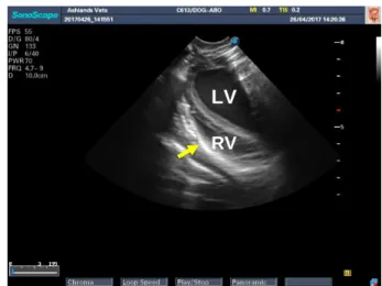
Figure 13 PCS in long axis view in a TFAST exam. Figure 14 PCS left side long axis view in right lateral recumbecy. It is possible to identify both left ventricle (LV) and right ventricle (RV) surrounded by the pericardium (yellow arrow).
LV
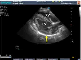
RVFigure 15 PCS right side long axis view in right lateral recumbecy. Pericardium sac is the hyperechoic line that surrounds the heart chambers (yellow arrow).
Figure 16 PCS left side short axis view in right lateral recumbecy. Both right (RV) and left ventricle (LV) are observed surrounded by the pericardium (yellow arrow).
Diaphragmatico-hepatic view
It is performed the same way as in AFAST scan.
Figure 17 The template for registration of TFAST exam findings (Lisciandro, et al., 2008).
LV RV
LV RV
How to diagnose pneumothorax with TFAST
In blunt trauma, 10% to 18% of the animals die from pulmonary injury and 13% to 50% develop PTX. Therefore, it’s important to detect and act fast during these situations. Usually, the veterinary surgeon resorts to physical examination and chest radiographies to detect this condition, but these tools are inconsistent and sometimes not practical.
Human studies have determined, using computer tomography as the gold standard, that chest ultrasounds have better Se (88-100% versus 36-75%) and a similar Sp (94% versus 100%) in detecting PTX comparing to chest radiographies (Boysen & Lisciandro, 2013).
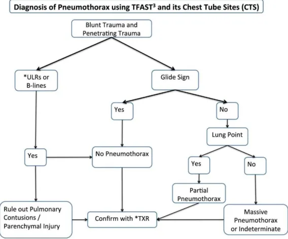
In the ultrasound exam, PTX is diagnosed by the presence of A-lines without the glide sign and it’s ruled out by the presence of the glide sign or URLs. Generally, the probe is placed in the highest point of the chest, because is where the air accumulates. And it’s important to wait for the first 5 breaths after moving the animal, so, if present, the air can settle in and be observed. The degree of the PTX is also assessed by finding the lung point. The lung point is the place where the collapsed lung meets the thoracic wall and it is identified by the presence of the glide sign, URLs or the lung pulse. The lung pulse is rarely seen, and it’s also the to-and-fro movement between the visceral and parietal pleura like the glide sing, but the rhythm is set by the heartbeat and not by the respiration movements. The degree of PTX is measure by the distance between the CTS to the lung point, and it can be classified as 1) mild to partial (if the lung point is near the CTS); 2) severe (if the lung point is found in the middle of the thorax wall) and 3) massive (if no lung point is found).
The reliability of using TFAST protocol in the detection of PTX still needs more research in small animals practice. One clinical study described that Se and Sp of TFAST in the diagnostic of PTX compared to chest radiographies was 78% e 93%, respectively. These values are lower than those found in Humans. However, the author concluded that: 1) they should have used CT as comparison method, 2) TFAST requires more experience to obtain proficiency than AFAST (one experience sonographer reported Se and Sp higher than 90%); 3) the presence of concurrent lung or pleural injuries can lead to false negatives, and 4) fast breathing rates might affect the ability of seeing the glide sign and, consequently, detecting PTX (so, it’s advisable to repeat the TFAST after patient analgesia). In spite of the results, TFAST has important advantages over radiography, because it’s portable, doesn’t required a lot of patient restrain, it’s rapidly performed, it’s not invasive and it is radiation free (Lisciandro, et al., 2008).
Figure 18 Algorithm for the use of TFAST in the diagnosis of PTX (Boysen & Lisciandro, 2013).
How to diagnose lung contusions with TFAST
The “wet” and “dry lung” are easy concepts to learn and very helpful in the diagnostic of several and diverse lung injuries. The “wet lung” is characterised by the presence of URLs and the dry lung by the presence of glide sign with A-lines. When URLs are present, the majority of underlying pathology are cardiogenic and non-cardiogenic pulmonary oedema, lung contusions, lung haemorrhage, and acute pneumonias. They can be discriminated based on its locations in the thorax. In the CTS view, generally speaking, the URLs in trauma patients represent lung contusions and in non-traumatic patients they represent cardiogenic and non-cardiogenic pulmonary oedema. In addition to what was previously referred, the TFAST using the wet and dry lung concept can be used to monitoring patients with risk of volume overload and development of pulmonary oedema (Lisciandro, 2014).
How to diagnose pleural and pericardial effusion with TFAST
Ultrasound is arguably (vs CT scan) the gold standard for pericardial effusion in human medicine. Mostly because, in humans the heart lies on the diaphragm. So, when using the DH view there’s little air interference when trying to see the heart. In dogs, the heart position is variable and depends on the patient body conformation and position. Nevertheless, if the animal is in standing
or sternal position or there’s a clinically relevant pericardium effusion present, the canine heart might get closer to the diaphragm and can be seen easily through the acoustic window made by the gall bladder and liver (Lisciandro, 2014).
As already mentioned, 83% of the pleural and pericardial effusion are diagnosed in the DH view. At this site, pericardium effusion is identified by the presence of fluid contained within the pericardium sac (a hyperechoic line), which surrounds the apex of the heart. On the other hand, pleural effusion is identified by a more angular, uncontained, irregular, sharper-angle image and better visualise in the PCS (Lisciandro, 2016).
At the PCS, the pericardial effusion is better diagnosed in the right pericardial view. In a long-axis view, the four chambers of the heart can be assessed, which may help to prevent mistaken them for pericardial effusion. The short-axis view is not advised, but, if it’s used, pericardial effusion should only be diagnosed at the level of the muscular apex, but never at the level of the left and right ventricle or neither at the base of the heart, because they are easily mistaken. Pleural effusion in this site is identified as uncontained fluid with the shape of triangles (Lisciandro, 2016). The underlying causes of pericardial effusions are left atrial tear secondary to mitral valve disease, haemorrhage from right atrial neoplasia, heart base tumour, idiopathic pericardial effusion and haemorrhage caused by anticoagulant rodenticide toxicity (Boysen & Lisciandro, 2013).
How to diagnose of cardiac tamponade with TFAST
Cardiac tamponade is a life-threatening condition and is an indication for immediate pericardiocentesis. It occurs when the intra-pericardial pressure exceeds right and ventricular pressures, causing the walls of this chambers to paradoxically move inward during the cardiac cycle. This condition can be readily diagnosed using TFAST either in DH or PCS views (long- and short-axis) (Boysen & Lisciandro, 2013).
How to diagnose diaphragmatic herniation with TFAST
In the DH view, usually, it is expected to see a continuous and curvilinear line of the diaphragm. When that is lost, congenital or traumatic diaphragmatic herniation should be considered. Moreover, the diagnosis is supported, if in the same exam, the gallbladder isn’t found in its normal location (also consider rupture). At the PCS, diaphragmatic herniation is suspected when the heart is obscured by solid organs viscous structures (Boysen & Lisciandro, 2013).
C
LINICALC
ASESSpike’s case
Characterization of patient and reason of appointment:
Spike was a five years old male cat, neutered, 5kg, Balinese crossbreed. Spike was dewormed and vaccinated against feline herpesvirus type 1, feline parvovirus, feline calicivirus and feline leukaemia virus. His owner brought him to Ashlands Veterinary Centre in Skipton because she was worried about his respiratory pattern.
Anamnesis:
The owners reported that Spike was eating and drinking well, but he seemed less energetic and more lethargic during the previous couple of weeks. Spike was also an indoor cat, who had access to a private garden, and he lived with another female cat that was his mother.
Physical exam:
Spike was alert during the examination. An increased effort to breathe was detected, especially in the abdominal component. The breathing rate was 60 breaths per minute. The pulse was 200 beats per minute and weak and the mucosa colour was pink, with a CRT of less than 2 seconds. Through auscultation, a gallop and louder breathing sounds were heard. All other parameters of physical examination were unremarkable.
TFAST exam:
CTS glide sign Present
CTS lung rockets Present
CTS step sign Absent
PCS view Pleural fluid
Cardiac tamponade Absent
LV filling (short axis) Indeterminate
DH view Pleural effusion
Comments: Pulmonary oedema and pleural effusion are the reason for the respiratory distress. Oxygen supplementation and furosemide IV. A TFAST scan is also advice after 2 hour for monitoring the diuretic therapy. An echocardiography exam is advised to determining the underlying reason.
Figure 19 CTS view during Spike TFAST. Presence of URLs (yellow arrow) and the glide sign was also present.
Figure 20 PCS view during Spike TFAST scan. The pericardium (yellow arrow) is easily identified by the hyperechoic line that surrounds the 4 heart chambers. Pleura fluid present (red arrow).
Echocardiography exam:
The results were an enlargement of the right side of the heart (without septum bowing), left atrium enlarged with smoke (LA:AO = 2.9:1), and multifocal left ventricle wall thickness (0.6 cm) with a normal interventricular septum (0.45 cm). Pleural effusion was present.
Figure 21 Right parasternal short-axis (aortic valve
level) view. Measurement of the LA:Ao ratio. Figure 22 Right parasternal short-axis (papillary muscle level). Measurement of ventricle wall thickness.
Diagnosis:
Feline unclassified cardiomyopathy in congestive heart failure.
Treatment:
In order to improve the respiratory distress, oxygen supplementation and furosemide IV at 1 mg/kg (0.1 mL of Dimazon® 5 % Solution IV every 2 or 4 hours as required) was advised as an emergency treatment.
For long course treatment: furosemide at 2 mg/kg PO BID (1/4 tablet Frusedale® 40 mg everyday); enalapril at 0.5 mg/kg PO SID (1 tablet of Enacard® 2.5 mg everyday); spironolactone at 2 mg/kg PO SID (1 tablet of Prilactone® 10 mg everday); pimobendane at 0.25 mg/kg BID (1 tablet of
Cardisure® 1.25 mg twice daily) and clopidogrel at 18.75 mg/kg SID (1 + ¼ tablet of Plavix® everyday).
Follow-up:
The owner was contacted after echocardiographic exam and was informed about Spike’s diagnosis and the needed treatment, and decided to euthanize Spike, because she wasn’t able to give any of the necessary treatment.
Discussion:
Respiratory distress is a common presentation in veterinary emergency medicine. Treating these cases is very challenging, because excessive handling might exacerbate the respiratory distress. When managing these patients, it’s important to reduce the number of diagnostic testing and to be limited to the ones that give more information and require less handling. (Fuentes, et al., 2010) The current guidelines advise to wait until the patient is stable to proceed with the diagnostic tests, which consists in oxygen supplementation and, if possible, a peripheral intravenous catheter placed at presentation to provide vascular access, as well as a very careful sedation in anxious animals (King & Clarke, 2010). There is, however, an exception to this rule for cases when lung sounds are dull, because presence of pleural space disease is suspected. In this situation, thoracentesis should be performed promptly to remove pleural air or fluid, even before radiographies are obtain (King & Boag, 2007). Therefore, after oxygen supplementation, a more careful physical examination must be performed in order to locate the origin of the problem. Based on that, the causes of respiratory distress can be classified in five categories: 1) airway obstruction (e.g. nasopharyngeal polyps and aspirated foreign bodies); 2) pulmonary parenchymal disease (e.g. pneumonia, acute respiratory distress and pulmonary oedema); 3) pleural space disease (e.g. PTX and pleural effusion); 4) thoracic wall abnormalities (traumatic injuries and spinal cord or phrenic nerve disease); and 5) “look-alikes” (hyperthermia and pain). Further treatment is applied based on this classification (King & Clarke, 2010).
In practical terms, the physical examination might be unreliable and misleading, especially in animals where handling might deteriorate their condition. For instance, an animal can be in a state of moderate hypoxia and still have pink mucous membranes because mucous membranes become blue or cyanotic when at least 50 g/L of haemoglobin is denatured (Fuentes, et al., 2010). Another example of physical exam unreliability happens during cats’ auscultation. Arrhythmias and heart murmurs might be absent even when a heart condition is present (Fuentes, et al., 2010). Likewise, the contrary is also true, there was estimated that 15% to 34% of healthy cats have a systolic heart murmur. Although, when gallop sounds are heard, they’re associated with serious heart condition in cats (Nelson & Couto, 2014). So, contrary to the bibliography, the veterinary surgeon, in practice, waits for a radiographic or ultrasound confirmation before proceeding with treatment.
During Spike’s case, its anamnesis and physical examination permitted to classify it in pulmonary parenchyma disease. It wasn’t air way obstruction because no stridor sounds were heard nor it was an inspiratory dyspnoea. Thoracic wall abnormality wasn’t a possible classification either because in the history there weren’t any indicative of trauma in a recent past. The class of pleural space diseases was ruled out as well because no muffle sounds were heard, but instead louder breathing sound were detected during auscultation. Finally, the class “look alike” wasn’t consider because the temperature was within the normal range.
Classifying Spike into the pulmonary parenchyma diseases’ class wasn’t enough to administer treatment, it was also important to be sure that was a heart condition. So, the TFAST scan was performed and it detected pleural effusion, pulmonary oedema and heart enlargement, which confirmed the suspicion, but also add a new condition that was undetected by the physical exam. It concluded that the respiratory distress was a result of both the pulmonary oedema and the pleural effusion.
But, because in the TFAST scan the pleural effusion was considered mild, it wasn’t the main cause of respiratory distress, which explains why wasn’t detected on the physical exam.
Apart from oxygen supplementation, it was also advised furosemide and TFAST exams after 2 hours to monitor the resolution of the pulmonary oedema and the pleural effusion. If the heart condition wasn’t confirmed, the diuretic therapy wouldn’t be indicated, and when the pleural effusion was detected (by radiographies e.g.), thoracentesis would be advised to classify the type of fluid, firstly macroscopically and then cytologic.
The TFAST scan, in a short amount of time after presentation, permitted to direct to the best treatment approach and could had been performed at the same time of oxygen supplementation and catheter placement. Be It also avoids the initial use of radiography, which reduce the exposure to radiation and the risks associated with animal movement and sedation. Finally, the TFAST scan could have been used to monitoring the diuretic therapy with the dry lung and wet lung principle and, if the thoracentesis end up being necessary, it could have been used to monitoring PTX development.
Ruby’s case
Characterization of patient and reason of appointment:
Ruby was a 9 years old neutered bitch, 10 kg, cavalier king Charles spaniel. She was dewormed and vaccinated against canine distemper virus, canine adenovirus, and canine parvovirus type 2 and 4 strains of Leptospira. Ruby’s owner brought her to the veterinary surgery, because she had a single collapse episode at the groomers the day before.
Anamnesis:
Apart from the collapse episode Ruby’s owner didn’t report any problems with her. Since the owners weren’t present at the time of the episode, a part from knowing that she lost consciousness, a good description of it wasn’t possible to obtain. But after the episode she behaved normally as if nothing had happened.
Physical exam:
During the exam, Ruby was conscious and relaxed. An increased respiratory effort during expiration was detected, although the breathing rate was 25 breaths per minute. During thoracic auscultation, a high and loud systolic murmur was heard (grade V/VI) on both sides with equal intensity. The pulse was 180 beats per minute and the mucosa colour was pale pink, with a CRT of less than 2 seconds. Abdominal distension was also present. All other parameters of physical examination were unremarkable.
AFAST exam:
Patient position Right lateral recumbecy
Gallbladder Halo sign present
Urinary bladder Present and normal appearance
DH view - Pleural fluid absent
- Pericardial fluid absent - Hepatic veins distended Positive or negative at the four views (0 negative, 1 positive)
DH 1
SR 1
CC 1
HR 1
AFS 4
Comments: Since the hepatic veins appear distended and the halo sign present, it suggests that the ascites has a cardiogenic origin, more precisely right side congestive heart failure.
Figure 23 DH view during Ruby AFAST scan. Presence of the “halo” sign (yellow arrow) and free fluid (red arrow).
Figure 24 SR view during Ruby AFAST scan. Presence of free fluid (red arrow).
Figure 25 CC view during Ruby AFAST scan. Present of free fluid (red arrow) .
Figure 26 HR view during Ruby AFAST scan. Presence of big amounts of free fluid (red arrow). It’s important to notice the hamburger like shape of the small intestines (yellow arrow).
TFAST exam:
CTS glide sign Present
CTS lung rockets Present
CTS step sign Absent
PCS view Absent
Cardiac tamponade Absent
LV filling (short axis) Indeterminate
DH view Nothing more to report
Comments: At the PCS view, an enlargement of the heart chambers was seen and a pulmonary oedema observed, which supports the right-side heart failure, but also a left side as well.
As an emergency treatment: furosemide IV and as TFAST after 2h to monitor. An echocardiography is advised for determining the underlying reason.
Echocardiography exam:
The results showed a 4 chamber dilatation with no wall thickness and intraventricular septum bowed to the right side, the presence of an atrial septum with left to right flow through it up to 2
m/s. There was a marked mitral valve leaflet clubbing and prolapse and the maximal mitral regurgitation with a large eccentric jet, directed towards septum. The LA:Ao ratio was 3.5:1. There was also a tricuspid regurgitation with a maximal regurgitation velocity of 3.4 m/s, which corresponds to pulmonary pressure of 46 mmHg, classified as mild. There were also present pulmonic and aortic insufficiency. The iso-volumetric relaxation time was 10 milliseconds (normal is between 45 and 65 milliseconds) and the ratio of early transmitral flow velocity to left ventricle iso-volumetric relaxation time. No evidence of any congenital defect was seen. These findings were consistent with degenerative mitral valve disease stage C according to American College of Veterinary Internal Medicine’s classification system for canine chronic valvular heart disease. The mitral valve disease was most likely the primary problem and the atrial septal defect is an acquired tear due to the impact of the mitral regurgitation jet. Left and right congestive heart failure was confirmed.
Figure 27 Right parasternal long-axis four chamber view. Left side enlargement with the intraventricular septum bowed to the right side. Marked mitral valve leaflet clubbing and prolapse (yellow arrow) and the atrial septum (red arrow)
Figure 28 Right parasternal long-axis four chamber view. Atrial septum with left to right flow, evident with colour Doppler.
Figure 29 Right parasternal long-axis four chamber view. Measurement with spectral Doppler of the flow velocity between the atrium septum.
Figure 30 Spectral Doppler of the caudal vena cava. It was dilated and didn't collapse on inspiration.
Diagnosis:
Treatment:
For the congestive heart failure was advised a quadruple therapy with pimobendam at 0.25 mg/kg PO BID (1 tablet of Cardisure® Flavoured 2.5 mg twice daily); torasemide at 0.3 mg/kg PO SID (1 tablet of UpCard® 3.0 mg everyday); enalapril at 0.5 mg/kg PO SID (1 tablet of Enacard® everyday); and spironolactone at 0.2 mg/kg PO SID (half a tablet of Prilactone® 40 mg everyday). For the pulmonary hypertension, it was prescribed sildenafil at 0.25 mg/kg TID PO (1 tablet of Viagra® 25 mg every 8 hours).
Discussion:
The history of a collapse episode with loss of consciousness is very important, because distinguishing syncope from seizure can be challenging. The features that are most helpful to differentiate between these two conditions are the precipitants of the episode, the prodromal behavioural changes, the clinical signs that occur during the episode, and the subsequent events. One episode that’s precipitated by exercise, stress, coughing, excessive barking, micturition, defecation, or pain is more likely to be a syncope than a seizure. On the other hand, disorientation before and after the episode and a long recovery period it is suggestive of seizure (Lorenz, et al., 2009). In Ruby’s case, even though there wasn’t a very good description of the collapse episode, it was possible to classify it as syncope, because she lost consciousness during a stressful situation (at the groomers) and she recovered from it very quickly, without any neurologic deficits. Although through the clinical history it was possible to associate the syncope observed in Ruby’s episode, this condition is not diagnostic, but a clinical sign of various possible diseases/ pathologies, and the choice of the treatment should depend of the underlying cause. So, syncope is a transient loss of consciousness and muscle tone caused by inadequate cerebral oxygenation or perfusion of the brain. It can be assigned to five different pathophysiologic causes in dogs and cats: 1) cardiac arrhythmias, 2) structural cardiac and pulmonary causes, 3) reflex-mediated syncope, 4) orthostatic hypotensive syncope, and 5) severe and abrupt hypoxemia.
The pathophysiology of cardiac arrhythmias’ syncope is markedly decreasing cardiac output provoked by bradyarrhythmias or tachyarrhythmias. The bradyarrhythmias is achieved by pausing the electrical activity and the tachyarrhythmias, by increasing the heart rate, which cause inadequate ventricular filling in diastole. Structural cardiac and pulmonary pathological conditions can cause syncope when the circulatory demands outweigh the heart’s ability to increase cardiac output. The diseases associated with this type of syncope are aortic stenosis, pulmonic stenosis, tetralogy of Fallot, atrial septal defect, hypertrophic obstructive cardiomyopathy, and cardiac neoplasia causing obstruction to blood flow, pericardial effusion/tamponade causing decreased venous return, pulmonary embolism, and pulmonary hypertension with or without Dirofilaria
immitis or Angiostrongylus vasorum infestation. It’s important to know that heart failure itself won’t
syncope is characterised by withdrawal of sympathetic tone with abrupt increase in vagal tone. The unset of this mechanism is controversial, and the triggering events are variable and determined by which afferent nerve or receptor is stimulated. The orthostatic hypotensive syncope is very rare in animals and is related to failure to maintain the blood pressure in an upright position. Finally, acute hypoxemia has been described as a cause of syncope in dogs with severe laryngeal paralysis or upper airway obstruction (Ettinger & Feldman, 2010).
The traditional diagnostic approach is based on the physical examination, especially the auscultation parameter. In Ruby’s case, she had dyspnoea (without tachypnoea), a bilateral heart murmur, tachycardia and ascites. Therefore, according to some bibliography, low output failure should be suspected and the next step would be to determine if the animal had pulmonary oedema or not. Typically, that would require some thoracic radiographs, but syncope is triggered by stressful situations and x-rays require a degree of handling that might lead to a new syncope episode even if the patient is stable. Likewise, it is time consuming and entails some risk regarding sedation (King & Boag, 2007).
For Ruby, the AFAST and TFAST were the diagnostic tools of choice. The AFAST exam detected free fluid in the abdomen and the halo sign in the gallbladder, which confirmed a congestive right-side heart failure. After that, a TFAST scan was performed and it detected pulmonary oedema, leading to the suspicion of a left side heart failure. A diuretic therapy was implemented.
Even though the FAST technique wasn’t able to determine the cause of collapse, it helped to achieve a better patient stabilization, by narrowing the differential diagnose list. The FAST technique also saved time, spared radiation exposure and reduced risks. A serial TFAST and AFAST scan was also advised to monitor the resolution of the pulmonary oedema and ascites, which could lead to a treatment change, if the first approach wasn’t enough. The contrary is also true because, if the pulmonary oedema and ascites were resolved, less aggressive diuresis should be applied.