Universidade da Beira Interior
Ciências da Saúde
Isolation and purification of STEAP1 protein
fragment in Escherichia coli cells: a potential
target for prostate cancer
Jorge Daniel Barroca Ferreira
Dissertação para obtenção do grau de Mestre em
Ciências Biomédicas
(2º ciclo de estudos)
Orientador: Prof. Doutor Cláudio Jorge Maia Batista
Coorientador: Prof. Doutor Luís António Paulino Passarinha
Acknowledgments
Firstly, I would like to thank to my supervisors, Professor Cláudio Maia and Professor Luís Passarinha for the opportunity that was given me to develop this project and for the constant availability to receive me, clear all my doubts and drive me through the best possible way. Also, thank to my colleagues at CICS for the support, mutual aid and for sharing a lot of headaches and endless hours of tough work.
To all my dear friends with whom I shared the greatest and the worse moments of my life during these last five years, and specially to my girlfriend, a huge thank for all the support and care. My boys and girls, believe that you had inspired me and were an extra motivation throughout this dissertation. A huge thank to Desertuna for being an important daily distraction outside the lab, for letting me relief stress over the most beautiful stages and places in Portugal, and essentially for preparing me to face the real world challenges.
There are no words to described my gratitude to my family, in special my mom and dad. Their daily effort for keeping me study all these years away from home and for always bringing a smile to my face through a special word of incentive and courage is priceless. I will never be able to return what you have done for me. This dissertation is entirely dedicated to you all.
Resumo
O cancro da próstata é o segundo tipo de cancro mais prevalente em todo mundo e a sexta causa de morte relacionada com a doença. As terapias atualmente aplicadas nomeadamente em estádios avançados não são completamente eficazes e apresentam diversas limitações. Por conseguinte, é necessário desenvolver alternativas recorrendo, por exemplo, a tecnologias vanguardistas que permitam identificar genes codificantes de proteínas membranares, abundantemente expressas em tecidos cancerígenos. The six-transmembrane epithelial
antigen of the prostate 1 (STEAP1) é uma proteína constituída por seis domínios
transmembranares interligados por loops extracelulares, geralmente localizada na membrana plasmática, como por exemplo em junções de comunicação ou mesmo em membranas endossomais, sugerindo que atua como um canal membranar ou proteína transportadora ao nível da comunicação intercelular entre células tumorais. Em associação com a elevada especificidade e níveis de expressão significativos em tecidos cancerígenos prostáticos, a STEAP1 é um candidato promissor para ser imposto como alvo terapêutico, em especial para imunoterapia. No entanto, o facto de não ser possível a sua produção em quantidades significativas impede o desenvolvimento de estudos que permitam estabelecer a estrutura tridimensional e estudar o seu comportamento in vivo. Assim, os objetivos principais consistiram em definir o meio de cultura e as condições ótimas de fermentação para obter o máximo de rentabilidade na biossíntese do primeiro loop da STEAP1 (STEAP11-142), para além de
avaliar diferentes condições inerentes ao método immobilized metal affinity chromatography (IMAC), de forma a obter níveis consideráveis de péptido purificado. Os resultados demonstraram que a fermentação em meio TB a 37 ºC, 250 rpm e pH 7.2 ao longo de 8 horas aumentou os níveis de biossíntese de STEAP11-142. O detergente Triton X-100 a 1 % (v/v) provou
ser o mais eficaz ao nível da recuperação proteica, resultado validado por Western-blot pela presença de uma banda única imunorreativa com o peso molecular expectável (17-25 kDa). No passo de purificação foram testadas duas resinas carregadas com níquel e cobalto. Os resultados evidenciaram um típico perfil cromatográfico, no qual a proteína alvo foi completamente retida. No entanto, uma quantidade considerável de proteínas provenientes do hospedeiro
Escherichia coli (E. coli) eluíram juntamente com o fragmento STEAP11-142, sendo necessário
desenvolver uma estratégia alternativa de eluição e otimizar a pureza das frações alvo.
Palavras-chave
Resumo alargado
O cancro da próstata é o segundo tipo de cancro mais prevalente em todo mundo e a sexta causa de morte relacionada com esta doença, apresentando especial incidência em homens situados numa faixa etária entre os 45 e 80 anos de idade. As terapias atualmente aplicadas no tratamento do cancro da próstata, nomeadamente em estádios avançados não são completamente eficazes e apresentam diversas limitações. Por conseguinte, é necessário desenvolver alternativas recorrendo, por exemplo, a tecnologias vanguardistas de biologia molecular que permitam a identificação de genes codificantes de proteínas membranares, abundantemente expressas em tecidos cancerígenos. The six-transmembrane epithelial
antigen of the prostate 1 (STEAP1) é uma proteína constituída por seis domínios
transmembranares interligados por loops extracelulares, geralmente localizada na membrana plasmática, como por exemplo em junções de comunicação ou mesmo em membranas endossomais, sugerindo que atua como um canal membranar ou proteína transportadora ao nível da comunicação intercelular entre células tumorais. Em associação com a elevada especificidade e níveis de expressão significativos em tecidos cancerígenos prostáticos, a STEAP1 é atualmente um candidato mais promissor para ser utilizado como alvo terapêutico. No entanto, o facto desta proteína não ser até ao momento produzida em elevadas quantidades impede o desenvolvimento de estudos de biointeração, estruturais e construção de antagonistas. Assim, o primeiro objetivo deste trabalho passou pela construção do vetor de expressão pET101/D-STEAP11-142 e a posterior transformação da bactéria Escherichia coli (E.
coli) BL21 (DE3). Posteriormente, foram testados diferentes meios líquidos de cultura (LB, TB,
2YT, SOC, SOB e semi-definido), com monitorização contínua do perfil de crescimento do hospedeiro e ainda definidas as condições ótimas de fermentação, considerando temperatura, agitação orbital e pH, com o intuito de alcançar o máximo de rendimento no processo de biossíntese do primeiro loop da STEAP1 (STEAP11-142). O passo seguinte consistiu em isolar o
péptido de interesse no seu estado nativo adotando o método de lise por esferas de vidro, sendo que na etapa de solubilização foi estabelecido o detergente (SDS, Triton X-100, Tween-20, Digitonina, CHAPS) que melhor permitiu obter uma maior recuperação proteica. Após o vasto leque de estudos desenvolvidos, foi possível definir a cultura de E. coli BL21 (DE3) em meio TB a 37 ºC, 250 rpm e pH 7.2 ao longo de 8 horas, apresenta os níveis de de STEAP11-142 mais
elevados, sendo o detergente Triton X-100 o que possibilitou uma maior solubilização da proteína de interesse. A análise por Wester- blot dos resultados alcançados evidenciou a presença de uma única banda imunorreativa com o peso molecular previsto para o nosso péptido de interesse (17-25 kDa), estando deste modo reunidas as condições para avançar com as etapas de purificação. Assim sendo, na etapa de purificação por Immobilized Metal Affinity
Chromatography (IMAC) foram testadas colunas carregadas com duas matrizes distintas,
de ligação e uma metodologia de gradiente linear de imidazole durante a etapa de eluição, na tentativa de obter níveis consideráveis de péptido purificado. No entanto, os resultados demonstraram ainda a presença de quantidades consideráveis de contaminantes, pelo que será necessário adotar estratégias alternativas para que num futuro próximo seja possível desenvolver estudos que permitam não só estabelecer a estrutura tridimensional, mas também estudos de biointeração para avaliar o comportamento in vivo do da STEAP11-142.
Abstract
Prostate cancer is the second most prevalent type of cancer worldwide and the sixth cancer-related death. The current applied therapies in advanced cancer stages are limited and not completely efficient. Thus, it is necessary to develop alternatives using, for example, molecular biology vanguard technologies that allow to identify genes that encode transmembrane proteins, abundantly expressed in cancer tissues. The six-transmembrane epithelial antigen of the prostate (STEAP1) was recognized as a protein presenting a structure with six transmembrane domains connected by extracellular loops. It is commonly located in plasma membrane, for example in communication junctions or even in endosomal membranes, suggesting a role as ion channel or transporter protein in intracellular communication between tumor cells. In association with its high specificity and significant expression levels in prostatic cancer tissues, STEAP1 is a promising candidate to be imposed as a therapeutic target, especially for immunotherapy. However, the impossibility of large-scale production hinders the development of structural and biointeraction studies, not only to establish the tridimensional structure of STEAP1 but also to understand its vivo behavior. Therefore, the main objectives consisted in establish the culture medium formulation and optimal fermentation conditions in order to achieve a maximum yield for the STEAP1 first loop (STEAP11-142) biosynthesis, besides
access several conditions inherent to immobilized metal affinity chromatography (IMAC) to obtain considerable levels of purified peptide. The main results showed that TB medium fermentation at 37 ºC, 250 rpm and pH 7.2 over 8 hours increased the STEAP11-142 biosynthesis
levels. Moreover, Triton X-100 at 1 % (v/v) demonstrated to be the most effective detergent for protein recovery. This result was validated by Western-blotting analysis which revealed a single immunoreactive band with the expected molecular weight (17-25 kDa). Concerning the purification step by IMAC, we tested two resins charged with nickel and cobalt. The results lead to a typical chromatographic profile where the target fragment was completed retained. Nevertheless, a considerable amount of heterologous proteins from Escherichia coli (E. coli) host co-eluted with the STEAP11-142 fragment, being necessary to develop an alternative elution
strategy and optimize the purity of the target fractions.
Keywords
Index
CHAPTER 1 – INTRODUCTION
1 Anatomy and physiology of the prostate 1
2 Prostate cancer 3
2.1 The role of androgens in carcinogenesis 4
2.2 Diagnosis and treatment 6
2.3 Transmembrane proteins over-expressed in prostate cancer 8
3 Six Transmembrane Epithelial of the Prostate 1 11
3.1 Expression and function in human tissues 12
3.2 Therapeutic target 13
4 Aims 16
CHAPTER 2 – MATERIALS AND METHODS
1 Materials 17
2 Strains, plasmids and media 17
3 Construction of the pET101/D-STEAP11-142 expression vector 18
4 Escherichia coli transformation and selection of positive clones 18
5 Biosynthesis of the STEAP1 fragment 19
6 Cell lysis and STEAP11-142 solubilization 19
7 STEAP11-142 purification in immobilized metal affinity chromatography 20
8 Agarose cell electrophoresis 20
9 Total protein quantification 20
10 SDS-PAGE and Western Blot analysis 21
CHAPTER 3 – RESULTS AND DISCUSSION
1 Construction of the pET101/D-STEAP11-142 expression vector 23
2 STEAP11-142 biosynthesis 24
3 STEAP11-142 solubilization 28
4 STEAP11-142 purification by an immobilized metal affinity
chromatography
29
CHAPTER 4 – CONCLUSION AND FUTURE PERSPECTIVES 33
List of figures
Figure 1: Diagrams of frontal and sagittal sections of the mal urogenital complex, illustration the anatomical position of the adult prostate and associated structures.
Figure 2: Progression pathway for human PCa, including pathological stages and the most relevant genetic alteration at each stage.
Figure 3: Schematic representation of the AR gene and protein structure. Figure 4: Mechanisms of androgen independence in PCa.
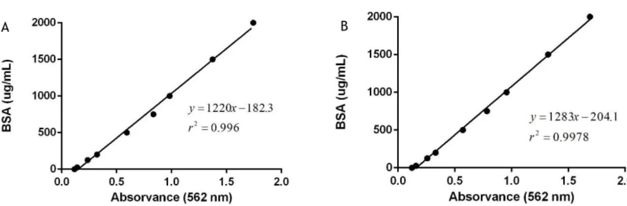
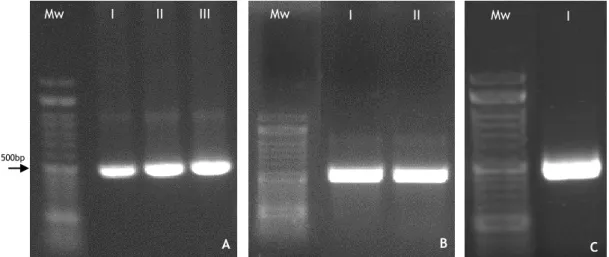
Figure 5: Schematic of STEAP1 protein structure, cellular localization and physiologic functions. Figure 6: A: BSA (lysis buffer) calibration curve; B: BSA (elution buffer) calibration curve. Figure 7: Agarose gel electrophoresis of two critical moments during the construction of the expression vector pET101/D-STEAP11-142. A: PCR products of STEAP11-142 amplification at
different annealing temperatures, 60 ºC, 62 ºC and 65 ºC, respectively; B: Highly purified plasmid pET101/D-STEAP11-142 from E. coli TOP10 competent cells; C: Highly purified pET101
vector cloned with STEAP11-142 DNA insert from E. coli BL21(DE3).
Figure 8: E. coli BL21 (DE3) growth profile of pre-inoculum cells in LB, TB, SOB, SOC and 2YT liquid culture media.
Figure 9: A direct comparison between E. coli BL21 (DE3) growth profile in TB, SOB and SOC liquid culture media.
Figure 10: Western blot analysis of human STEAP11-142 peptide production and cellular
compartmentalization in TB, SOC and SOC liquid culture media during the biosynthesis process, under the presence (w – with) and absence (w/ no – without) of inducer IPTG.
Figure 11: Western blot analysis of human STEAP11-142 production in TB medium, in the presence
of lactose inducer and an integrated comparison with IPTG induce (w – with) and non-induced (w/ no – without) cultures.
Figure 12: A: Western blot analysis of STEAP11-142 production in TB medium, omitting the
addition of an inducer, in time intervals of 2 h during 12 h; B: Western blot analysis of STEAP1 1-142 production in TB medium, omitting the addition of an inducer, in time intervals of 2 h during
12 h, using fermentation conditions previously described as ideal (351 rpm, 35 ºC and pH 6.2). Figure 13: Western blot analysis of the STEAP11-142 recovery using 1 % (v/v) of several common
detergents in the resolubilization of the target protein membrane fraction.
Figure 14: A: Chromatographic profile of STEAP11-142 purification from E. coli lysates on a
HisTrap FF crude (5 mL column volume) with nickel ions immobilized; B: Western blot/SDS-PAGE analysis of collected fractions. STEAP11-142 position in the western blot gel is at the correct
position (17-25 kDa); C: STEAP11-142 fraction in SDS-PAGE gel (Comassie staining) is represented
by an arrow. Blank line represents absorbance at 280 nm, while dashed line the imidazole concentration. Binding process was performed at 50 mM imidazole at 0.5 mL/min flow rate followed by an increasing elution linear gradient to 500 mM imidazole at 1.0 mL/min flow rate. A final step at 500 mM imidazole, 500 mM NaCl, 50 mM Tris and 1 mM MgCl2 buffer at pH 7.8 and 1.0 mL/ min flow rate was also performed.
Figure 15: A: Chromatographic profile of STEAP11-142 purification from E. coli lysates on a
HisTrap FF crude (5 mL column volume) with cobalt ions immobilized; B: Western blot/SDS-PAGE analysis of collected fractions. STEAP11-142 position in the western blot gel is at the correct
position (17-25 kDa); C: STEAP11-142 fraction in SDS-PAGE gel (Comassie staining) is represented
by an arrow. Blank line represents absorbance at 280 nm, while dashed line the imidazole concentration. Binding process was performed at 50 mM imidazole at 0.5 mL/min flow rate followed by an increasing elution linear gradient to 500 mM imidazole at 1.0 mL/min flow rate. A final step at 500 mM imidazole, 500 mM NaCl, 50 mM Tris and 1 mM MgCl2 buffer at pH 7.8 and 1.0 mL/ min flow rate was also observed.
List of tables
Table 1: Brief description of main FDA-approved drugs used to treat PCa and their respective effects.
List of abbreviations
ABC Adenosine triphosphate-binding cassette ADT Androgen deprivation therapy
AR Androgen-receptor
ATP Adenosine triphosphate
BPH Prostatic hyperplasia
BSA Bovine serum albumin
CHAPS 3-[(3-cholamidopropyl) dimethyl-ammonio]-1-propanesulfonate COMT Catecol O-Metiltransferase
CRPC Castration-resistant prostate cancer
CTL Cytotoxic T lymphocytes
CVs Column volumes
CZ Central zone
DHT 5α-dihydrotestosterone
DNA Deoxyribonucleic acid
DNase Deoxyribonuclease I
E. coli Escherichia coli
E2 17β-estradiol
EPCA Early prostate cancer antigen ETS E26 transformation specific EZH2 Enhancer of zeste homolog gene 2
FDA United States Food and Drug Administration GltP Glutamate proton symporter
GnRH Gonadotropin releasing hormone GPCRs G protein-coupled receptors hSGLT1 Human sodium-glucose transport 1
HTL Helper T lymphocytes
IMAC Immobilized metal affinity chromatography IPTG Isopropyl-β-D-1-thiogalactopyranoside
LB Luria-bertani
LHRH Luteinizing hormone-releasing hormone
mAb Monoclonal antibody
MAPEG Membrane-associated proteins in eicosanoid and glutathione MCTs Monocarboxylate transporters
NMR Nuclear magnetic resonance
OD Optical density
PCa Prostate cancer
PCA3 Prostate cancer antigen 3 PCR Polymerase chain reaction
pET101/D ChampionTM pET101 Directional TOPO®
PIN Prostatic intraepithelial neoplasia PSA Prostate specific antigen
PSCA Prostate stem cell antigen
PSMA Prostate specific membrane antigen PVDF Polyvinylidene difluoride
PZ Peripheral zone
SDS Sodium dodecyl sulfate
SDS-PAGE Sodium dodecyl sulphate-polyacrylamide gel electrophoresis
SPR Surface plasmon resonance
STD-NMR Saturation-transfer difference NMR
STEAP Six-transmembrane epithelial antigen of the prostate
TAA Tumor-associated antigen
TB Terrific-broth
TMPRSS2 Transmembrane protease serine 3
TZ Transition zone
UGS Urogenital sinus
YidC Integral membrane chaperone uPA Urokinase plasminogen activator
Chapter 1
Introduction
1. Anatomy and Physiology of the Prostate
The development of the prostate begins at 10-12 weeks of gestation, being the first molecular modification the growth of prostatic bunds from its embryonic precursor, the urogenital sinus (UGS). It includes organ determination, epithelial budding, duct elongation, branching morphogenesis, cellular differentiation and maturation (1-4). The mutual interactions between the stromal and epithelial compartments are also crucial to initiate the postnatal development and maturation of the prostate (5).
The process inherent to prostate development is dependent on the action of sex steroid hormones that occurs in response to testicular androgen-mediated interactions, wherein the biological active 5α-dihydrotestosterone (DHT), derived from local conversion of fetal testosterone by 5α-reductase plays the main role (1-8). Testosterone also acts in order to maintain a totally differentiated epithelium with secretory functions, being then required for the maintenance of prostatic and glandular functions(4, 8).
After birth, serum testosterone levels decrease and the prostate stagnates its growth, weighing about 2g in childhood (4, 7). Only at puberty, its exponential growth due to augmented levels of androgens, secreted from the testes, stimulate the last morphofunctional changes needed to establish the adult phenotype, which is constituted by a multicellular mesenchymal layer surrounding secretory ducts with basal and luminal epithelial cells as well as neuroendocrine cells (2, 4, 5, 7). Until near 25 to 30 years of age, the prostatic weight remains similar and starts to rise slowly, reaching about 20 g, presenting a shape similar of a walnut and measuring 3 cm in length, 4 cm in width and 2 cm in depth (2, 6).
McNeal has established the most accepted model for the anatomy and physiology of the prostate. The adult male human prostate is composed by: i) peripheral zone (PZ), ii) central zone (CZ), iii) transition zone (TZ) and iv) the anterior fibromuscular stroma zone. This last structure is surrounded by a capsule of stromal fibromuscular tissue, within an inner layer of smooth muscular and an outer covered of collagen (2, 6, 9-13). Regarding the anatomic location, the prostate base is connected to the bladder neck, surrounding the proximal urethra in the anteromedial prostate, and the apex is under the urogenital diaphragm up to the midprostate, between the rectum and the symphysis pubis, in a subperitoneal region essentially composed by skeletal muscle fibers (2, 6, 10). The seminal vesicles constitutes an important accessory gland located superiorly to the prostate base, towards the vas deferens to form the
ejaculatory ducts, performing a connection with the vasa or ducti deferentia through a layer from the bladder muscle, also covering the prostate (2, 6).
Figure 1: Diagrams of frontal and sagittal sections of the male urogenital complex, illustrating the
anatomical position of the adult prostate and associated structures (14).
The glandular prostate comprises the UGS-derived PZ, which constitutes 65% of a normal organ mass and it is divided into an anterior and posterior branches, exiting posterolaterally from the verumontanum, in the urethral wall, to the prostate apex, wherein the carcinoma is more susceptible to occur (2, 9-12, 15). The CZ is a cone shape region derived from the Wolffian duct, comprising 30% of the total prostate mass, with ducts arising on the verumontanum and then projected to cover the confluence region of the ejaculatory ducts and the prostatic urethra (2, 9, 10, 15). The transition zone TZ encompasses only 5%, contains the urethral sphincter and a segment of the proximal urethra, from the verumontanum to the bladder neck, being also organized into two similar lobes of glandular tissue and muscle fibers, whose ducts leave the posterolateral gap of the urethral wall and extend laterally (2, 9, 11, 12, 16). The anterior fibromuscular stroma has an apical half rich in striated muscle, converging into the prostate and muscles of the pelvic diaphragm, stimulating cell proliferation and presenting secretory functions, being also composed by a base predominant in smooth muscle cells, extending into the fibers of the bladder neck (1, 2, 4). The distal portion is relevant for the voluntary sphincter functions, whilst the involuntary ones are attributed essentially to the proximal region (2).
The main functions of the prostate consist in providing proteins, ions and nutrients needed for a correct composition of the prostatic secretions, which are then mixed with the fluids released from the seminal vesicles, creating the final ejaculate (4, 6). This is crucial in further processes
related to semen gelation, coagulation and liquefaction, in the coating and uncoating of spermatozoa and its interaction with cervical mucus, essential for the fertility success (4).
2. Prostate Cancer
Prostate cancer (PCa) is the second most prevalent type of cancer among man and the sixth leading cause of male cancer-related death worldwide (5, 17). In 2013, 238.590 new cases of PCa were diagnosed in man ranging 45 to 80 years of age and an estimated value of 29.729 patients died, particularly in underdeveloped countries (17, 18). The risk factors are a combination of endogenous factors, including hormones, aging, oxidative stress and exogenous, related to environmental risk factors, such as lifestyle habits, geographic residence area and socioeconomic standing (19-21). The genetic factors also has a direct role in the incidence of PCa and individual susceptibility to acquire this disease, mainly the family history and ethnicity, with special incidence in Afro-Americans, followed by Caucasians and Asians (3, 19, 20, 22). PCa is divided into 2 different types, familial and sporadic. Familial disease is characterized by the appearance of cancer at early ages, less than 55 years old, in members of the same family. Sporadic PCa is triggered in cases when the genetic material is damaged by environmental constrains during life (20). The pathophysiology of PCa englobes benign lesions, namely benign prostatic hyperplasia (BPH) or malignant, such as prostatic intraepithelial neoplasia (PIN) or adenocarcinoma (23), as represented in figure 2.
Figure 2: Progression pathway for human PCa, including pathological stages and the most relevant genetic
alterations at each stage (24).
The tumor growth starts generally slow and asymptomatic and may involve an indolent course without metastatic disease development (25). As cancer develop, the prostate has multiple tumor foci that become progressively less organized, with smaller ducts, until occur a complete
loss of these structures (26). Likewise, gene expression is changed according to cancer stage and prostatic lesion type (27). Therefore, the cancer is capable to invade surrounding tissues and contribute to the development of metastasis (26, 27). Usually, cancer-related death is attributed to metastatic process, where tumor cells acquire the ability to spread from a primary to a secondary site. This is a complex process that includes the degradation of the basement membrane, the invasion, intravasation, anoikis evasion and extravasation of the extracellular matrix (28, 29).
2.1. The Role of Androgens in Carcinogenesis
The normal prostate cells are dependent of androgens, which maintain their growth, survival and terminal differentiation. In addition, androgens play an important role for the normal architecture, homeostasis and physiological function of the prostate (10, 30). The mechanism of androgens action is mediated through the androgen receptor (AR), which is expressed in several adult and fetal reproductive tissues, such as prostate, testis and seminal vesicle. The main androgen with biological action in the prostate is DHT, which presents higher affinity and activity than testosterone, and it plays both stimulatory and inhibitory roles in order to regulate the normal prostate growth and biologic functions (3, 5, 31-33).
The AR is a member of the nuclear steroid receptor superfamily of transcription factors, with a molecular weight of 110 kDa (30, 31). It is located on the X-chromosome (Xq11-12), composed up to eight exons, encoding approximately 919 amino acids, with different functional motifs namely an amino-terminal domain with a transactivation domain 1, crucial for the regulation of primary transcription, a DNA-binding domain containing two zinc fingers responsible for DNA recognition, dimerization and stabilization, which can be connected to the hinge region, being responsible for the regulation of receptor nuclear import through a nuclear localization signal. Moreover, it has a C-terminal domain, comprising a ligand binding domain and the transactivation domain 2, also involved in regulation of transcription (31, 34-36)
The initial state of AR is inactive, but becomes active when bound to DHT. Conformational changes allow AR dimerization, which is accompanied with the dissociation of cytoplasmic heat shock proteins, and consequently, it may translocate into the nucleus. Then, it binds to androgen-response elements within the gene regulatory region, stimulating or inhibiting the expression of several genes (31, 37-39).
Figure 3: Schematic representation of the AR gene and protein structure (37).
In PCa cells, the AR is broadly expressed in the majority of initially diagnosed patients and the androgen/AR signaling axis has an active function in early stages of the disease. Taking into account the role of AR in cell proliferation, it is considered the main driver of prostate malignant cells development and progression (3, 5, 38, 40, 41). It is well documented that many patients will eventually become castration-resistant prostate cancer (CRPC), and there are several studies showing the involvement of AR, such as AR mutations, imbalance of AR co-regulators, alteration of selective androgen/AR signal transduction pathways and changes in the surrounding microenvironment and stromal cells or de novo synthesis (figure 3) (36, 38, 42).
Figure 4: Mechanisms of androgen independence in PCa (38).
Actually, over 300 AR mutations have been identified in PCa patients and cancer cell lines, being its frequency and nature dependent on the cancer stage. In localized primary PCa, mutations of the AR are uncommon (0%-4%), but their frequency is significantly increased in advanced or metastasized profile (8, 38, 43, 44). The most relevant essentially result in
alteration or loss of AR function or represent a disruption of signaling pathways. Functional studies revealed a gain of function mutations, characterized by increased binding affinity and the consequent activation of the AR by estrogenic and progestagenic steroids, adrenal androgens and DHT metabolites. Cells carrying this type of mutations present a growth advantage in androgen-deprived environment, essentially due to alteration in cofactor recruitment and reduction of ligand specificity (38, 43-45).
The functional levels of androgens can be maintained through the conversion of adrenal androgens or intratumoral de novo synthesis of androgen, which is considered a relevant mechanism contributing to reactivate and restore the AR transcriptional activity (46, 47) Then,
de novo synthesis of steroids in PCa cells is a possible reason for the progression into an
castration-resistant profile, contributing to tumorigenesis in the absence of testicular androgens (5, 8, 38, 48).
2.2. Diagnosis and Treatment
At early stages, the prostate specific antigen (PSA) is used to detect patients with PCa, which is increased in serum levels (19, 20). However, there are several limitations because serum PSA levels may also be increased in cases of BPH (49). In addition, the quantity of PSA levels in serum are not capable to predict disease aggressiveness or if the tumor will progress into a metastatic stage (19, 50-52). Another diagnostic marker is the urinary prostate cancer antigen 3 (PCA3), which was firstly identified in 1999 as overexpressed in more than 95% of primary cancer tissues and associated with metastasis (53). Although there is a correlation between the PCA3 levels and PCa risk, it does not ensure tumor existence, since many cancerous patients may present low PCA3 levels (54, 55).
Therefore, it is needed to identify novel biomarkers to detect aggressive prostatic lesions. For that, several complementary methods are usually performed, including the screening of molecular differentiation between PCa stages, application of adjuvant therapies to enhance cancer regression, imaging techniques, trans-rectal examination with ultrasound guidance and biopsy (19). Together with the assessment of several clinical features of patients such as age, family history, tumor stage, serum PSA, Gleason score, number of positive prostate biopsies and the amount of malignant tissue, it is possible to establish a most suitable and appropriated treatment (19).
Usually, active surveillance is applied to patients with Gleason score of 6 or less, avoiding unnecessary or potentially harmful treatment, since this type of PCa is considered inoffensive. Regarding the PCa with low recurrence risk or in early stages, the primary treatment consists in radiation-based therapy or radical prostatectomy, which demonstrated to be effective in cancer reverse by destruction or excision of damaged tissue (19, 31, 38, 56). However, PCa can undergo recurrence and progress into a metastatic profile. In this context, the standard
treatment applied in the last 70 years has been surgical or chemical castration, also known as androgen deprivation therapy (ADT) (31, 57, 58).
Briefly, the ADT treatment consists in blocking the AR/androgen pathway or trigger an improperly activation of the AR, using radical radiotherapy as a complement. For that, it is necessary to submit patients to hormonal manipulations through androgen antagonists or agonists for gonadotropin releasing hormone (GnRH), which cause tumor shrinkage, retard it growth and progression, besides prevent testosterone production, based on the androgen dependency of prostate cells to survive. Surgical methods can also be applied, namely orchiectomy or chemically with luteinizing hormone-releasing hormone (LHRH) agonists, LHRH antagonists or anti-androgens in combination with castration by combined androgen blockade for reducing tumor burden and associated pain (31, 59-61). The newest upgrades on the biologic mechanisms underlying PCa allow to apply novel chemotherapeutic drugs approved for the United States Food and Drug Administration (FDA) as a complementary strategy, which have been effective in improving the survival time of man with advanced PCa, as summarized in table 1.
Table 1: Brief description of main FDA-approved drugs used to treat PCa and their respective effects.
Drug Class Effect References
Abiraterone acetate A n d ro gen antag on is t
Inhibit androgen biosynthesis
Increase average survival time of patients Suppress testosterone concentration in tumor microenvironment
(19, 59-62)
Bicalutamide
Decrease pain
Minimize sexual dysfunction Antitumor activity
Improve cancer-associated symptoms
(19, 30, 61, 62) Cyproterone
acetate
Suppress androgen binding to AR
Increase programmed necrosis (19, 40)
Enzalutamide Increase average survival time of patients
Prevent nuclear translocation and chromatin binding of AR (19, 60, 61) Flutamide
Decrease PSA levels and relieve cancer-associated symptoms
Suppress androgen binding to AR
(30, 37) Cabazitaxel Ch em ot h erapeu ti c d ru g
Increase average survival time of patients Decrease PSA levels and relieve cancer-associated symptoms
(19, 60, 62)
Docetaxel
Increase average survival time of patients Decrease PSA levels and relieve cancer-associated symptoms (19, 39, 60, 62) Leuprolide G n RH ag on is
t Persistent stimulation of pituitary gland
Suppression of testosterone production
(57, 60) Goserelin (56) Lupron LH RH ag on is t
Diminish androgen levels in local cancerous tissues (63)
Despite the initial PCa remission induced by ADT, this therapy is not completely efficient in eradicate the entire population of affected cell, besides negatively select subpopulations that survive in the absence of androgens. Moreover, after 2 to 3 years of treatment, a significant percentage (10%-40%) of patients experience disease relapse and starts to progress into widespread metastasis or transit from the initial androgen-dependent state to CRPC, which is characterized by the maintenance of the AR signaling and expression of AR-regulated genes, and it is also associated with a poor prognosis and short survival time (37-39, 60, 64).
Considering that the current established therapies are limited and induce side effects that impairs the life quality of patients, it is necessary to develop alternatives, which should be more effective and less toxic, in order to manage those who develop recurrent disease or present advanced tumor stage or metastatic disease. Therefore, innovative molecular biology
technologies, including differential display analysis, subtraction approaches and microarrays, which allow to identify several genes that encode transmembrane proteins (65, 66).
The discovery of novel membrane protein commonly founded in several human pathologies are nowadays a promising tool for a deeper understanding of biologic mechanisms underlying the diseases. Consequently, it is possible to perform a thorough study of relevant interactions relaying the target protein and 3D structure analysis. The ultimate goal is to develop therapeutic drugs with high binding affinity or design a proficient therapy capable to manage and revert the injurious effects induced by its overexpression in human tissues.
2.3. Transmembrane Proteins over-expressed in Prostate Cancer
The prostate-specific membrane antigen (PSMA) was identified in 1987 as a type II integral membrane glycoprotein overexpressed in PCa epithelial cells involved in hydrolyzing peptides in prostatic fluids (65, 67). It can be used to follow the progress of the disease in a posttreatment phase due to considerable sensitivity and specificity in distinguish PCa from other type of malignancies or as a target for monoclonal antibodies (mAb) (65).
The prostate stem cell antigen (PSCA) is a prostate-specific glycosyl phosphatidylinositol-anchored glycoprotein expressed on the cell surface, being its increased levels related with PCa presence, stage, progression and metastasis (65, 68). Despite its function is still unclear, PSCA may play a role in progenitor cell function, tumorigenesis and clinical progression (67). The early prostate cancer antigen (EPCA) is a nuclear structural protein associated with PCa presenting high sensitivity and specificity, suggesting its use as a significant biomarker (65, 69). The function of EPCA is still unknown, however it is suggested a relation with and early step in prostate carcinogenesis (67).
The enhancer of zeste homolog gene 2 (EZH2) is a member of the polycomb group of proteins, commonly overexpressed in PCa and promoter of cancer growth. EZH2 acts as a gene silencer by maintaining the transcription repressive state of genes. However, it also silences the expression of tumor suppressor genes triggering an increased overexpression in metastatic PCa, in comparison with localized and BPH (65, 67). Therefore, it could be useful as a biological marker to identify patients at risk of metastasis (70).
The urokinase plasminogen activator (uPA) axis represents a potential target for PCa due to its involvement in various phases of tumor development and progression (65, 71) Consequently, its raised levels are correlated with cancer stage and metastatic onset (65).
The transmembrane protease serine 2 (TMPRSS2) regulates the overexpression of E26 transformation-specific (ETS) transcription factors, which are considered a persistent feature
in approximately 50% of patients, and are involved in PCa progression by disrupting AR signaling (20, 65, 72).
The monocarboxylate transporters (MCTs), particularly MCT2 and MCT4, are transmembrane proteins with consistent overexpression in the majority of androgen unresponsiveness PCa, being then considered important biomarkers that allow to distinguish between affected tissues from non-neoplastic (73-75). Likewise, some general markers can also be correlated with PCa, including N-cadherin which provides information for PCa treatment, since it predicts cancer recurrence after ADT and also ZEB1, being its overexpression correlated with high Gleason scores (65, 76, 77).
Table 2: Description of main biomarkers for PCa and their respective functions.
Marker Description Function References
PSMA Type II integral membrane glycoprotein
Hydrolyze peptides in prostatic fluids Follow the progress of the disease in a posttreatment phase
Target for monoclonal antibodies
(65, 67)
PSCA
Prostate cell suface-specific glycosyl phosphatidylinositol-anchored glycoprotein
Related with cancer presence, stage, progression and metastases
Progenitor cell function, tumorigenesis and clinical progression
(65, 67, 68)
EPCA Nuclear structural
protein Early prostate carcinogenesis (65, 67, 69) EZH2 Polycomb group of
proteins
Promote cancer growth Gene silencer
Biomarker for metastatic patients
(65, 67, 70)
uPA
Serine protease Cancer development and progression
(65, 71) TMPRSS2 (20, 65, 72) MCTs Transmembrane monocarboxylate transporter
Biomarker for unresponsiveness PCa (73-75)
ADAM9 and CDON are also found overexpressed in PCa, despite the last present low specificity. Therefore, only ADAM9 is considered a therapeutic targets for PCa, contrary to CDON that need more studies regarding expression and function in human cancer (66, 78). Nowadays, the six-transmembrane epithelial antigen of prostate 1 (STEAP1) is considered the most proper candidate to be imposed as a potential therapeutic target, since it fulfills all the desirable characteristics, namely high expression associated with a high PCa specificity (66, 79).
3. Six Transmembrane Epithelial of the Prostate 1
The STEAP family comprises four members, namely STEAP1 to STEAP4, sharing a similar structure with six transmembrane domains, edged by a carboxy- and an amino-terminal intracellular domains (80-82). The majority of STEAP proteins has an YXXØ (in which Ø is a large hydrophobic amino acid) consensus sequence and the Rossman fold (GXGXXG/A motif) responsible for targeting transmembrane proteins to lysosomes and endosomes. These domains are intrinsic characteristics of proteins with oxidoreductase and dehydrogenase functions, respectively, and have the ability to bind nucleotides such as NAD and FMN (80, 81). Despite all the common features and high homology degree between STEAPs, their cellular functions, expression patters and subcellular localization are distinct (81, 82).
STEAP1 gene is located close to the telomeric region at the long arm of chromosome 7q21.13, includes 5 exons and 4 introns along 10.4 kb size and contains a gene cluster predicted to encode transmembrane proteins. The transcription mechanism origins 2 different mRNA of 1.3 kb and 4.0 kb (80, 81, 83). However, only the first one is translated into a protein with 399 amino acids and a molecular weight of 39.72 kDa (80-83).
Figure 5: Schematic of STEAP1 protein structure, cellular localization, and pathophysiologic functions
(80)
.
STEAP1 is not homologous to any other familiar proteins, since its structure suggests that it is a cell-surface molecule composed by six-transmembrane domains with the COOH- and N-terminals located in the cytosol, 3 extracellular and 2 intracellular loops (80, 84, 85). The transmembrane domains are located at 73-95, 117-139, 164-182, 218-240, 252-274, 289-311 base pairs of the amino acid sequence (83). In contrast with the other proteins, STEAP1 lacks N-terminal NADPH-oxidoreductase, FNO-like domain and Rossman-fold, turning it the smallest of the classical STEAPs (80-82, 86). Additionally, this protein keeps two conserved histidine residues implicated in FRE heme-binding proteins and contains a flavin-NADPH-binding domain, despite structurally distinct and located at C-terminal (87). It also presents another
heme-binding domain called “apoptosis, cancer and redox associated transmembrane”, which facilitates the transmembrane electron transfer, and consequently, affects cell growth and its metabolism, as represented in figure 5 (25, 88).
Concerning the ferric oxidoreductase activity of STEAP1, it contributes to the generation, metabolism and increased levels of intracellular reactive oxygen species (ROS), which induces the expression of redox-sensitive and pro-invasive genes (80, 81, 83, 86, 89). In addition, it activates several signaling pathways involved in metastatic and proliferative cancer cells. Thus, the upregulation of ROS-levels, as a consequence of STEAP1 overexpression, is a hallmark of several cancers, including PCa (86, 89).
3.1. Expression and Function in Human Tissues
STEAP1 is highly expressed in several types of human cancers as well in cancer cell lines, namely in prostate, bladder, breast, colon, cervix, ovary, pancreas, testis and Ewing sarcoma (80, 84, 86, 90). The expression of STEAP1 is almost restricted to the prostate gland, mainly at cell-cell junctions located in the plasma membrane of prostate epithelial cells or in basal layer, which may behave as a prostate stem cells reservoir (23, 80). Although at very low levels, this protein is also present in non-tumoral tissues such as breast, colon, fallopian tubes, pancreas, pituitary, stomach, ureter and uterus (83, 84, 90).
The primary location of this protein is in the columnar epithelial cells at the apical plasma membrane of cell-cell junctions, besides being also dispersed in the cytoplasm and endosomal membranes (84, 90, 91). Herein, it plays a role in iron metabolism caused by the ubiquitous expression and partial co-localization with transferrin, transferrin receptor 1 and endosomes, which are specialized in iron and cooper uptake (82, 86). Therefore, due to its location and secondary structure, STEAP1 acts as an ion channel or transporter protein in both tight and gap junctions or even in cell adhesion processes, playing an important role in intracellular communication between tumor cells and surrounding stromal cells (79, 84, 90). Likewise, it has the capability to modulate the proliferation and invasion of cancer cells through the adjustment of intracellular concentration of few ions such as Na+, K+, Ca2+ and then triggering an enhanced
effect in cell growth and, consequently in cancer development (79, 80, 92).
The cellular localization and expression patterns of STEAP1 may depend on histologic diagnosis (25, 84). Regarding BPH lesions, this protein is found in basal cells, while in PIN lesions it is located in luminal, basal and stroma cells (23, 25, 84). High levels of STEAP1 are associated with worse prognosis in PCa, being associated with cancer recurrence and aggressiveness (25). STEAP1 constitutes an important target for bisphosphonates, since they are capable to reduce cell viability, in a dose-dependent manner and also decrease STEAP1 mRNA expression in PCa cells, due to its cytostatic effects (93, 94). During the last few years, the use of bisphosphonates
adhesion and invasion processes (95, 96). As Zoledronic Acid is the most powerful amino-bisphosphonate, which exerts its action by reducing cell proliferation or inducing cell apoptosis through low and high doses, respectively (97).
A similar study showed that a prolonged exposure to zinc induces an aggressive behavior of PCa cells, therefore enhancing tumor cells proliferation and simultaneously increasing the expression of STEAP1 genes (98). In normal prostate gland, zinc is highly expressed and it is essential for normal functions of prostate cells. On the other hand, this compound is significantly reduced in prostate malignancies and any alteration in its homeostasis triggers an uncontrolled growth of tumor cells and leads to cancer development. This is essentially due to an impairment in the expression of regulatory genes in several biologic processes and signal transduction pathways, regulated by zinc (98-100).
Considering the role of sex steroid hormones in PCa, STEAP1 regulation was evaluated by DHT and 17β-estradiol (E2) in LNCaP cells. Surprisingly, STEAP1 expression decrease in time- and dose-dependent manner in both cases. However, this effect is not mediated by their cognate receptors, and thus it is possible that the levels of STEAP1 decrease in response to sex steroid hormones in order to overcome the proliferation effects triggered by these hormones (91). Therefore, cells presenting higher expression levels of STEAP1 have a more aggressive behavior, confirming a relationship between with the AR at transcriptional level, which is reinforced by the genetic or pharmacologic suppression of STEAP1 mRNA by AR inhibitors (72, 101, 102). Expectations about immunotherapy have been raising, transforming this practice a more attractive and achievable. Herein, it is crucial to establish human cancer and cancer-associated cytotoxic T lymphocytes (CTL) lines, which represent only the anergized immune system and trigger a cancer-specific response (103). However, the absence of tumor-associated antigen (TAA) capable of inducing this sort of response constitutes the central problem and invalidate the development of effective immune-based therapies (104).
3.2. Therapeutic Target
As referred before, STEAP1 expression levels in human PCa are 5- to 10- fold higher, comparing to other cancer types. Also, considering the location of STEAP1 in cell membrane associated to its low or absent expression in normal tissues, its epitopes became strong tools to develop novel methodologies focusing human cancer and a potential target for immunotherapy (84, 85, 103-105).
Likewise, cancer immunotherapy mainly occurs through immunogenic binding epitopes, and some studies have demonstrated that STEAP186-94 and STEAP1262-270 arecapable to stimulate
antigen-specific CTL and helper T lymphocytes (HTL). Therefore, CTL and HTL recognize and destroy STEAP1-expressing cancer cells via TAA recognition mechanism, without elicit any significant autoimmune response (19, 103-105). Therefore, this is a promising method because
consists in a noninvasive option to treat minimal residual disease, to prevent metastatic spread, to inhibit the growth of prostate cancer cells or even to delay recurrences without compromising life quality (104). Nowadays, Sipuleucel-T is an active cellular immunotherapeutic agent constituted by antigen-presenting cells and several study cases demonstrated that it increase the average survival time of cancerous patients (19, 106, 107). DNA vaccines are another promising tool for immunotherapy due to their ability to induce a highly specific T cell response and simultaneously capable to overcome the immune system surveillance and tolerance mechanisms employed by cancer cells (108-110). Several vaccines were already used against overexpressed proteins in PCa, including STEAP1, with genes encoding cytokines or co-stimulatory molecules to enhance the specificity of the immune response. The results obtained demonstrated that these vaccines induce a delay or even a inhibition in tumor progression (110, 111).
Nowadays, antibody-based therapies have become a preferred choice for targeting cancer. STEAP1 immunogenicity encouraged the development of antibody-drug conjugates, combining anti-STEAP1 specific mAb with chemotherapeutic drugs with cytotoxic potential or a detectable label agent to be used as therapeutic approach (112). The connection between anti-STEAP1 mAbs and external loops of the protein suppresses its biological function and thereafter compromises all the oncogenic functions (79, 101, 113). The mAb needs to bind preferentially to malignant cells at a relatively high density, and internalize in a manner that allows for release of the drug from the linker in the appropriate intracellular compartment. The linker needs to attach the drug to the mAb in a manner that is stable in the circulation, but releases the drug once it is internalized. The cytotoxic drug needs to be nontoxic when linked to the mAb in the circulation, but capable of killing tumor cells once it is internalized and released (114).
Moreover, its capacity to recognize antigens with a high selective degree specific, creates the idea of targeting malignant cells whit minimal undesired collateral toxicity to normal tissue, as it is proven by the successful mAb: trastuzumab, rituximab, cetuximan and bevacizumab (79, 101, 114, 115). Recently, novel anti-STEAP1 mAbs with stronger binding capacity and higher affinity to cell surface epitopes were capable to immunoprecipitate STEAP1 protein, modulate its functions as transporter through the blockage of intercellular communications and induced a significant inhibition of tumor growth (79). Also, a recent technology consisting in a novel diagnostic agent, consisting in a mAb radiolabeled complex immunoreactive for full-length human STEAP1 was capable to accumulate itself in the bloodstream at tumor site and detect precisely metastatic cells (101).
Currently, STEAP1 is not produced and isolated in considerable amounts to perform complementary structural studies, which constitute a significant limitation. In order to
structure are achieved through in silico analysis. Once established the optimized conditions for STEAP1 production, techniques such as X-ray crystallography, circular dichroism, protein nuclear magnetic resonance (NMR), saturation-transfer difference NMR (STD-NMR), surface plasmon resonance (SPR) could be applied to provide complete information regarding STEAP1 crystalized structure and unspecific interactions (116-121).
4. Aims
To our best knowledge, there are no published studies in the literature focusing the implementation of a laboratory hands-on platform from the biosynthesis to purification of the STEAP11-142 immunodomain. A most effective manner to manage harmful effects of
overexpressed STEAP1 levels in human PCa can be achieved by understanding the interactions between STEAP11-142 purified extracts and immunotherapeutic agents. For that, it is necessary
to develop a biotechnology procedure capable of producing significant amounts of STEAP11-142
with considerable purity levels.
The main goals of this master thesis were:
- Construct the pET101/D-STEAP11-142 expression vector and transformation of E. coli
BL21 (DE3) host;
- Define ideal culture medium formulation and optimal fermentation conditions to achieve maximum STEAP11-142 yield;
- Discover STEAP11-142 cellular compartmentalization;
- Isolate the STEAP11-142 in its native state from heterologous system and establish the
most effective detergent for protein recover concerning the solubilization step; - Develop IMAC chromatographic profiles in order to obtain considerable levels of purified
Chapter 2
Materials and Methods
1. Materials
Ultrapure reagent-grade water was obtained with a Mili-Q system as well as lactose from Merck Millipore (Darmstadt, Germany). Agar and European bacteriological agar were acquired from Laboratorios CONDA (Madrid, Spain). Tryptone and yeast extract were obtained from BIOKAR Diagnostics (Allonne, France) Dipotassium phosphate, lysozyme and sodium dodecyl sulfate (SDS) were bought from AppliChem (Darmstadt, Germany). Monopotassium phosphate, magnesium chloride, magnesium sulfate, acid-washed glass beads, digitonin, ampicillin (sodium salt), isopropyl-β-D-1-thiogalactopyranoside (IPTG) and deoxyribonuclease I (DNase) were purchased from Sigma-Aldrich (St. Louis, MO, USA). Sodium chloride, glucose, tris base, Triton X-100, Tween 20, methanol and acetic acid glacial 99 % were acquired from Fisher Scientific UK (Loughborough, UK). Potassium chloride was obtained from Panreac (Barcelona, Spain). Glycerol was obtained from HiMedia Group (Mumbai, India). 3-[(3-cholamidopropyl) dimethyl-ammonio]-1-propanesulfonate (CHAPS) was acquired from AMRESCO (Solon, OH, USA). The NZY ladder VI and NZYColour protein marker II for estimation of peptide base pairs (bp) and mass, respectively and GreenSafe Premium were purchased from NZYTech (Lisbon, Portugal). All other chemicals were of analytical grade commercial available and used without further purification.
2. Strains, plasmids and media
The Escherichia coli (E. coli) TOP10F’ (Invitrogen, Carlsbad, USA) cells were used for deoxyribonucleic acid (DNA) manipulations. The E. coli transformants were selected on Luria-Bertani (LB) plates (10.0 g/L tryptone, 5.0 g/L yeast extract, 10.0 g/L sodium chloride, 35.0 g/L agar, pH 7.5) supplemented with 100 μg/mL ampicillin at 37 ºC. The plasmids pIRES-STEAP1 and pET101/D-hCOMT were applied on this purpose, while the pET101/D-STEAP1 was used as expression vehicle in E. coli BL21-Star (DE3) (Invitrogen) host. These cells were used for protein production in typical complex media, such as LB (10.0 g/L tryptone, 5.0 g/L yeast extract, 10.0 g/L sodium chloride, pH 7.5), SOB (20.0 g/L tryptone, 5.0 g/L yeast extract, 0.25 g/L sodium chloride, 10.0 mL/L magnesium chloride 1 M, 10.0 mL/L magnesium sulfate 1 M), SOC (20.0 g/L tryptone, 5.0 g/L yeast extract, 0.5 g/L sodium chloride, 0.2 g/L potassium chloride, 20.0 mL/L glucose 1 M, 10.0 mL/L magnesium chloride 1 M), 2xYT (8.0 g/L tryptone, 5.0 g/L yeast extract, 5.0 g/L sodium chloride) and Terrific-Broth (TB) (12.0 g/L tryptone, 24.0 g/L yeast extract, 4.0 mL/L glycerol, 100.0 mL/L fermentation salts, pH 7.2). All media were supplemented with ampicillin 100.0 μg/mL.
3. Construction of the pET101/D-STEAP1
1-142expression vector
The ChampionTM pET101 Directional TOPO® (pET101/D) expression kit (Invitrogen) was used for
the expression of human STEAP11-142 peptide in its native form, according to manufacturer’s
instructions. Briefly, DNA fragment with approximately 500 bp coding for STEAP11-142 was cloned
into another vector used as template for polymerase chain reaction (PCR). pET101/D-STEAP1142
was constructed by PCR in T100TM Thermal Cycler (Bio-Rad, Hercules, USA) using specific cloning
primers (forward primer: 5’-AAGG AATT CAGG AGCC CTTC ACCA TGGA AAGC AGAA AAGA CATC-3’; reverse primer: 3’-AATG AGCT CCTA ATGG TGAT GGTG ATGT GCAT GAAG TTGG ACAA TTGA TGCT-5’) with EcoR I and Sac I restriction site for directional cloning. PCR was conducted in the following conditions: initial denaturation at 95ºC for 5 minutes, 30 cycles denaturation at 95 ºC for 30 s, annealing for 65 ºC for 30 s and extension at 72 ºC for 1 min and final elongation step at 72ºC for 5 minutes. The amplified cDNA fragment was confirmed by agarose gel electrophoresis and purified using NZYMiniprep kit (NZYTech). Previously, we digested STEAP1 1-142 with EcoR I (Takara Bio Inc., Shiga, Japan) and Sac I (Takara Bio Inc.) and then cloned into
pET101/D, also digested with the same restriction enzymes, by T4 DNA ligase (NZYTech) over 4 hours. This construct was transformed into E. coli TOP10F’ cells, grown overnight at 37 ºC in LB-Agar plates containing 100 μg/mL ampicillin and colonies were screened by PCR. Several positive colonies were inoculated in 4.0 mL of LB medium and grown overnight at 37 ºC and 250 rpm. From these cultures, highly purified plasmids were extracted using NZYMiniprep kit and subsequently subjected to DNA sequence analysis to confirm the identity of the target gene.
4. Escherichia coli transformation and selection of positive clones
Since the target plasmid pET101/D-STEAP11-142 was confirmed to correspond to human STEAP1 1-142, it was introduced into E. coli BL21-Star (DE3) competent cells, used as host for STEAP11-142
overexpression under the effect of ampicillin as a selection marker. Cell lyse of a single half colony at 95 ºC for 10 min was previously performed and the transformation process was carried out using 2 μL of a sequenced positive plasmid pET101/D-STEAP11-142 per 40 μL vial host solution.
Furthermore, cells were plated in LB agar containing 100 μg/mL ampicillin and grown overnight at 37 ºC. High expression level of transformants were screened by PCR, conducted with the specific cloning primers (forward primer: 5’-AAGG AATT CAGG AGCC CTTC ACCA TGGA AAGC AGAA AAGA CATC-3’; reverse primer: 3’-AATG AGCT CCTA ATGG TGAT GGTG ATGT GCAT GAAG TTGG ACAA TTGA TGCT-5’) using the following conditions: initial denaturation at 95ºC for 5 minutes, 30 cycles denaturation at 95 ºC for 30 s, annealing for 65 ºC for 30 s and extension at 72 ºC for 1 min and final elongation step at 72ºC for 5 minutes. The amplified DNA was assessed by agarose gel electrophoresis. The other colony half was inoculated in 62.5 mL LB pre inoculum medium, supplemented with 100 µg/mL ampicillin and cells were grown at 37ºC and 250 rpm overnight. The inoculation volume was fixed to achieve an initial cell density at 600nm (OD600)
equal to 0.2, measured by spectrophotometry (Pharmacia Biotech, Ultrospec 3000, Denmark) and a specific pre inoculum volume was transferred to 62.5 mL LB inoculum medium, also
formula:
V𝑝𝑟𝑒 𝑖𝑛𝑜𝑐𝑢𝑙𝑢𝑚 × OD𝑝𝑟𝑒 𝑖𝑛𝑜𝑐𝑢𝑙𝑢𝑚 = OD𝑖𝑛𝑜𝑐𝑢𝑙𝑢𝑚 × (V𝑝𝑟𝑒 𝑖𝑛𝑜𝑐𝑢𝑙𝑢𝑚 + V𝑖𝑛𝑜𝑐𝑢𝑙𝑢𝑚)
These cells grown at 37ºC and 250 rpm, until OD600 reach the phase between 0.6 and 0.8. At
this moment, a solution containing 70% inoculum medium with pET101/D-STEAP11-142 plasmid
and 30% glycerol was prepared. Then, it was divided into criostem tubes and stored frozen at -80.0ºC until further use.
5. Biosynthesis of the STEAP1 fragment
Initially, cells containing the expression plasmid were grown at 37ºC in LB plates. Then, several colonies were inoculated in 25.0 mL of LB, S.O.B, S.O.C, 2xYT and TB media in 100-mL shake flasks. In initial assays, cells were grown at 37ºC and 250 rpm until OD600 reached 2.6. The
inoculation volume was fixed to achieve an initial OD600 of 0.2 and the fermentations were
conducted until an OD600 ranging 0.6-0.8. At this moment, IPTG and lactose were added to a
final concentration of 1 mM, in 1000 mL shake flasks. The bacterial growth profile was accessed till stationary phase. Once established the ideal fermentation conditions, the studies were focused on TB medium, over 8 hours of fermentation without applying any inducer. An additional test considering previous results obtained in a semi-defined medium by Silva and coworkers, was performed applying the predicted temperature and stirring rate ideal conditions of 35 ºC and 351 rpm, respectively (122). Finally, cells were harvested by centrifugation (3500 g, 20 min, 4 ºC) and stored frozen at -80.0 ºC until use.
6. Cell lysis and STEAP1
1-142solubilization
The bacterial cells pellet was resuspended in appropriate lysis buffer (500 mM NaCl, 50 mM Tris, 1 mM MgCl2, pH 7.8) and disrupted by 0.5 mg/mL lysozyme treatment during 15 min at
room temperature and mechanical action of acid-washed glass beads adapted from a previously defined ratio (1 g cellular mass : 2 g glass beads : 2 mL lysis buffer) (123). The mixture was vortexed 7 times for 1 min with an interval of 1 min on ice. Then, 0.10 mg/mL DNase I was added to the lysate immediately followed by centrifugation (16000g, 30 min, 4 ºC). After the centrifugation step, the solubilization was carried out incubating the 16000g pellet (P16000g) in 1% (v/v) Triton X-100 plus lysis buffer at 4 ºC overnight, until full solubilization. The 16000g supernatant (S16000g) was also collected, in order to understand the protein cellular compartmentalization, moment when these samples were discarded. In addition, four detergents namely SDS, Tween-20, CHAPS and Digitonin were tested for P16000 solubilization in order to compare their efficiency in STEAP11-142 fragment recovery. The samples were
conserved at -80 ºC until further purity and immunoreactivity analysis by SDS-PAGE and Western-blotting, respectively. The protein contents were measured by Pierce BCA Protein Assay Kit (Thermo Fisher Scientific) using BSA as standard and calibration control samples according to the manufacturer’s instructions.
7. STEAP1
1-142purification in immobilized metal ion affinity chromatography
All chromatographic separation assays were performed at room temperature on ÄKTA avant
purifier system with UNICORN 6.1 software (GE Healthcare) and all buffers pumped in the system were prepared with Mili-Q system water, filtered through a 0.20 µM pore size membrane (Schleicher Schuell, Dassel, Germany) and degassed ultrasonically. Two different Immobilized Metal Ion Affinity Chromatography (IMAC) commercial HisTrapTM FF crude 5 mL columns (GE
Healthcare) were packed according to company guidelines with two distinct metals, nickel and cobalt.
In initial trials, the columns were equilibrated with binding buffer (500 mM NaCl, 50 mM Tris-base, 1 mM MgCl2, 5 mM Imidazole, pH 7.8). Then, aliquots containing STEAP11-142 resuspended
pellet in binding buffer (diluted 1:2) were injected onto the column using 2 mL at a 0.5 mL/min flow rate along 5 column volumes (CVs). The elution undergone through a linear gradient using an elution buffer (500 mM NaCl, 50 mM Tris-base, 1 mM MgCl2, 500 mM Imidazole, pH 7.8) of 0
% to 100 % imidazole at 1.0 mL/min flow rate over 5 CVs. A final elution step was maintaining during approximately 2 CVs to perform a complete elution of the bounded. In all chromatography assays, pH profile, absorbance at 280 nm, conductivity and pressure were continuously monitored. The fractions of 2-6 mL corresponding to peak 1 and 2 were collected in Pierce™ Protein Concentrator PES (3.000 MWCO) and then concentrated at 4 ºC. The samples were conserved at -80 ºC until further purity and immunoreactivity analysis by SDS-PAGE and Western-blotting, respectively. The protein contents were measured by Pierce BCA Protein Assay Kit (Thermo Fisher Scientific) using BSA as standard and calibration control samples according to the manufacturer’s instructions.
8. Agarose gel electrophoresis
The DNA electrophoresis was performed on a gel containing 1% agarose (GRiSP, Oporto, Portugal) and carried out in Tris-acetic acid (TAE) buffer (40 mM Tris base, 20 mM acetic acid and 1 mM EDTA, pH 8.0). The run undergoes at 120 V for 30 min and the bands corresponding to STEAP11-142 DNA, containing Green Safe reagent, were visualized under ultra violet (UV) light
after using UVITEC FireReader (UVItec, Cambridge, UK).
9. Total protein quantification
The protein content in samples was measured by the Pierce BCA Protein Assay Kit (Thermo Fisher Scientific, Waltham, MA, USA), using bovine serum albumin (BSA) as the standards (0.025-2.0 mg/mL), according to manufacturer’s instructions in xMark™ Microplate Absorbance Spectrophotometer (Bio-Rad). The lysis buffer curve was applied to quantify solubilized STEAP11-142 pellets while elution buffer curve was used for purified samples.
10. SDS-PAGE and Western Blot Analysis
A total protein mass ranging 2 µg and 5 µg were submitted to reducing sodium dodecyl sulphate-polyacrylamide gel electrophoresis (SDS-PAGE). Samples were boiled in loading buffer containing 500 mM Tris-Cl (pH 6,8), 10 % (w/v) SDS, 0.02 % bromophenol blue (w/v), 0.2 % glycerol (v/v), 0.02 % β-mercaptoethanol (v/v) for 5 min and then run on 12.5 % stacking and resolving acrylamide (GRiSP) gels containing 0.1 % SDS, with a running buffer (25 mM Tris, 192 mM glycine, 0.1 % (w/v) SDS) at 120 V for 105 min. Then, one gel was stained by Coomassie brilliant blue overnight at room temperature over constant stirring followed by 1 h period in discoloration solution I (320 mL/L methanol, 56 mL/L acetic acid glacial 99%) and overnight in discoloration solution II (50 mL/L methanol, 70 mL/L acetic acid glacial 99%) at same conditions. The other gel was electrotransferred to a polyvinylidene difluoride (PVDF) membrane (GE Healthcare, Buckinghamshire, UK), for 30 min at 4 ºC in a buffer containing 10 mM CAPS and 10 % (v/v) of methanol. Subsequently, the membranes were blocked with washing buffer containing 5 % (w/v) milk for 60 min at room temperature and incubated overnight at 4 ºC with rabbit anti-STEAP1 at 1:300 dilution in washing buffer (Santa Cruz Biotechnology, Dallas, TX, USA) polyclonal primary antibody, followed by an incubation with an anti-rabbit IgG alkaline phosphatase secondary antibody at a 1:40000 dilution in washing buffer (Santa Cruz Biotechnology). The PVDF membranes were incubated with ECF substrate (GE Healthcare) for 5 min and immune-reactive proteins were detected by exposure to chemiluminescence detection with the Molecular Imager FX Pro Plus MultiImager (Bio-Rad).
A B