doi: 10.1590/0037-8682-0464-2017
Major Article
Corresponding author: Armando de Oliveira Schubach. e-mail: armando.schubach@ini.fiocruz.br
Received 12 December 2017 Accepted 8 October 2018
Favorable responses to treatment with 5 mg
Sb
v/kg/day meglumine antimoniate in patients with
American tegumentary leishmaniasis acquired in
different Brazilian regions
Jamyra Iglesias Cataldo
[1],
Fátima Conceição-Silva
[2],
Liliane de Fátima Antônio
[1],
Armando de Oliveira Schubach
[1],
Mauro Célio de Almeida Marzochi
[1],
Cláudia Maria Valete-Rosalino
[1],[3],
Maria Inês Fernandes Pimentel
[1],
Marcelo Rosandiski Lyra
[1],
Raquel de Vasconcellos Carvalhaes de Oliveira
[4],
Juliana Helena da Silva Barros
[5],
Raquel da Silva Pacheco
[1]and
Maria de Fátima Madeira
[1][1]. Fundação Oswaldo Cruz, Instituto Nacional de Infectologia Evandro Chagas, Laboratório de Pesquisa Clínica e Vigilância em Leishmanioses, Rio de Janeiro, RJ, Brasil.
[2]. Fundação Oswaldo Cruz, Instituto Oswaldo Cruz, Laboratório de Imunoparasitologia, Rio de Janeiro, RJ, Brasil.
[3]. Universidade Federal do Rio de Janeiro, Faculdade de Medicina, Departamento de Otorrinolaringologia e Oftalmologia, Rio de Janeiro, RJ, Brasil. [4]. Fundação Oswaldo Cruz, Instituto Nacional de Infectologia Evandro Chagas, Laboratório de Epidemiologia Clínica, Rio de Janeiro, RJ, Brasil.
[5]. Fundação Oswaldo Cruz, Instituto Oswaldo Cruz, Laboratório de Biologia de Tripanossomatídeos, Rio de Janeiro, RJ, Brasil.
Abstract
Introduction: Favorable responses in American tegumentary leishmaniasis (ATL) patients to treatment with 5 mg Sbv/kg/day
meglumine antimoniate (MA) has been reported in Rio de Janeiro, but little is known regarding the therapeutic response to low doses in patients from other locations. Methods: A retrospective review of medical records was conducted to compare the therapeutic
response to 5 mg Sbv/kg/day MA treatment among 36 patients who acquired ATL in Brazilian states other than Rio de Janeiro (OS
group) and 72 patients from Rio de Janeiro (RJ group). Results: One course of 5 mg Sbv/kg/day MA cured 72.8% of 81 cutaneous
(CL) and 66.6% of 27 mucosal (ML) leishmaniasis-infected patients: 70% in the CL/RJ group, 81% in the CL/OS group, 50% in the ML/RJ group, and 80% in the ML/OS group. After up to two additional treatment courses at the same dose, 88.9% and 85.2% of the CL and ML patients were cured, respectively. Adverse events were observed in 40% of patients in the CL/RJ group, 57% of the CL/OS group, 58% of the ML/RJ group, and 80% of the ML/OS group. No significant differences were observed in the cure rates or adverse effects between the RJ and OS groups. No patients required permanent discontinuation of treatment due to adverse events.
Conclusions: Patients with ATL acquired in both RJ and OS may respond to low-dose MA. While high-dose MA should remain the standard treatment for ATL, low-dose MA might be preferred when toxicity is a primary concern.
Keywords: American tegumentary leishmaniasis. Leishmania spp.; Leishmania (Viannia) braziliensis subpopulations.
Therapy. Meglumine antimoniate. Low-dose.
INTRODUCTION
American tegumentary leishmaniasis (ATL) is a group of complex diseases caused by different Leishmania species,
transmitted by insects of the Phlebotominae family. It is distributed in all the Brazilian states. ATL can affect the skin (cutaneous leishmaniasis - CL) and the mucosa of the upper airways and digestive tract (mucosal leishmaniasis - ML).
Leishmania (Viannia) braziliensis is the most frequent and
dispersed etiological agent in the Brazilian territory; L. (V.) guyanensis is a prominent agent in the Amazon basin; L. (Leishmania) amazonensis is less frequent in all Brazilian
regions1,2.
For decades, the World Health Organization (WHO) has recommended the use of systemic pentavalent antimonials (Sbv) for the treatment of CL. Although their therapeutic
response may vary in different regions, the use of 20 mg Sbv/
770
Cataldo JI et al. - Tegumentary leishmaniasis low-dose treatment
uncommon. Therefore, treatments for CL should also not be life-threatening, and adverse effects should be monitored3,4. In 2013, the Pan American Health Organization (PAHO) updated those recommendations and highlighted the need to incorporate scientific evidence available in each country regarding their respective national control programs5.
Intramuscular (IM) or intravenous (IV) meglumine antimoniate (MA) is recommended in Brazil as the first option for the treatment of ATL: for ML, 20 mg Sbv/kg/day for 30 days,
preferably concurrent with 400 mg pentoxifylline three times a day, and for CL, 10 to 20 mg Sbv / kg / day for 20 days. In both
cases, the limit of 3 ampoules (1,215 mg Sbv) per day is advised.
The Brazilian Ministry of Health defines therapeutic failure as an unsatisfactory response to two regular and sequential courses of treatment2. Although the therapeutic response to MA may present
regional differences6,7,8 resistance to antimonials is not a national public health problem, and 50 to 100% of the patients with an unfavorable response to the initial treatment respond satisfactorily to a second course of treatment with the same therapeutic scheme2,9. If a therapeutic failure ensues, the use of a second-choice drug such as amphotericin B (desoxycholate or liposomal forms) or pentamidine isethionate2 is suggested. In areas where
L. (V.) guyanensis is the main etiological agent, pentamidine
isethionate is the first-choice drug2. All three recommended
medicines are considered effective, although they may cause adverse effects and occasionally may lead to death. From 2001 to 2013, 337,336 cases of ATL were reported in Brazil, with 1,522 deaths (lethality = 0.45%)10,11. Since ATL not typically a life-threatening disease, this seems to be a high lethality rate when compared to other potentially lethal diseases such as tuberculosis (2.05%) and visceral leishmaniasis (6.56%)12,13. Several of these deaths may be consequent to the MA treatment administered in standard doses, with poor patient follow-up during treatment10.
Until the mid-1980s, patients with ATL were treated with a daily MA ampoule, regardless of body weight, in the Evandro Chagas National Institute of Infectious Diseases (INI), Oswaldo Cruz Foundation (Fiocruz), located in Rio de Janeiro city14,15. Such a practice was at the time recommended by compendia of infectious diseases16 and dermatology17. A retrospective study
of 151 patients treated with a daily MA ampoule between 1967 and 1982 allowed the average administered dose to be calculated according to patient body weight. Fifty-one of these patients were re-evaluated between 5 and 14 years post-treatment and remained clinically cured14. The therapeutic regimen of IM or IV 5 mg Sbv/
kg/day MA for 30 days was subsequently adopted in INI/Fiocruz as the first-choice treatment for CL18,19. However, adverse effects continued to cause temporary interruption of treatment in the elderly and patients with comorbidities. These patients, however, sustained improvement during the period without medication20.
Other authors reported that intermittent MA schedules were better tolerated than continuous regimens21. Based on these
observations, MA 5 mg Sbv/kg/day IM or IV was successfully
administered in up to three 10-day series interspersed with 10-day periods without medication for CL treatment in patients above 60 years, those with comorbidities, or after discontinuation of systemic treatment due to adverse effects22. In different series of
cases of CL18,19,20,23 or ML20 patients treated with 5 mg Sbv/kg /
day MA, the frequency of successful treatment varied between 80 and 86%. These responses were similar to the efficacy of 76.5% reported for the conventional dose of 20 mg Sbv / kg / day9, but
with a lower frequency of adverse effects24.
Clinical trials are the most appropriate studies to evaluate treatments5,25. However, published clinical trials comparing low-dose MA (5 mg Sbv/kg/day) with the standard high-dose
(20 mg Sbv/kg/day) regimen are rare and restricted to Rio de
Janeiro19,26. In 2017 the Brazilian Ministry of Health considered
that the therapeutic regimen with MA 5 mg Sbv/kg/day as used in
Rio de Janeiro could be utilized in other regions of the country when the use of 20 mg Sbv/kg/day poses a risk for the patients
due to their clinical conditions2. However, it was noted that the
available studies were developed with patients infected with the genotypes of Leishmania sp. in the state of Rio de Janeiro.
However, in the last three decades, the conventional dose of 20 mg Sbv/kg/day has not been commonly used at INI/
Fiocruz for the treatment of ATL22, regardless of the Brazilian
state in which patients acquired the infection23. Between 2001
and 2013, 777 patients with ATL were treated at INI/Fiocruz10,
comprising 581 (74.8%) CL and 196 (25.2%) ML cases. In 86.1% of patients infections were acquired in Rio de Janeiro, and in other Brazilian states in 13.9%. Patients with poor therapeutic responses, and those who relapsed after an apparent good initial response were generally treated with 5 mg Sbv/kg/day MA or
with intralesional MA, once or twice before attempting treatment with another drug, thus accomplishing a total of 997 courses of treatment in the 777 patients. Out of the 997 courses of treatment performed, 91.7% were executed with MA: 73.1% with 5 mg Sbv/kg/day, 12.2% via the intralesional route, and only 6.4%
with 20 mg Sbv/kg/day10. Most of the patients in the last group
had been included in a clinical trial for CL26 or ML (ongoing).
Amphotericin B (desoxycholate or liposomal forms) was used in 6.3%, pentamidine isethionate in 1.2% and other drugs in 0.8% of the patients. The lethality coefficient was 0.1%, the relapse incidence was 5.8%, and the late ML incidence post-CL treatment was 0.25%. In regard to the outcomes for the 777 patients, 95.9% were cured, 0.1% died (one patient treated with 20 mg Sbv/kg/
day), and 4.0% abandoned follow-up10. A high lethality would
be expected considering INI/Fiocruz is a reference center which receives serious and complicated cases from different Brazilian regions; however such a lethality did not occur. Furthermore, if alternative low-dose regimens with MA were inadequate, a high incidence of skin lesion reactivations and late mucosal lesion development would be expected, but those did not occur either.
In this study, we compared responses to treatment with 5 mg Sbv/kg/day MA administered to patients from the state of Rio
de Janeiro with treated patients from other Brazilian states. In addition, we characterized the species of Leishmania and the subpopulations of L. (V.) braziliensis acting as etiological agents.
METHODS
Study design and patients
ATL: those who acquired Leishmania infection in the state
of Rio de Janeiro (the RJ group), and those who acquired
Leishmania infection in Brazilian states other than Rio de
Janeiro (the OS group).
The studied population was composed of ATL patients treated between 2000 and 2011 at INI. During the study period, the same protocol was used for diagnosis, treatment, and follow-up by a medical team composed of dermatologists, ear, nose, and throat specialists, and infectious diseases specialists. Mucous membranes were evaluated by nasal endoscopy and video-laryngoscopy with the aid of rigid endoscopes at 30° or 70° (Storz, Germany), in each medical appointment. For diagnostic purposes, patients underwent biopsy of skin and mucosal lesions for isolation of promastigote forms of Leishmania sp. in biphasic
Novy, MacNeal, and Nicolle medium (NNN) plus Schneider's
Drosophila medium (Sigma-Aldrich) supplemented with 10%
fetal bovine serum. All the isolates were cryopreserved in a sample bank for later identification of the involved species.
Patients with CL were treated with 5 mg Sbv/kg/day MA
intramuscularly or intravenously over 30 days26.Patients with ML were treated with MA 5 mg Sbv/kg/day intramuscularly or
intravenously for 30 to 120 days until epithelialization and a non-infiltrated, or a minimally infiltrated mucous membrane was observed20. During the treatment period, MA vials were
dispensed in a sufficient amount to be administered daily until the next scheduled medical appointment.
Treatment responses were checked in CL and ML patients, respectively, by dermatologic and ear, nose, and throat examination every 7 to 10 days during treatment, and in 1, 3, 6, 9, 12, 18, 24, 36, 48 and 60 months after the end of treatment. For both CL and ML patients, epithelialization was defined as ulcer closure without any erosion. For CL patients, complete healing (scarring) was characterized by epithelialization and absence of crusts, desquamation, infiltration, or erythema2,26. For ML patients, a cure was defined as epithelialization and absence of mucous membrane infiltration without post-treatment recurrence2,20. Epithelialization time and complete healing time were measured in days and compared between the groups.
For both CL and ML patients, clinical worsening was defined as the deterioration of the presenting signs or the appearance of new lesions, verified at any medical appointment. A poor therapeutic response was defined as an inability to achieve a clinical cure2,26. The patients with poor therapeutic responses or those whose lesions relapsed after apparently positive initial responses were typically treated subsequently with 5 mg Sbv/kg/
day MA, intralesional MA, or amphotericin B (desoxycholate or liposomal forms).
Clinical evaluation of adverse events was performed every 7 to 10 days during treatment and 1 month after the end of treatment. Electrocardiograms and peripheral blood samples for routine laboratory tests (complete blood count, and serum aspartate transaminase, alanine transaminase, gamma-glutamyl transferase, alkaline phosphatase, amylase, lipase, glucose, urea, and creatinine) were obtained at baseline and each clinical assessment2,20,26. Clinical, laboratory, and electrocardiographic adverse events were graded using a toxicity scale adapted from
the Division of AIDS Table for Grading of Severity of Adult and Pediatric Adverse Events27.
Criteria for the selection of patients in this study
All patients that acquired Leishmania infection in Brazilian
states other than Rio de Janeiro and: 1) were diagnosed by the isolation of Leishmania spp. in culture; 2) the isolates remained
cryopreserved for species identification, and 3) who initiated and concluded treatment with 5 mg Sbv/kg/day MA at INI/Fiocruz
were included in the OS group. For each patient selected for the OS group, we randomly selected two patients that acquired
Leishmania infection in the state of Rio de Janeiro and which
fulfill above criteria 1, 2 and 3 (the RJ group).
Each patient was questioned regarding the site of acquisition of Leishmania infection during a careful interview, with
questions regarding current and previous places of residence, trips for work or leisure reasons, and possible exposures in endemic areas.
Sample preparation
Leishmania promastigotes isolated from each patient were
stored in liquid nitrogen (N2) and subsequently recovered after two passages in Schneider’s Drosophila medium (Sigma,
Steinhein, Germany) supplemented with 10% fetal bovine serum (WL - Inmunoquimica) at 26ºC. Parasites in the stationary phase were harvested by centrifugation at 4.000 rpm for 10 min at 4ºC and washed several times with sterilized phosphate-buffered saline, pH 7.2. The L. (V.) braziliensis reference strain MHOM/
BR/75/M2903 was used as a control in all experiments.
Leishmania sample identification
We used multi-locus enzyme electrophoresis (MLEE) for
Leishmania identification28. Five enzyme systems were used:
6PGDH (6-phosphogluconate dehydrogenase, EC.1.1.1.43); GPI (phosphoglucose isomerase, EC.5.3.1.9); NH (nucleoside hydrolase, 2 loci, EC. 3.2.2.1); G6PDH (glucose-6-phosphate dehydrogenase, EC.1.1.1.49), and ME (malic enzyme, EC. 1.4.1.9) (Sigma, Steinhein, Germany). Considering the most frequent Brazilian dermotropic species, L. (V.) braziliensis
(MHOM/BR/75/M2903), L. (L.) amazonensis (IFLA/BR/67/
PH8) and L. (V.) guyanensis (MHOM/BR/1975/M4147)
reference strains were used as controls in all MLEE experiments.
Evaluation of ITS2 region heterogeneity in
Leishmania braziliensis
We used PCR assays and amplified product sequencing to compare a representative panel of L. (V.) braziliensis samples
isolated from RJ and OS patients. DNA from promastigotes was extracted using a DNAzol (Invitrogen, Life Sciences, USA) kit according to the manufacturer’s instructions. Then, DNA was stored at −20°C until tests were performed.
772
triphosphate (dNTP), and 3 mM MgCl2, and 1 U AmpliTaq Gold® PCR polymerase (Applied Biosystems). PCR was
performed using the following thermal cycling: 95°C for 10 min, followed by 95°C; 33 x 20 s cycles at 53°C for 30 s, and 72°C for 1 min, with a final cycle at 72°C for 6 min. All PCR products were analyzed using 2% agarose gel electrophoresis. In addition, all products were purified using a QIAquick Gel Extraction kit (Qiagen, Crawley, United Kingdom) according to the manufacturer’s instructions.
An automated sequencer (3730 DNA Analyzer, Applied Biosystems) was used for sequencing DNA and all nucleotide sequences. All nucleotide sequences obtained for molecular targets were aligned and compared using Mega 6.0v software29. The same software was used for estimating genetic divergence among sequences from the patients belonging to the RJ and OS groups, generated per Tamura-Nei’s algorithm, and considering uniform rates and a partial deletion of 95%.
Statistical analysis
Exploratory data analysis was performed by analyzing frequencies of qualitative (gender, Leishmania species, clinical
form, and lesions location), and quantitative variables (number of lesions and their evolution time), in addition to summary statistics (mean, standard deviation) of age and the median, minimum, and maximum treatment time, follow-up time, and the time between the first and second treatment.
The Pearson’s Chi-squared test or Fisher’s exact test (for 2 x 2 tables or expected counts < 5) were used to evaluate
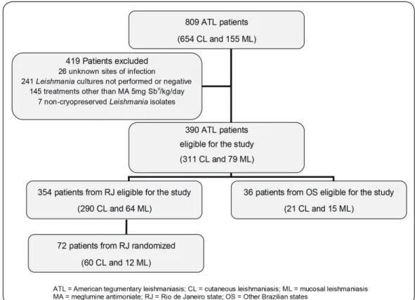
FIGURE 1: Flow chart of inclusion and exclusion of the studied patients, from the population of patients with American tegumentary leishmaniasis treated at Evandro Chagas National Institute of Infectious Diseases, Oswaldo Cruz Foundation, Rio de Janeiro, between 2000 and 2011.
demographic, clinical, and therapeutic factors (total, moderate, and severe clinical, laboratory, and electrocardiographic adverse events) associated with the patient’s origin of infection (RJ x OS groups) adjusted by the clinical form (CL or ML). A comparison between a ‘cure’ and ‘poor treatment response’ was performed within each group of infection origin adjusted by clinical form. The Kolmogorov-Smirnov test did not reject the null hypothesis at a 5% significance level for the association of age with the infection origin group; thus, a t-test was used.
Statistical Package for Social Sciences (SPSS) 16.0v
software was used for data analysis. Test values p < 0.05 were considered statistically significant. Due to some small cell counts, we additionally provided the frequency counts.
Ethical Approval
The Research Ethics Committee of INI (code CAAE 18473213.5.0000.5262) approved this study, and the study was conducted in accordance with the Helsinki Declaration of 1964, as revised in 1975, 1983, 1989, 1996, and 2000. All patients provided informed consent.
RESULTS
Patients
During the study period, 809 ATL patients were attended at INI. According to the inclusion criteria, 36 patients were selected for the OS group. For the RJ group, we randomly selected 72 patients from the 354 eligible patients (Figure 1).
Among the OS group, 42% were from the northern region (according to the Brazilian state where the infection occurred, 25.2% patients from Amazonas, 11.2% from Pará, 2.8% from Rondônia, and 2.8% from Roraima); 33% were from the northeastern region (8.3% from Ceará, 8.3% from Bahia, 5.6% from Maranhão, 2.7% from Paraíba, 2.7% from Pernambuco, 2.7% from Sergipe, and 2.7% possibly from Alagoas or Ceará); 22% were from the southeastern region (19.2% from Minas Gerais and 2.8% from Espírito Santo); and 3% were from the central-west region (3% from Goiás). In the RJ group, 58.3% of the patients were from the city of Rio de Janeiro.
TABLE 1: Clinical features and responses to treatment with 5 mg Sbv/kg/day meglumine antimoniate in 108 patients with American tegumentary leishmaniasis
according to the clinical form: cutaneous leishmaniasis (CL) or mucosal leishmaniasis (ML), and to the site where infection was acquired: in Rio de Janeiro (RJ) or other Brazilian states (OS).
CL (N=81) ML (N=27)
Clinical features RJ (N=60) OS (N=21) p-valuea RJ (N=12) OS (N=15) p-valuea
Age (years)
Median (Min - Max)b 30 (0-75) 30 (8-75) 0.406 52 (12-75) 52 (12-80) 0.954
Evolution time (months)
Median (Min - Max)b 2 (0.25-12) 2 (0.50-18) 0.687 3 (1-24) 24 (1-704) 0.004
Male
n (%)c 36 (60) 16 (76) 0.183 12 (100) 10 (67) *
With Co-morbiditiesd,e
n (%)c 20 (33) 5 (28) 0.658 7 (64) 10 (71) 1.000
Site of the lesions n (%)c
Nasal exclusivef - - - 3 (25) 10 (67) 0.054
Legs and feet 19 (32) 13 (62) 0.015 - -
-Ulcerative cutaneous lesions
n (%)c 56 (93) 15 (71) 0.016 - -
-Number of cutaneous lesions
Median (Min - Max)b 1 (1-6) 1 (1-5) 0.855 - -
-Diameter of the greater cutaneous lesion(mm)
Median (Min - Max)b 28 (7-110) 20 (5-55) 0.141 - -
-Therapeutic Response
Cure after first treatment
n/N (%)g 42/60 (70) 17/21 (81) 0.404 6/12 (50) 12/15 (80) 0.127
Cure after second treatment
n/N(%)g 10/15 (67) 3/4 (75) 1.000 4/5 (80) 1/3 (33) 0.464
Cumulative cure after second treatment
n/N (%)g 52/60 (87) 20/21 (95) 0.434 10/12 (83) 13/15 (87) 1.000
Cumulative cure after third treatment
n/N (%)g 53/60 (88) 20/21 (95) 0.673 10/12 (83) 14/15 (93) 0.569
Epithelialization time (days) after first treatment
Median (Min - Max)b 54 (14-174) 35 (23-72) 0.254 - -
-Complete healing time (days) after first treatment
Median (Min - Max)b 131 (41-348) 68 (47-341) 0.013 98 (54-150) 153 (79-465) 0.095 aSignificant p-values of < 0.05 are highlighted in bold; b Min - Max: Minimum - Maximum; cn: absolute number; d missing: CL / OS 3 patients; ML/RJ 1 patient; ML / OS 1 patient; e CL / RJ – asthma, cardiopathy, diabetes mellitus, dyslipidemia, rheumatic fever, arterial hypertension, and hypothyroidism; CL/ OS – cardiopathy, hyperthyroidism, chronic renal failure, psoriasis, Hashimoto’s thyroiditis, and talassemic trace; ML/RJ – cardiopathy, diabetes mellitus, psychiatric disorder, alcoholism, arterial hypertension, chronic renal failure, syphilis, and tuberculosis; ML/OS – bronchitis, cardiopathy, diabetes mellitus, alcoholism, strongyloidiasis, arterial hypertension, hypothyroidism, chronic renal failure, and syphilis; f Lesions exclusively located in the nasal mucosa were
considered less severe. Lesions in mucous membranes other than the nasal mucosa, associated or not with lesions in the nasal mucosa, were considered
severe; gn/N: cured/treated. * Statistical tests were impossible.
The distribution of the 108 patients by age, sex, and clinical features according to clinical form (CL or ML) and the site where the infection was acquired (RJ or OS) are shown in Table 1.
Therapeutic outcome
Four out of the 108 patients received antimonial treatment prior to the study: one in the CL / RJ group (one month before), two in the CL / OS group (respectively, 3 and 9 months before), and one in the ML/OS group (3 months earlier).
One course of 5 mg Sbv/kg/day MA resulted in cures in
774
FIGURE 2: Responses of 108 patients with American tegumentary leishmaniasis (ATL) to treatment with 5 mg Sbv/kg/day meglumine antimoniate, according to the clinical form: cutaneous leishmaniasis (CL), or mucosal leishmaniasis (ML), and to the area where infection was acquired: in Rio de Janeiro (RJ), or in other Brazilian states (OS).
Cataldo JI et al.
- T
The cure rates, epithelialization time, and complete healing time, according to the clinical form (CL or ML) and to the site where the infection was acquired (RJ or OS) are shown in Table 1. The complete healing time was lower in the CL/OS group than in the CL/RJ group (p=0.013). No other statistically significant differences were observed between the RJ and OS groups.
Nineteen patients with CL and eight patients with ML received a second course of treatment with 5 mg Sbv/kg/day
MA treatment. The mean time between the first and second treatments in patients with CL was 81 days (minimum 12 and maximum 713 days), and in patients with ML, it was 166 days (minimum 47 and maximum 700 days). Complete healing was observed in 13 of these 19 patients (68%) with CL, and in five of these eight patients (62%) with ML (Figure 2). The
cure rates according to the clinical form (CL or ML) and to the site where the infection was acquired (RJ or OS) is shown in
Table 1. No significant differences were observed between
the RJ and OS groups. It was not possible to recover the epithelialization time of these patients from their medical records. The complete healing time was 107 days (minimum 13 and maximum 295 days) in the CL/RJ group (eight patients evaluated and two missing), and in the CL/OS group (three patients) it was 42, 61, and 63 days, respectively.
Three patients in the CL/RJ group and one patient in the ML/ OS group received a third course of treatment with 5 mg Sbv/kg/
UI
-
e G):;:, os
Q.
...
G).0 E
::::i z
Adverse events by grade of severity
70 �---�
60 +--�
50
40
-30
20
10
o +---+---+----f--+--+--�---+---,1---+---+---+----f--�-+---+---+---,--+----l
CURJ CUOS MURJ MUOS
Clinica!
CURJ CUOS MURJ MUOS
Laboratory
o Absent o Mild o Moderate ■ Severe
CURJ CUOS MURJ MUOS
Electrocardiographic
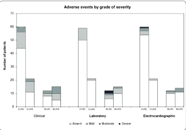
FIGURE 3: Distribution of clinical, laboratory, and electrocardiographic adverse events, by grade of severity (mild, moderate and severe) in 108 patients with American tegumentary leishmaniasis treated with 5 mg Sbv/kg/day meglumine antimoniate according
to the clinical form: cutaneous leishmaniasis (CL) or mucosal leishmaniasis (ML), and to the area where infection was acquired: in Rio de Janeiro (RJ) or in other Brazilian states (OS).
day MA. The time elapsed after the second treatment was 76, 86, and 108 days for the patients with CL, and 422 days for the patient with ML, respectively. Complete healing was observed in two patients (one CL and one ML) (Figure 2). It was not possible
to recover the epithelialization time of the medical records from these patients. The complete healing time was 145 and 67 days for the patient in the CL/RJ and ML/OS groups, respectively. The small number of patients submitted to the third treatment did not allow statistical verification between RJ and OS groups. After up to two additional treatments with the same dose, 88.9% and 85.2% of the CL and ML patients were respectively cured (Figure 2). The cumulative cure rates according to the
clinical form (CL or ML), and to the site where the infection was acquired (RJ or OS) is shown in Table 1.
Adverse events were observed in 40% of CL patients in the RJ group and 57% in the OS group. Regarding ML patients, the occurrence of adverse events was 58% in the RJ group and 80% in the OS group. The differences between the RJ and OS groups were not statistically significant for both CL and ML (p = 0.398 and 0.174, respectively). Permanent discontinuation of treatment due to adverse events was not required for any patient.
Figure 3 demonstrates the occurrence or absence of clinical,
776
clinical form (CL or ML), and to the origin of patients (RJ or OS). There were no statistically significant differences regarding the occurrence of clinical, laboratory, and electrocardiographic adverse events between RJ and OS groups in CL (p = 0.077, 0.278, 0.670, respectively) and in ML patients (p= 0.128, 0.452, 1,000, respectively). The description and the amount of clinical, laboratory, and electrocardiographic adverse events is shown in Table 2.
Out of 27 patients who required a second treatment with MA 5 mg Sbv/kg/day, ten presented with some adverse event. Eight
presented clinical adverse events (six graded as mild and two as moderate). Three presented laboratory adverse events (one mild, one moderate, and one severe). One patient presented a moderate electrocardiographic adverse event. All four patients who required a third treatment with MA 5 mg Sbv/kg/day did
not present any adverse events.
The median follow-up time after the end of the last course of treatment with MA 5 mg Sbv /kg/day until the date of the last evaluation
was 1,174 days (a minimum of 10 and a maximum of 5,642 days).
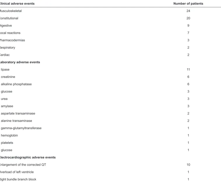
TABLE 2: Clinical, laboratory, and electrocardiographic adverse events observed in 108 patients with American tegumentary leishmaniasis treated with 5 mg Sbv/kg/day meglumine antimoniate, in descending order of occurrence.
Clinical adverse events Number of patients
Musculoskeletal 24
Constitutional 20
Digestive 9
Local reactions 7
Pharmacodermias 3
Respiratory 2
Cardiac 2
Laboratory adverse events
↑ lipase 11
↑ creatinine 6
↑ alkaline phosphatase 6
↑ glucose 3
↑ urea 3
↑ amylase 3
↑ aspartate transaminase 2
↑ alanine transaminase 2
↑ gamma-glutamyltransferase 1
↓ hemoglobin 1
↓ platelets 1
↓ glucose 1
Electrocardiographic adverse events
Enlargement of the corrected QT 10
Overload of left ventricle 1
Right bundle branch block 1
Species etiological identification
Twenty-one (58%) out of 36 samples could be identified in the OS group. Leishmania (L.) amazonensis was identified in
one patient from Maranhão, and L. (V.) guyanensis was identified
in two patients from Amazonas. Leishmania (V.) braziliensis was identified in 18 (86%) out of the 21 patients, of whom seven were from the northern region (four from Amazonas, one from Rondônia, one from Roraima, and one from Pará), six from the northeastern region (three from Bahia, one from Ceará, one from Maranhão, and one probably from Alagoas or Ceará), four from the southeastern region (all from Minas Gerais), and one from the central-western region (Goiás). Among those 18 samples identified as L. (V.) braziliensis, 16 presented Z27 isozyme
patterns, and two samples were iso-enzymatic variants for the ME, 6PGDH and the GPI loci when compared with the L. (V.) braziliensis reference strain. These two iso-enzymatic variants
were found to be identical to each other, and both patients were from the state of Amazonas.
In the RJ group, 72 (100%) samples could be identified as L. (V.) braziliensis and classified as Zymodeme Z2728. No isozyme
variants were detected in this group.
Leishmania (V.) braziliensis was the only species identified
in all 29 patients with ML (six out of 15 patients with ML in the OS group and all 14 patients with ML in the RJ group).
Amplification of the ITS region performed in a representative panel of 12 L. (V.) braziliensis samples presented bands
of approximately 750 bp (all samples). Comparisons with sequences stored in the GenBank database confirmed L. (V.) braziliensis in these samples, including two variants detected
by isoenzymes. Estimates of mean evolutionary divergence among nucleotide sequences revealed that the evaluated L. (V.) braziliensis samples presented low intraspecific genetic
diversity (0.006) when comparing the sequences. Estimates of mean evolutionary divergence among the base pair sequences of L. (V.) braziliensis from RJ and OS groups were 0.001793
and 0.006162, respectively.
DISCUSSION
This study compared the therapeutic responses to 5 mg Sbv/
kg/day MA treatment among ATL cases from Rio de Janeiro to those in patients from other Brazilian states, and verified which
Leishmania species and L. (V.) braziliensis subpopulations were
involved as etiological agents. However, this is a retrospective review of medical records with a limited power of inference, since the sample size is not sufficient to compare the proportions of the therapeutic responses to MA. Although the patients selected for the RJ group were randomized in the proportion of 2 for each patient selected for the OS group, the randomization was not sufficient to enable an equitable distribution of some clinical characteristics between both groups. The time of evolution of mucosal lesions was higher in the ML/OS group, which could have resulted in a worse therapeutic response than in the RJ group. However, this did not occur. Likewise, the higher proportion of lesions located on the legs and feet of patients in the CL/OS group, and the higher proportion of ulcerative lesions in the CL/RJ group could have negatively influenced the therapeutic responses in these groups, but again this did not occur. No difference was observed between cure rates and epithelialization time in both groups. Interestingly, the complete healing time was lower in the CL/OS group than in the CL/RJ group.
Leishmania (V.) braziliensis was found in 100% of samples
from the RJ group, and in 86% of samples from the OS group. Two L. (V.) braziliensis isozyme variants were identified in the
OS group. Three other analyzed cases (1 patient with ML and 2 with CL) from Bahia were caused by L. (V.) braziliensis with isozyme profiles similar to the L. (V.) braziliensis reference
strain. Some authors suggested that different ATL clinical forms, including localized or disseminated leishmaniasis, are caused by distinct L. (V.) braziliensis subpopulations distributed among
various locations of the state of Bahia8,31,32. Previous studies performed in Rio de Janeiro observed no association between
Leishmania’s genotypic patterns and the clinical manifestations
or therapeutic responses33,34,35.
Leishmania (L.) amazonensis (1 case) and L. (V.) guyanensis
(2 cases) were isolated from patients who acquired infections in the states of Maranhão and Amazonas, respectively, corroborating epidemiological data from these regions2,36,37. As 42% of the OS individuals came from the northern region, it is interesting to comment on the possibility of the presence of other species causing the lesions, especially L. (V.) shawi, and
L. (V.) naiffi38, which could not be retrieved. The presence of
a single cutaneous lesion was prevalent in our patients (79%), and this coincides with the classic clinical patterns described for ATL in Brazil when caused by L. (V.) braziliensis2,39. ML is
a rare but serious metastatic complication of ATL, presenting in less than 5% of all patients2,40. If we consider the status of INI as a center where severe and complicated cases from several Brazilian regions are referred, we would expect to find an increased proportion of patients with ML, as described in this study10. Leishmania (V.) braziliensis is the primary causative
agent of mucocutaneous forms in South America2,3,5, and was the only species identified in the patients with ML in this study.
Since this is a retrospective review of medical records and not a clinical trial, it is not possible to certify that the prescribed treatment doses were necessarily administered, nor that the consultation protocol was strictly followed by all patients. However, during the treatment period, patients received a sufficient number of MA vials for daily treatment until the next scheduled medical appointment. This routine prevents patients from using a greater amount of medication than prescribed during the treatment period. We posit that some patients may have used less than the recommended amount of medication if they did not use all the received ampoules. Another possibility is the interruption of the treatment for a few days due to missed appointments. Unfortunately, there was no strict control of adherence to treatment among this study cohort. However, the medical records display the date of the beginning and the end of the treatments, as well as instances of therapeutic interruptions due to adverse events or any other reasons.
One or two additional courses of 5 mg Sbv/kg/day MA or
intralesional MA are usually performed at INI/Fiocruz before changing to a second-line drug10. Ninety percent of the 108
patients with ATL responded favorably to up to three courses of treatment with 5 mg Sbv/kg/day MA, without significant
adverse events. This response reaches 92.6% if we consider the courses of treatment with intralesional MA. It is important to highlight that the therapeutic failure criteria proposed by the Brazilian Ministry of Health include two subsequent regular courses of treatment2.
The response to prolonged treatments is sometimes questioned and attributed to eventual spontaneous healing. However, some studies suggested a low rate of spontaneous cure for ATL after no treatment or use of placebo, particularly in L. (V.) braziliensis infection41,42. In addition, spontaneous healing
is not an expected occurrence in ML2,40.
778
Between 2001 and 2013, only 4% of 777 patients treated at INI were lost to follow-up10. Another study found a significant
association between greater adherence to treatment and the use of 5 mg Sbv/kg/day MA treatments43. Other authors reported that
an intermittent schedule (longer than the standard schedule) was associated with a lower frequency of discontinuation of treatment and loss to follow-up21. Most adverse events in this
study were mild or moderate. No patient required permanent discontinuation of treatment due to adverse events.
In most patients undergoing more than one 5 mg Sbv/kg/day
MA course of treatment, the time elapsed between the courses of treatment was relatively long. Although previous courses of treatment may have contributed to the success of the next treatment, apparently there was no increase in the frequency or severity of adverse events.
Previous evidence suggests that a 5 mg Sbv/kg/day schedule
can promote a decrease in both the incidence and severity of adverse events, without relevant impairment of treatment effectiveness14,15,19,20,44. A blind, non-inferiority, randomized, controlled clinical trial included 72 patients from Rio de Janeiro. In the intention-to-treat analysis, clinical cure was observed in 77.8% of patients treated with the alternative low-dose of 5 mg Sbv/ kg/day, and in 94.4% of patients treated with the standard
high-dose regimen of 20 mg Sbv/kg/day. However, in the group
treated with 20 mg Sbv/kg/day, there was a higher frequency
of serious adverse events and a higher frequency of treatment interruptions. Out of the patients allocated in the group treated with 5 mg Sbv/kg/day MA who presented with an unfavorable
therapeutic response, 85.7% were cured after a second treatment with 5 mg Sbv/kg/day MA or with intralesional MA. These
results suggest that an alternative low-dose of 5mg Sbv/kg/
day MA may be a viable treatment option, particularly when treatment toxicity is a concern26.
However, there is no consensus whether patients infected by different Leishmania species respond differently to treatment
using MA2,6,45. Among 90 patients infected by L. (V.) braziliensis in this study, 71% were cured after a single course of treatment, and the other 19% after a second or third course. The only patient infected by L. (L.) amazonensis in this study presented
with the classic localized cutaneous form with a single lesion, which usually responds satisfactorily to antimonial drugs2; he
was cured after the first course of treatment. Despite the poor expected response of L. (V.) guyanensis to MA2,6,7,37, one out
of the two patients infected by this species was cured after the second treatment using 5 mg Sbv/kg/day. Although MA is the
first-choice drug for treating ATL in Brazil, pentamidine has been the drug of choice in the Brazilian Amazon basin for the treatment of patients infected with this species2.
Forty-four patients with CL caused by L. (V.) braziliensis
were grouped into responders or non-responders to the initial treatment with 5mg Sbv/kg/day MA in a previous study.
However, 81% of the non-responders progressed to cure after one to three additional courses of treatment (median=1) with MA 5 mg Sbv/kg/day or intralesional MA. There was no association
between the genotype of the isolated parasites and the responder or non-responder status33.
Two other studies evaluated the genetic polymorphism and the
in vitro sensitivity in pairs of L. (V.) braziliensis isolates obtained from CL patients before treatment with MA (5 mg Sbv/kg/day or
intralesional) and after reactivation or treatment failure46,47. The first study revealed genetic polymorphism between the samples isolated before treatment and after reactivation or treatment failure, suggesting a possible differentiation of the structure of the original parasite population46. In the second study, an increase in the median lethal dose (LD50) values for the samples obtained after
treatment was observed, after exposure for both promastigotes and amastigotes to the MA. However, in both studies patients responded adequately to the second treatment using the same initial therapeutic regimen, independent of the in vitro findings46,47.
Overall, the results of these studies do not support the hypothesis that treatment with 5 mg Sbv/kg/day MA may induce
the selection of clinically resistant strains and suggest that other factors may influence the therapeutic outcome in patients with unsatisfactory response to the initial treatment33,46,47.
As expected, in our study the RJ group L. (V.) braziliensis
isolates were shown to be a homogeneous population. In the OS group, we found two variants of isoenzymes among the analyzed samples of L. (V.) braziliensis. Additionally, analyzing
the ITS region of the DNA of 12 L. (V.) braziliensis isolates,
higher genetic variability was found in the OS group samples, when compared to the RJ samples. It is uncertain if the genetic variability observed in L. (V.) braziliensis populations is an
important factor affecting the therapeutic outcome48,49. Results from our study and other studies30,34,35 indicate that the L. (V.)
braziliensis population endemic to the state of Rio de Janeiro
presents a homogeneous genetic pattern with low variability, which could explain the favorable therapeutic response to MA observed in patients who were infected in this state. However, therapeutic response to antimonial drugs may vary in states other than Rio de Janeiro, where ATL may be caused by different
Leishmania species or by an L. (V.) braziliensis subpopulation
with a high genetic variability6,8,50. Despite this fact, the results of the current study confirmed the findings of previous studies performed at INI/Fiocruz that included patients from other Brazilian states,10,20,23,33 and suggest that patients infected by L.
(V.) braziliensis or other Leishmania species may respond to
low-dose MA, regardless of their geographic origin in Brazil. Pentavalent antimony treatment can lead to significant toxicity that implies potentially severe adverse events with serious risks of morbidity and death. Therefore, such treatment should be avoided in a subgroup of patients, including very young children, elderly persons, and those with hepatic, renal, or cardiovascular diseases2,3. While the standard dose of antimony should remain the recommended treatment for ATL2,3, low-dose antimony treatment might be preferred when toxicity is a primary concern2,26.
A randomized clinical trial, using standard treatment as the control, and involving centers from different Brazilian states may be required to confirm our results.
Conflict of interest
Financial Support
This research is part of a doctoral thesis regarding clinical research of infectious diseases authored by Jamyra Iglesias Cataldo, Evandro Chagas National Institute of Infectious Diseases (NIID), Oswaldo Cruz Foundation, Rio de Janeiro, Brazil.
This study was partially funded by CNPq (National Council for Scientific and Technological Development, protocol no. 485333/2013-9), by PAEF-IOC-Fiocruz (Program of Strategic Actions for Development and Strengthening of Accredited Laboratories and Areas of Research Support, Oswaldo Cruz Institute, Oswaldo Cruz Foundation protocol no. IOC-008-FIO-15) and by FAPERJ (Research Support Foundation of the State of Rio de Janeiro - State’s Young Scientists Program, protocol no. 102.183/2013, E26-201.537/2014; and State’s Scientists Program protocol no. E26-202.011/2015). Maria de Fatima Madeira, Armando de Oliveira Schubach, and Mauro Celio de Almeida Marzochi are recipients of productivity grants from CNPq (National Council for Scientific and Technological Development).
REFERENCES
1. Camara Coelho LI, Paes M, Guerra JA, d, Coelho C, Lima B, et al. Characterization of Leishmania spp. causing cutaneous leishmaniasis in Manaus, Amazonas, Brazil. Parasitol Res. 2011;108(3):671-7.
2. Ministério da Saúde. Secretaria de Vigilância em Saúde. Departamento de Vigilância das Doenças Transmissíveis. Manual de Vigilância da Leishmaniose Tegumentar [Internet]. Brasília: SVS/ MS; 2017 [cited 2017 November 9]. Available from: http://bvsms. saude.gov.br/bvs/publicacoes/manual_vigilancia_leishmaniose_ tegumentar.pdf.
3. World Health Organization (WHO). Control of the Leishmaniases. WHO Technical Report Series No.: 949. Geneva: World Health Organization; 2010. 186p.
4. World Health Organization (WHO). Control of Leishmaniases: report of a WHO Expert Committee. WHO technical report series No.: 793. Geneva: World Health Organization; 1990. 158p. 5. Organización Panamericana de la Salud (OPAS). Leishmaniases en
las Américas: recomendaciones para el tratamiento. Washington, DC: OPAS; 2013. 43pp.
6. Romero GA, Guerra MV, Paes MG, Macêdo VO. Comparison of cutaneous leishmaniasis due to Leishmania (Viannia) braziliensis and L. (V.) guyanensis in Brazil: therapheutic response to meglumine antimoniate. Am J Trop Med Hyg. 2001;65(5):456-65.
7. Neves LO, Talhari AC, Gadelha EP, Silva Junior RM, Guerra JA, Ferreira LC, et al. A randomized clinical trial comparing meglumine antimoniate, pentamidine and amphotericin B for the treatment of cutaneous leishmaniasis by Leishmania guyanensis. An Bras Dermatol. 2011;86(6):1092-101.
8. Guimarães LH, Queiroz A, Silva JA, Silva SC, Magalhães V, Lago EL, et al. Atypical Manifestations of Cutaneous Leishmaniasis in a Region Endemic for Leishmania braziliensis: Clinical, Immunological and Parasitological Aspects. PLoS Negl Trop Dis. 2016;10(12):e0005100.
9. Tuon FF, Amato VS, Graf ME, Siqueira AM, Nicodemo AC, Amato Neto V. Treatment of New World cutaneous leishmaniasis - a systematic review with a meta-analysis. Int J Dermatol. 2008;47(2):109-24.
10. Brahim LR, Valete-Rosalino CM, Antônio LF, Pimentel MIF, Lyra MR, Paes LEC, et al. Low dose systemic or intralesional meglumine antimoniate for American tegumentary leishmaniasis results in low lethality, low incidence of relapses, and low late mucosal involvement in a referral centre in Rio de Janeiro, Brazil (2001-2013). Mem Inst Oswaldo Cruz. 2017;112(12):838-43.
11. Ministério da Saúde [Internet]. Sistema de Informação de Agravos de Notificação SINAN – Brasil. 2001-2013 [access on 28 Apr 2015]. Available from: http://dtr2004.saude.gov.br/sinanweb
12. Ministério da Saúde [Internet]. DATASUS - Tecnologia da informação a serviço do SUS. Tuberculose - casos confirmados notificados no sistema de informação de agravos de notificação SINAN - Brasil. 2001-2013 [access on 19 July 2017]. Available from: http://tabnet. datasus.gov.br/cgi/tabcgi.exe?sinannet/cnv/tubercbr.def
13. Ministério da Saúde [Internet]. DATASUS - Tecnologia da informação a serviço do SUS. Leishmaniose Visceral - casos confirmados notificados no sistema de informação de agravos de notificação SINAN - Brasil. 2001-2013 [access on 19 July 2017]. Available from: http://tabnet.datasus.gov.br/cgi/deftohtm. exe?sinannet/cnv/leishvbr.def
14. Schubach AO, Marzochi KB, Moreira JS, Schubach TM, Araújo ML,Vale AC, et al. Retrospective study of 151 patients with cutaneous leishmaniasis treated with meglumine antimoniate. Rev Soc Bras Med Trop.2005;38(3):213-7.
15. Oliveira-Neto MP, Schubach A, Araujo ML, Pirmez C. High and low doses of antimony (Sbv) in American cutaneous leishmaniasis. A five years follow-up study of 15 patients. Mem Inst Oswaldo Cruz. 1996;91(2):207-9.
16. Zamith VA. Leishmaniose Tegumentar Americana. In: Veronesi R, editor. Doenças Infecciosas e Parasitárias. 6th ed. Rio de Janeiro:
Guanabara Koogan; 1976. p. 701-11.
17. Sampaio SAP, Castro RM, Riviti EA. Dermatologia Básica. 2nd ed. São Paulo: Artes Médicas; 1981.
18. Oliveira-Neto MP, Schubach A, Mattos M, Goncalves-Costa SC, Pirmez C. A low dose antimony treatment In 159 patients with American cutaneous leishmaniasis. Extensive follow-up studies (up to 10 years). Am J Trop Med Hyg. 1997;57(6):651-5.
19. Oliveira-Neto MP, Schubach A, Mattos M, Gonçalves-Costa SC, Pirmez C. Treatment of American cutaneous leishmaniasis: a comparison between low dosage (5 mg/kg/day) and high dosage (20 mg/kg/day) antimony regimens. Pathol Biol (Paris). 1997;45(6):496-9. 20. Vasconcellos EFC, de Oliveira Schubach A, Valete-Rosalino CM,
de Souza Coutinho R, Conceição-Silva F, de Matos Salgueiro M, et al. American tegumentary leishmaniasis in older adults: 44 cases treated with an intermittent low-dose antimonial schedule in Rio de Janeiro, Brazil.J Am Geriatr Soc. 2010;58(3):614-6.
21. Azeredo-Coutinho RBG, Mendonça SCF. An intermittent schedule is better than continuous regimen of antimonial therapy for cutaneous leishmaniasis in the municipality of Rio de Janeiro, Brazil. Rev Soc Bras Med Trop. 2002;35(5):477-81.
22. Schubach AO, Conceição-Silva F. Estado da Arte no Tratamento da Leishmaniose Tegumentar Americana no Brasil. In: Conceição-Silva F, Alves CA, editors. Leishmanioses do Continente Americano. 1st ed. Rio de Janeiro: Editora FIOCRUZ; 2014. p. 391-412. 23. Antônio LF, Fagundes A, Oliveira R, Pinto P, Vasconcellos EFC,
Bedoya-Pacheco SJ, et al. Montenegro skin test and age of skin lesion as predictors of treatment failure in cutaneous leishmaniasis. Rev Inst Med Trop São Paulo. 2014;56(5):375-80.
24. Oliveira LF, Schubach AO, Martins MM, Passos SRL, Oliveira RV, Marzochi MCA, et al. Systematic review of the adverse effects of cutaneous leishmaniasis treatment in the New World. Acta Trop. 2011;118(2):87-96.
25. Olliaro P, Vaillant M, Arana B, Grogl M, Modabber F, Magill A, et al. Methodology of clinical trials aimed at assessing interventions for cutaneous leishmaniasis. PLoS Negl Trop Dis. 2013;7(3):e2130. 26. Saheki MN, Lyra MR, Bedoya-Pacheco SJ, Antônio LF,
780
antimony for American cutaneous leishmaniasis: a randomized blind non-inferiority trial in Rio de Janeiro, Brazil. PLoS ONE. 2017;12(5):e0178592.
27. DAIDS Division of AIDS, National Institute of Allergy Infectious Diseases, National Institutes of Health. Division of AIDS table for grading the severity of adult and pediatric adverse events 2004 [cited 12 May 2015]. Available from: https://rsc.tech-res.com/docs/ default-source/safety/daids_ae_grading_table_v2_nov2014.pdf 28. Cupolillo E, Grimaldi G Jr, Momen H. A general classification of
New World Leishmania using numerical zymotaxonomy. Am J Trop Med Hyg. 1994;50(3):296-311.
29. Tamura K, Peterson D, Peterson N, Stecher G, Nei M, Kumar S. MEGA 5: Molecular Evolutionary Genetics Analysis using Maximum Likelihood, Evolutionary Distance, and Maximum Parsimony Methods. Mol Biol Evol. 2011;28(10):2731-9.
30. Cupolillo E, Brahin LR, Toaldo CB, de Oliveira-Neto MP, de Brito ME, Falqueto A, et al. Genetic polymorphism and molecular epidemiology of Leishmania (Viannia) braziliensis from different hosts and geographic areas in Brazil. J Clin Microbiol. 2003;41(7):3126-32.
31. Schriefer A, Schriefer AL, Goes-Neto A, Guimaraes LH, Carvalho LP, Almeida RP, et al. Multiclonal Leishmania braziliensis population structure and its clinical implication in a region of endemicity for American tegumentary leishmaniasis. Infect Immun. 2004;72(1):508-14. 32. Queiroz A, Sousa R, Heine C, Cardoso M, Guimarães LH, Machado
PRL, et al. Association between an Emerging Disseminated form of Leishmaniasis and Leishmania (Viannia) braziliensis Strain Polymorphisms. J Clin Microbiol. 2012;50(12):4028-34.
33. Gagini T, Schubach AO, Madeira MF, Valete-Rosalino CM, Pimentel MIF, Pacheco RS. Genotypic profiles of Leishmania (Viannia) braziliensis strains from cutaneous leishmaniasis patients and their relationship with the response to meglumine antimoniate treatment: a pilot study. Parasite. 2017;24:34.
34. Baptista C, Schubach AO, Madeira MF, Leal CA, Pires MQ, Oliveira FS, et al. Leishmania (Viannia) braziliensis genotypes identified in lesions of patients with atypical or typical manifestations of tegumentary leishmaniasis: evaluation by two molecular markers. Exp Parasitol. 2009;121(4):317-22.
35. Oliveira FS, Valete-Rosalino CM, Pacheco SJ, Costa FA, Schubach AO, Pacheco RS. American tegumentary leishmaniasis caused by Leishmania (Viannia) braziliensis: assessment of parasite genetic variability at intra- and inter-patient levels. Parasit Vectors. 2013;6:189. 36. Oliveira JPC, Fernandes F, Cruz AK, Trombela V, Monteiro E,
Camargo AA, et al. Genetic diversity of Leishmania amazonensis strains isolated in northeastern Brazil as revealed by DNA sequencing, PCR-based analyses and molecular karyotyping. Kinetoplastid Biol Dis. 2007;6:5.
37. Francesconi VA, Francesconi F, Ramasawmy R, Romero GAS, Alecrim MdGC. Failure of fluconazole in treating cutaneous leishmaniasis caused by Leishmania guyanensis in the Brazilian Amazon: An open, nonrandomized phase 2 trial. PLoS Negl Trop Dis. 2018; 12(2):e0006225.
38. Fagundes-Silva GA, Romero GAS, Cupolillo E, Yamashita EPG, Gomes-Silva A, Guerra JAO, et al. Leishmania (Viannia) naiffi:
rare enough to be neglected? Mem Inst Oswaldo Cruz. 2015;110(6):797-800.
39. Oliveira-Neto MP, Mattos MS, Perez MA, Da-Cruz AM, Fernandes O, Moreira J, et al. American tegumentary leishmaniasis (ATL) in Rio de Janeiro State, Brazil: main clinical and epidemiologic characteristics. Int J Dermatol. 2000;39(7):506-14.
40. Blum J, Lockwood DN, Visser L, Harms G, Bailey MS, Caumes E, et al. Local or systemic treatment for New World cutaneous leishmaniasis? Re-evaluating the evidence for the risk of mucosal leishmaniasis. Int Health. 2012;4(3):153-63.
41. Cota GF, de Sousa MR, Fereguetti TO, Saleme PS, Alvarisa TK, Rabello A. The Cure Rate after Placebo or No Therapy in American Cutaneous Leishmaniasis: A Systematic Review and Meta-Analysis. PLoS ONE. 2016;11(2):e0149697.
42. Oliveira-Ribeiro C, Pimentel MIF, Oliveira RVC, Fagundes A, Madeira MF, Mello CX, et al. Clinical and laboratory profiles of patients with early spontaneous healing in cutaneous localized leishmaniasis: a historical cohort study. BMC Infect Dis. 2017;17:1-8.
43. Ribeiro MN, Pimentel MI, Schubach AO, Oliveira RV, Teixeira JL, Leite MP, et al. Factors associated to adherence to different treatment schemes with meglumine antimoniate in a clinical trial for cutaneous leishmaniasis. Rev Inst Med Trop São Paulo. 2014;56(4):291-6.
44. Oliveira-Neto MP, Mattos M, Pirmez C, Fernandes O, Goncalves-Costa SC, Souza CF, et al. Mucosal leishmaniasis ("espundia") responsive to low dose of N-methylglucamine (Glucantime) in Rio de Janeiro, Brazil. Rev Inst Med Trop Sao Paulo. 2000;42(6):321-5. 45. Rodrigues AM, Hueb M, Santos TA, Fontes CJ. Factors
associated with treatment failure of cutaneous leishmaniasis with meglumine antimoniate [in Portuguese]. Rev Soc Bras Med Trop. 2006;39(2):139-45.
46. Baptista C, Schubach AO, Madeira MF, Miranda LFC, Pinto AGS, Barros JHS, et al. Evaluation of genetic polymorphism of Leishmania (V.) braziliensis isolates obtained from the same patient before and after therapeutic failure or reactivation of cutaneous lesions. J Trop Med. 2012; Article ID 808132.
47. Baptista C, Miranda LFC, Madeira MF, Leon LLP, Conceição-Silva F, Schubach AO. In vitro sensitivity of paired Leishmania (Viannia) braziliensis samples isolated before meglumine antimoniate treatment and after treatment failure or reactivation of cutaneous leishmaniasis. Disease Markers. 2015; Article ID 943236.
48. Rojas R, Valderrama L, Valderrama M, Varona MX, Ouellette M, Saravia NG. Resistance to antimony and treatment failure in human Leishmania (Viannia) infection. J Infect Dis. 2006;193(10):1375-83. 49. Torres DC, Adaui V, Ribeiro-Alves M, Romero GA, Arévalo J,
Cupolillo E, et al. Targeted gene expression profiling in Leishmania braziliensis and Leishmania guyanensis parasites isolated from Brazilian patients with different antimonial treatment outcomes. Infect Genet Evol. 2010;10(6):727-33.
50. Torres DC, Ribeiro-Alves M, Romero GA, Dávila AM, Cupolillo E. Assessment of drug resistance related genes as candidate markers for treatment outcome prediction of cutaneous leishmaniasis in Brazil. Acta Trop. 2013;126(2):132-41.