Review
Caroline J. Sheeba
a, Gregory Marslin
a, Ann Mary Revina and Gregory Franklin*
Signaling pathways influencing tumor
microenvironment and their exploitation for
targeted drug delivery
Abstract: In the recent years, the “tumor
microenviron-ment” has been receiving growing attention due to its
involvement in neoplastic transformation, tumor growth,
invasion, and protection of tumor cells from host immune
response. All these events are facilitated by chemical
sig-nals produced by the tumor as well as the surrounding
stromal cells. This review is divided into two main parts in
which the first part discusses the receptor tyrosine kinase
(RTK)-mediated growth factor signaling, steroid hormone
(SH) signaling, ancient signaling pathways, and other
molecules that are involved in tumorigenesis and how
they interact with each other to create a complex tumor
microenvironment. In the second part, we bring together
the recent nanocarrier-mediated drug delivery approaches
to target the signaling pathways/molecules present in the
tumor microenvironment.
Keywords: active targeting; functionalization;
nanopar-ticle-mediated drug delivery; signaling pathways; tumor
microenvironment.
aBoth authors contributed equally to this work.
*Corresponding author: Gregory Franklin, Departamento de Biologia (CITAB-UM), Universidade do Minho, 4710-057 Braga, Portugal, e-mail: [email protected]
Caroline J. Sheeba: Regenerative Medicine Program, Departamento de Ciências Biomédicas e Medicina, Universidade do Algarve, 8005-139 Faro, Portugal; IBB-Institute for Biotechnology and Bioengineering, Centro de Biomedicina Molecular e Estrutural, Universidade do Algarve, 8005-139 Faro, Portugal; Life and Health Sciences Research Institute (ICVS), School of Health Sciences, University of Minho, 4710-057 Braga, Portugal; and ICVS/3B’s, PT Government Associate Laboratory, Braga/Guimarães, Portugal Gregory Marslin: Departamento de Biologia (CITAB-UM), Universidade do Minho, 4710-057 Braga, Portugal
Ann Mary Revina: Life and Health Sciences Research Institute (ICVS), School of Health Sciences, University of Minho, 4710-057 Braga, Portugal; and ICVS/3B’s, PT Government Associate Laboratory, Braga/Guimarães, Portugal
1 Introduction
Cancer can be considered as a developmental disorder
because most of the signaling pathways responsible for
tumor formation are the ones involved in embryo
devel-opment. This is evident by the resemblance of aggressive
tumor cells with embryonic stem cells by means of their
plastic, multipotent nature. Deregulation/dysfunction
of developmental pathways in and around tumor cells
as well as the absence of many regulatory checkpoints
results in aberrant uncontrolled growth of tumor cells.
Research over years has contributed substantially to our
understanding of the cellular and molecular interactions
in the tumor microenvironment that orchestrates
tumo-rigenesis. The constantly evolving tumor
microenviron-ment is rich in growth factors, which elicit a cascade of
signaling events through specific cell-surface receptors,
leading to rapid proliferation, angiogenesis, resistance
to cell death, and endure epithelial-mesenchymal
transi-tion (EMT) and metastasis. Our knowledge about the role
of tumor microenvironment in cancer has improved
sig-nificantly, moving from a conceptual framework toward
the development of novel strategies to treat cancer.
Com-bining therapies that target not only the tumor cells but
also the tumor microenvironment and/or the signaling
pathways providing resistance to the cancer cells from
responding to chemotherapy, have greater degree of
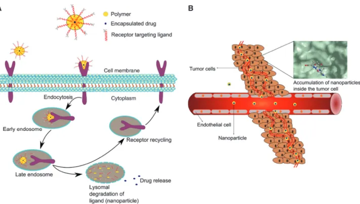
success in cancer treatment [1]. Nanoparticles designed
based on the characteristics and specific signaling
inter-action of the tumor microenvironment is a promising
strategy to combat cancer. For instance, nanoparticles
sensitive to the acidic pH of the tumor microenvironment
provides selectivity to tumor cells over the normal ones,
thus enhances specificity and drug delivery efficiency [2,
3]. The first part of this review presents a holistic
discus-sion about the important signaling molecules/pathways
such as the receptor tyrosine kinases (RTK), steroid
hor-mones (SH), and the ancient signaling pathways that are
altered during cancer and signaling interactions enriching
the tumor microenvironment. Interested readers are also
referred to other in-depth reviews on specific topics under
most of the sections. The second part consolidates how
the signaling molecules discussed in the previous part are
exploited to functionalize nanoparticle-mediated
thera-peutic strategies to treat cancer effectively.
2 Receptor tyrosine kinase (RTK)
signaling
Signaling via mutated or constitutively active variant
of receptor tyrosine kinase (RTK) function as a
poten-tial means for cancer cells to evade host mechanisms
and develop tumors. A huge deal of attention has been
diverted toward RTK signaling because of their
overex-pression commonly found in many cancers, their ability
to crosstalk between themselves, and importantly, they
connect the extracellular cues with intracellular
effec-tor pathways. As a result, RTK recepeffec-tor expression has
been extensively used as a prognostic biomarker in
many malignancies. There are several RTKs, and only the
primary ones upregulated in cancer are reviewed here
(Figure 1).
2.1 ErbB family of receptors
The epidermal growth factor receptor (EGFR) is a member
of the ErbB family, a subfamily comprised of ErbB1/HER1/
EGFR, ErbB2/HER2, ErbB3/HER3, and ErbB4/HER4. The
ErbB receptors are prominent cancer drivers, which
form active homo- or heterodimers upon ligand binding
[4]. ErbB receptors bind to EGF produced by the same
cell (autocrine) or other cells (paracrine). After ligand
binding, the dimerized receptor’s intracellular tyrosine
kinase domain will be activated causing phosphorylation
of specific tyrosine residues that serve as docking sites
for proteins containing Src homology 2 (SH2) domains
such as Grb2, Shc1, p85, PLCγ, and JAK1, leading to the
activation of several intracellular signaling pathways.
These downstream signaling cascades include the Ras/
MAPK/extracellular signal-regulated kinase (ERK), PI3K/
Akt, JAK/ STAT, and PLCγ/protein kinase-C (PKC)
path-ways for cell proliferation, survival, and mobility [5, 6].
The intracellular kinase domain of HER3 is thought to be
an inactive pseudokinase that lacks several catalytically
important residues and so it primarily signals by
heter-odimerizing with HER2 [7]. However, it was reported to
have sufficient kinase activity to trans-autophosphorylate
its intracellular region [8]. Recently, HER3 overexpression
in various tumors including colorectal, gastric, breast,
and ovarian cancers has been associated with worse
sur-vival, and its effect on overall survival was significantly
higher when HER2 was co-overexpressed [9]. Similarly,
ErbB receptors are also expressed at high levels in
differ-ent cancers, and the levels of gene/protein expression is
correlated with the growth, state, and aggressiveness of
cancer [10, 11]. For instance, HER2 amplification occurs
in 20% of breast cancers [11], and 54% of glioblastoma
exhibit EGFR overexpression [12]. Glioblastoma cells
often present both the wild-type EGFR gene amplification
and the constitutively active variant EGFRvIII, resulting in
increased EGFR signaling [12]. However, EGFRvIII
expres-sion without EGFR gene amplification is fairly
uncom-mon, suggesting that EGFR gene amplification may
precede EGFRvIII mutation [13]. All the aforementioned
features make ErbB receptors a potential therapeutic
target to treat tumors. A detailed review on targeting ErbB
receptors can be found in [14].
2.2 Fibroblast growth factor receptor
(FGFR) family
Fibroblast growth factor receptors (FGFR) are
transmem-brane tyrosine-kinase receptors that coordinate a variety
of cellular functions. There are 4 FGFRs (FGFR1-4) and 22
FGF ligands [15]. Binding of FGF ligands to FGFRs activate
several downstream signaling pathways, including Ras/
MAPK/ERK, PLCγ/PKC, PI3K/Akt, and JAK/STAT. Being
a crucial signaling for basic processes such as
prolifera-tion, survival, angiogenesis, and migraprolifera-tion, deregulated
FGF signaling can contribute to the development and
pro-gression of tumors [16]. FGFR signaling is altered in many
cancers including benign skin tumors [17], prostate [18],
bladder, and breast cancers [19–21]. Breast cancer cells
have been reported to overexpress FGFR1, 2, 4 and display
mutations in FGFR2 and 4 [21]. Moreover, emerging data
suggest that in addition to the known functions of FGF
signaling in promoting tumor cell proliferation and
sur-vival, FGF signaling might also regulate EMT [22], tumor
metastasis and lymphangiogenesis in a vascular
endothe-lial growth factor-C (VEGF-C)-dependent mechanism [23].
Overexpression of FGFR1 and its altered splicing
mecha-nisms, leading to increased expression of FGFR1β isoform
has been associated with high-grade/stage bladder cancer
[24, 25]. Although, activating mutation and
overexpres-sion of FGFR3 is a common phenomenon observed in
low-grade bladder cancer [19], a switch from its
epithe-lial to mesenchymal isoform with wider ligand affinity is
thought to have more deleterious effects [19, 26].
Particu-larly, FGFR1 has been considered as a potential oncogene
in breast cancer because its deregulated signaling
contrib-utes to cell proliferation, growth, angiogenesis, EMT, and
cell migration in S115 breast cancer [20]. Overall, FGFRs
stands as an attractive target for therapeutic intervention
in cancer [19, 21, 27].
2.3 Insulin receptors (IR) and insulin-like
growth factor receptors (IGFR) family
The insulin receptors (IR-A and IR-B) and the
insulin-like growth factor receptors (IGF1R and IGF2R) are
tyros-ine kinase membrane-bound receptors that share ∼60%
sequence homology and regulates glucose homeostasis
and growth in response to nutrient availability in cells.
IR has two isoforms, IR-A and IR-B, which are
predomi-nantly expressed in the fetal and adult tissues,
respec-tively. However, cancer cells preferably overexpress the
fetal isoform IR-A, which has the advantages of
generat-ing hybrid receptors with IGFIR and to have equal affinity
to IGF1/IGF2 like that of IGF1R [28–30]. In fact, the hybrid
receptors are reported to possess higher affinity for IGF1
than insulin and function predominantly as an IGF1
recep-tor [31]. IR-mediated nonmetabolic insulin signaling has
been found in human myosarcoma cells [32], colon cancer
cells [33], breast, prostate, and colorectal cancers [34–36].
Moreover, IR-associated obesity, -type 2 diabetes mellitus
(T2DM) and -hyperinsulinemia are some important risk
factors for several malignancies including breast cancer
[37]. Upon insulin binding to IR, the activated RTK will
phosphorylate insulin receptor substrate proteins
(IRS1-4), providing docking sites for effectors/adapter proteins,
containing SH2 domains. This triggers a cascade of
reac-tions causing the activation of PI3K/Akt and Ras/MAPK
pathways that mediate the metabolic and mitogenic
activ-ities of insulin, respectively [38, 39]. The antiapoptotic
activity of insulin is reported to involve both the PI3K/
Akt and MAPK pathways [40, 41]. Insulin also possesses
angiogenic properties in a VEGF-dependent or
-independ-ent manner through PI3K/Akt and MAPK pathways [42,
43]. While the ability of insulin to stimulate PI3K is lost in
the presence of insulin resistance and hyperinsulinemia,
its capacity to activate MAPK pathway is enhanced [39].
Thus, hyperinsulinemia-mediated increased levels of
cir-culating insulin in association with IR-A overexpression
in cancer cells may cause abnormal nonmetabolic effects
of IR, such as cell survival, proliferation, migration, and
angiogenesis, the key events that occur during tumor
growth and metastasis [38, 43], making the circulating
insulin a risk factor of colorectal, pancreatic, and breast
cancers [44, 45].
IGF1R is a potential cellular oncogene through which
both IGF1 and IGF2 exert their mitogenic, antiapoptotic,
and transforming activities [46]. IGF1R expression is
seen as a prerequisite for tumor formation because
mouse fibroblasts deprived of IGF1R were unable to be
transformed by a number of oncogenes [47, 48]. IGF1R
signaling plays critical steps, namely, cell adhesion,
migration, invasion, and angiogenesis during the
meta-static cascade and is involved in a wide range of cancers
including the breast, prostate, pediatric, cervix, and
ovarian cancers [37]. Ligand binding to the
extracellu-lar subunit of IGF1R causes autophosphorylation and
conformational changes of its tyrosine kinase domain,
leading to the binding of IRS1-4 and Shc proteins.
Phos-phorylation of these proteins eventually activates at least
two signaling pathways: PI3K/Akt and Ras/Raf/MEK/
ERK. The antiapoptotic effect of IGF1R is mainly exerted
by the PI3K/Akt pathway activation. Phosphorylated IRS
activates PI3K, which helps the conversion of
phosphati-dylinositol-4,5-bisphosphate (PIP2) to PIP3, the reaction
inhibited by phosphatase and tensin homolog (PTEN).
PIP3 phosphorylates Akt as well as PKC proteins, both of
which regulate the metabolic activities of the cell such as
glucose uptake [49, 50]. Importantly, activated Akt
inter-feres with the antiapoptotic and proapoptotic functions
of several proteins. Upon phosphorylation by Akt,
Bcl-2-associated death promoter (BAD) becomes inactivated
and allows the antiapoptotic activity of Bcl-2, promoting
cell survival. In addition, phosphorylated-Akt also
inhib-its the proapoptotic protein caspase-9 and prevents cell
death [51]. By activating nuclear factor-κB- (NF-κB), Akt
can also regulate the expression of antiapoptotic genes
[52]. On the other hand, phosphorylated Shc protein
binds to Grb2 that recruits Son of Sevenless (SOS), which
in turn activates Ras/Raf/MER/ERK pathway. Activated
ERK get translocated to the nucleus and regulates target
gene expression, influencing cell proliferation and
sur-vival [53].
IGF1 and IGF2 are single-chain polypeptides that
share 62% sequence homology and generate multiple
transcripts depending on their transcription initiation
promoter sites and alternative splicing mechanisms. The
availability of free IGF1 to interact with IGF1R is regulated
by the levels of the six IGF-binding proteins
(IGFBP1-6). Under normal physiological conditions, only 1% of
the IGFs circulate freely, while others are bound to the
IGFBPs [54]. In addition to IGFBPs, their associated
pro-teases are also important in IGFR signaling because they
hydrolyzes IGFBPs, causing the release of bound IGFs,
enabling them to interact with IGF1R. Diet, nutrition, and
growth hormones have an influence on IGF1 expression
[55]. Similarly, IGF1R expression is also affected by
nutri-tion, growth factors, and SHs [56]. Although other growth
factors stimulate IGF1R production, IGF1 functions as its
negative regulator [57]. Hyperinsulinemia can also favor
the production of IGF1 and increases its bioavailability
and IGF1R signaling by modulating IGFBPs [58]. Both IGF1
and IGF2 are overexpressed in an array of cancers such as
the colon, prostate, breast, colorectal, thyroid, lung,
pan-creatic cancers, and several sarcomas [59, 60]. Insulin and
IGF1 have the ability to cross-bind to each other’s
recep-tor, although with much less affinity than that of their
preferred ligand [61]. Unlike IGF1R, IGF2R has no tyrosine
kinase activity, and it binds to IGF2 and reduces its
bio-availability by sending it for lysosomal degradation [62].
Because of this effect, IGF2R has been considered as a
potential tumor-suppressor molecule. In-depth reviews
on IR, IGF, and IGF1R in cancer can be found elsewhere
[37, 39, 63, 64].
2.4 Platelet-derived growth factor receptors
(PDGFR)
There are two types of the platelet-derived growth factor
receptors: PDGFRα and PDGFRβ that are activated by
five different disulfide-linked dimer ligands: PDGF-AA,
-BB, -AB, -CC, and -DD with varying specificity. Although
all PDGFs except the PDGF-DD interact with PDGFRα
and induce receptor dimer formation, PDGF-AA is the
most potent ligand of PDGFRα. PDGF-BB and PDGF-DD
interacts with PDGFRβ [65]. Ligand-binding to receptors
induces homo- or heteroreceptor dimerization, leading to
the activation of their intrinsic tyrosine kinase domain and
subsequent recruitment of SH2-domain-containing
signal-ing proteins, which activates the downstream pathways
that cause the basic cellular processes like, proliferation,
migration, and transformation [66]. Both, PDGFs (-BB and
-DD) and the receptors (PDGFRα and PDGFRβ) are
over-expressed in the breast [67], prostate [68], kidney [69], lung
[70], ovarian [71], glioma [72], melanoma [73], and bone
[74] tumors. Expression of PDGFs and PDGFRs are found
even in low-grade gliomas, unlike the EGFR expression
found only in high-grade tumors, suggesting an early
role for PDGF signaling in gliomas [75]. PDGFR signaling
in tumor is primarily associated with angiogenesis and
metastasis, like in the case of gliomas and breast cancer
[67, 76]. PDGF-B, -C, and -D has been reported to enhance
tumor angiogenesis through enhanced VEGF expression
[77–79]. Tumor cell-secreted PDGF-B also functions to
determine the fate of the mesenchymal stem cells in vitro
through a transmembrane glycoprotein receptor,
neuro-pilin-1 (NRP-1) signaling [80], and it should be noted that
NRP-1 expression is positively correlated with the
inva-sion ability of cancer cells. Recently, it was demonstrated
that the knockdown of PDGFRβ in glioblastoma stem cells
downregulates the critical angiogenesis regulator VEGF
[81]. In this context, VEGF165 has been reported to bind to
NRP-1 and trigger the NRP-1/VEGFR2/PI3K/Akt signaling
pathway causing tumor angiogenesis, cancer cell invasion,
and tumorigenesis [82]. PDGFR also influence the cancer
microenvironment by recruiting nearby stromal cells,
which facilitate tumor-stromal cell interaction that
deter-mines tumor development [83, 84]. The role of PDGFR in
cancer has been critically reviewed before [85].
2.5 Vascular endothelial growth factor
receptors (VEGFR)
The vascular endothelial growth factor (VEGF) family is
crucial for angiogenesis, lymphangiogenesis, and
vasculo-genesis, and it consists of six members: VEGF (or VEGF-A),
VEGF-B, VEGF-C, VEGF-D, VEGF-E, and placental growth
factor (PlGF). The biological effects of VEGF are mediated
Figure 1 RTK and SHR signaling.
The activity of growth factors (GF) such as EGF, FGF, IGF, PDGF, and VEGF family members are mediated by the RTK signaling. These receptors are made up of an extracellular region, a single transmembrane spanning region, and a cytoplasmic tyrosine kinase domain. The extracellu-lar domain of the RTK binds to the respective GF ligands that cause receptor dimerization and subsequent autophosphorylation on multiple specific intracellular tyrosine residues, creating binding sites for specific proteins. Autophosphorylated RTKs stimulate small GTP-binding protein, Ras by recruiting SOS and its adapter protein GRB2 to the membrane. This initiates a series of signal transduction cascade. Ras activates PLCγ, which can also be activated by Src in a RTK-dependent or -independent manner through steroid hormone receptors (SHR). Activated PLCγ hydrolyses PIP2 to release the second messengers 1,2-diacylglycerol (DAG) and IP3, in which DAG is the activator of PKC that activates Ras/Raf and thus ERK signaling, leading to the expression of transcription factors related to cell proliferation, migration, and angiogenesis. In addition, PKC also activates PLD that catalyzes the hydrolysis of PC to PA, activator of signaling cascades like mTOR. PA also inhibits PTEN, a tumor suppressor that negatively regulate mTORC1 activity. IP3 activates Ca2+ release from the endoplasmic reticulum by binding to its intracellular receptor (IP3R). Thus, accumulated intracellular calcium displaces the inhibitory binding of caveolin to eNOS and induces NO production, which increases angiogenesis and vasopermeability. Another important intracellular pathway activated upon RTK signaling is the PI3K/Akt, which starts with the recruitment of PI3K (p85α/p110α) to the receptor, enabling p110α to phosphorylate PIP2 and PIP3. Binding of PIP3 to Akt, allows Akt phosphorylation and partial activation by PDK1. Thus, partly activated Akt is fully activated by mTORC2. In turn, phosphorylated/fully activated Akt activates mTORC1 either directly or through its inhibitory action on TSC1/TSC2, which inhibits mTOR. mTORC1 regulates S6K and HIF1α, inducing translation of several genes including the ones participating in homeostatic responses to hypoxia. Although Akt signaling can promote cell proliferation, metabolism, migration, and angiogenesis, its important role is to function as an antiapoptotic signal by exerting its effect by phosphorylating a variety of downstream targets including mTOR, NF-κB, eNOS, FOXO1, GSK3, etc. reviewed in [95]. Here, the activities of FOXO1 and GSK3 are suppressed by p-Akt, reliving their inhibitory func-tion on cell proliferafunc-tion and survival. The activity of Akt is negatively regulated by PTEN, which inhibits phosphorylafunc-tion of PIP2 to PIP3. Erk/MAPK is an important proliferative pathway, which is activated by Ras/Raf. Phosphorylated Erk dimer can function in the cytosol as well as in the nucleus where it activates many transcription factors related to cell proliferation. GFs may also activate ERK through PLCγ/ PKC signals. The JNK pathway is a subgroup of MAP kinases that is phosphorylated/activated by MAP2K isoforms MKK4 and MKK7, which themselves are phosphorylated by MEKK1-4. Phosphorylated JNKs are translocated to the nucleus where it will activate its well-known target, c-Jun and other transcription factors, namely, activating transcription factor 2 (ATF2) and activator protein 1 (AP1). The JNK pathway can either have a pro-oncogenic role by promoting cell proliferation or can behave as a tumor suppressor by its proapoptotic effects or by employing tumor surveillance through the involvement of the immune system in a context-dependent manner (reviewed in [96, 97]). The JAK/STAT pathway also plays significant role in cell growth, survival, and differentiation. Activated RTK dimers allow phosphorylation of JAK proteins, which will activate STATs to form dimers. These dimers then get translocated into the nucleus and activate transcription of specific genes, related to survival and proliferation. Src is a nonreceptor cytoplasmic tyrosine kinase, which gets activated following RTK and/or integrins/FAK stimulation (FAK is a tyrosine kinase, which acts both as a signaling molecule and a scaffold protein). Src could induce activa-tion of different transducactiva-tion cascades including Ras/MAPK, PI3K/Akt, and STAT pathways [98] and inhibit PTEN [99]. Dysregulated steroid hormone (such as androgen, estrogen, and progesterone) signaling through their respective receptors results in uncontrolled proliferation and survival, leading to tumor initiation and progression. Ligand-induced receptor dimers bind either directly to specific DNA response ele-ments or through other DNA-bound transcription factors to alter the transcription of specific genes. Integration of steroid hormone (SH) and GF signaling occur through Erk/MAPK, Akt/PI3K, PKC, PLC, and STAT pathways (reviewed in [100]).
by their interaction with the three protein-tyrosine kinase
vascular endothelial growth factor receptors (VEGFR1,
VEGFR2, and VEGFR3). The two non-enzymatic receptors,
NRP-1 and NRP-2, are proposed to facilitate the binding
of various VEGF ligands to their primary receptors [86].
During tumorogenesis, it is vital that the rapidly
proliferat-ing tumor grown beyond 1–2 mm
3receive adequate blood
supply through newly generated tumor blood vessels.
VEGFs overproduced by tumor cells are essential to drive
angiogenesis that enables tumor growth and metastasis
[87]. Binding of VEGFs to their appropriate VEGFR induces
receptor dimerization that leads to autophosphorylation
of the receptor’s intrinsic tyrosine residues within the
kinase domain-stimulating catalytic activity. This will
ultimately activate the intracellular Ras/Raf/MEK, PLCγ,
and PI3K/Akt pathways resulting in the survival of
imma-ture endothelial cells, growth and migration of vascular
endothelial cells, and enhanced capillary vascular
perme-ability through different mechanisms [88]. VEGF signaling
through the PI3K/Akt pathway is also known to regulate
the expression of metastasis- and fibrosis-related genes
belonging to the TGF-β and connective tissue growth
factor family [89, 90]. Endothelial isoform of nitric oxide
synthase (eNOS), the major source of nitric oxide (NO) can
also be stimulated by VEGFR signaling downstream of Akt
activation to increase vascular permeability [91, 92]. VEGFs
and VEGFRs are overexpressed in various human primary
solid tumors including the ovarian, breast, non-small-cell
lung carcinomas, colon, and colorectal cancers. Although
VEGFR is primarily expressed in tumor vessels and
associ-ated with tumor-angiogenesis [93], they are also expressed
in tumor cells [93], enabling tumor growth [94].
VEGF-A exerts its activity by binding to VEGFR1 and
VEGFR2. VEGFR1 expressed in the endothelial cells
primar-ily functions during development and tumor angiogenesis
by binding to VEGF-A, -B, and PIGF [101, 102], and it is
overexpressed in tumor cells [103]. Although the
expres-sion level of the VEGFR1-specific ligand, PIGF, is increased
in many tumors [104], the function of this protein in tumor
development is controversial because it has been
associ-ated with both tumor suppression [105, 106] as well as
enhanced tumor growth [107, 108]. Accordingly, PIGF
block-age did not display tumor inhibition in all the tested mouse
models for tumor [109]. Although, VEGFR2 has lower
affin-ity for VEGF-A than VEGFR1, VEGFR2 exhibits stronger
tyrosine kinase activity in response to its ligands, which
makes VEGFR2 the major receptor of VEGF-A [110], and it
can function both in an autocrine and paracrine fashion
[94]. VEGFR3 expression in the vascular endothelium
begins with the purpose of remodeling the primary
capil-lary plexus during embryonic development. But, along
development and in adult life, VEGFR3 expression gets
restricted to the lymphatic endothelial cells and mainly
contributes to lymphangiogenesis [111]. VEGFR3 exerts its
signaling by binding to VEGF-C and -D, which are
overex-pressed in tumors [112]. Signaling through VEGF/VEGFR3 in
lymphatic vessels is worth investing because the lymphatic
vasculature is a route for tumor metastasis. Recently,
Karn-ezis et al. [113] have shown that the collecting lymphatics
serve as an important place for cancer metastasis by linking
the signals via the VEGF-D/VEGFR2/VEGFR3 and the
pros-taglandin pathways. Contrary to its role in tumorigenesis,
a soluble form of VEGFR2 (splice variant) was found as an
inhibitor of lymphangiogenesis by sequestering VEGF-C
and preventing it from activating VEGFR3 [114]. To have
a deeper understanding of VEGF signaling in tumor, the
readers can refer to Rastogi (2008) [88].
3 Steroid hormones (SH)
Steroid hormones (SH) that are associated with cancer
are the ones that can elicit cell proliferation and enable
cancer progression. Deregulated estrogen and androgen
(also progesterone) signaling is the predominant
causa-tive agent of breast, ovarian, testis, and prostate cancers.
The role of estrogen and androgen receptors in tumor
for-mation are briefed here (Figure 1).
3.1 Estrogen receptor (ER)
The signaling pathways activated downstream of the
estrogen receptor (ER) is critical for the development and
growth of breast cancer. Classically, upon binding of the
ligand 17β-estradiol (E2) to ER, the dimerized receptor gets
translocated into the nucleus. Genomic action of ER is
triggered by the binding of the dimerized ERs to the DNA
directly in the estrogen response element or indirectly
by tethering to other DNA-bound transcription factors,
leading to ER target activation. During this process, the
E2-ER complex recruits functionally diverse coregulators
such as SRC1, AIB1, MTA1, etc. to form multiprotein
com-plexes, which will modulate ER function [115]. In addition,
ER can also exert nongenomic signaling through its
inter-action with cytosolic/membrane-associated signaling
pro-teins [100]. Among the two ER transcription factors (ERα
and ERβ), ERα is overexpressed up to 70% in breast tumors
compared to normal tissues [100]. Both the genomic and
nongenomic actions of ERα play a significant role in breast
tumors because of their role in proliferation and
metasta-sis [116, 117]. In fact, bone and lung metastametasta-sis of tumor
has been associated with their ERα expression levels [118,
119]. On the other hand, ERβ-mediated signaling in breast
tumor cells play a distinct role of antiproliferative [120] and
antimigratory function, and its expression level is inversely
correlated with invasive breast cancer [121]. EMT is a key
process that occurs during the invasion of tumor cells to
the surrounding tissues, and ER can influence this process
by interacting with the major regulators of EMT, the Snail
and Slug [122, 123]. Collectively, deregulated genomic and
nongenomic signaling through ERs and their coregulators
underlie a majority of human breast cancers, which causes
a huge percentage of cancer-related deaths in women.
3.2 Androgen receptor (AR)
Androgen is a SH that stimulates growth, development,
and maintenance of prostate cells by binding to the
andro-gen receptor (AR), which is a member of the
steroid-thy-roid-retinoid nuclear-receptor superfamily. Prostate cancer
is one of the most common forms of cancer in men, and
its development and growth mainly depend on androgen
in such a way that the ablation of androgen can suppress
prostate tumor. However, overtime, they can develop into
androgen-independent prostate cancers (AIPC), which is
a lethal form that progresses and metastasizes. Although,
these are hormone-refractory tumors, they still overexpress
AR [124]. Basically, androgens regulate the ratio of
prolifer-ating cells over the dying cells by promoting proliferation
and inhibiting apoptosis. Testosterone is the main
circulat-ing androgen, whose free form is converted into
dihydrotes-tosterone (DHT) by the enzyme 5α-reductase (SRD5A2) in
the prostate. DHT is the most active hormonal ligand for
AR, and upon its binding, AR homo-dimerizes and bind
to the androgen response elements (AREs) in the promoter
regions of its target genes. This AR homo-dimer complex
will further recruit coregulatory proteins, which can be
either coactivators or corepressors depending on which the
target genes will be activated or repressed [125]. Most of the
AIPCs still express AR but signal in a non-androgen-bound
manner [126] through their crosstalk with growth factor
(GF) signaling pathways. GFs, such as IGF1, EGF,
keratino-cyte growth factor (KGF), and FGFs can activate AR in the
absence of androgen [127]. For instance, in mice, HER2 is
overexpressed in AIPC condition, and it is shown to convert
androgen-dependent cell lines into androgen-independent
cells upon overexpression [128]. HER2 might mediate this
action through the antiapoptotic PI3K/Akt pathway
activa-tion [129]. A crosstalk between AR and ERK has also been
reported in prostate and molecular apocrine breast cancer,
contributing to disease progression [130–132].
4 Ancient signaling pathways
in tumor
There are three important highly conserved signaling
pathways that are hyperactive in the tumor cells. They
are the multifunctional Hedgehog (Hh), Notch, and WNT
signaling (Figure 2), which regulate the basic cellular
pro-cesses such as proliferation, differentiation and survival
that underlie most of the critical cell fate decisions.
4.1 Hedgehog (Hh) signaling
Hyperactive Hedgehog (Hh) signaling is an important
hallmark of a large number of human cancers,
includ-ing those of the brain [133], skin [134], lung [135], prostate
[136], gastrointestinal track [137], and pancreatic cancer
[138]. Hh is a morphogen that can act in a short- and
long-range manner. There are three Hh proteins: Sonic
Hh, Indian Hh, and Desert Hh, which transduce their
signaling through glioma-associated (Gli) family of zinc
finger transcription factors (Gli1-3). Gli1 always functions
as a strong transcriptional activator; Gli2 and Gli3 have
both activator and repressor functions, although Gli2
mostly functions as an activator and Gli3 as a repressor.
In the absence of Hh ligand, Gli1 is not transcribed, but
Gli2 and Gli3 are expressed; however, they will be
sub-jected to proteolytic cleavage to form the short
repres-sor forms [139]. Different ratios of Gli-activator (Gli-A) to
Gli-repressor (Gli-R) have the potential to differentially
regulate gene expression during embryo development
[140, 141] and tumorigenesis [139]. This combination of Gli
proteins is defined as the Gli code, and it is proposed to
underlie specific cellular fates [139, 142]. Patched
(PTCH1-2) is the major receptor for Hh proteins. Binding of Hh to
PTCH, releases PTCH-mediated inhibition on smoothened
(SMO), allowing it to transduce Hh signaling
intracellu-larly, causing Gli-A accumulation and nuclear
transloca-tion to turn on Hh target gene expression. Hh signaling in
vertebrates requires the presence of a nonmotile primary
cilium where SMO is accumulated upon Hh signaling
acti-vation [143]. Under tumorous conditions, hyperactiacti-vation
of Hh pathway happens either by mutation of pathway
components, namely, PTCH, (receptor and negative
regu-lator); SMO, (signaling mediator); or supressor of fused
(SUFU), (prevents nuclear translocation of Gli molecules
and also inhibits Gli1-mediated transcriptional activity
[144]) or by PTCH [145] or SMO [146] or Hh
overexpres-sion [147–149]. Mutation of pathway components results
in ligand-independent constitutive pathway activation,
and the latter causes ligand-dependent pathway
activa-tion. When the tumor cell overexpresses the ligand, it can
promote growth and survival of the neighboring tumor
cell by signaling in an autocrine fashion. By this means,
the tumor can be controlled by adding pathway
inhibi-tors [135] or can be accelerated by supplementing ligands
[137]. Alternatively, Hh-dependent signaling can also
occur in a paracrine manner where the ligand produced
by the epithelial cells signals to the underlying
mesenchy-mal or stromesenchy-mal cells, which in turn signals back to regulate
epithelial cell proliferation and survival, by producing
various signaling molecules. Apart from being activated
in cancerous cells, hyperactive Gli code is the key factor
of human glioma cancer stem cells [133]. Stecca and Ruiz
[139] proposed that the naturally repressed form of Gli
code is reverted when the tumor suppressors are lost upon
mutations/epigenetic changes, resulting in uncontrolled
proliferation of the cancer stem cells. Expression of the
Hh pathway components has also been detected in human
breast cancer stem cells [150], overall pointing to the
pos-sibility of therapeutic targeting of the stem cell population
that ultimately cause tumor. Detailed reviews on Hh
sign-aling can be found elsewhere [139, 151].
4.2 Notch signaling
Notch is an evolutionarily conserved
fundamen-tal signaling pathway that regulates several events
during embryo development and tissue homeostasis
Figure 2 Schematic representation of the ancient signaling pathways, Hh, Notch, and WNT.
Members of the Gli family of transcriptional factors are the effectors of Hh signaling. In the absence of Hh ligand (SHH, DHH, and IHH), the full length Gli proteins (Gli- activator: Gli-A) are proteolytically cleaved into a lower molecular weight transcription repressor forms (Gli- repres-sor: Gli-R). Binding of Hh to its receptor, PTCH relieves its inhibition on SMO, allowing SMO-mediated accumulation of the full-length Gli-A form and its translocation into the nucleus where it activates Hh target genes. In the absence of Hh ligand, SUFU interacts with Gli proteins, sequestering the Gli-A from in the cytoplasm, preventing their nuclear translocation. Notch is a cell-cell communication pathway in which one cell expresses the plasma transmembrane ligand (Delta/Jagged) and the other expresses the receptor (Notch). Upon ligand binding, a series of proteolytic cleavage events occur, ultimately releasing the NICD into the cytoplasm and subsequent translocation into the nucleus. In the nucleus, NICD binds to RBPjk, a DNA-binding protein along with the transcriptional coactivator MAML1 to recruit transcriptional coactivators (CoAs) in order to initiate transcription of Notch target genes. In the absence of NICD, RBPjk will be in association with corepressors (CoRs) that inhibits Notch target gene transcription. Activation of the WNT signaling cascade begins when the secreted WNT ligands bind to FZD receptor and LRP5/6 coreceptors resulting in downstream stabilization and nuclear translocation of the transcriptional coactivator β-catenin through the activity of Dvl. In the nucleus, prior to WNT signaling, lymphoid-enhancing factor (LEF) and T-cell factor (TCF) are bound to the promoter/enhancer regions of WNT target genes, repressing their expression. Accumulation of β-catenin by WNT signaling leads to binding of β-catenin to TCF/LEF, promoting transcriptional activation of several target genes. In the absence of WNT ligand, β-catenin is associated with a cytoplasmic complex containing CK1α, GSK3, AXIN, and the APC protein. This complex promotes phosphorylation of β-catenin and targets it for ubiquitination and subsequent degradation.