UNIVERSIDADE DE LISBOA
Faculdade de Medicina Veterinária
Comparison of clinical and physiologic parameters, complications, and techniques, between laparoscopic ovariectomy and ovariohysterectomy in dogs
João Filipe Antunes Mendes
CONSTITUIÇÃO DO JÚRI ORIENTADORES
Doutora Luísa Maria Freire Leal Mateus
Dr. David Robinson
Doutor José Manuel Chéu Limão Oliveira
Doutora Berta Maria Fernandes Ferreira São Braz Doutora Berta Maria Fernandes
Ferreira São Braz
2019 LISBOA
UNIVERSIDADE DE LISBOA
Faculdade de Medicina Veterinária
Comparison of clinical and physiologic parameters, complications, and techniques, between laparoscopic ovariectomy and ovariohysterectomy in dogs
João Filipe Antunes Mendes
DISSERTAÇÃO DE MESTRADO INTEGRADO EM MEDICINA VETERINÁRIA
CONSTITUIÇÃO DO JÚRI ORIENTADORES
Doutora Luísa Maria Freire Leal Mateus
Dr. David Robinson
Doutor José Manuel Chéu Limão Oliveira
Doutora Berta Maria Fernandes Ferreira São Braz Doutora Berta Maria Fernandes
Ferreira São Braz
2019 LISBOA
i ACKNOWLEDGEMENTS
Gostaria de começar por agradecer às pessoas mais importantes da minha vida, ao meu pai e à minha mãe, por terem sempre acreditado em mim e por me darem todo o apoio deste mundo em todos aspetos da minha vida, permitindo-me chegar a esta etapa.
À professora Berta São Braz, por todo o apoio, paciência, carinho e boa disposição que teve para me conseguir aturar ao longo deste curso e em especial nesta fase (feito dos deuses). A professora é uma inspiração e um modelo a seguir, o meu profundo obrigado.
Ao professor Telmo Nunes, que independentemente de quão ocupado estava, conseguia sempre arranjar um tempinho para me dar uma ajuda vital na parte estatística.
Aos meus grandes colegas: Francisco Coelho; Gonçalo Reis; Margarida Silva; Inês Pinto; Elisa Jackson; Sandrine Silva; Miguel Serra; João Oliveira; Diogo Pires; Cristina Barroso; Mariana Abreu e Joana Bastos; que me acompanharam ao longo destes 6 anos. Pessoas incríveis e únicas como nunca tinha conhecido antes.
Vocês são incríveis, a faculdade não seria a mesma sem vocês, o meu sincero agradecimento. Aos meus grandes amigos conterrâneos João Lourenço, João Supico, João Bernardo e Ricardo Santos, por já me acompanharem há largos anos, e embora atualmente cada um se encontre no seu sítio, é algo que é para a vida.
A special thanks to David Robinson my supervisor and mentor, for all the knowledge you thought me, and the patience and dedication you had with me. Nothing compares to that feeling of a late night orthopaedic surgery and the kebab following that.
To all the Kingston Veterinary Group for all the support they gave me, special thanks to the veterinary surgeons that thought me so much, Kate Maguire, Leomi Hill, Richard Jones (Mr. Jay, you are an amazing person, and an excellent teacher) and to the wonderful nurses Nikki Brown, Elaine Ninnes, Rach Butler, Laura Scooter, Emma Leach, Becky P-V and Emily Chapman your dedication is unmatched. To my friends Mariana Borges, Marta Mariano, Rita Pires, João Escalda, Hugo Martins, Keith Chang, it wouldn’t be same without you, it was like I never left home, couldn’t have asked for better friends/co-workers.
Queria também agradecer aos professores que duma maneira ou outra, tiveram um grande impacto na minha vida acadêmica: Prof. Miguel Carreira, Doutor. Rodrigo Bom, Prof. Luís Lamas, Prof. José Henriques, Prof. Rodolfo Leal, Prof. Luís Madeira de Carvalho, Doutor David Ramilo, Prof, Manuela Oliveira
À AEFML e ao Hospital de Santa Maria, por ao longo de 5 anos terem funcionado como uma segunda casa na época de exames, ao providenciarem espaços de estudo e convívio.
ii RESUMO
Comparação de parâmetros clínicos e fisiológicos, complicações e técnicas entre ovariectomia por laparoscopia e ovariohisterectomia em cães.
A gonadectomia é um dos procedimentos cirúrgicos realizados com maior frequência na medicina veterinária, podendo ser realizado por várias técnicas como por exemplo, a ovariohisterectomia (OVH) ou ovariectomia por laparoscopia (LapOVE). Uma vez que estes procedimentos são realizados por rotina na prática clínica, este trabalho tem por objetivo comparar os parâmetros temperatura e glucose, as complicações (intraoperatórias e pós-operatórias), os tempos de execução das técnicas cirúrgicas e dor para avaliar se alguma delas poderá ser superior à outra..
Este estudo decorreu ao longo de seis meses do estágio intracurricular no “Kingston Veterinary Group”, no Hospital de Park Street. Para o realizar utilizaram-se dois grupos, - o da LapOVE com 14 animais e o da OVH com 10 animais, nos quais se registaram e de seguida compararam os parâmetros já mencionados.
Os resultados obtidos permitem verificar que o tempo necessário para preparar o paciente e para realizar a cirurgia, bem como o tempo total do procedimento foram superiores no grupo LapOVE do que no grupo OVH. Para se avaliar se houve um efeito significativo do procedimento sobre a temperatura e a glucose realizou-se uma análise com modelos lineares mistos, tendo-se verificado um efeito significativo do procedimento ao longo do tempo na temperatura (P <0.0003) tendo a OVH um menor impacto sobre o paciente pois a temperatura antes e depois da cirurgia variou menos. O procedimento escolhido teve um efeito significativo na glucose (P<0.016), o que poderá ser indicativo de menor dor cirúrgica no procedimento da LapOVE. Em relação à dor pós-cirúrgica, apesar de existir uma pequena diferença nas primeiras três horas após os pacientes serem extubados, não houve diferença pronunciada entre os dois procedimentos, mesmo quando a pontuação da dor no grupo OVH foi superior ao grupo LapOVE. No grupo LapOVE houve mais complicações intraoperatórias e pós-operatórias. Assim e apesar da técnica laparoscópica, apresentar algumas vantagens para este procedimento específico, a gonadectomia, as mesmas não são suficientemente fortes ou importantes para que se prefira a realização da LapOVE em vez de OVH convencional.
Palavras-Chave: Ovariectomia por laparoscopia; Ovariohisterectomia; Glucose; Pontuação de dor; cirurgia
iii ABSTRACT
Comparison of clinical and physiologic parameters, complications, and techniques, between laparoscopic ovariectomy and ovariohysterectomy in dogs
Gonadectomy is one of the most frequently performed surgical procedures in veterinary medicine, this can be achieved by several techniques, for example ovariohysterectomy (OVH) or laparoscopic ovariectomy (LapOVE). Given that these procedures are performed routinely, the objective of this work is to compare the parameters temperature and glucose, complications (intraoperative and post-operative), the time it takes to execute the surgical techniques and pain to evaluate if one is superior to the other.
This study was done throughout the six months of traineeship at Kingston Veterinary Group at Park Street Hospital. To accomplish it, two groups were used, - the LapOVE with 14 animals and the OVH with 10 animals, in which the parameters above mention, were recorded and compared.
We can conclude from the results obtained, that the time to prepare the patient, perform the surgical procedure and the total procedure is longer for the LapOVE group as opposed to the OVH group. To evaluate if there was a significant effect of the procedure over temperature and glucose a linear mixed model analysis was performed. There was a significant effect of the procedures over time on temperature levels (P <0.0003) with OVH having a less impact on the patient, given that the temperature before and after the surgery varied less.
The procedure chosen had a significant effect on glucose P (<0.016). Which can mean less operative pain in the LapOVE procedure. Regarding post-operative pain, although a very slight difference existed in the first three hours after the patients were extubated, there were no major differences between the two procedures, even when the pain score in the OVH group was higher than the LapOVE. In the LapOVE group there were more intraoperative and post-operative complications. Even though the laparoscopic technique presented several advantages, for this specific procedure, gonadectomy, they were not substantial or important enough to choose performing a LapOVE over a conventional OVH.
iv TABLE OF CONTENTS
ACKNOWLEDGEMENTS i
RESUMO ii
ABSTRACT iii
LIST OF FIGURES vii
LIST OF TABLES viii
LIST OF GRAPHICS viii
LIST OF ABBREVIATIONS viii
Internship activity description 1
I. Comparison of clinical and physiologic parameters, complications, and techniques, between laparoscopic ovariectomy and ovariohysterectomy in dogs 3
1. Introduction to the dissertation subject 3
2. Bibliographic review 4
2.1 Anatomy of the female dog reproductive system ... 4
2.1.1 Ovaries 4 2.1.2 Uterine Tube 5 2.1.3 Uterus 6 2.1.4 Vagina 6 2.1.5 Vestibule 6 2.1.6 Vulva 7
2.2 Blood supply, lymphatic drainage and innervation of the female genital organs ... 7 2.3 Sterilization of the female dog... 8
2.3.1 Advantages 8 2.3.2 Disadvantages 9 2.3.3 Techniques 10 2.3.3.1 Ovariohysterectomy 10 2.3.3.1.1 Technique 10 2.3.3.1.2 Complications 11
2.3.3.2 Lateral flank approach for ovariohysterectomy 12
2.3.3.2.1 Technique 12 2.3.3.2.2 Contraindications 13 2.3.3.3 Ovariectomy 13 2.3.3.4 Technique 14 2.4 Laparoscopy ... 14 2.4.1 Indications 14 2.4.2 Advantages 15
v 2.4.3 Contraindications 16 2.4.4 Equipment 17 2.4.4.1 Imaging Chain 17 2.4.4.2 Laparoscope (Telescope) 17 2.4.4.3 Light source 18
2.4.4.4 Endoscopy video cameras 18
2.4.4.5 Monitors 18 2.4.4.6 Insufflator 19 2.4.4.7 Operating table 19 2.4.5 Instruments 19 2.4.5.1 Cannula-Trocar assembly 19 2.4.5.2 Forceps 20 2.4.5.3 Scissors 20 2.4.5.4 Tissue Retractors 20 2.4.5.5 Others 21 2.4.6 Electrosurgery 21 2.4.7 Laparoscopic Surgery 21 2.4.7.1 Anaesthesia 21 2.4.7.2 Animal preparation 22 2.4.7.3 Insufflation 22 2.4.7.3.1 Types of Gas 23 2.4.7.3.2 CO2 23 2.4.7.4 Complications 24 2.4.7.4.1 Pneumoperitoneum 24 2.4.7.4.2 CO2 24 2.4.7.4.2.1 Cardiovascular effects 25 2.4.7.4.2.2 Respiratory effects 25 2.4.7.4.2.3 Neurological changes 26
2.4.7.5 Gonadectomy using laparoscopy 26
2.4.7.5.1 LapOVE 26
2.4.7.5.1.1 Technique 27
2.4.7.5.2 LapOVH 27
2.4.7.5.2.1 Technique 27
2.4.7.5.3 Challenges and complications to both procedures 28
2.4.7.6 Learning Curve 29
vi
2.5 Pain ... 30
2.5.1 Surgical Stress 31 2.5.2 Glucose 32 2.5.3 Pain Scores 32 2.5.3.1 Glasgow Composite Measure Pain Scale 34 II. Study Case 35 1. Objectives 35 2. Experimental design 35 3. Materials and methods 35 3.1 Materials ... 35 3.1.1 Animals 35 3.1.2 Surgical Material 35 3.2 Methods ... 37 3.2.1 Patient preparation 37 3.2.2 Anaesthetic Protocol 38 3.2.3 Patient Position and surgical field preparation 38 3.2.3.1 LapOVE 38 3.2.3.2 OVH 39 3.2.4 Surgical procedure - LapOVE 39 3.2.4.1 Pneumoperitoneum 40 3.2.4.2 Trocar – cannula position 40 3.2.4.3 Surgery 41 3.2.5 Surgical procedure - OVH 43 3.2.6 Surgical time 44 3.2.7 Temperature 44 3.2.8 Glucose 44 3.2.9 Pain scores 44 3.2.10 Complications 45 3.2.11 Post-operative 45 3.3 Statistical analysis ... 45 4. Results 45 4.1 Populational sample ... 45 4.2 Surgical Time ... 46 4.3 Temperature ... 46 4.4 Glucose ... 49 4.5 Surgical Pain ... 52
vii 4.6 Complications ... 53 5. Discussion 54 5.1 Surgical time ... 54 5.2 Temperature ... 54 5.3 Glucose ... 55 5.4 Pain scores ... 56 5.5 Complications ... 57 6. Conclusion 58 7. Bibliography 60 8. APPENDIX 67 8.1 Appendix 1 ... 67 8.2 Appendix 2 ... 68 LIST OF FIGURES Figure 1 Diagram of peritoneal reflections and the female genitalia of the dog (adapted from Evans & De Lahunta, 2013) ... 4
Figure 2 Female urogenital system in situ, ventral aspect. (Adapted from Evans & De Lahunta, 2013) ... 5
Figure 3 Basic endoscopic imaging chain, (2014 Photo Courtesy of Karl STORZ GmbH & Co.KG) ... 17
Figure 4 Traditional optical system (top figure) vs. Hopkins rod system (bottom figure) (adapted from Chamness, (2005) ... 18
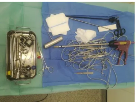
Figure 5 Surgical set up for LapOVE (Original) ... 36
Figure 6 Electrocautery and bipolar (With authorization from Freelance Surgical Veterinary Divison) ... 37
Figure 7 Laparoscopic tower (original)... 37
Figure 8 Clipping area for LapOVE (original) ... 37
Figure 9 Surgical theatre and staff disposition (original)... 38
Figure 10 Patient position (original) ... 39
Figure 11 Soft tissue surgical theatre (original) ... 39
Figure 12 Set up before the first incision is made (original) ... 40
Figure 13 Veress needle and CO2 tube in place ... 40
Figure 14 Both trocar’s positioned ... 41
Figure 15 Ovary being held by the spay hook (original) ... 42
Figure 16 Advantage VetSeal sealing and cutting the surrounding tissues (original) ... 42
Figure 17 Ovary ready to be removed (original) ... 42
Figure 18 Ovary being removed with artery (original) forceps... 43
Figure 19 Both ovaries once removed ... 43
Figure 20 Aspect of the wound after closure ... 43
viii LIST OF TABLES
Table 1 Side effects of ovariectomy/ovariohysterectomy in the bitch, adapted from (Romagnoli,
2008) ... 9
Table 2 Type and number of complications that occurred during and after OVH in 141 bitches performed by final-year students (adaptation from Burrow et al., 2004) ... 11
Table 3 Diagnostic indications for laparoscopy and treatments performed by laparoscopy or laparoscopic assistance (adapted from Rawlings, 2011)... 15
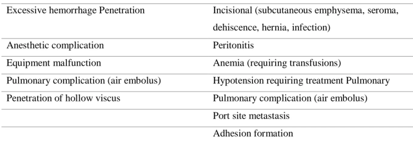
Table 4 Potential complications with laparoscopy adapted from McClaran & Buote, (2009) 24 Table 5 Specific Laparoscopic equipment ... 36
Table 6 Age and weight of the animals of both study groups ... 46
LIST OF GRAPHICS Graph 1 Surgical times of the different steps according to the procedure with error bars of the interquartile range ... 46
Graph 2 Plot of means of temperature evolution according to the procedure with the error bars of the standard deviation ... 47
Graph 3 Boxplot of the end of surgery temperature by procedure ... 48
Graph 4 Quantile-Comparison Plot of the temperature ... 48
Graph 5 Plot of means of the glucose evolution difference according to the procedure with the error bars of the standard deviation ... 49
Graph 6 Boxplot of glucose after extubation by procedure ... 50
Graph 7 Quantile-Comparison plot of glucose... 51
Graph 8 Plot of means of the evolution of the pain score ... 52
Graph 9 Complications intraoperative and post-operative of both procedures ... 53
LIST OF ABBREVIATIONS 3D = Three dimensional
CCU = Camera control unit CCD = Semiconductors
CMPS = Glasgow Composite Measure Pain Scale
CMPS-SF = Glasgow Composite Measure Pain Scale and its short form HALO = Harmonic scalpel-assisted laparoscopy
IASP = International Association for the Study of Pain IAP = Intra-abdominal pressure
i.e. = id est
LapOVE = Laparoscopic ovariectomy
ix MIS = Minimally invasive surgery
NRS = Numerical rating scale OVE = Ovariectomy
PATs = Pain assessment tools SDS = Simple descriptive scale
SILS = Single incision laparoscopic surgery VAS = Visual analogue scale
1 Internship activity description
The current dissertation under the subject of “Comparison of clinical and physiologic parameters, complications, and techniques, between Laparoscopic ovariectomy and ovariohysterectomy in dogs”, was elaborated in the context of the curricular internship of the Integrated Master’s degree in Veterinary Medicine from the “Faculdade de Medicina Veterinária - Universidade de Lisboa”. The internship was carried out at Kingston Veterinary Group mainly at the Park Street Hospital and the several branches, located in Kingston Upon Hull, for the period of 6 months between September 2018 and March 2019, with a workload of more than 45 hours per week accounting for over 1000 hours. It took place under the supervision of Dr. David Robinson and the co-supervision of Professor Berta São Braz. During the internship, I assisted and participated in numerous procedures carried out in different areas of small animal veterinary medicine. In the Surgery department, led by Dr. David Robinson, I cooperated by helping in several orthopaedic procedures (e.g. CWO, Fabello-tibial Suture, Tibial Tuberosity Transposition, Fracture repairs, stabilization of coxofemoral luxation using the Toggle Pin Method), soft tissue surgeries (e.g. exploratory laparotomies, laparoscopic ovariectomy, cystotomies, splenectomies, gastrotomy, enterotomy, perineal hernia repair, anal sacculectomy, incisional/excisional biopsies, BOAS surgery, grid keratectomy, temporary eyelid tacking, cherry eye repair, caesarean section, gastric dilatation volvulus) and post-operative care of the patients. Furthermore, I was also given the opportunity to develop my surgical skills by performing several surgical procedures under the supervision of senior surgeons, such as dog and cat castrations, dog and cat ovariohysterectomies, dewclaw removal, superficial nodule/mass excisions, dentistry procedures, scaling and polishing. The same applies in terms of anaesthesia, considering I was able to practice procedures such as induction, intubation and general anaesthetic monitoring. In the Internal Medicine department, I improved my clinical case solving skills and participated in the care and treatment of the inpatients, practicing procedures such as drugs administration, blood sampling, blood typing, collection and transfusion, venous and urinary catheterization, cystocentesis and running several diagnostic tests (e.g. haematology, biochemistry, urinalysis). In addition, I accompanied clinicians during consultations and was also able to take the lead and develop my consulting skills. Finally, with regards to the Diagnostic Imaging department, I assisted the surgeons during endoscopy (rhinoscopy, bronchoscopy, gastroscopy) and ultrasound, MRI and CT scans. Moreover, I was able to practice patient positioning for radiographic examination. During and after these procedures, I was taught by senior vets how to interpret the results and to take conclusions towards diagnostic and treatment planning in specific clinical cases. During
2
this period, I was also able to write a case report titled “Case report of an anal gland malignant melanoma in a dog” with Dr. Hugo Martins which is currently up to revision.
3
I. Comparison of clinical and physiologic parameters, complications, and techniques, between laparoscopic ovariectomy and ovariohysterectomy in dogs
1. Introduction to the dissertation subject
Gonadectomy is one of the most frequently performed surgical procedures in veterinary practice
given that it is the most reliable means of controlling a pet population (Goethem, Schaefers-Okkens, & Kirpensteijn, 2006).
That result can be achieved by different techniques, although in this dissertation we will emphasize on comparing ovariohysterectomy (OVH) and laparoscopic ovariectomy (LapOVE), given that these were the main procedures that were used at the hospital where the results for this dissertation were obtained.
OVH is the preferred technique for gonadectomy in dogs and cats, and for most uterine diseases, that includes but are not restricted to: pyometra, congenital anomalies and uterine prolapse, uterine rupture, among others that will be discussed in more detail more ahead, it is also described among other reports that it is the preferred technique for gonadectomy in this species (Goethem et al., 2006), and it is the most common surgical procedure performed in veterinary medicine (Davidson, Moll, & Payton, 2004). The combined incidence of intraoperative complications associated with traditional open OVH in healthy female dogs has been reported to be as high as 20.6% (Burrow, Batchelor, & Cripps, 2005).
Laparoscopic procedures have become quite common in human medicine, awareness of these techniques has increased amongst the general public. Pet owners now expect these minimally invasive techniques to also be an choice for their animals (Howe, 2006).
There are several studies that document the advantages for patients undergoing laparoscopic
versus open surgery that have been published for both human and veterinary patients. In female
dogs, laparoscopic sterilization provides a greatly improved view of the surgical field given the enhanced illumination and magnification provided by the instrumentation, which in return may decrease the risk of several intraoperative complications such as incomplete removal of ovarian tissue or haemorrhage (Corriveau, Giuffrida, Mayhew, & Runge, 2017).
Significantly lower pain scores, plasma cortisol levels, plasma glucose levels and plasma creatine phosphokinase levels have been reported in medium-breed dogs undergoing LapOVE (Rosewell, 2016).
The purpose of this dissertation is to compare both, clinical and physiologic parameters, complications, and techniques, to evaluate if one is superior to the other.
4 2. Bibliographic review
2.1 Anatomy of the female dog reproductive system
The female genital organs consist of ovaries, uterine tubes, uterus, vagina, vestibule, pudendum femininum (vulva), and clitoris, figure 1. The ovaries contain the oocytes, which are intermittently ovulated, engulfed by the infundibulum, fertilized or not in the uterine tube, and carried to the uterus. If fertilization takes place the blastocysts develop in the uterine tube and are passed into the uterine horns to be distributed and implanted in either uterine horn (Evans & De Lahunta, 2013).
Figure 1 Diagram of peritoneal reflections and the female genitalia of the dog (adapted from Evans & De Lahunta, 2013)
2.1.1 Ovaries
The ovary, is a paired oval organ, attached by a mesovarium to the body wall and the mesosalpinx. The mesovarium proximale extends from the body wall to the origin of the mesosalpinx, and the mesovarium distale extends from the origin of the mesosalpinx to the ovary and forms part of the wall of the ovarian bursa. The ovary lies caudal to the kidney (as can be seen in figure 2) and contains all of the oocytes that the female will ovulate in her lifetime. It is also the source of several hormones. In its normal position, an ovary may be described as having tubal and uterine extremities, a free border, and medial and lateral surfaces. The tubal end is nearest the infundibulum. The uterine end is the end attached to the uterus by the proper ligament of the ovary. The ovary is smooth in appearance before estrus, which
5
occurs for the first time between 6 and 9 months of age. In multiparous bitches, the surface may be rough and nodular (Evans & De Lahunta, 2013).
The ovary usually lies between the abdominal wall and the descending colon. The ventral border and medial surface of the ovary are in contact with the mesovarium. Frequently fat is deposited within the mesosalpinx partially obscuring the ovary (Evans & De Lahunta, 2013).
Figure 2 Female urogenital system in situ, ventral aspect. (Adapted from Evans & De Lahunta, 2013)
2.1.2 Uterine Tube
The paired uterine tubes (also termed oviducts or salpinx) receive and transport the oocytes to the uterus. They also carry the sperm in their ascent. Fertilization normally happens within the tubes. Each tube is suspended by the mesosalpinx and connects the peritoneal cavity with the uterine cavity and therefore with the external environment. The ovarian extremity of the uterine tube; which receives the oocyte after ovulation, takes the form of a funnel and is termed the infundibulum. The free edges of the infundibulum are bordered by numerous diverging processes, called fimbria, which contact and sometimes adhere to the surface of the ovary. The inside of the funnel is marked by folds, which converge to border a small opening in the depths of the funnel, the abdominal ostium (Konig & Liebich, 2004).
The abdominal ostium leads to the ampulla (ampulla tubae uterinae), where fertilisation normally takes place. The embryo stays in the ampulla for a few days before it is transported to the apex of the horn of the uterus through the narrower, more convoluted distal part of the tube, the isthmus. The uterine tube opens into the uterine horn through the uterine ostium
6
(ostium uterinum tubae) and marks the site of the uterotubal junction. It´s an abrupt junction in carnivores, in which the uterine ostium is located on top of a papilla, thus forming a barrier against ascending infections (Konig & Liebich, 2004).
2.1.3 Uterus
The uterus, which lies mainly dorsal to the small intestine, consists of a very short body from which two long and slender horns diverge. The body is close to the pubic brim but may be abdominal or pelvic in position. The cervix is also quite short however, the tissue thickening extends beyond the external ostium as a fold on the roof of the vagina. Transverse grooves often divide this fold into cranial, middle, and caudal tubercles; these become much swollen at certain stages of the cycle. The ostium of the cervix generally faces caudoventrally, and this orientation, combined with the asymmetry of the fornix and the fissuration of the cervical prolongation, could make its identification rather difficult, even with the aid of an endoscope (Dyce, Sack, & Wensing, 2010).
The uterus is composed of three tunics or layers: serosa, muscularis (myometrium), and mucosa (endometrium) with the last one being the thickest of the three (Fransson, 2018).
2.1.4 Vagina
The vagina is a dilatable canal, extending from the uterus to the vestibule. Cranially, the vagina is limited by the fornix, which extends ventral to the cervix. The fornix is the deepest part of the vagina and lies ventral and cranial to the cervix. The length of the dorsal vaginal wall is less than that of the ventral wall because of the oblique position of the cervix. The vagina ends just cranial to the urethral opening. It is delineated from the vestibule by a transverse mucosal ridge that extends dorsally on each side of the midventral line. No definite hymen is present at this point in the bitch, although its vestige may sometimes be found at the vaginovestibular junction. Both the diameter and length of the vagina increase significantly during pregnancy and during parturition. The longitudinal folds of the vaginal mucosa are high, permitting an expansion in diameter. Smaller transverse folds connecting the longitudinal folds allow craniocaudal stretching of the vagina (Evans & De Lahunta, 2013).
2.1.5 Vestibule
The vestibule constitutes the caudal part of the copulatory organ. It spreads from the external urethral opening to the external vulva and combines reproductive and urinary functions. It´s shorter than the vagina and lies mostly behind the ischial arch, which allows it to slope ventrally to its opening at the vulva. The resulting inflection of the axis of the genital passage must be taken into consideration when introducing a vaginal speculum or other instruments. The wall
7
of the vestibule contains vestibular glands, the secretion of which retains the mucosa of the vestibule moist and facilitates coitus and parturition. At oestrus, the odour of the secretion has a sexually stimulating effect upon the male animal. In the bitch the glands are small, but numerous and the duct openings are arranged in linear series. Darker patches of the lateral walls betray the position of the vestibular bulbs, a concentration of veins forming erectile tissue, regarded as the homologue of the bulb of the penis (Dyce et al., 2010). The entire clitoris in normal bitches is a relatively small, with the glans clitoris projecting into the fossa clitoridis.The clitoral fossa is encountered when the vulvar labia are separated. The clitoral fossa should not be confused with the vestibule, which lies dorsal and cranial to the clitoral fossa (Johnston, Kustritz, & Olson, 2001). The clitoris the homologue of the male penis, is composed of paired roots, a body, and a glans. The roots and body are homologues of the male corpora cavernosa penis, and the glans clitoridis is homologous with the glans penis, although it is not bipartite in structure. The body of the clitoris in the dog has both erectile and fatty tissue. It is covered by the tunica albuginea (Evans & De Lahunta, 2013).
2.1.6 Vulva
The vulva lies caudal to the vestibule and consists of two lips, labii joined dorsally and ventrally by commissures and separated by a narrow cleft, the rima pudendi. The labia form the external boundary of the vulva and in part are homologous with the scrotum of the male. The labia are soft and pliable, composed of fibrous and elastic connective tissue, striated muscle fibers, and an abundance of fat. The vaginal processes, containing the round ligaments of the uterus, often end in the subcutaneous connective tissue of the labia. The dorsal commissure lies at or slightly ventral to the dorsal plane passing through the symphysis pelvis. The ventral portions of the labia, with their uniting commissure, form a pointed projection extending ventrally and caudally from the body, usually with a tuft of hair (Evans & De Lahunta, 2013).
2.2 Blood supply, lymphatic drainage and innervation of the female genital organs Blood supply to the female genital organs is provided by four paired arteries: ovarian artery; uterine artery; vaginal artery and internal pudendal artery. After dividing from the aorta, the ovarian artery follows a convoluted course to the ovary. It supplies the ovary and detaches branches to the uterine tube and to the tip of the uterine horn. The uterine branch anastomoses with the uterine artery within the broad ligament. The rest of the female genital tract is supplied by the uterine and vaginal arteries, which are branches of the internal iliac artery and by the continuation of the vaginal arteries, the internal pudendal artery. The uterine artery passes to
8
the uterus within the broad ligament. It detaches a series of branches to the body and horn of the uterus (Konig & Liebich, 2004).
In dogs and cats, the uterine artery is a branch of the vaginal artery. The major blood supply to the uterus is provided by the uterine branch of the ovarian artery. The caudal parts of the female genital tract are supplied by branches of the internal pudendal and vaginal arteries; the pattern of branching varies in different animals (Konig & Liebich, 2004).
The ovarian vein drains the majority of the uterus. The adjacent vessel walls are substantially thinner than the others and facilitate transmural transportation of Prostaglandin F2α from the vein into the artery. The vaginal vein vascularizes an extensive plexus in the walls of the vagina and vestibule (Konig & Liebich, 2004).
The lymphatics of the female genital tract drain primarily into the medial iliac lymph nodes and to the lumbar aortic lymph nodes (Konig & Liebich, 2004).
Innervation of the female genital organs is provided by the autonomic nervous system. The ovaries receive sympathetic fibers from the intermesenteric and the caudal mesenteric plexus and parasympathetic fibers from the vagus. The rest of the female genital tract receives parasympathetic and sympathetic innervation via the pelvic plexus (Konig & Liebich, 2004).
2.3 Sterilization of the female dog 2.3.1 Advantages
There are innumerous reasons supporting the gonadectomy of female dogs, that range from prevention of diseases and pregnancy to population control.
Elective sterilization of dogs has a dual benefit given that it counteracts overpopulation by preventing reproduction and lowers the likelihood that an individual animal will be relinquished to a humane organization. It may also correct sexually dimorphic aggression, a negative behavioral interaction that occurs between females or between males housed with females (Fransson, 2018).
The removal of the ovaries in bitches is associated with absence of ovarian diseases (such as, ovarian tumors and ovarian cysts), as well with a decreased risk of mammary and uterine diseases like: mammary neoplasia and pyometra, respectively, and also false pregnancy, estrogen-related diseases (vaginal hyperplasia/prolapse, persistent estrus, bone marrow aplasia), pregnancy-related diseases (pregnancy complications, uterine prolapse) or parturition-related diseases (dystocia, uterine prolapse, subinvolution of placental sites (Romagnoli, 2008).
9
A positive effect on survival was also noted when ovariohysterectomy was performed as an adjunctive treatment for canine mammary gland carcinoma. In the case of pyometra, that is quite a common disease in intact female dogs, it is estimated that 23% to 24% of intact bitches require treatment for pyometra by 10 years of age (Fransson, 2018).
Female dogs gonadectomized prior to puberty have a 95% reduction of the risk of developing mammary tumors as opposed to bitches spayed after the first heat (8% risk), after second heat (26% risk) or bitches spayed after 2.5 years of age or left intact (100% or full risk) (Romagnoli, 2008).
It is also well documented that neutered animals have a longer life span than intact animals. The increased life expectancy in gonadectomized animals may be due to the preventive effect on diseases of the reproductive tract and ⁄or the reduced risk-associated behavior (Reichler, 2009).
2.3.2 Disadvantages
The benefits of spaying pets cannot be overstated. Nonetheless, there are some conditions that have been reported with increased frequency in gonadectomized dogs compared with sexually intact animals (Kustritz, 2007).
Disadvantages of gonadectomy include anaesthetic and surgical complications, increased risk of neoplasia of various organ systems, increased incidence of some musculoskeletal and endocrinologic disorders, obesity and urinary incontinence in bitches, as it can be seen on, table 1 (Kustritz, 2012).
Table 1 Side effects of ovariectomy/ovariohysterectomy in the bitch, adapted from (Romagnoli, 2008)
The most commonly reported factor for increased risk of obesity is gonadectomy. Spayed female dogs have increased food intake and appetite after ovariohysterectomy, most likely resulting from the loss of estrogen, which may act as a satiety factor (Fransson, 2018).
Side Effects Bitch
Generic surgical risk 7-27%
Obesity 25-50%
Urinary incontinence 5-12%
Osteoporosis Reported
10
However, a study showed that differences related to obesity in entire neutered dogs, were not associated with differences in diet or exercise duration, frequency and intensity, which means it can be managed (Robertson, 2003).
Urinary incontinence is also a problem of spayed female dogs it can have an incidence of 5.1% (Angioletti, De Francesco, Vergottini, & Battocchio, 2004). These authors also showed that the relationship between acquired urinary incontinence and the type of surgery performed did not show meaningful values. Others disadvantages vary according to the surgical technique that is used.
2.3.3 Techniques
There are several of techniques for surgically sterilizing of female dogs that have been described. Each one has advantages and disadvantages to both the surgeon and patient. Techniques that have been described include traditional midline ovariohysterectomy, lateral flank ovariohysterectomy or ovariectomy, laparoscopic ovariohysterectomy or ovariectomy. However, no matter the technique used, strict adherence to sound surgical technique and asepsis is mandatory for good surgical outcome with minimal complications (Howe, 2006). 2.3.3.1 Ovariohysterectomy
2.3.3.1.1 Technique
In dogs, ovariohysterectomy is traditionally performed through a small ventral midline incision. In adult female dogs, the incision typically begins at, or not more than 1 cm caudal to, the umbilicus. Once the reproductive tract has been identified, the suspensory ligament should be carefully broken using caudolateral or caudomedial traction with the index finger of the dominant hand, while holding traction on the proper ligament using the nondominant hand. Several techniques have been described for clamping and ligating the ovarian and uterine pedicles, including the single-, double-, and triple-clamp methods. Regardless of the technique selected, it is important to visualize or carefully palpate the ovary between the thumb and index finger, and then to ‘‘pinch’’ the thumb and forefinger together (while holding and protecting the ovarian tissue) deep to the ovary prior to placement of the clamp. This will prevent inadvertent clamping of the ovarian tissue, which could result in ovarian remnant tissue syndrome. Double ligations, utilizing an encircling and a transfixing ligature on all ovarian pedicles in adult dogs are recommended. When ligating the uterine body, it is important to place the most caudal ligature at the junction of the cervix and uterine body, so to avoid leaving any viable uterine body tissue that could result in a stump pyometra in the future. The second ligature is placed cranial to the first and is appropriately spaced so as to avoid leaving excessive devitalized tissue (Howe, 2006).
11 2.3.3.1.2 Complications
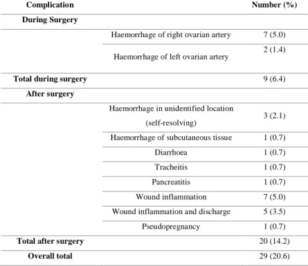
Burrow et al., (2005), the authors of a study involving 142 ovariohysterectomy of female dogs reported complications similar to those published in previous studies: haemorrhage of the ovarian arteries during surgery (nine out of 141), haemorrhage after surgery (four out of 141), wound inflammation (12 out of 141) and other types of complication (four out of 141), as can be seen on table 2. The rates of intraoperative, post-operative and total complications were 6.3, 14.1 and 20.6 per cent, respectively. Haemorrhage is one of the most common complications secondary to an ovariohysterectomy, and death of the patient can occur if it is severe (Howe, 2006). Common causes of intraoperative bleeding in elective ovariohysterectomy performed by inexperienced surgeons include tearing of the perirenal peritoneum while strumming the suspensory ligament, incomplete ligation of the mesovarium and associated ovarian vessels, and loose ligatures on the mesovarium (Fransson, 2018).
Careful use of technique while breaking the suspensory ligament, handling and manipulating the ovarian and uterine pedicles, and placing ligatures is important in preventing haemorrhage. Proper placement, spacing, and tightness of ligatures is also critical to prevent intraoperative and post-operative haemorrhage. Careful examination of each pedicle with tension relieved, prior to release of the pedicle, is also an important step to prevent this type of complication (Howe, 2006).
Table 2 Type and number of complications that occurred during and after OVH in 141 bitches performed by final-year students (adaptation from Burrow et al., 2004)
Complication Number (%)
During Surgery
Haemorrhage of right ovarian artery 7 (5.0)
Haemorrhage of left ovarian artery 2 (1.4)
Total during surgery 9 (6.4)
After surgery
Haemorrhage in unidentified location
(self-resolving) 3 (2.1)
Haemorrhage of subcutaneous tissue 1 (0.7)
Diarrhoea 1 (0.7)
Tracheitis 1 (0.7)
Pancreatitis 1 (0.7)
Wound inflammation 7 (5.0)
Wound inflammation and discharge 5 (3.5)
Pseudopregnancy 1 (0.7)
Total after surgery 20 (14.2)
12
Ovarian remnant syndrome is the presence of functional ovarian tissue in the abdomen following ovariohysterectomy, due to not removing all ovarian tissue as a result of inappropriate ovariohysterectomy technique, that may result in signs of proestrus, estrus, and rarely false pregnancy. Stump pyometra may occur following ovariohysterectomy if a portion of the uterine horns or uterine body is not removed and the animal has increased progesterone concentrations (Howe, 2006).
Granuloma formation at uterine or ovarian pedicle remnants has been reported in up to 28% of dogs undergoing ovariohysterectomy (Fransson, 2018).
Accidental ligation of a ureter, which can result in hydronephrosis or atrophy of the kidney, is quite preventable by careful identification of the uterine horns, uterine body, and cervix prior to ligation of the uterine body, and avoidance of ligation of any extraneous peribladder fat which may contain a ureter (Howe, 2006).
2.3.3.2 Lateral flank approach for ovariohysterectomy
There are certain conditions for which the lateral flank approach technique is preferred for ovariohysterectomy, they include excessive mammary gland development due to lactation or mammary gland hyperplasia. On a lactating animal, using the lateral flank approach can avoid potential complications that may be associated with the ventral midline approach, such as excessive hemorrhage from the skin and subcutaneous tissue, wound inflammation or infection, and leakage from mammary tissue. In addition, using the lateral flank approach in lactating animals minimizes disruption to the mammary glands so that those animals are more likely to continue nursing appropriately after surgery (Mcgrath, Hardie, & Davis, 2004). Advantages of this technique include the reduced potential for eventration if wound dehiscence occurs (Mcgrath et al., 2004).
2.3.3.2.1 Technique
The lateral approach is performed through a dorsoventral incision that is placed just caudal to the midpoint between the last rib and iliac crest. The incision length is approximately 3 cm in dogs, however, it can differ depending upon the size of the animal. The abdominal wall is entered via a grid approach using blunt dissection through the separate layers of muscle. Once the uterus and ovary have been identified, the ovarian pedicle is isolated and ligated in standard fashion. After the ovarian pedicle is ligated, and the broad ligament to that side is torn, the uterine horn is traced to the bifurcation, and the second uterine horn identified and traced cranially to the second ovary. Visualization of the contralateral ovarian pedicle can be difficult through a small flank incision, and it may be necessary to enlarge the incision. Once the second ovarian pedicle has been ligated, and the broad ligament divided, traction is applied to both
13
uterine horns to expose the ligation site on the uterine body. The uterus is then ligated in standard fashion. After verifying lack of hemorrhage, the body wall musculature should be closed in two layers. Subcutaneous tissue and skin closure is routine (Howe, 2006).
2.3.3.2.2 Contraindications
Contraindications for the lateral flank approach for ovariohysterectomy include uterine distention due to pregnancy or pyometra, obesity, or patient age younger than 12 weeks. It can also cause visible scarring or imperfections in hair color or regrowth. The primary disadvantage of the flank approach is limited exposure to the abdomen if complications arise. It is also difficult to properly identify animals that already had an ovariohysterectomy because the incision scar may be in the flank region and not in the typical ventral midline location. The exposure of the uterine stump and contralateral ovarian pedicle is generally more limited, making it difficult to achieve hemostasis if a pedicle is accidentally dropped or if bleeding occurs in these areas (Mcgrath et al., 2004).
2.3.3.3 Ovariectomy
The most common indication for ovariectomy (OVE) in veterinary medicine is elective sterilization in animals with a normal uterus. Usually, the technique used is ovariohysterectomy, but long-term studies have failed to show significant advantage of the ovariohysterectomy compared with ovariectomy alone unless the uterus has pathologic changes (Fransson, 2018).
The main argument against ovariectomy state that the uterus should be removed to prevent pyometra from occurring later in life. However, the incomplete uterine body removal which is often performed in ventral ovariohysterectomy creates no smaller risk. In a study with 72 dogs, no stump pyometras were described in dogs that underwent the ventral midline technique and in which proper ovarian removal had been performed (Janssens & Janssens, 1991).
Also, there is no evidence that conditions such as cystic endometrial hyperplasia or other conditions, develop in the ovariectomized bitch unless progestagens are administered, compared with ovariohysterectomy (Okkens, Kooistra, & Nickel, 1997).
A major advantage of OVE is that it can be performed through a smaller celiotomy and with less traction on the female genital tract. With respect to long-term urogenital problems, including endometritis/pyometra and urinary incontinence, it has been clearly established that they do not occur more frequently with either technique. Most evidence extracted from the literature report no benefit and thus no indication for removing the uterus during routine neutering in healthy bitches. Thus, reinforcing the idea that OVE should be the procedure of choice for canine gonadectomy (Goethem et al., 2006).
14 2.3.3.4 Technique
The technique is performed using a ventral midline abdominal approach that starts at the umbilicus and extends caudally. The ovary is identified and the ovarian pedicle is ligated using traditional techniques and materials. Once ligated, the ovarian pedicle is severed. The uterine artery and vein are then ligated and severed at the proper ligament (cranial tip of the uterine horn), and the ovary removed. Closure is routine (Howe, 2006).
2.4 Laparoscopy
Laparoscopy is a technique to look into the abdominal cavity via a tiny incision using a (rigid) telescope, allowing visual exploration of the internal organs.(Schneider & Feussner, 2017) It is also a minimally invasive surgical technique that achieves many of the same maneuvers as the traditional surgical procedure of laparotomy. However, laparoscopy is done by using the optical space produced by a gas that causes insufflation. Initial insufflation is done with the use of a Veress needle or a catheter placed through a minilaparotomy incision or by the placement of a trocar-cannula with the use of a Hasson technique. As soon as the peritoneal cavity is distended, the laparoscope is placed through the cannula and used to observe the placement of additional trocar-cannulae. With the use of these additional cannula, biopsy and surgical instruments are passed into the abdomen. An air-tight seal must be maintained by laparoscopic cannula sites to prevent loss of carbon dioxide and optical space (Rawlings, 2011).
2.4.1 Indications
Laparoscopic treatments, which have been in constant development, evidently expand the types and numbers of small animal procedures. Furthermore, the number of indications that can be treated with laparoscopy has extended are also shown on table 3, (Rawlings, 2011).
Virtually all of the organ biopsy specimens taken by traditional laparotomy can also be obtained by laparoscopy, examples of this are on table 3 (Rawlings, 2011).
Commonly performed laparoscopic or laparoscopic-assisted procedures in dogs and cats include liver, spleen, intestinal, and lymph node biopsies; feeding tube placement; ovariohysterectomy and ovariectomy (Buote, Kovak-McClaran, & Schold, 2011).
Additional applications of diagnostic laparoscopy include evaluation of abdominal trauma. This use is expanding in human medicine, along with operator experience can be quite accurate. In veterinary medicine, this represents a noninvasive method to evaluate blunt abdominal trauma. Injuries such as splenic and hepatic lacerations, diaphragmatic hernia, bladder rupture, renal rupture and abdominal hernia can be assessed. This often dictates the potential need for open abdominal surgery (Richter, 2001).
15
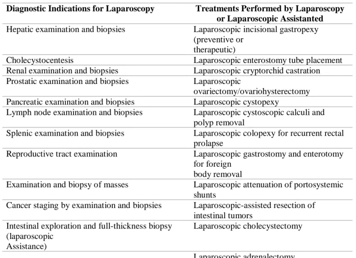
Table 3 Diagnostic indications for laparoscopy and treatments performed by laparoscopy or laparoscopic assistance (adapted from Rawlings, 2011)
Diagnostic Indications for Laparoscopy Treatments Performed by Laparoscopy or Laparoscopic Assistanted Hepatic examination and biopsies Laparoscopic incisional gastropexy
(preventive or therapeutic)
Cholecystocentesis Laparoscopic enterostomy tube placement
Renal examination and biopsies Laparoscopic cryptorchid castration Prostatic examination and biopsies Laparoscopic
ovariectomy/ovariohysterectomy Pancreatic examination and biopsies Laparoscopic cystopexy
Lymph node examination and biopsies Laparoscopic cystoscopic calculi and polyp removal
Splenic examination and biopsies Laparoscopic colopexy for recurrent rectal prolapse
Reproductive tract examination Laparoscopic gastrostomy and enterotomy for foreign
body removal
Examination and biopsy of masses Laparoscopic attenuation of portosystemic shunts
Cancer staging by examination and biopsies Laparoscopic-assisted resection of intestinal tumors
Intestinal exploration and full-thickness biopsy (laparoscopic
Assistance)
Laparoscopic cholecystectomy
Laparoscopic adrenalectomy
2.4.2 Advantages
The advantages of minimally invasive surgery (MIS) have been found in studies of laparoscopic colectomy, appendectomy, gastric bypass, and splenectomy in human patients. At the moment, few of these reported advantages have been scientifically evaluated in veterinary patients. Also, several studies in human medicine have evaluated postoperative surgical site infection rate in procedures in which MIS is considered the reasonable standard of care. MIS have a decrease in pain and discomfort and a more rapid return to normal activity after surgery, with fewer wound-healing and other complications. It is usually correlated that the reported advantages of MIS in humans might also be realized in companion animals (Mayhew, Freeman, Kwan, & Brown, 2012).
Nonetheless there are some studies that report a reduction in pain and more rapid return to normal life in veterinary patients after MIS (Mayhew et al., 2012), such as, in procedures corresponding to laparoscopic ovariohysterectomy, ovariectomy (Culp, Mayhew, & Brown, 2009; Devitt, Cox, & Hailey, 2005).
16
It is generally accepted that the immune system is better preserved following laparoscopic than open surgery, what is demonstrated by the lower release of several biomarkers including interleukin 6 and C-reactive protein. This decreased of the immune response is most likely a consequence of a significantly smaller tissue injury (Targarona, Balague, Knook, & Trías, 2000).
The effect of insufflation gas is also considerable. It has been shown that air is actually more damaging to local cell-mediated immunity than CO2, the most frequently used gas in
laparoscopy. (Mayhew et al., 2012). In a study performed in a murine model, it was reported that laparotomy or air-insufflation laparoscopy impaired macrophage phagocytosis to a greater extent than did CO2 laparoscopy (Watson, Redmond, McCarthy, Burke, & Bouchier-Hayes,
1995).
Also, images and movies can also be recorded during the procedure. These can be used to monitor disease severity and to communicate with the client and veterinary colleagues (Rawlings, 2011).
2.4.3 Contraindications
A traditional laparotomy procedure is frequently favored if the addition of endoscopy significantly complicates the procedure. An open laparotomy is usually required in the case of a major mass resection. Unstable cardiopulmonary or renal systems, endocrinopathies and neurologic problems can be contraindications for the same reason that they would be in traditional laparotomy.
The major contraindication for laparoscopy is a lack of equipment or experience on the part of the operator to perform the procedure. Training is required, and experience with multiple procedures reduces complications during laparoscopy. The primary missing examination tool is digital palpation, such as in the liver and spleen, during a laparotomy though this may be overrated (Rawlings, 2011).
In open surgery, instruments are easily sterilized by conventional methods (ethlylene oxide or autoclave). Though, for laparoscopic work the kit is mechanically more complex and so its complete sterilization can be more difficult. Several infectious complications, especially at the level of the abdominal wall, have been reported after laparoscopic cholecystectomy, perforated duodenal ulcer surgery and hysterectomy (Targarona et al., 2000).
Insufficient training and experience are one of the most important limitations/contraindications, given the long learning curve (Buote et al., 2011).
17 2.4.4 Equipment
2.4.4.1 Imaging Chain
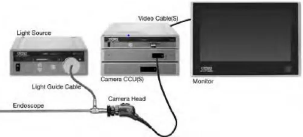
A basic video endoscopy imaging system consists of the following components: a light source, light‐transmitting cable, endoscope, camera, and monitor; (figure 3) each component is essential and the quality of the image obtained is dependent of each element (Brandão & Chamness, 2015).
The light generated by the light source is transmitted by the optic fiber light cable, and farther down the telescope, by optics fiber to illuminate the anatomy being observed. The image is transmitted through a series of lenses from the distal end of the telescope to the eyepiece, where the chip in the video camera head senses the image and transmits it to the camera control unit (CCU), which processes the endoscopic image and transmits it to a monitor for viewing (Brandão & Chamness, 2015).
Figure 3 Basic endoscopic imaging chain, (2014 Photo Courtesy of Karl STORZ GmbH & Co.KG)
2.4.4.2 Laparoscope (Telescope)
The telescope uses glass lenses to direct light by way of a fiberoptic bundle to illuminate the target area and the eyepiece. In conventional telescopes, a series of lenses are embedded in an air medium, whereas in the Hopkins rod lens telescopes (that are considered the gold standard, Schneider & Feussner, 2017) the lenses have been substituted with glass rods divided by small negative air lenses, see figure 4 (Van Lue & Van Lue, 2009).
In a rod lens system, air acts as a negative lens, within a glass medium, as opposed to the glass lenses within an air medium found in conventional telescopes. Rod lens telescopes convey significantly more light and have a wider field of view (Chamness, 2005).
18
Figure 4 Traditional optical system (top figure) vs. Hopkins rod system (bottom figure) (adapted from Chamness, (2005)
2.4.4.3 Light source
Light is conveyed from a remote light source through a fiberoptic light cable to the rigid fiberoptic laparoscope (telescope). The light source needs to be bright enough to illuminate a large cavity like the abdomen of a large deep chested dog. Currently, the Xenon light source is the most popular given that it offers excellent tissue color reproduction with light closely approximating that of pure sunlight (Brandão & Chamness, 2015).
2.4.4.4 Endoscopy video cameras
The camera head encompasses either one or three semiconductors (CCDs), which sense the image and convert it to an electronic signal. Modern video cameras have a variety of features that make them lightweight, soakable, gas sterilizable, and, certain models are autoclavable. They may have automatic exposure control, a zoom lens, contrast enhancement capability, and buttons on the camera heads to control various settings or activate peripheral devices. The CCD is the “chip” referred to in single-chip vs. three-chip cameras (Chamness, 2005).
The optical quality of modern single-chip cameras is high, but the ones that make use of three-chip is better. Three-three-chip cameras have horizontal resolution and superior accuracy of color reproduction (Chamness, 2005), and have a chip for each of the primary colors of the image (red, green, and blue) and provide a superior image compared with one-chip cameras (Richter, 2001).
2.4.4.5 Monitors
The final display for viewing the endoscopic image is provided by the video monitor and its connected via a cable, either directly from the camera processor or from any number of various recording devices that may be placed in between the camera processor and the monitor. The video chain should always terminate with the monitor (Chamness, 2008).
19
Optimal positioning of the screen during the operation is crucial, the monitor height should always be attuned to the eye level of the surgeon, and positioned in such a way as to minimize neck strain (Chamness, 2008).
2.4.4.6 Insufflator
The insufflator is a pump that provides CO2 into the abdomen, which is needed to achieve the
artificial space that allows the surgeon to perform the surgery. It is designed to yield adequate pressure (ca. 15 mmHg) and to maintain it during the procedure even in the event of gas leaks, but, at the same time, it needs to avoid pressure peaks which would be harmful for the patients. Therefore, pressure and flow sensors are vital components of an insufflator. Insufflators are equipped with displays, that indicate the effective and the preselected intra-abdominal pressure (IAP) along with gas flow and the total amount of insufflated gas (Schneider & Feussner, 2017).
In the case of using a Veress needle approach, at the beginning of initiating insufflation, the pressure should be low, with a high flow rate on the insufflator display, hence reflecting the instillation of the CO2 gas into the large potential space of the peritoneal cavity. If the pressure
is initially high, the surgeon should check in the first place all stopcocks to guarantee that they are in the open position. If pressure still remains high, another possible reason is the Veress needle or cannula is not being in the desired tissue space (Van Lue & Van Lue, 2009).
2.4.4.7 Operating table
The ideal table allows the surgeon to tilt the patient laterally in either direction as well as vary the degrees of head-up (known as reverse Trendelenburg position) or head-down (Trendelenburg position) positions. The table height will be lower during a laparoscopic procedure than during a conventional procedure given the long length of the instruments (Van Lue & Van Lue, 2009).
2.4.5 Instruments
Endoscopic instruments tend to be the same as used for traditional surgery with the exception that they are long and have a narrow shaft that is suitable for passing through the cannula. Reusable instruments can be properly cleaned and sterilized in a steam autoclave. (Rawlings, 2011)
2.4.5.1 Cannula-Trocar assembly
The trocar is a sharp-pointed stylet that penetrates the abdominal wall. It is removed when the cannula enters the abdominal cavity, leaving the cannula in place which enables the laparoscope to be introduced later. (Richter, 2001).
20
When there is a need to penetrate the muscles and peritoneum a sharp trocar is used while the blunt trocars are used where there is no need to cut tissues. It is vital to avoid injury to internal structures such as the heart and lungs (Bennett, 2009).
To address the significant morbidity that can follow a trocar injury to a vascular structure or bowel, new features have been introduced such as trocars with optical viewing capability during insertion, use of a fascial- or tissue- separating blunt tip, threaded cannulas placed with a twisting motion, or combinations thereof, which are now widely used (Van Lue & Van Lue, 2009).
2.4.5.2 Forceps
Laparoscopic forceps can be divided into three categories: grasping forceps, dissecting forceps, and biopsy forceps, and like in any open surgery, the kind of forceps used will depend on its intended use (Swanson & Millard, 2015).
The jaws of laparoscopic forceps can be traumatic or atraumatic; grasping forceps can also be fenestrated or closed, with varying strength that is proportional to their teeth size; dissection forceps can be bent or straight (Prisco, 2002). Given the loss of tactile feedback occurring in any laparoscopy procedure, even atraumatic forceps can damage tissues if too much force is applied, so one must be careful (Moore & Ragni, 2012).
2.4.5.3 Scissors
Compared to open surgery, the use of scissors in minor access surgery is more limited, as one would expect. The skill required is much greater since they are potentially harmful. Scissors come in a variety of straight, curved, or hook blades. The edges can be serrated to prevent tissue slipping out of the blades. Curved scissors allow a better visual control during cutting and are generally preferred in laparoscopic surgery (Schneider & Feussner, 2017).
2.4.5.4 Tissue Retractors
One of the most important disadvantages to overcome in the use of laparoscopic instrumentation is the loss of tactile feedback (Van Lue & Van Lue, 2009). Palpation probes are used to palpate organs and structures within a body cavity, and to move and retract out of the viewing field (Swanson & Millard, 2015). Ovariectomy hooks are used during laparoscopic ovariectomy and ovariohysterectomy to suspend the ovary against the body wall to ease ligation and transection of the ovarian pedicle. The small puncture that is created from the hook does not need to be closed (Swanson & Millard, 2015).
21 2.4.5.5 Others
There are many other instruments used for additional maneuvers, and should be acquired depending on the procedure intended (Tapia-Araya, Martin-Portugués, & Sánchez-Margallo, 2015).
2.4.6 Electrosurgery
Electrosurgery is a valuable tool to make surgery much safer and faster. One must always take into account, the disruptive processes that can result from electric current running through tissue (Schneider & Feussner, 2017). In the monopolar system, current is generated in the cautery device to the active electrode, moving on throw the tissue, returning to the return electrode and back to the cautery (Prisco, 2002). The use of bipolar and ultrasonic sealing devices has resulted in more efficient and cost-effective surgery given that they limit the application of mechanical devices such as clips an and staples to larger vessels and more vascular tissues (Phillips et al., 2008). B. E. B. J. Van Goethem, Rosenveldt, & Kirpensteijn, (2003) also concluded that laparoscopic ovariectomy can be performed more rapidly when using bipolar instead of monopolar electrosurgery and with less risk of mesovarial haemorrhage. Although the advantages of bipolar based energy-based surgical devices, there are reports that certain ultrasonic energy-based surgical devices produce significantly less thermal damage. (Phillips et al., 2008).
2.4.7 Laparoscopic Surgery 2.4.7.1 Anaesthesia
In small animals, laparoscopy procedures will generally require the use of general anesthesia and it is used for invasive or longer duration procedures and for patients with significant respiratory disease. General anesthesia allows patients to easily be ventilated and provides analgesia, good muscle relaxation, and a quiet surgical field. It will also allow to convert the surgery to a laparotomy in case of a major complication or unexpected finding. Each type of laparoscopic procedures have unique requirements, which must be considered when anesthetizing patients to prevent potential complications from occurring (Quandt, 1999). General anesthesia, using balanced anesthesia technique including several parenteral and inhalational agents with the use of muscle relaxants, showed a rapid recovery and cardiovascular stability (Gerges, Kanazi, & Jabbour-Khoury, 2006).
For abdominal laparoscopy, veterinary surgeons must be mindful of the main hemodynamic and respiratory consequences of laparoscopic procedures on the patient, such as increased IAP formed by the establishment of the pneumoperitoneum, the type of gas used and the position
22
of the patient on the operating table for easy maneuvering of the surgeon (Dörfelt, Ambrisko, & Moens, 2012).
2.4.7.2 Animal preparation
The urinary bladder should be expressed prior to MIS to minimize the risk of accidentally traumatizing it and to increase visualization. MIS is always performed under standard aseptic conditions just like any open procedure. The owner must always be prepared and aware for the possibility of needing to convert the surgery into an open procedure as a result of complications or an inability to accomplish the procedure through a MIS technique, hence the patient being clipped for a complete abdominal laparotomy. For an ovariohysterectomy, tipping the head down will improve visualization of structures in the caudal abdomen while for a liver biopsy, tipping the rear end down will displace viscera away from the liver improving visualization (Bennett, 2009).
Position of the patient depends on the procedure to be performed; dorsal recumbency is used in most cases, at least to place the first port and the telescope. Thermal support is also crucial, particularly in smaller patients (Sladakovic & Divers, 2016).
2.4.7.3 Insufflation
In the typical anatomy of small animals, the mucosal surfaces are in close contact and the peritoneal space contains just a little fluid. In the case of the endoscope being placed into this space, the image obtained would be a diffuse pale red image resulting from the various organs and tissues. In order to have a proper image that enables the laparoscopic procedure, it is necessary to fill the peritoneal cavity with an inert gas and create a space to work – the pneumoperitoneum. In cats and small dogs, it is suggested to insufflate the abdomen to a maximum of 12 mmHg, and in larger dogs no higher than 14 mmHg. Once the operating cannulae is inserted, IAP may be lowered to 10 mmHg or below, as all that is required is a sufficient space to visualize the site of interest (Lhermette & Sobel, 2008).
In a recent study, the levels of pressure and influence of body fat, were studied, found that patients with a higher abdominal fat thickness would need a lower pneumoperitoneum pressure to maintain adequate laparoscopic working space, whereas patients with less abdominal fat thickness may need higher pressures. Thus the insufflator pressure could be tailored to the patient based on their body composition (Becker et al., 2016).
To attain a pneumoperitoneum there are two possible main techniques: a closed technique with the use of a Veress needle, and the open or Hasson technique performed through a full wall incision with a blunt trocar-cannula (Ferrão, 2016; Tapia-Araya et al., 2015).
23
Veress needle technique, uses as the name says, a Veress needle which consists of an outer sharp cutting tip with a spring-loaded blunt obturator within the needle that retracts into the needle once the needle as it passes through the tough body wall then advances past the sharp tip once inside avoiding injury to internal structures (Bennett, 2009). This is the most common method for insufflating the abdominal cavity. A skin puncture incision is performed in the selected abdominal area, and the abdominal wall is lifted and tensed upwards. The Veress needle is then inserted and directed caudally at a 50° angle from the skin, preferably towards the right caudal quadrant and away from the spleen. This insertion should be preferably carried out in the same site intended for the introduction of a trocar-cannula; the site is often caudal or cranial to the umbilicus. Usually the Veress needle insertion site corresponds to the second trocar-cannula (Tapia-Araya et al., 2015).
2.4.7.3.1 Types of Gas
The ideal laparoscopic insufflation gas should be easily available, relatively inexpensive, colorless, highly soluble in plasma, and suitable to use for most patients and procedures. It also should be chemically stable, physiologically inert, and nonexplosive. There are several of types of gases whose use have been studied to be used in laparoscopic procedures, carbon dioxide (CO2), nitrous oxide (N2O), helium (He), air, nitrogen (N2), and argon (Ar) (Menes & Spivak,
2000).
2.4.7.3.2 CO2
Carbon dioxide (CO2) is currently the insufflation gas of choice for laparoscopy. It satisfies
most of the requirements for an ideal insufflation gas, being colorless, noninflammable, and rapidly excreted from the circulation (Neuhaus, Gupta, & Watson, 2001).
The hemoglobin carried by the red blood cells has a higher affinity for CO2 than for other
laparoscopic gases, adding a higher safety margin in the rare event of gas embolism. The high solubility of CO2 in plasma is accountable for its recognized, yet overemphasized
disadvantage: some degree of serum CO2 elevation and a decrease in serum pH. In fact, severe
hypercarbia (pCO2 > 55 mm Hg) rarely seen, except in patients with depressed
cardiopulmonary function. Mild hypercarbia has little or no hemodynamic effect and is well tolerated by almost all patients. Yet, with the rapid expansion of laparoscopic procedures and the change in patient demography, the potential for severe hypercarbia in some cases should be kept in mind (Menes & Spivak, 2000). Warmed and humidified CO2, can also be used with
minimal additional equipment and cost, which had been reported to improve temperature control (Dean et al., 2017).