Acute and developmental toxicity of gold
nanorods on zebrafish (Danio rerio) embryos
Bárbara Sofia Maia Mesquita
Dissertation submitted in partial fulfillment of the requirements
For the Degree of Integrated Master in Bioengineering, Molecular Biotechnology
Supervisor: Dr. Sónia Fraga, PhD Co-Supervisor: Dr. João Paulo Teixeira
Acute and developmental toxicity of gold
nanorods on zebrafish (Danio rerio) embryos
Bárbara Sofia Maia Mesquita
Submitted in partial fulfillment of the requirements
For the Degree of Integrated Master in Bioengineering, Molecular Biotechnology
Supervisor: Sónia Fraga, PhD (Postdoctoral Researcher, Department of Environmental Health, National Institute of Health Doutor Ricardo Jorge, Porto, Portugal and EPIUnit- Institute of Public Health, University of Porto, Porto, Portugal)
Co-Supervisor: João Paulo Teixeira, PhD (Assistant Researcher, Department of Environmental Health, National Institute of Health Doutor Ricardo Jorge, Porto, Portugal and EPIUnit- Institute of Public Health, University of Porto, Porto, Portugal)
É autorizada a reprodução parcial desta dissertação, apenas para efeitos de investigação, mediante declaração escrita do interessado, que a tal se compromete.
Some of the results included in this dissertation have been presented in scientific meetings:
Toxicity of gold nanorods on zebrafish (Danio rerio) embryos. Mesquita B, Fraga S, Simões AM, Lopes I, Teixeira JP. International Conference on Occupational & Environmental Toxicology, 21st-23rd June 2016, Porto, Portugal.
Toxicity of gold nanorods on zebrafish (Danio rerio) embryos. Mesquita B, Fraga S, Simões AM, Lopes I, Teixeira JP. XIV International Congress of Toxicology (IUTOX) 2nd-6th October 2016, Merida, Mexico.
Acknowledgments
I would like to start by thanking my research supervisor Dr. Sónia Fraga for the support, encouragement, friendship and for never letting me give up even when everything seemed to be falling apart. Undoubtedly, Dr. Sónia Fraga has contributed to my growth as a young scientist and her advices will follow me throughout my career.
I am also grateful to Dr. Isabel Lopes for the welcoming at the Biology Department of Aveiro University and the endless guidance and patience throughout the project. Additional thanks to Dr. João Paulo Teixeira for the opportunity that has given me to join his research team.
I could not forget to mention Dr. Isabel Sousa, Dr. Jorge Carneiro and Dr. Tiago Galvão from the CICECO-Centre for Research in Ceramics and Composite Materials, Aveiro University, for the valuable assistance on the characterization of the materials under evaluation. Additional thanks to Anabela Simões, Rita Almeida, Joana Santos and Abel Ferreira for teaching me all I should know about zebrafish and helping me throughout my experience in Aveiro University.
Thanks to all the staff of Environmental Health Department from National Institute of Health D. Ricardo Jorge (Porto) for the support and friendship. And an especial thank you to Dr. Susana Silva and Dr. Solange Costa, for their support, advice, encouragement and mostly for their friendship and kindness, which I will never forget.
I am also indebted to all my friends (Andreia Granja, Beatriz Queirós, Eva Carvalho, Lúcia Rebelo, Mariana Neves, Rita Pinto, Sílvia Fonseca, Sofia Assis and Sofia Moreira) that accompanied me in these 5 years and always pushed me to be the best I can possibly be.
At last, thanks to my family: my father, Alexandre; my mother, Alice and my sister, Filipa, who supported me throughout this whole academic journey, handled all my tears, my fears, my dramas and always believed in me. Mostly, thank you for doing everything you could to make sure nothing ever was missing. I would be nothing without you.
Abstract
Nanotechnology is one of the fastest growing areas and is expected to have a huge economic and social impact in the upcoming years. Gold nanomaterials (AuNMs), due to their unique optical-electronic properties, offer an opportunity for wide-ranging applications in diverse fields such as biomedicine, catalysis and electronics, and therefore are being focus of great attention.The large-volume manufacturing predicted for the next decades, with its subsequent release into the environment, coupled with the reactivity that arise at nanoscale have fostered the necessity to evaluate AuNMs risk for humans and ecosystems.
Accordingly, this study aimed to evaluate the acute and developmental toxicity of a commercial suspension of Au nanorods (AuNRs) capped with the cationic surfactant cetyltrimethylammonium bromide (CTAB), herein denominated as CTAB-AuNRs, to zebrafish (Danio rerio) early life stages. Zebrafish embryos were exposed to CTAB-AuNRs at concentrations ranging between 50 and 150 µg/L. Lethality and developmental endpoints such as hatching, edemas, malformations, heart rate, body length and development delays were assessed until 96 hours post fertilization (hpf). Sublethal concentrations were then used to investigate the internalization and the genotoxic potential of CTAB-AuNRs at 48 and 96 hpf zebrafish embryos. Uptake of the tested AuNRs was evaluated by quantifying the embryos Au content by Inductively Coupled Plasma-Optical Emission Spectrometry (ICP-OES), whereas DNA damage was assessed by the comet assay.
The CTAB-AuNRs induced 50% of mortality (LC50,96hpf) at a concentration of
110.2 µg/L. In addition, at sublethal concentrations it was found to elicit development abnormalities such as tail deformities, pericardial edema, decreased body length and delays in the development of the eyes, head and tail elongation. Moreover, about 1% of the initial concentration of CTAB-AuNRs present in the exposure media was internalized by zebrafish embryos before (48 hpf) and after hatching (96 hpf). However, no DNA damage was induced by CTAB-AuNRs exposure. While mild malformations were observed, with a general all or nothing effect, the developmental delay observed coupled with the internalization of CTAB-AuNRs in zebrafish tissue might induce structural and functional changes that will only be unfolded later on with possible repercussions in the fitness of adult stages.
Overall, CTAB-AuNRs caused significant lethal and sublethal effects at low concentrations, highlighting the need to perform predictive risk assessment of these
nanomaterials in order to establish environmental safety values, support regulatory decisions and ultimately, assist the development of safer NMs and manufacturing processes.
Keywords: Nanotechnology, gold nanorods, ecotoxicity, genotoxicity, zebrafish embryos
Resumo
A nanotecnologia é uma das indústrias em maior crescimento e é expectável que tenha um grande impacto económico e social nos próximos anos. Os nanomateriais de ouro (AuNMs), devido às suas propriedades ótico-eletrónicas únicas, apresentam aplicabilidade em várias áreas como a biomedicina, catálise e eletrónica e, portanto, têm sido foco de grande atenção. A grande reatividade que surge à nano-escala aliada ao aumento do volume de produção de AuNMs e a sua subsequente libertação no meio ambiente, justifica a necessidade de avaliar o risco destes NMs para os humanos e o ecossistema.
Desta forma, este estudo teve como objetivo avaliar a toxicidade aguda e os efeitos nível do desenvolvimento embrionário de uma suspensão comercial de nano-bastonetes de Au revestidos com o surfactante catiónico brometo de acetiltrimetilamónio (CTAB), aqui designadas por CTAB-AuNRs, em embriões de peixe-zebra (Danio rerio). Estes foram expostos a concentrações entre 50 e 150 µg/L de CTAB-AuNRs e a letalidade e parâmetros de avaliação do desenvolvimento como a eclosão, edemas, malformações, batimento cardíaco, tamanho do corpo e atrasos no desenvolvimento, foram avaliados até às 96 horas pós-fertilização (hpf). Posteriormente, foram utilizadas concentrações subletais para investigar o potencial de internalização e genotoxicidade das CTAB-AuNRs em embriões às 48 e 96 hpf. A captação das AuNRs foi estimada através da quantificação de ouro nos embriões por Espectrometria de Emissão Atómica com Plasma Indutivamente Acoplado (ICP-OES) enquanto o dano no DNA foi aferido pelo ensaio do cometa.
A concentração letal média (CL50) das CTAB-AuNRs às 96 hpf foi 110.2 µg/L.
Ademais, concentrações subletais de CTAB-AuNRs induziram malformações como deformidades na cauda, edema no pericárdio, diminuição do tamanho e atrasos no desenvolvimento da cauda, olhos e cabeça dos peixe-zebra. Para além disso, cerca de 1% da concentração inicial de CTAB-AuNRs presente no meio de exposição foi detetada nos embriões tanto antes (48 hpf) como depois (96 hpf) da sua eclosão. Todavia, as CTAB-AuNRs não provocaram dano no DNA. Apesar das malformações observadas terem sido moderadas, os atrasos no desenvolvimento observados e a presença das CTAB-AuNRs no tecido dos peixes-zebra, poderá acarretar alterações estruturais e funcionais que apenas se irão manifestar mais tarde, com possíveis repercussões no fitness do peixe-zebra em adulto.
Resumindo, as suspensões de CTAB-AuNRs causaram letalidade e efeitos subletais significativos a concentrações baixas, o que realça a necessidade de realizar ensaios para prever o risco associado aos NMs de forma a estabelecer valores ambientais seguros, auxiliar na tomada de decisões regulamentares e por últimos, colaborar no desenvolvimento de NMs e processos de produção mais amigos de ambiente.
Palavras-chave: Nanotecnologia, nano-bastonetes de ouro, ecotoxicidade, genotoxicidade, embriões de peixe-zebra.
Table of Contents
List of Figures ... IX List of Tables ... XI List of Abbreviations and Symbols ... XIII
1. Introduction ... 1
Gold Nanomaterials (AuNMs): properties, applications and synthesis methods ……….. 3
Toxicology of AuNMs ... 6
Human Exposure to AuNMs and the associated risks ... 8
Ecotoxicology of AuNMs in aquatic environments ... 9
Mechanism of AuNMs toxicity: focus on genotoxicity ... 10
Zebrafish (Danio rerio) as an alternative animal model to assess toxicity .... 13
Stages of embryonic development ... 14
Toxicity of AuNMs towards more susceptible life stages ... 15
2. Scope and Aims ... 19
3. Materials and Methods... 23
Reagents ... 25
Physicochemical characterization of the gold nanorods (AuNRs) ... 25
Handling and preparation of the AuNRs suspensions ... 26
Zebrafish (Danio rerio) maintenance and embryo collection ... 26
Acute toxicity assessment ... 26
Uptake of the AuNRs by zebrafish embryos ... 27
Genotoxicity assessment ... 28
Statistical Analysis ... 29
4. Results ... 31
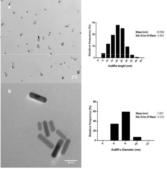
Physicochemical characterization of CTAB-AuNRs ... 33
Lethality and developmental effects of CTAB-AuNRs on zebrafish embryos ……… 35
Genotoxicity assessment ... 43
5. Discussion ... 45
6. Conclusion and Future Perspectives... 53
List of Figures
Figure 1 Scheme representing gold nanorods growth in the seed-mediated synthesis using CTAB that promotes anisotropic growth by adsorbing preferentially to Au [100] and Au [110] crystal facets, over the Au [111] facets [adapted from (Murphy, Thompson et al. 2011)]. ... 6
Figure 2 Nano-enable products life-cycle and possible stages for environmental and human exposure [adapted from (Initiative 2016)]. ... 8
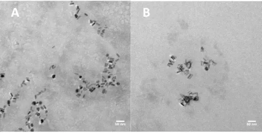
Figure 3 Development stages and main structural features of zebrafish. Zebrafish embryos at 24 hpf (1), 48 hpf (2), 72 hpf (3) and 96 hpf (4 and 5) (Kimmel, Ballard et al. 1995). ... 15 Figure 4 Representative TEM micrographs of the CTAB-AuNRs stock suspension with magnification of 30000x (A) and 200000x (B) and the corresponding histograms of size distribution. Scale bars: 50 nm (A) and 20 nm (B). ... 33 Figure 5 EDX spectrum (A) and X-ray pattern (B) of the CTAB-AuNRs stock suspension. Scale bar: 5 µm (B). ... 34 Figure 6 Representative TEM micrographs of the CTAB-AuNRs working suspensions dispersed in zebrafish water (ZW) at 104 µg/L (A) and 150 µg/L (B) with magnification of 60000x Scale bars: 50 nm (A and B). ... 35
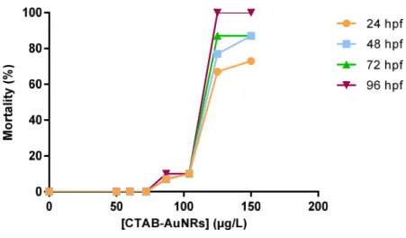
Figure 7 Zebrafish embryos mortality at different time-points (24, 48, 72 and 96 hpf) following exposure to CTAB-AuNRs. ... 36
Figure 8 Zebrafish embryos abnormalities during exposure to CTAB-AuNRs. At 24 hpf: Control embryos (A) and embryos exposed to 104 µg/L (B), 125 µg/L (C) and 150 µg/L (D). At 48 hpf: control embryos (E) and embryos exposed to 104 µg/L (F), 125 µg/L (G) and 150 µg/L (H). At 96 hpf: Control embryos (I) and embryos exposed to 104 µg/L (J and K). Dead embryo at 96 hpf (L). ... 39
Figure 9 Effects of CTAB solutions in 6 hpf embryos. Micrographs of control (non-exposed) (A) and exposed to 0.017 mM CTAB (B), which corresponds to the CTAB content present at the highest concentration of CTAB-AuNRs tested (150 µg/L). ... 40 Figure 10 Representative eight-point (0-200 µg/L) calibration curve used for Au content analysis by ICP-OES. ... 41 Figure 11 Au content of embryos exposed to different concentrations of CTAB-AuNRs at 48 and 96 hpf quantified by ICP-OES and expressed as μg Au/g fresh weight. ... 42
List of Tables
Table 1 Summary of the main physicochemical features of the tested AuNRs dispersed in water. ... 34 Table 2 Zeta potential of the stock suspension of CTAB-AuNRs and working suspensions in ZW. ... 35 Table 3 Effects of CTAB-AuNRs on the developmental parameters of zebrafish embryos. ... 38
Table 4 Effects of CTAB-AuNRs exposure on zebrafish’s heart rate (measured at 48 hpf) and body length (measured at 96 hpf). ... 39
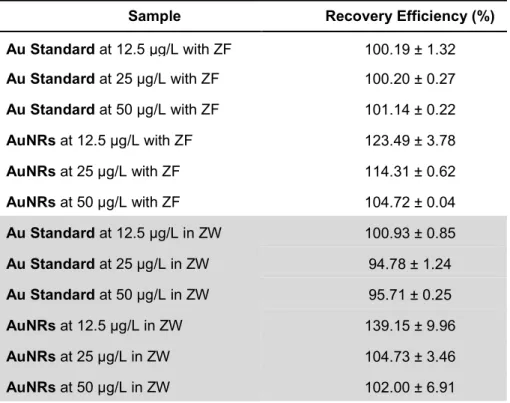
Table 5 Recovery efficiency of Au in zebrafish embryos (ZF) and zebrafish water (ZW) samples spiked with different concentrations (12.5, 25 and 50 μg/L) of Au or AuNRs as assessed by ICP-OES. ... 41 Table 6 Fraction of elemental gold in zebrafish tissues comparing to the initial concentration of CTAB-AuNRs in media expressed as percentage (%) of uptake. ... 42 Table 7 Comet assay analysis of DNA damage in zebrafish embryos exposed to different concentrations of CTAB-AuNRs at 48 and 96 hpf... 43
List of Acronyms and Symbols
ADME Absorption-Distribution-Metabolism-Elimination
Ag Silver
Au Gold
AuNMs Gold nanomaterials AuNRs Gold nanorods
bw Body weight
Cit Citrate
CNS Central Nervous System
CTAB Cetyltrimethylammonium bromide CTAB-AuNRs AuNRs capped with CTAB
DLS Dynamic Light Scattering DMSO Dimethyl sulfoxide dpf Days post-fertilization
EC Environmental Concentration EET Extraembryonic tissues EMEA European Medicines Agency FBS Fetal bovine serum
FDA Food and Drug Administration FET Fish Embryo Acute Toxicity Test GSH Glutathione
hESC Human Embryonic Stem Cells hpf Hours post-fertilization
IM Intramuscular
IV Intravenous
LMP Low melting point
LSPR Localized Surface Plasmon Resonance MEEE 2-(2-(2-mercaptoethoxy)ethoxy) ethanol MES Mercaptoethane sulfonic acid
NM Nanomaterial
OECD Organization for Economic Co-operation and Development PBS Phosphate-buffered saline
PEN Project on Emerging Nanotechnologies PTFE Polytetrafluoroethylene
PVP Polyvinylpyrrolidone ROS Reactive Oxygen Species SEM Standard error of the mean
TEM Transmission Electron Microscopy TG Test guideline
TiO2 Titanium dioxide
TMAT Trimethylammonium ethanethiol TPPMS Mono-sulfonated triphenylphosphine ZF ZW Zebrafish Zn Zinc ZW Zebrafish water
Gold Nanomaterials (AuNMs): properties, applications
and synthesis methods
Nanomaterials (NMs) are defined by the European Commission as “natural, incidental or manufactured materials containing particles, in an unbound state or as an aggregate or as an agglomerate and where, for 50 % or more of the particles in the number size distribution, one or more external dimensions is in the size range 1 nm - 100 nm” (in 2011/696/EU) (Commission 2011). Nanomaterials can be classified according to their chemical nature as carbon-based (e.g. fullerenes, carbon nanotubes, carbon black); metal-based (e.g. gold, silver, metal oxides, quantum dots) and organic-based (e.g. dendrimers and polymers). They can also be categorized according to their geometric configuration in spheres, shells, rods, wires, tubes, horns, thin films, coils and cones (Tiwari, Tiwari et al. 2012).
At nanoscale, the NMs exhibit a larger surface to volume ratio, which increases the number of active sites and surface area available to interact with diverse chemical species enhancing its chemical/catalytic reactivity, and its mechanic, optical, electrical and magnetic behavior (Oberdörster, Oberdörster et al. 2005). Due to these new or improved physicochemical properties compared to their bulk counterparts, engineered NMs hold great promise for an assortment of applications in a wide variety of fields, including biomedical, electronics, energy, environmental, and pharmaceutical industries. In fact, the global market for nanotechnology is expected to grow to $64.2 billion by 2019 as report by BCC Research (McWilliams 2014).
According to the Project on Emerging Nanotechnologies (PEN) online inventory, there are 1827 nano-enabled products already available on the market. Metal NMs,in particular silver (Ag) and titanium dioxide (TiO2), are among the most used NMs found
in consumer products (www.nanotechproject.org, last accessed September 13, 2016). The most recent update of PEN’s inventory of 2016 lists 442 and 92 Ag- and TiO2
NMs-based products, respectively. On the other hand, only 25 products containing gold nanomaterials (AuNMs) are listed in PEN, most of them in the cosmetic category (www.nanotechproject.org, last accessed September 13, 2016). However, AuNMs have been attracting great interest in the biomedical field as diagnostic and therapeutic tools due to their compatibility, and non-toxic and non-immunogenic nature (Howes, Rana et al. 2014) and in the electronics industry to enhance solar cells, liquid crystal displays and flash memory devices (Manheller 2012). Their global market size is likely to be worth 7 billion euros by 2020 (Global Markert Insights Insights 2016).
The AuNMs are characterized by localized surface plasmon resonance (LSPR), which occurs when an electromagnetic field drives the collective oscillations of a NM’s free electrons into resonance, high molar extinction coefficients, broad energy bandwidth, excellent conductivity, catalytic activity, high surface area and stability (Saha, Agasti et al. 2012). These properties are easily tunable by varying particle size, shape, dispersity, and chemical environment. Shape anisotropy of gold nanorods (AuNRs) is a classic example of how geometry configuration remarkably influence the properties of NMs. Comparing to Au nanospheres, which possess a single LSPR peaks, AuNRs have two distinct plasmon resonances peaks owing to the two different axes (longitudinal and transverse) of the rods. Therefore, by varying the aspect ratio (length/diameter), the longitudinal LSPR can be tuned throughout the visible region of the spectrum and into the near-infrared region, which renders AuNRs with significantly different optical properties from Au nanospheres and long-term photostability (Burrows, Vartanian et al. 2016, Hinman, Stork et al. 2016).
Overall, these properties make AuNMs an exceptional component for bio-sensing and bio-imaging technologies (Saha, Agasti et al. 2012). Furthermore, as AuNMs can be easily functionalized with selective and specific recognition molecules (e.g. antibodies, natural ligands for certain receptors or peptides), they are being increasingly exploited as diagnostic tools to detect biomarkers of several diseases (Baptista, Doria et al. 2010, Zhou, Gao et al. 2015) and as drug delivery systems (Alkilany, Thompson et al. 2012, Kumar, Zhang et al. 2013). By loading the cargo on Au’s surface through covalent or non-covalent binding and with proper functionalization, AuNMs can carry pharmaceutical drugs or other therapeutic molecules directly to the target site, which is of particular importance in cancer treatment as it enables the reduction of side effects and increases the therapeutic index. Recently, Amreddy, Narsireddy, et al. have successfully developed an AuNR-based drug delivery system, where the AuNRs were loaded with doxorubicin, a chemotherapeutic agent, conjugated with a pH-sensitive linker, and transferrin. Transferrin was used to target the overexpressed transferrin receptors in human lung cancer cells, whereas the pH-sensitive linker confines the release of doxorubicin to the acidic conditions of endosomes or lysosomes upon internalization of the AuNR by cancer cells (Amreddy, Muralidharan et al. 2015). Similar strategies such as PEGylation of AuNMs have also been applied to avoid phagocytic clearance by the reticuloendothelial system and immunogenic reactions, thus, enhancing circulation time (Ghosh, Han et al. 2008, Boccalon, Bidoggia et al. 2015). AuNMs are also suitable for hyperthermia in cancer treatment as they are able to convert absorbed light into heat
through a series of nonradiative processes (Huang and El-Sayed 2010, Khan, Vishakante et al. 2013).
A number of diagnostic devices comprising AuNMs have been already approved by the Food and Drug Administration (FDA) such as Verigene (Nanosphere) diagnostic tests to detect infectious pathogens and drug resistance markers, and First Response™ Pregnancy Tests from Church & Dwight Co., Inc. Although up to date no AuNM-base drug delivery system has been approved to clinical use, there are several of those currently under clinical trial. For instance, AurImmuneTM (Cytimmune) has
successfully attainned phase II clinical trial. This therapeutic platform, comprised by spherical AuNMs carrying tumor necrosis factor alfa (TNF-α), is design to penetrate the leaky blood vessels of tumors and specifically bind to TNF-α receptors on endothelial cells, acting as a Trojan Horse that enables follow-on chemotherapy to reach the tumor efficiently.
In the past few years, AuNMs have also been drawing attention to address environmental problems such as water pollution. AuNMs can be used as a platform to monitor the levels of toxic ions (e.g. arsenic, mercury, chromium), pesticides (e.g. atrazine, methylparathion) and pharmaceutical drugs (e.g. paracetamol, atenolol), and for aquatic environmental remediation (Saha, Agasti et al. 2012, Qian, Pretzer et al. 2013). Other potential applications of AuNMs include visual display technologies, such as touch sensitive screens, and advanced data storage technologies namely advanced flash memory devices.
Nanotechnology industry has been focused on the production of NMs with tailored size, shape, chemical composition and dispersity as NM’s properties, and consequently their applications, are strongly dependent on these characteristics. Therefore, the optimization of synthesis methods has been object of intense research. The production of NMs, namely AuNMs, requires fine tuning of various parameters that influence the thermodynamic and kinetic aspects, and consequently determines the quality and yield of the synthesis (Scarabelli, Sánchez-Iglesias et al. 2015). Numerous strategies have been developed to synthesize AuNMs, the firstly reported was the reduction of Au salts in the presence of a reducing agent such as sodium citrate that initiates the nucleation of the Au ions, thus forming NMs. To prevent aggregation, a stabilizing agent is often added during synthesis (Turkevich, Stevenson et al. 1951, Frens 1973). To produce anisotropic AuNRs, the seed-mediated growth developed by Gearheart et al. is the most widely used method (Jana, Gearheart et al. 2001). In this technique, there is a temporal and spatial separation between the nucleation and
growth steps. Briefly, small spherical seeds are first produced from tetrachloroauric acid (HAuCl4), an Au salt, and a strong reducing agent (e.g. sodium borohydride) that
induces nucleation. Subsequently, these seeds are added to a growth solution that contains additional gold salt, a weak reducing agent (e.g. ascorbic acid) and a ‘‘soft template’’ [e.g. cetyltrimethylammonium bromide (CTAB)] that directs anisotropic growth. The ascorbic acid is commonly used to reduce the Au salt to elemental Au but this reaction only occurs in the presence of the seeds, otherwise it only reduces Au3+ to
Au+ (Grzelczak, Pérez-Juste et al. 2008, Xia, Zhang et al. 2015). The CTAB, a cationic
surfactant, guides the growth of Au seeds into rod-like shape by adsorbing preferentially to Au [100] and Au [110] crystal facets, over the Au [111] facets, which blocks the AuNM growth at the sides and induces the growth along the longitudinal axis as shown in Figure 1 (Murphy, Thompson et al. 2011). Moreover, the positively charged bilayer of CTAB on AuNR’s surface creates mutual repulsions and prevents the aggregation of AuNRs, promoting their dispersity. However, CTAB is highly cytotoxic and for biomedical applications other strategies have been studied to replace this compound in the seed-mediated growth synthesis and still preserve the AuNRs properties (Gui and Cui 2012). Other production methods for anisotropic AuNMs include photochemical synthesis (Kim, Song et al. 2002) and electrochemical synthesis in solution (Yu, Chang et al. 1997) or in hard templates (Foss Jr, Hornyak et al. 1992).
Figure 1 Scheme representing gold nanorods growth in the seed-mediated synthesis using CTAB that promotes anisotropic growth by adsorbing preferentially to Au [100] and Au [110] crystal facets, over the Au [111] facets [adapted from (Murphy, Thompson et al. 2011)].
Toxicology of AuNMs
Gold in the bulk form has for long been considered chemically inert, biocompatible, non-toxic and non-immunogenic (Howes et al., 2014). However, at nanoscale these metallic particles emerge as a catalyst with substantially different properties comparing to their bulk counterparts, which calls into question their safety.
Moreover, the increasing production and widespread application of AuNMs lead to their release into the different environmental compartments (e.g. soil, air and water), endangering human and environmental health.
As described in Figure 2, the release of NMs into the environment can occur at any stage of their life cycle, which begins with the processing of the raw materials and their transformation into NMs, followed by their incorporation into products, usage and ultimately disposal (e.g. landfills and waste incineration plants) (Sun, Gottschalk et al. 2014). The Ag- and TiO2-NMs, for example, have been shown to be released from
paints on building facades into urban runoff (Kaegi, Ulrich et al. 2008, Kaegi, Sinnet et al. 2010). Currently, there is no consistent data on environmental concentrations (EC) of NMs mainly due to the lack of appropriated separation and analytical methods. The few studies performed so far, mostly with Ag- and TiO2-NMs, have used probabilistic
material-flow modelling to predict the EC of NMs in surface water, waste water treatment plant, soils, sediments and atmosphere. For instance, gathering the data of the current literature on the topic, the modeled concentrations of TiO2-NMs in surface
water ranges from 3 to 1600 ng/L, whereas for Ag-NMs the predicted EC is slightly lower (between 0.1 and 1000 ng/L) (Gottschalk, Sun et al. 2013). In the case of AuNMs, Mahapatra et al. has reported that the estimated EC of AuNMs derived from nanomedicine products in surface water is approximately 0.468 and 0.0047 ng/L, respectively for the United Kingdom and United States of America (Mahapatra, Sun et al. 2015). On the other hand, according to Boxall et al., AuNMs derived from consumer products are present in water in concentrations ranging from 100 to 1430 ng/L (Boxall, Chaudhry et al. 2007). The high disparity of the predicted concentrations obtained in the different studies reflects the different estimations of NMs production and emission rates, as well as in NM product’s market penetration. Moreover, the material flow analysis for NMs requires a fine understanding and description of the NM’s flow chain, from resource extraction to final waste disposal. In addition, once introduced into the environment, NMs will establish diverse interactions with biotic and abiotic systems that will change their intrinsic characteristics and will further influence their fate and behavior in the ecosystem. These variables have not been taken into account in most of the material-flow analyses performed so far (Gottschalk, Sun et al. 2013) and thus further studies are required to fill in these gaps.
The employees of NM’s companies, as shown in Figure 2, are the primary individuals at risk of exposure to NMs during manufacturing, packaging, handling, transport and disposal, followed by the consumers of nano-products. Therefore, it is
occupational exposure limited values. Maynard et al., who inspected a single-walled carbon nanotube-generating operation, reported workplace air concentrations up to 53 µg/m3 (Maynard, Baron et al. 2004). Furthermore, a study performed to assess the
inhalation exposure of workers to metal oxides NMs associated with industrial wastewater treatment processes in a semiconductor research and development facility showed that NMs of 20.5 nm were present at concentrations greater than 100 particles/cm3 during the execution of a specific task (Brenner, Neu-Baker et al. 2015).
However, the available data regarding occupational exposure assessment of NMs is so far very limited, again due to the methodology required that is still under development. Up to date no study has been performed with AuNMs.
Taken together, it is essential to evaluate AuNMs risk for humans and ecosystems, understand their mechanism of action and the properties responsible by their toxicity in order to safeguard workers and consumers, establish environment safety values, support regulatory decisions and ultimately, assist the development of safer NMs and manufacturing processes.
Figure 2 Nano-enable products life-cycle and possible stages for environmental and human exposure [adapted from (Initiative 2016)].
Human Exposure to AuNMs and the associated risks
Manufacturers and consumers of nano-enabled products are likely to be exposed to engineered NMs through different routes such as inhalation, dermal, oral, ocular, intravenous(IV) and intramuscular (IM), these latter two occurring almost exclusively in biomedical settings (Warheit and Sayes 2015). After exposure, the NMs
can translocate the biological barriers and reach the bloodstream, where they become coated with different proteins that adsorb to their surface, leading to the formation of a protein corona (Monopoli, Åberg et al. 2012). The nature of the corona formed is governed by the intrinsic characteristics of the NMs (e.g. chemical composition, surface chemistry, particle size, shape, charge and dispersion stability) (Mahmoudi, Lynch et al. 2011, Nel, Parak et al. 2015). These NM-protein complexes will subsequently dictate the tissue distribution of NMs and the interaction between NMs and cells. The biodistribution of AuNMs has also been reported to be dependent on their size, shape and surface chemistry (Janát-Amsbury, Ray et al. 2011, Morais, Soares et al. 2012, Han, Lee et al. 2015, Elci, Jiang et al. 2016). For instance, AuNMs of 13 and 105 nm were found to accumulate in lungs and translocate to bloodstream of Sprague–Dawley rats following inhalation of 12.8 ± 2.42 µg/m3 for 5 days, however only the smaller ones
were detected in the liver, spleen, brain and testes (Han, Lee et al. 2015). Furthermore, AuNMs have been shown to accumulate preferentially in the liver and spleen following intravenous injection, which might result from the capacity of the reticuloendothelial system to remove particulate matter from circulation (Semmler‐Behnke, Kreyling et al. 2008, Cho, Cho et al. 2009).
AuNMs have generally been considered biocompatible, however the potential biopersistence of AuNMs in the organism allied to their catalytic activity might result in long-term chronic effects. In some studies, these NMs have been reported to cause fatigue, decreased appetite, weight loss (Chen, Hung et al. 2009, Zhang, Wu et al. 2010), altered gene expression pattern (Balasubramanian, Jittiwat et al. 2010), spleen atrophy and mild anemia (Fraga, Brandao et al. 2014) in exposed rodent models. Nevertheless, it has been pointed out that the main reason for some of the toxic effects detected might be the adsorbed molecules on the AuNM’s surface, used to increase NMs dispersion and stability in aqueous media, or impurities derived from the synthesis process, rather than to the AuNMs per se (Alkilany and Murphy 2010). Therefore, the toxicity of pristine, surface-modified and functionalized NMs should be carefully distinguished.
Ecotoxicology of AuNMs in aquatic environments
In the aquatic environment, the NMs can be suspended in the water phase or deposited in the sediment, depending on their primary characteristic and their interaction with abiotic systems. For instance, factors such as salinity, temperature, pH and natural organic matter content will determine agglomeration and aggregation status, dissolution, redox reactions, surface transformation and sedimentation of NMs
and therefore their bioavailability for the aquatic species (Wang, Zhang et al. 2016). The NMs associated with the sediment will interact directly with benthic species, whereas NMs suspended in the water column will be bioavailable for algae, invertebrates and fish (Amiard-Triquet, Amiard et al. 2015). Moreover, organisms can interact directly with NMs through adsorption and subsequent internalization, or may be taken up via trophic transfer. Recently, it was demonstrated that AuNRs are able to pass from the water column to the marine food-web (Ferry, Craig et al. 2009). Moreover, AuNMs have been detected in Daphnia magna fed with the unicellular microorganisms Chlamydomonas reinhardtii and Euglena gracilis previously exposed to these nanoparticles (Lee, Yoon et al. 2015). These studies indicate that AuNMs are indeed able to accumulate at the lowest trophic levels (e.g. microorganisms and invertebrates) and to be transferred to the highest trophic levels (fish and possibly humans) through the food chain.
Up to date, few studies on the biodistribution and toxicity of AuNMs in aquatic organisms have been undertaken. Nevertheless, the current literature indicates that AuNMs are able to accumulate and induce toxicity at some extent in different levels of the trophic cascade. The AuNMs have been shown to accumulate in the digestive tract of both aquatic invertebrates and fish following exposure to concentrations ranging from 0.5 to 100 mg/L, and 20 mg/L, respectively, although no hazard has been reported (García-Cambero, García et al. 2013, Botha, Boodhia et al. 2016). On the other hand, juvenile marine fishes (Pomatoschistus microps) exposed to 0.2 mg/L of AuNMs were shown to have decreased predatory performance, which in the wild might reduce the individual fitness, compromising population growth and survival (Ferreira, Fonte et al. 2016). The exposure of marine bivalve Scrobicularia plana to 100 µg/L of differently sized (5, 15 and 40 nm) AuNMs has shown to negatively impact the burrowing speed and to increase the levels of the enzymes involved in the antioxidant defense mechanisms, although the feeding behavior was not affected (Pan, Buffet et al. 2012). Furthermore, 40 nm AuNMs coated with polyvinylpyrrolidone (PVP) at 4, 80 and 1600 μg/ L were reported to increase the hepatic expression of antioxidant, immune and apoptosis related-genes mRNA levels in exposed Sparus aurata fishes (Teles, Fierro-Castro et al. 2016).
Mechanism of AuNMs toxicity: focus on genotoxicity
Oxidative stress, inflammation and genotoxicity seem to be the three main mechanisms underlying NMs toxicity (Fadeel and Pietroiusti 2012). Oxidative stress is caused by an excessive reactive oxygen species (ROS) production and/or a decrease
in the antioxidant defenses, which can damage cells through lipid peroxidation, DNA/protein oxidation or by interfering with signaling pathways and gene function. Additionally, oxidative stress may initiate an inflammatory signaling cascade and ultimately induce cell death. Cytotoxicity of metallic NPs have been extensively explored in in vitro and in vivo studies and can be reviewed elsewhere (Lewinski, Colvin et al. 2008, Arora, Rajwade et al. 2012).
Genotoxicity induced by NMs, which encompasses all types of DNA or chromosome damage (e.g. strand breaks, adducts rearrangements, mutations, chromosome aberrations and aneuploidy), is less explored and data obtained are frequently controversial. Nevertheless, it appears that genotoxic effects induced by metal NMs can arise either through direct interaction of the NMs with the genetic material or indirectly by elevation of ROS levels (Dusinska, Magdolenova et al. 2013). By interacting directly with DNA, NMs can produce a variety of DNA lesions including simple base modifications, base mismatches, double-strand breaks and bulky DNA adducts. These lesions can give rise to further gene mutations and chromosomal damage (numerical or structural) if not repaired (Golbamaki, Rasulev et al. 2015). Aneugenic events may also be caused during cell division by interaction of NMs with the mitotic spindle apparatus, centrioles or their associated proteins (Huang, Chueh et al. 2009). Di Bucchianico et al. conducted a study to investigate the ability of 5 and 15 nm AuNMs to induce genotoxic events in human primary lymphocytes and murine macrophages and observed a significant increase in both primary and oxidative DNA damage after 2 and 24 h of exposure to different concentrations (0.1, 1, 10, and 100 μg/mL) of these NMs. In addition, a concentration dependent increase in the frequency of micronuclei (MN) was observed in both cell types. In order to distinguish between MN originated from chromosome fragments or whole chromosomes that lag behind at anaphase during nuclear division, the authors used Fluorescent In Situ Hybridization (FISH)-pancentromeric probes. Data showed that MNs were centromere-positive, which indicate that aneugenic rather than clastogenic events were induced by tha AuNMs (Di Bucchianico, Fabbrizi et al. 2014).
Interaction with DNA-related proteins, namely the ones involved in the replication process, transcription and damage repair, has also been shown to play a role in the genotoxic potential of metal oxide NMs (Magdolenova, Collins et al. 2014, Golbamaki, Rasulev et al. 2015). Indeed, DNA damage as well as altered expression of proteins associated with cell cycle regulation and DNA repair were observed in MRC-5 human fetal lung fibroblast cells exposed to AuNMs (Li, Lo et al. 2011).
NMs have also been shown to induce DNA damage through secondary mechanisms such as oxidative stress. Indeed, ROS generation has been considered the main mechanism of DNA damage resulting from exposure to NMs. Excessive ROS levels can cause the depletion of the antioxidant defenses together with oxidative modifications in the DNA, including strand breaks and base oxidation [e.g. 8-hydroxy-2′-deoxyguanosine (8-OHdG)] (Wan, Mo et al. 2012, Magdolenova, Collins et al. 2014). Moreover, DNA damage in vivo can also be induced as a secondary response to inflammation triggered by NMs (Downs, Crosby et al. 2012).
Following DNA damage cells activate complex signaling networks which promote either DNA repair/survival or cell death (Roos, Thomas et al. 2015). The final outcome, i.e. survival or death, is dependent on several factors, namely cell tolerance to damage and/or magnitude of the impairment in DNA repair mechanisms caused by the NMs, which as mentioned above may have an important part in genotoxic events (Magdolenova, Collins et al. 2014).
Although there are only few in vivo studies focused on the genotoxicity of AuNMs, the results in general are negative for both DNA damage and clastogenic/aneugenic potential. Downs et al. showed accumulation of AuNMs of different sizes (2, 20, 200 nm) in the liver and lung tissues of rats exposed through IV injection to 0.030 mg/kg body weight (bw). However, this tissue accumulation was neither translated into an increase in DNA damage in liver, lung and white blood cells nor into an increase in the % of MN in circulating reticulocytes (Downs, Crosby et al. 2012). In another study, neither DNA damage in lung cells nor augmented frequency of MN in polychromatic erythrocytes was identified 3 days after single intratracheal instillation of 36 µg/mL AuNMs of different sizes (2, 20 or 200 nm) in male rats (Schulz, Ma-Hock et al. 2012). In contrast, oral exposure to AuNMs for 7 or 14 days increased the frequency of MN in polychromatic erythrocytes at 320 mg/kg bw and the formation of DNA adducts in hepatic cells at 160 and 320 mg/kg bw. These findings suggests that AuNMs exposure can induce oxidative stress-mediated genomic instability (Girgis, Khalil et al. 2012). Nevertheless, adequate in vivo studies using doses and exposure periods that represent realistic scenarios are still lacking.
Considering the potential application of AuNMs in the medical field, the surface modification of NMs can be a good strategy to protect cells from the undesirable genotoxic effects of AuNMs. Bearing that in mind, Fraga et al. studied the genotoxicity of AuNMs coated with citrate (Cit) or 11-mercaptoundecanoic acid (11-MUA) in human liver HepG2 cells. Both differently coated AuNMs were taken up by HepG2 cells,
however only Cit-AuNMs induced significant DNA damage. Interestingly, the genotoxicity observed was inversely proportional to the tested concentrations (0.1, 1 and 10 µM) (Fraga, Faria et al. 2013). On the other hand, Hashimoto et al. did not find any significant DNA damage either in L929 fibroblast or Raw macrophage cells exposed to 100 and 400 µg/mL of citrate-coated AuNMs (Hashimoto, Kawai et al. 2016). In addition, sophorolipid reduced AuNMs, which have been explored as drug delivery systems, have not induced DNA breaks at 0.5, 5 and 50 µg/mL in HepG2 cells (Singh, D’Britto et al. 2010).
Zebrafish (Danio rerio) as an alternative animal model to
assess toxicity
Zebrafish (Danio rerio) is a small tropical fish, originally found in the rivers of India and South Asia, that has become one of the most popular model organism for (eco)toxicology, vertebrate development and human disease studies. Zebrafish offers several technical advantages over other animals used in experimental research, such as rapid development, high fecundity, generation of transparent embryos that develop outside the mother’s body (allowing the in vivo visualization of cell-biological events), low maintenance costs and ease of manipulation (Scholz, Fischer et al. 2008). Furthermore, the genomic sequencing of zebrafish is highly advanced and various DNA libraries, microarray resources, and protocols for generating transgenic fish and knocking down gene expression in embryos have been developed and widely established (Stegeman, Goldstone et al. 2010).
The adult zebrafish is used as a model in environmental toxicology for hazard identification and risk assessment of chemicals, plant protection products, pharmaceuticals, biocides, feed additives, and effluents. In 2013, the fish embryo acute toxicity test (FET) with zebrafish embryos was approved by the Working Group of the National Coordinators of the OECD Test Guideline Program and published as OECD test guideline (TG) no. 236. This test guideline is intended to determine the acute or lethal toxicity of chemicals in early-life stages of zebrafish, but can also be used as an alternative to the fish acute test [OECD TG 203; (OECD 1992)] as zebrafish embryos were shown to have similar toxicological profiles for hazardous agents with various adult fish species commonly tested (Braunbeck, Kais et al. 2015).
Zebrafish has a high degree of genetic, molecular and physiological similarity with mammals and has been shown to exhibit very similar Absorption-Distribution-Metabolism-Elimination (ADME) profiles to rodent models and humans, which explains
different compounds (Yang, Ho et al. 2009, Bailey, Oliveri et al. 2013, MacRae and Peterson 2015). In that respect, the FDA and European Agency for the Evaluation of Medicinal Products (EMEA) have recently accepted zebrafish’s pharmacology and toxicology data for preclinical trials to evaluate whether a new drug is safe or not for human testing, a requirement to obtain regulatory approval for a clinical trial (He, Gao et al. 2014). One example of a disease-relevant compound that has been tested in zebrafish is ProHema, a stabilized derivative of prostaglandin E2, which was found to increase the numbers of hematopoietic stem cells (North, Goessling et al. 2007). This compound is currently under Phase II trials in patients undergoing umbilical cord blood transplantation for leukemia or lymphoma as it might improve the effectiveness of transplantation by enhancing the rate of bone marrow recovery (MacRae and Peterson 2015).
Stages of embryonic development
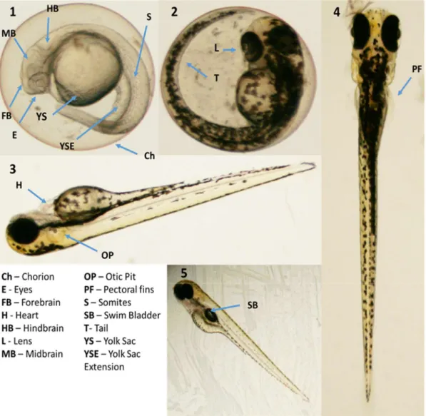
The embryonic development of zebrafish starts when the eggs, spawned by females, are fertilized by sperm released by males into the water. After fertilization, the zygote undergoes a series of cleavage stages at the animal pole, which results in a flattened blastodisc at the periphery. The following period named gastrula is characterized by the transformation of the blastodisc to ball-like shape and the beginning of the epiboly, where the blastodisc spreads over the yolk in the vegetal pole direction. In the gastrulation stage, which occurs between 5-10 hours post fertilization (hpf), the embryo is converted into a structure of three germ layers - ectoderm, endoderm and mesoderm. These germ layers will give rise to all tissues in the organism. At 10 hpf, the gastrulation and epiboly are completed and the embryo has an anteroposterior axis with the contours of head and tail. During the segmentation period (10-24 hpf) takes place the development of somites, which are blocks of segmental mesoderm that establish the segmental organization of the body. Moreover, rudimentary organs, such the eyes and the brain, become evident and the embryo elongates with the detachment of the tail as illustrated in Figure 3 (1). The heartbeat of zebrafish embryo begins at 22 hpf, followed by the blood flow at 24 hpf. Between 24 and 48 hpf, pigmentation becomes observable in the eyes and body and the skeletal muscle starts to contract spontaneously [Figure 3 (2)]. At the end of 48 hpf, the organogenesis is almost complete and the embryo is motile and responsive to external stimuli. The embryos hatch from the chorion approximately between 48 and 96 hpf, and eleutheroembryo stage ensues but the embryo is still dependent on the yolk for food supply [Figure 3 (3)]. At 96 hpf occurs the formation of the pectoral fins [Figure 3 (4)] and the inflation of swim bladder [Figure 3 (5)], essential for fish to achieve neutral
buoyancy with minimal energy expenditure (Kimmel, Ballard et al. 1995, Rosenthal and Harvey 2010). At 5 days post fertilization (dpf), the zebrafish embryos fully depleted the yolk and start the external feeding, therefore reaching the larvae stage. The adult period begins with the sexual maturation, which can be attained at the age of 3 months (Scholz, Fischer et al. 2008).
Figure 3 Development stages and main structural features of zebrafish. Zebrafish embryos at 24 hpf (1), 48 hpf (2), 72 hpf (3) and 96 hpf (4 and 5) (Kimmel, Ballard et al. 1995).
Toxicity of AuNMs towards more susceptible life stages
As the likelihood of organisms being exposed to NMs increases, there are raising concerns regarding their potential to cause reproductive toxicity, which includes adverse effects on sexual function and fertility in adult males and females, as well as developmental toxicity in the progeny. Developmental toxicity might manifest as death,
structural abnormalities (malformations), growth retardation, behavioral and functional abnormalities. These effects can result from exposure of either parent prior to conception or during prenatal development (OCDE 2008). The embryonic period, more specifically during pre-differentiation, early-organogenesis and late-organogenesis, is when the developing organs are most vulnerable to toxic substances as embryonic cells have immature repair and detoxification mechanisms, and the development process requires precise temporal-spatial sequencing (Hansen and Harris 2013, Dutta 2015). Moreover, in the case of mammals, placenta per se does not guaranty safety for the fetus, as it is permeable to many substances. In fact, it has been shown both in vivo, in animal models, and ex vivo, in the human placental perfusion model, that NMs may indeed pass through the placenta (Myllynen, Loughran et al. 2008, Chu, Wu et al. 2010, Grafmüller, Manser et al. 2013). Therefore, exposure to NMs might interfere with the normal developmental course of the embryo and cause permanent defects. In the case of aquatic organisms, the exposure of early-life stages to these xenobiotics can further result in reduced fitness, susceptibility to predation in the wild, lower reproductive rates, or to the development of carcinogenesis, endocrine, and immune system defects (Embry, Belanger et al. 2010).
Interestingly, so far no effects were detected on mammalian embryos after AuNMs exposure. According to Yang et al., there is a higher accumulation of AuNMs in the fetus at early pregnancy stages (below gestational day 9.5) than at gestational day 11.5 or above, while the accumulation in the extraembryonic tissues (EET) increases with the gestational age. The time frame at which these change occurs coincide with the maturation of placenta that seems to decrease the fetal exposure to NPs (Yang, Sun et al. 2012). Moreover, it has been shown a higher accumulation in fetus and EET of ferritin and polyethylene glycol (PEG)-coated AuNMs than of citrate-capped NPs. The latter coating has a negative charge, which might explain its reduced accumulation (Yang, Sun et al. 2012). Furthermore, Semmler-Behnke et al. reported that after a single IV administration of three differently sized AuNMs (14, 18 and 80 nm at 5.2, 3.2 and 26.5 µg/rat, respectively) at gestational day 18, all three AuNMs were detected in the EET, while only 14 and 18 nm AuNMs were observed in the fetus. These results seem to indicate that the AuNMs accumulation in the fetus is size-dependent. In that matter, the authors suggested that AuNMs translocation across the placental tissues occurs through transtrophoblastic channels and/or via transcellular processes, which further indicates the important role of placenta in embryo toxicity (Semmler-Behnke, Lipka et al. 2014). Altogether, these results showed that the gestational age at which pregnant mice are exposed, NM size and surface composition greatly impact AuNMs
distribution in fetus and EET. Considering these results it would be expected that the extent of impairment of fetal and postnatal development would be higher at early gestational exposure as it results in higher accumulation in the fetus. However, it has been shown that prenatal exposure to AuNMs did not affect the offspring independently of the gestational age (Yang, Sun et al. 2012). Furthermore, an in vitro study revealed that injection of 50 µg/mL of AuNMs into 2-cell mice embryo have not interfered with blastocyst rate or the expression of genes involved in embryo development (Taylor, Garrels et al. 2014). Nevertheless, it should be highlighted that this study lack of the evaluation of important parameters to determine embryo quality for implantation success such as cleavage rate, cell number, symmetry and shape of the blastomeres and cytoplasmic fragmentation extent in the perivitelline space.
In regard to the potential toxicity of AuNMs towards early-life stages of fish, AuNMs have been reported to diffuse through the chorionic pore canals and reach the inner cell mass of zebrafish embryos, remaining inside them throughout the entire development (Browning, Lee et al. 2009, Browning, Huang et al. 2013). Furthermore, Browning et al. reported that the effects of AuNMs in the zebrafish embryo development are minimal and dependent of NM size with smaller NMs (11.6±0.9 nm) inducing higher mortality and deformities than larger AuNMs (86.2±10.8 nm) at the same administered doses (Browning, Lee et al. 2009, Browning, Huang et al. 2013). Other studies reported no obvious abnormalities in the zebrafish embryos development after exposure to bare or polyvinyl alcohol (PVA)-capped AuNMs (Asharani, Lianwu et al. 2011, Kim, Zaikova et al. 2013). On the other hand, Truong et al. investigated the influence of functional groups on the toxicity induced by AuNMs. AuNMs functionalized with the positively charged trimethylammonium ethanethiol (TMAT) were shown to induce embryo lethality, while AuNMs functionalized with the negatively charged mercaptoethane sulfonic acid (MES) caused sub-lethal malformations to the embryos (Truong, Tilton et al. 2013). Moreover, both types of NMs were shown to cause misregulation of genes associated with immune response, and inflammation processes and behavioral abnormalities that were extended to the adulthood (Truong, Saili et al. 2012, Truong, Tilton et al. 2013). On the contrary, AuNPs functionalized with the neutral 2-(2-(2-mercaptoethoxy)ethoxy) ethanol (MEEE) did not induce toxicity (Truong, Tilton et al. 2013). Furthermore, TMAT ligands were also shown to affect eye development through an increase in the apoptotic process and downregulation of genes involved in eye formation, and to impair swimming behavior and axonal growth (Kim, Zaikova et al. 2013). Mono-sulfonated triphenylphosphine (TPPMS) and glutathione (GSH) ligands are being explored for AuNMs stabilization and for
therapeutic purposes (Leifert, Pan-Bartnek et al. 2013). Yu Pan et al. evaluated the teratogenicity of these coatings in zebrafish embryos and verified that AuNMs carrying TPPMS where much more toxicity than AuNMs carrying GSH. These authors observed that TPPMS-AuNMs caused 100% lethality at 400 µM and peripheral edema, cardiac malformations and hypopigmentation at sub-lethal doses. In addition, co-incubation of TPPMS-AuNMs with GSH, a ROS scavenger, decreased significantly the malformations, which suggests that toxicity of TPPMS-AuNMs was due to oxidative stress (Pan, Leifert et al. 2013). These studies demonstrated that AuNMs functionalization and particularly the charge, either positive or negative, have a significant impact on zebrafish development. Ionic concentration of the exposure medium was also shown to influence AuNMs toxicity. As ionic concentration decreases, the dispersity of AuNMs increases, which causes an increase in mortality, malformation and behavioral deficits of zebrafish embryos (Truong, Zaikova et al. 2012).
Although up to date it has not been reported noteworthy teratogenic effects of AuNMs in vivo, a few studies have shown that these NMs might interfere with Central Nervous System (CNS) development. The AuNMs were found to be highly toxic for human embryonic stem cells (hESC), whose differentiation into neurons mimics early stages of human brain development, in a size-dependent manner. The AuNMs of 1.5 nm adversely affected hESC survival and neuronal differentiation at 0.6 or 10 µg/mL, whereas 4 or 14 nm AuNMs did not induce toxicity. Exposure to 1.5 nm AuNP have also resulted in cell death of hESC-derived neural progenitor cells (Senut, Zhang et al. 2015). Furthermore, Söderstjerna et al. reported that AuNMs of 20 and 80 nm significantly affected the sphere size and morphology of human embryonic neural precursor cells (Söderstjerna, Johansson et al. 2013). Consequently, further studies are needed to confirm the real extent of AuNMs toxicity in embryo development as although no obvious malformations were observed, its accumulation may induce more subtle alterations particularly at cellular level.
Nanotechnology is expected to have a huge economic and societal impact worldwide within the next few years. Due to their unique optical-electronic properties, AuNMs have received great attention and therefore are being increasingly investigated and used in various commercial, industrial and biomedical applications. The high reactivity that may arise at nanoscale and the comparable dimensions between cellular components and NMs might lead to harmful interactions between them, which have brought into question NMs safety. Owing to the inert and biocompatible nature of Au in the bulk form, AuNMs were initially regarded as nontoxic and less scrutinized in terms of their safety evaluation and risk assessment. Nevertheless, considering the socioeconomic impact that AuNMs are expected to reach in the upcoming years and their release into the environment, it is crucial to determine the implications of exposure to these NMs, particularly the ones already available in the market, on both organisms and the ecosystems. Indeed, NMs may enter the aquatic system, accumulate in sediments and result in multi-component mixtures posing different threats not only to wildlife but also to humans.
In this context, the main goal of this study was to evaluate the acute and developmental toxicity of a commercial suspension of Au nanorods (AuNRs) capped with the cationic surfactant cetyltrimethylammonium bromide (CTAB), herein designated as CTAB-AuNRs, to early life stages of biota, which are the most sensitive life cycle stage and often highly predictive of xenobiotics toxicity in the adult stage. Zebrafish (Danio rerio) embryos were chosen since they are recognized as a suitable and relevant model for (eco)toxicological studies, with a high degree of genetic, molecular and physiological similarity to humans and a good alternative to animal testing following the 3R’S principle. Therefore, zebrafish embryos allow the analysis of multiple endpoints ranging from acute to developmental toxicity determination.
To achieve this main objective, four specific goals were established:
i. To characterize the colloidal suspensions in terms of particle size, size distribution, morphology and zeta potential as the toxicological potential of the NMs is highly dependent on their physicochemical characteristics; ii. To assess the lethality, acute toxicity and developmental effects, including embryo development delays and malformations, of CTAB-AuNRs to zebrafish embryos;
iii. To confirm whether or not CTAB-AuNRs are internalized by the exposed zebrafish by assessing the embryo Au content at different time-points (before and shortly after hatching);
iv. To investigate the genotoxic potential of CTAB-AuNRs at sublethal concentrations since genotoxic agents can induce carcinogenesis and/or heritable defects that may severely impact the health of an individual or the population.
3. Materials and
Methods
Reagents
All chemicals used were of high purity or analytical grade. Dimethyl sulfoxide (DMSO; CAS no. 37-68-5), Triton X-100 (CAS no. 9002-93-1), low melting point (LMP) agarose (CAS no. 39346-81-1), Tris-HCl (CAS no. 1185-53-1), 30% (w/w) hydrogen peroxide (H2O2; CAS no. 7722-84-1) solution, 10 mg/mL ethidium bromide (CAS no
1239-45-8) solution and cetyltrimethylammonium bromide (CTAB; CAS no. 57-09-0) were purchased from Sigma-Aldrich (Madrid, Spain). Ethanol absolute (EtOH; CAS no. 64-17-5), sodium hydroxide (NaOH; CAS No. 1310-73-2), sodium chloride (NaCl; CAS no. 7647-14-5), hydrochloric acid (HCl, Cas no.7647-01-0) and Tris base (CAS no. 77-86-1) were bought from Merck (Darmstadt, Germany). Fetal bovine serum (FBS) and Molecular Probes® SYBR® Gold were purchased from Thermo Fisher Scientific
(Madrid, Spain), while ethylenediaminetetraacetic acid disodium salt (Na2EDTA; CAS
no. 6381-92-6) and nitric acid (HNO3; CAS no. 7697-37-2) were purchased from Prolab
(Laval, Canada). Phosphate-buffered saline (PBS; CAS no. 10049-21-5) and normal melting point (NMP) agarose were supplied by Lonza (Basel, Swiss) and Bioline (London, UK), respectively. A gold pure calibration standard was obtained from Perkin Elmer (Waltham, MA, USA).
Physicochemical characterization of the gold nanorods
(AuNRs)
CTAB-stabilized AuNRs (catalogue no. A12-10-750) with axial dimension=10 nm, long size dimension=35 nm and absorbance peak= 750 nm were supplied by NanopartzTM (Salt Lake City, UT, USA) and stored according to manufacturer’s
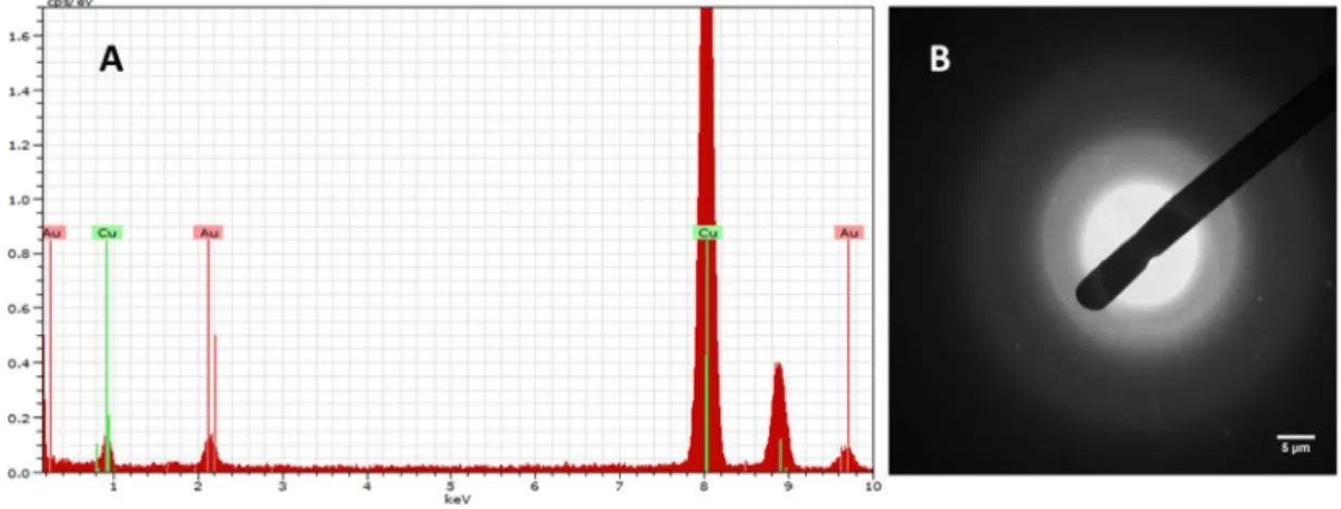
recommendations. Size and morphology of the CTAB-AuNRs were assessed by Transmission Electron Microscopy (TEM) either in the stock or working suspensions using a Hitachi H-9000 microscope operated at 300 kV. For this analysis, a drop of the suspensions under study was placed on a carbon-coated copper grid and the solvent was left to evaporate at room temperature. Particle size distribution of CTAB-AuNRs was measured in 5 TEM images (30 AuNRs/image) using the ImageJ software (NIH, USA). To determine the crystallographic nature and purity of the tested AuNRs, a Hitachi H-9000 microscope equipped with x-ray diffraction mode and a spectrometer was used. Zeta potential was measured using a Zetasizer Nano ZS (Malvern Instruments, Worcestershire, UK). Measurements were performed in triplicate, either in distilled water or water from the Zebrafish facility (ZW).
Handling and preparation of the AuNRs suspensions
All procedures of handling and preparation of the CTAB-AuNRs suspensions were standardized to minimize within-experiment variations. The experiments were performed using the same batch of AuNRs. The stock suspension was kept at 4ºC, protected from light and remained stable without any detectable sign of precipitation or change of color throughout the study. Different concentrations of CTAB-AuNRs were freshly prepared from the stock suspension by direct dilution in autoclaved ZW.
Zebrafish (Danio rerio) maintenance and embryo
collection
Danio rerio embryos used in this study were provided by the zebrafish facility established at the Department of Biology, University of Aveiro (Portugal). Adult zebrafish were maintained under standard controlled conditions (26.0 ± 1◦C, 80%
humidity, photoperiod cycle of 16 h light:8 h dark) in tanks equipped with recirculating systems. The fishes were fed with a commercially artificial diet (ZM 400 Granular), and maintained in carbon-filtered water with the following characteristics: 0.34 mg/L of Instant Ocean® synthetic sea salt (Spectrum Brands, USA), 26.0 ± 1◦C, 750 ± 50
µS/cm, pH 7.5 ± 0.5 and dissolved oxygen saturation ≥ 95%.
For the experiments, zebrafish eggs were obtained by natural crossbreeding. Before the onset of darkness on the day prior to the test, zebrafish females and males were separated to guaranty that all the embryos will be in the same development stage and spawn traps (marbles) were deposited in the tanks to avoid the predation of the eggs by adult zebrafish. At the onset of light on the day of the test, zebrafish females and males were rejoined. Zebrafish eggs were carefully collected within 1 h after natural mating, rinsed in ZW and observed under a stereomicroscope (Stereoscopic Zoom MicroscopeSMZ 1500, Nikon Corporation, Japan). Unfertilized eggs, eggs with irregularities during cleavage and eggs with injuries or other kind of malformations were discarded.
Acute toxicity assessment
To assess the toxicity of CTAB-AuNRs on zebrafish embryos experiments were performed according to the OECD testing guideline 236 on Fish Embryo Acute Toxicity (FET) Test (OECD 2013). At 6 hours postfertilization (hpf) embryos were exposed to different concentrations of CTAB-AuNRs. A toxicity range finding test prior to the acute
toxicity definitive test was conducted to select the appropriate concentrations. The definitive study was conducted to determine the concentration producing 50% of mortality at 96 hpf (LC50,96hpf). Lethality was therefore the first parameter to be
evaluated. The embryo is considered dead if there is coagulation of the embryo, lack of somite formation after 48 h or lack of heart beat, which should be visible at 48 hpf. For the definitive test, embryos at 6 hpf were exposed to different concentrations of CTAB-AuNRs (50 to 150 µg/L) and CTAB (0.008 to 0.017 mM). Embryos exposed to ZW were used as negative controls. Ten embryos were used per replicate and 3 replicates were used per treatment, and distributed individually in 24-wells microplates (2 mL of test solution per well) containing 4 internal controls. Embryos were observed under a stereomicroscope (Stereoscopic Zoom MicroscopeSMZ 1500, Nikon Corporation, Japan) at 24, 48, 72 and 96 hpf and the following parameters were evaluated: survival, somite formation, incidence of pericardial edema, lack of heartbeat, malformations (general, spinal, tail and head), hatching, total body length (snout to tail tip) and developmental delay. At 48 hpf, the heart rate (beats/15 s) was measured by counting heart beats under a stereomicroscope in 3 randomly selected embryos of each replicate. The body length was measured in digital images taken from zebrafish embryos using the ImageJ software (NIH, USA). Development delay was obtained by matching the developmental stage of a given embryo with the developmental stages defined by Kimmel et al. (1995).
Uptake of the AuNRs by zebrafish embryos
To investigate the degree of uptake of CTAB-AuNRs, zebrafish embryos (6 hpf) were exposed to sublethal concentrations of the CTAB-AuNRs (42, 50, 60, 72 and 87 μg/L). For this analysis, 30 embryos/treatment were used and distributed individually in 24-wells microplates (2 mL of test solution per well). Three independent uptake experiments were performed. An aliquot (10 mL) of the incubation media was collected prior incubation. At the end of the exposure period (48 and 96 hpf), zebrafish embryos were rinsed twice in ZW to remove unspecific binding of AuNRs and weighted. The embryos were stored at -20°C until analysis.
AuNRs uptake by zebrafish embryos was estimated based on embryo Au content quantified by Inductively Coupled Plasma-Optical Emission Spectrometry (ICP-OES). The collected samples were transferred into polytetrafluoroethylene (PTFE) vessels and digested in a mixture of 1 mL aqua regia (3 HCl:1 HNO3), 1 mL 30% H2O2
and 6 mL deionized water for 1.5 h at 220 °C, 4 bar, 1200 watts using an Ethos Advanced Microwave Digestion System (Milestone, Bergamo, Italy). After cooling, the
![Figure 1 Scheme representing gold nanorods growth in the seed-mediated synthesis using CTAB that promotes anisotropic growth by adsorbing preferentially to Au [100] and Au [110] crystal facets, over the Au [111] facets [adapted from (Murphy,](https://thumb-eu.123doks.com/thumbv2/123dok_br/18888819.933594/26.892.186.731.668.869/representing-nanorods-mediated-synthesis-promotes-anisotropic-adsorbing-preferentially.webp)
![Figure 2 Nano-enable products life-cycle and possible stages for environmental and human exposure [adapted from (Initiative 2016)]](https://thumb-eu.123doks.com/thumbv2/123dok_br/18888819.933594/28.892.127.755.555.919/figure-enable-products-possible-environmental-exposure-adapted-initiative.webp)